33,99 €
Mehr erfahren.
- Herausgeber: John Wiley & Sons
- Kategorie: Fachliteratur
- Serie: At a Glance
- Sprache: Englisch
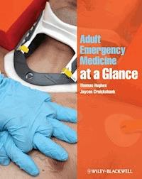
Following the familiar, easy-to-use at a Glance format, and in full-colour, this brand new title provides an accessible introduction and revision aid for medical students and junior doctors. Reflecting the increased profile of Emergency Medicine in clinical practice and the medical school curriculum, Adult Emergency Medicine at a Glance provides a user-friendly overview of the key subjects that will enable any student or junior doctor to 'hit the ground running' when they enter one of the most exciting areas of clinical medicine.
Adult Emergency Medicine at a Glance is:
- A concise, visually orientated course in emergency medicine that is perfect for both study and revision
- Organised around symptoms: 'Short of Breath', rather than diagnoses: 'Pneumonia'
- Focused on the most common or dangerous conditions you will see in the Emergency Department and includes the latest cardiac resuscitation guidelines
- Comprehensively illustrated throughout with over 47 full-page colour illustrations
Sie lesen das E-Book in den Legimi-Apps auf:
Seitenzahl: 285
Veröffentlichungsjahr: 2011
Ähnliche


Contents
preface and ack
List of Abbreviations
1 Life in the Emergency Department
What happens when apatient arrives at the Emergency Department?
Treatment areas in the Emergency Department
Other areas
Culture of the Emergency Department
Emergency Department rules
2 Diagnosis
Using tests
Treatment orders
3 Shock and intravenous fluids
Intravenous access
Types of intravenous fluids
Temperature
Shock
4 Imaging in the Emergency Department
Plain radiography
Clinical ultrasound
Computed tomography scan
Magnetic resonance scan
Interventional imaging
5 Analgesia
Non-pharmacological analgesia
Nitrous oxide
Paracetamol (acetaminophen) and compound analgesics
Moderate opiates
Major opiates: morphine, fentanyl, pethidine (meperidine)
Non-steroidal anti-inflammatory drugs
Local anaesthesia and nerve blocks
Intravenous regional anaesthesia (Bier's block)
6 Airway management and sedation
Airway, breathing and cervical spine
Circulation
Disability
Management
7 Blood gas analysis
Arterial blood gases
Oxygenation
Acid-base disturbance
8 Trauma: primary survey
Treatment priority
Trauma team
Penetrating vs non-penetrating trauma
Ambulance transfer and handover
A: Airway and cervical spine protection
B: Breathing and ventilation
C: Circulation
9 Trauma: secondary survey
Log roll, perineal injury
Patterns of injury
Imaging
Fluid resuscitation
Surgical resuscitation
10 Major head and neck injury
Airway, breathing and cervical spine
Circulation
Disability
Investigations
Management
11 Minor head and neck injury
Imaging
Head injury: clinical assessment
Neck injury: clinical assessment
Other investigations
Common diagnoses
Diagnoses not to miss
12 Wounds
Resuscitation
History
Examination
Management
Special situations
13 Burns
History
Resuscitation
The burn
Intravenous fluids
Minor burns
Carbon monoxide and cyanide poisoning
Electrical burns (including lightning)
Chemical burns
14 Hand injuries
History and examination
Management
Common injuries
Diagnoses not to miss
15 Wrist and forearm injuries
Examination: look, feel, move
Management of fractures: principles
Common diagnoses
Smith’s fracture
Diagnoses not to miss
16 Shoulder and elbow injuries
History
Examination
Imaging
Management
Common diagnoses
Diagnoses not to miss
17 Back pain, hip and knee injuries
Back pain
Hip and knee injury
18 Tibia, ankle and foot injuries
History
Examination
Imaging
Management
Disposal: who can go home?
Common diagnoses
Diagnoses not to miss
19 Abdominal pain
History
Examination
Investigations
Management
Disposal: who can go home?
20 Urology problems
History
Examination
Investigations
Common diagnoses
Diagnoses not to miss
21 Ear, nose, throat and dental problems
Ear
Nose and face
Throat
Dental
22 Eye problems
Examination
Slit lamp examination
Common diagnoses
Diagnoses not to miss
23 Obstetrics and gynaecology problems
Resuscitation
History
Examination
Investigations
Common diagnoses
Diagnoses not to miss
24 Toxicology: general principles
History
Resuscitation: ABCDE
Examination
Management
Decontamination
Elimination
25 Toxicology: specificpoisons
Paracetamol (acetaminophen)
Antidepressants
Opiates
Benzodiazepines
Alcohol
Salicylates
Digoxin
Iron
Stuffers and packers
26 Psychiatry: self-harm and capacity
Deliberate self-harm
Personality disorders
Capacity, consent and ethics
Advance Directives
27 Psychiatry: the disturbed patient
Mental state examination: ABCSMITH
The acutely disturbed patient
Delerium (organic) or psychiatric symptoms
Factitious disorders
28 Observational medicine
Minor injury in elderly patient
Homelessness
'Frequent flyers'
Domestic violence and elder abuse
Drugs and alcohol
Other patient groups
29 Loss of function and independence
History
Examination
Investigations
Management
Disposal: who can go home?
30 Syncope, collapse and falls
History
Examination
Investigations
Treatment: identify or stratify
Diagnoses not to miss
31 Slow heart rate
Investigations
Common diagnoses
Diagnoses not to miss
32 Fast heart rate
Clinical assessment
Investigations
Common diagnoses: atrial
Diagnoses not to miss: ventricular
33 Cardiac arrest
Cardiopulmonary r esuscitation
Drugs
Defibrillation
Return of spontaneous circulation
When to stop resuscitation?
34 Chest pain: cardiovascular
History
Investigations
Treatment
Acute coronary syndromes
Pericarditis
Aortic dissection
35 Chest pain: non-cardiovascular
History
Examination
Investigations
Common conditions
36 Shortness of breath
History
Examination
Investigations
Management
37 Anaphylaxis
Why give adrenaline?
Secondary drugs for anaphylaxis
Anaphylaxis mimics
Investigations
Management
Disposal: who can go home?
38 Sepsis
History
Examination
Investigations
Management
Disposal: who can go home?
Common diagnoses
Diagnoses not to miss
39 Endocrine emergencies
Diabetes
Investigations
Common diagnoses
Rare diagnoses
40 Gastroenterology
History
Examination
Investigations
Management
Common diagnoses
Diagnoses not to miss
41 Headache
History
Examination
Investigations
Management
Disposal: who can go home?
Common diagnoses
Diagnoses not to miss
42 Stroke and transient ischaemic attack
Clinical assessment
Investigations
Stroke
Treatment
Transient ischaemic attack
43 Seizures
Stop the seizure
Search for acause
Examination
Investigations
Disposal: who can go home?
44 Hypothermia and hyperthermia
Hypothermia
Hyperthermia
45 Pre-hospital medicine
At the scene
Phonetic alphabet
46 Major incident
Major incident triage
At the hospital
47 Chemical, biological, radiation, nuclear and explosive incidents
General approach
Chemical
Biological
Radiation
The ‘worried well’
Case studies: questions
Case studies: answers
Index
This edition first published 2011, © 2011 by Thomas Hughes and Jaycen Cruickshank
Blackwell Publishing was acquired by John Wiley & Sons in February 2007. Blackwell’s publishing program has been merged with Wiley’s global Scientific, Technical and Medical business to form Wiley-Blackwell.
Registered office: John Wiley & Sons Ltd, The Atrium, Southern Gate, Chichester, West Sussex, PO19 8SQ, UK
Editorial offices: 9600 Garsington Road, Oxford, OX4 2DQ, UKThe Atrium, Southern Gate, Chichester, West Sussex, PO19 8SQ, UK111 River Street, Hoboken, NJ 07030-5774, USA
For details of our global editorial offices, for customer services and for information about how to apply for permission to reuse the copyright material in this book please see our website at www. wiley.com/wiley-blackwell
The right of the author to be identified as the author of this work has been asserted in accordance with the Copyright, Designs and Patents Act 1988.
All rights reserved. No part of this publication may be reproduced, stored in a retrieval system, or transmitted, in any form or by any means, electronic, mechanical, photocopying, recording or otherwise, except as permitted by the UK Copyright, Designs and Patents Act 1988, without the prior permission of the publisher.
Designations used by companies to distinguish their products are often claimed as trademarks. All brand names and product names used in this book are trade names, service marks, trademarks or registered trademarks of their respective owners. The publisher is not associated with any product or vendor mentioned in this book. This publication is designed to provide accurate and authoritative information in regard to the subject matter covered. It is sold on the understanding that the publisher is not engaged in rendering professional services. If professional advice or other expert assistance is required, the services of a competent professional should be sought.
The contents of this work are intended to further general scientific research, understanding, and discussion only and are not intended and should not be relied upon as recommending or promoting a specific method, diagnosis, or treatment by physicians for any particular patient. The publisher and the author make no representations or warranties with respect to the accuracy or completeness of the contents of this work and specifically disclaim all warranties, including without limitation any implied warranties of fitness for a particular purpose. In view of ongoing research, equipment modifications, changes in governmental regulations, and the constant flow of information relating to the use of medicines, equipment, and devices, the reader is urged to review and evaluate the information provided in the package insert or instructions for each medicine, equipment, or device for, among other things, any changes in the instructions or indication of usage and for added warnings and precautions. Readers should consult with a specialist where appropriate. The fact that an organization or Website is referred to in this work as a citation and/or a potential source of further information does not mean that the author or the publisher endorses the information the organization or Website may provide or recommendations it may make. Further, readers should be aware that Internet Websites listed in this work may have changed or disappeared between when this work was written and when it is read. No warranty may be created or extended by any promotional statements for this work. Neither the publisher nor the author shall be liable for any damages arising herefrom.
Library of Congress Cataloging-in-Publication Data Hughes, Thomas, MSc.
Adult emergency medicine at a glance/Thomas Hughes, Jaycen Cruickshank.p.; cm.Includes index.ISBN 978-1-4051-8901-91. Emergency medicine--Handbooks, manuals, etc. I. Cruickshank, Jaycen. II. Title. [DNLM: 1. Emergency Medicine--methods--Handbooks. 2. Adult. WB 105 H894a 2011] RC86.8.H84 2011616.02'5--dc222010024551ISBN: 9781405189019A catalogue record for this book is available from the British Library.Set in 9 on 11.5 pt Times by Toppan Best-set Premedia Limited1 2011
Adult Emergency Medicine at a Glance
Preface
Emergency Medicine has undergone a quiet revolution over the past twenty years due to a variety of factors that have changed the way medicine is practiced.
Increasing demand and expectations of medical care.Reduction of junior doctors’ hours.An ageing population.Fragmentation of out of hours care.Reduced hospital bed-stay.Sub-specialisation of inpatient medical and surgical practice.Litigation.These factors have pushed expert decision-making towards the front door of the hospital so that the correct diagnosis and treatment start as soon as possible in the patient’s journey. As other specialties have moved away from the acute assessment and treatment of patients, Emergency Medicine has expanded to fill the vacuum left, and in doing so has increased its realm of practice substantially.
Emergency Medicine is exciting and confronting, intimidating and liberating – it is the chance to exercise and hone your diagnostic and practical skills in a well-supervised environment. Clinical staff who work in the ED have all been through the inevitable feelings of fear, uncertainty and doubt that come with the territory, and want you to experience the enjoyment and satisfaction of working in an area of medicine that is never boring.
When trainees start Emergency Medicine, it is often the first time they have seen patients before any other staff. To use a traditional analogy, they have seen plenty of needles, and may be very good at recognising them, but now they are faced with haystacks, in which may be hidden a variety of sharp shiny objects.
Medical textbooks usually describe topics by anatomy or pathology (needles), e.g. heart failure, which tends to assume the diagnostic process. In this book we have tried to organise topics by presentation (haystacks), e.g. ‘short of breath’ , and have tried to articulate the key features that help us find the needles.
We are both great fans of the ‘At a Glance’ series, and have enjoyed the challenge of combining the breadth of practice of adult Emergency Medicine with the concise nature of the ‘At a Glance’ format. We hope you enjoy this book and find it useful as you explore this most dynamic area of medicine.
Acknowledgements
We would like to thank Karen Moore and Laura Murphy at Wiley-Blackwell for their support and advice (and let’s face it, patience) during the elephantine gestation of the book. Also Helen Harvey for understanding deadlines, and Jane Fallows for doing such a great job with the illustrations. We thank the students of Oxford and Melbourne Universities, whose inquiring minds keep us on our toes, and whose questions stimulated us to think of new ways to present the information we have used here. We are both lucky enough to have worked with an exceptional colleague, Philip Catterson, whose teamwork, leadership and hard work have helped us to achieve success in our jobs, and from whom we have learned the few interpersonal skills we have.
In addition TH would like to thank: Professor Christopher Bulstrode who has been an inspiration and mentor and without whose support the book would not have happened. My family, and particularly my wife Marina for her support. My work colleagues for continuing to tolerate me most of the time, and Jackie and Tracey, the ED secretaries who keep me in order. Nic Weir, Rob Janas and David Bowden for their help in sourcing images and the final preparation.
JC would like to thank: All the people who have taught me along the way, particularly Trevor Jackson and Steven Pincus. My parents Ron and Christine for making everything possible, and my wife Kerry and sons Jesse and Flynn for making everything worthwhile.
Thomas Hughes Jaycen Cruickshank
1 Life in the Emergency Department
This chapter describes the way the Emergency Department operates, and some of the unwritten rules. The prevalence of Emergency Department-based drama generates plenty of misconceptions about what occurs in the Emergency Department. For instance, it is generally inadvisable to say ‘stat’ at the end of one’s sentences, and neither of the authors has been mistaken for George Clooney!
What happens when a patient arrives at the Emergency Department?
Alert phone
Also known as the ‘red phone’ or sometimes ‘the Bat-phone’ , this is the dedicated phone line that the ambulance service uses to prewarn the Emergency Department of incoming patients likely to need resuscitation.
Triage
The concept of triage comes from military medicine – doing the most good for the most people. This ensures the most effective use of limited resources, and that the most unwell patients are seen first.
Nurses rather than doctors are usually used to perform the triage because doctors tend to start treating patients. Systems of rapid assessment and early treatment by senior medical staff can be effective, but risk diverting attention from the most ill patients.
Reception/registration
The reception staff are essential to the function of the Emergency Department: they register patients on the hospital computer system, source old notes and keep an eye on the waiting room. They have to deal with difficult and demanding patients, and are good at spotting the sick or deteriorating patient in the waiting room.
Waiting room
Adult and paediatric patients should have separate waiting rooms, and some sort of entertainment is a good idea. Aggression and dissatisfaction in waiting patients has been largely eliminated in the UK by the 4-hour standard of care: all patients must be seen and discharged from the Emergency Department within 4 hours.
Treatment areas in the Emergency Department
Resuscitation bays
Resuscitation bays are used for critically ill and unstable patients with potentially life-threatening illness. They have advanced monitoring facilities, and plenty of space around the patient for clinical staff to perform procedures. X-rays can be performed within this area.
High acuity area
This is the area where patients who are unwell or injured, but who do not need a resuscitation bay, are managed. Medical conditions and elderly patients with falls are common presentations in this area.
Low acuity area
The ‘walking wounded’ – patients with non-life-threatening wounds and limb injuries – are seen here. Patients with minor illness are discouraged from coming to the Emergency Department, but continue to do so for a variety of reasons.
There is a common misconception that patients in this area are similar to general practice or family medicine patients. Numerous studies have found that there is an admission rate of about 5% and an appreciable mortality in low acuity patients, whereas only about 1% of GP consultations result in immediate hospital admission.
Other areas
Imaging
Imaging, such as X-rays and ultrasound, are integral to Emergency Department function. Larger Emergency Departments have their own CT scanner.
Relatives’ room
When dealing with the relatives of a critically ill patient and breaking bad news, doctors and relatives need a quiet area where information is communicated and digested. This room needs to be close to the resuscitation area.
Observation/short stay ward
This is a ward area close to the Emergency Department, run by Emergency Department staff. This unit treats patients who would otherwise need hospital admission for a short time, to enable them to be fully stabilised and assessed. The function of these units is described in Chapter 28.
Hospital in the home
Some hospitals run a ‘hospital in the home’ programme for patients who do not need to be in hospital but who need certain therapy, e.g. intravenous antibiotics, anticoagulation. The Emergency Department is the natural interface between home and hospital.
Culture of the Emergency Department
There is a much flatter (less hierarchical) organisational structure in the Emergency Department than most other areas in the hospital. This occurs because all levels of medical, nursing and other staff work together all the time, and the department cannot function without their cooperation. Ensuring good teamwork requires good leadership, an atmosphere of mutual respect and a bit of patience and understanding.
The resulting atmosphere can be one of the most enjoyable and satisfying places to work in a hospital. A of this less hierarchical culture that surprises junior doctors is that nurses will question their decisions; this is a sign of a healthy culture in which errors are less likely to occur, and is actively encouraged.
Emergency Department rules
Being a doctor in the Emergency Department is different from elsewhere in the hospital. There is nowhere to hide and, for the first time in most medical careers, you are responsible for making the decisions. On the positive side, there are plenty of people around to help you, who have all been through the same process.
Some basic advice:
Write legible, timed, dated notes.Show respect for other professional groups and be prepared to learn from them.Do not be late for your shifts; do not call in sick less than 6 hours before a shift.Patients who re-present are high risk and need senior review.Take your breaks. You need them.Keep calm.If in doubt, ask.Do not pick up so many patients that you cannot keep track of them.Do not avoid work or avoid seeing difficult patients. We do notice.The nurse in charge is usually right.With so many people working closely together in a stressful atmosphere, it is inevitable that conflicts will occur. Do not let them fester; some ground rules for resolving such conflicts are:
Resolve it now.Do it in private.Do it face to face.Focus on facts.Criticise action, not person.Agree why it is important.Agree on a remedy.Finish on a positive.4 Imaging in the Emergency Department
Imaging use in the Emergency Department has increased rapidly over the past few years due to technical advances and increasing pressure to move decision-making earlier in a patient’s journey, and to prevent unnecessary hospital admissions. Ultrasound is now a core skill for senior Emergency Department doctors and new hospitals often have a CT scanner in the Emergency Department.
Plain radiography
Plain radiographs interpreted by the treating clinician are used for the majority of Emergency Department imaging. The advent of digital radiography has made real-time reporting by radiologists easier.
X-rays are ionising radiation and cause damage to tissues through which they pass. The energy released is proportional to the density of the tissue. Abdominal or thoracolumbar radiographs should not be performed in young people, especially females, without a very good reason, as the gonads are very radiosensitive. In this book, X-ray doses are expressed in terms of chest radiographs (CXR). One CXR is approximately 3 days of background radiation.
X-rays are not therapeutic. If the result will not change management, radiographs should not be taken. Examples include uncomplicated rib fractures (when not worried about a pneumothorax), coccyx pain and stubbed toes other than the big toe. Soft tissues are poorly shown by plain films, making it an insensitive examination for joints that rely on these for stability, e.g. knee, shoulder.
Reading plain radiographs
1 Check the patient’s name and the date of the film, particularly on digital radiography systems, which offer many opportunities for confusion. 2 There should be two good views of limbs: anterior-posterior and lateral.
3 If requesting imaging of more than one area, ask yourself if this is necessary. If not urgent, it may be better to re-examine the patient once they have had some analgesia, or obtain a senior opinion.
4 You will learn more from your radiology department if you engage with them and ask their advice rather than expecting a purely technical service.
5 Many Emergency Departments operate a system whereby the radiographer can flag an abnormality on the radiograph. You should not dismiss something that the radiographer has flagged as abnormal without obtaining a senior opinion.
Clinical ultrasound
Clinical (bedside) ultrasound use has increased exponentially with the availability of cheap robust ultrasound machines, and is now a core skill for Emergency Department doctors. Ultrasound has been described as the ‘visual stethoscope’ and is revolutionising the assessment and management of patients in the Emergency Department.
Ultrasound was initially used in the Emergency Department in the resuscitation room for:
Detecting abdominal aortic aneurysms (AAA).Focused abdominal scanning in trauma (FAST) scans, searching for blood in the peritoneal cavity.Central venous line placement.However, ultrasound use is now expanding to include:
Shock assessment: cardiac function, vascular filling, signs of pulmonary embolus, together with the AAA and FAST scans.Basic echocardiography.Deep vein thrombosis (DVT) scanning.Early pregnancy scanning.Hepato-biliary scanning.Disadvantages are that ultrasound is operator dependent, requires training and skill validation, and can divert attention from more important problems.
Computed tomography scan
As resolution and availability have increased and acquisition times have dropped, computed tomography (CT) has become an increasingly useful tool for the Emergency Department. CT is very good for bony injuries, and the trauma CT has proved to be more sensitive and specific than clinical examination in major trauma, but requires a very large radiation dose (1000 CXR).
Neck imaging in high-risk trauma is routinely done by CT (100 CXR), as plain films are insufficiently sensitive at detecting significant injury. Examples of high-risk injuries are a high-speed rollover road traffic collision, and also the elderly patient who falls forward, hitting their face (‘fall on outstretched face’), who is at high risk of odontoid peg fracture, and in whom interpretation of plain radiographs is very difficult (see Chapter 11).
Modern CT scanners have enough resolution and speed to be able to resolve cardiac anatomy including the coronary arteries, pulmonary emboli and aortic dissection (400 CXR). CT brain scan (100 CXR) is an essential part of the assessment of stroke or the unconscious patient. CT KUB (kidneys, ureters and bladder; 400 CXRs) is the imaging of choice in renal colic.
Magnetic resonance scan
Magnetic resonance (MR) scanning is rarely used in the Emergency Department apart from possible cauda equina syndrome (acute central disc prolapse pressing on the cauda equina), giving bowel and bladder symptoms. MR scanning can be used to avoid the large radiation dose incurred by CT, e.g. investigating renal colic in young women.
Joints in which stability and function are mainly due to soft tissues, i.e. ligaments and cartilage such as the knee and shoulder, are well imaged by MR scanning, but it is generally difficult to access these directly from the Emergency Department.
Interventional imaging
Interventional imaging has an increasing role for a limited number of severe conditions. Interventional imaging is generally offered in larger hospitals, and together with trauma care, is one of the main drivers for centralisation of acute services into large hospitals.
Primary percutaneous cardiac intervention with stenting has become the treatment of choice for patients with myocardial infarction.


