BSL3 and BSL4 Agents E-Book
111,99 €
Mehr erfahren.
- Herausgeber: Wiley-VCH
- Kategorie: Wissenschaft und neue Technologien
- Sprache: Englisch
Unique coverage of proteomic and glycomic approaches to better distinguish highly dangerous pathogens, as well as using these to explore novel treatment and prevention options.
The editors and authors are either part of a specialized European network initiated to develop fast and reliable detection and therapy options, or are associated with the core military research complex of the United States.
With its description of the methods, their advantages and limitations, as well as the principle outcomes, this is a must-have resource for all professionals dealing with BSL3 and/or BSL 4 agents.
Sie lesen das E-Book in den Legimi-Apps auf:
Seitenzahl: 408
Veröffentlichungsjahr: 2012
Ähnliche


Table of Contents
Cover
Related Titles
Title page
Copyright page
Preface
List of Contributors
1 Introduction: Application of Proteomic Technologies for the Analysis of Microbial Infections
1.1 Introduction
1.2 Search for New Factors of Virulence and Potential Diagnostic Markers
1.3 Search for New Vaccine Candidates
1.4 Analysis of Post-Translational Modifications of Bacterial Proteins and Protein–Protein Interactions
1.5 Conclusions
Part One: Basic Proteomic Methods
2 Separation of Proteins and Peptides
2.1 Introduction
Acknowledgment
3 Basic Mass Spectrometric Approaches
3.1 Introduction
3.2 Ionization
3.3 Mass Analyzers
3.4 Protein Identification
3.5 Conclusion
Acknowledgments
4 Quantitative Mass Spectrometric Approaches
4.1 Introduction
4.2 iTRAQ Analysis of Bacterial Pathogens
5 BN-PAGE of Microbial Protein Complexes
5.1 Introduction
5.2 Methods for Studying Protein–Protein Interactions
5.3 Blue Native Polyacrylamide Gel Electophoresis
5.4 Evaluation of BN-PAGE – Staining, MS, Western Blotting
5.5 BN/SDS-PAGE of ATP Synthase of Francisella tularensis
5.6 Conclusion
Acknowledgment
6 Analysis of Francisella tularensis Glycoproteins
6.1 Introduction to Post-Translational Modifications in Prokaryotes
6.2 Methodology
6.3 Bioinformatics
6.4 Application of Glycoproteomic Approach Utilizing ProQ-Emerald and DIG Glycan Kits to Francisella tularensis (F. tularensis)
6.5 Results
6.6 Conclusion
Acknowledgments
Part Two: Identification of Proteins and Glycans from Microorganisms as Candidate Molecules for Use in Detection/Diagnosis, Therapy, and Prophylaxis
7 Comparative Proteome Analysis of Strains with Differential Virulence
7.1 Introduction
7.2 Methods
7.3 Results
7.4 Discussion
8 Analysis of Francisella tularensis Acetonitrile Extracts
8.1 Introduction
8.2 Material and Methods
8.3 Results
8.4 Conclusions
Acknowledgments
9 Analysis of Culture Filtrate Proteins of Francisella tularensis
9.1 Introduction
9.2 Materials and Methods
9.3 Results
9.4 Discussion
Acknowledgments
10 Lipopolysaccharides of Coxiella burnetii: Chemical Composition and Structure, and Their Role in Diagnosis of Q Fever
10.1 Introduction
10.2 Lipopolysaccharides of C. burnetii
10.3 Conclusion
Acknowledgments
11 Mimivirus Possesses Anonymous and Unique Gene Products Endowed for Antigenic Properties
11.1 Introduction
11.2 Material and Methods
11.3 Results
12 Detection of Differentially Modified Pathogen Proteins by Western Blot after 2D Gel Electrophoresis and Identification by MALDI-TOF/TOF
12.1 Introduction
12.2 Materials and Methods
12.3 Results
12.4 Discussion
13 Composition and Structure of Lipid A of the Intracellular Bacteria Piscirickettsia salmonis and Coxiella burnetii
13.1 Introduction
13.2 Composition and Structure of Lipid A of P. salmonis
13.3 Composition and Structure of Lipid A of C. burnetii
13.4 Conclusion
Acknowledgments
14 Proteins of Coxiella burnetii and Analysis of Their Function
14.1 Introduction
14.2 Proteins of C. burnetii and Their Functions
14.3 Conclusion and Perspectives
Note Added in Proof
Acknowledgments
15 Subtype and Toxin Variant Identification of Botulinum Neurotoxin Type A Using Proteomics Techniques
15.1 Introduction
15.2 Botulinum Neurotoxins
15.3 Differentiation of Botulinum Neurotoxins
15.4 Amino Acid Sequence Identification Using Proteomics
15.5 Extraction of BoNT from Complex Matrices
15.6 Identification of BoNT Serotype with Proteomics
15.7 Identification of BoNT/A Subtype with Proteomics
15.8 Identification of BoNT/A1 Strain with Proteomics
15.9 Conclusions
Disclaimer
16 Protein Microarrays for Antigen Discovery
16.1 Introduction
16.2 Microarray Assembly
16.3 Antibody Assays
16.4 Antigens and Proteomes of Viruses
16.5 Antigens and Proteomes of Pathogenic Bacteria
16.6 Conclusions
Acknowledgment
17 MALDI-TOF Mass Spectrometry for Rapid Identification of Highly Pathogenic Microorganisms
17.1 Introduction
17.2 Microbial Identification by MALDI-TOF Mass Spectrometry
17.3 Inactivation of Highly Pathogenic Microorganisms for MALDI-TOF Mass Spectrometry
17.4 Identification of Important Bacterial Pathogenes Using MALDI-TOF MS
17.5 Concluding Remarks
Acknowledgments
Part Three: Analysis of Host–Pathogen Interactions
18 Quantitative Proteomic Profiling of the Interaction of Francisella tularensis LVS with Macrophages Using J774.2 Cell Line
18.1 Introduction
18.2 Material and Methods
18.3 Results
19 Proteome Analysis of Bacterial Protein Expression after Ingestion of Microbes by Macrophages
19.1 Introduction
19.2 Material and Methods
19.3 Results
19.4 Discussion
Acknowledgment
Index
Related Titles
Katz, R., Zilinskas, R. A. (eds.)
Encyclopedia of Bioterrorism Defense
2011
ISBN: 978-0-470-50893-0
Shah, H., Gharbia, S. (eds.)
Mass Spectrometry for Microbial Proteomics
2010
ISBN: 978-0-470-68199-2
Krämer, R., Jung, K. (eds.)
Bacterial Signaling
2010
ISBN: 978-3-527-32365-4
Kostic, T., Butaye, P., Schrenzel, J. (eds.)
Detection of Highly Dangerous Pathogens
Microarray Methods for BSL 3 and BSL 4 Agents
2009
ISBN: 978-3-527-32275-6
Kwaik, Y. A., Metzger, D. W., Nano, F., Sjostedt, A., Titball, R. (eds.)
Francisella Tularensis
Biology, Pathogenicity, Epidemiology, and Biodefense
2007
ISBN: 978-1-57331-691-0
Jungblut, P. R., Hecker, M. (eds.)
Proteomics of Microbial Pathogens
2007
ISBN: 978-3-527-31759-2
The Editors
Prof. Jiri Stulik
Ministry of Defence
Faculty of Military Health Service
Trebesska 1575
500 01 Hradec Králové
Czech Republic
Prof. Rudolf Toman
Slovak Academy of Sciences
Institute of Virology
Dubravska cesta 9
845 05 Bratislava 45
Slovak Republic
Prof. Patrick Butaye
Veterimary and Agrochemical Research Centre
Department of Bacteriology and Immunology
Groeselenberg 99
1180 Bruxelles
Belgium
Dr. Robert G. Ulrich
Army Medical Research Inst.
of Infectious Diseases
1425 Porter Street
Frederick, MD 21702
USA
Cover
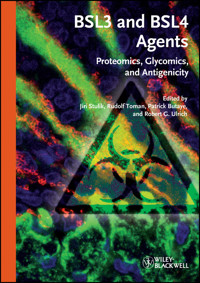
Section of a lung from a cynomolgus macaque exposed to aerosolized Yersinia pestis. The bacteria (red) are apparent throughout the lung, which is stained for fibrin (green). Cell nuclei are shown in blue. Courtesy of Derron Alves (staining by Christine Mech, microscopy by Gordon Ruthel).
Limit of Liability/Disclaimer of Warranty: While the publisher and author have used their best efforts in preparing this book, they make no representations or warranties with respect to the accuracy or completeness of the contents of this book and specifically disclaim any implied warranties of merchantability or fitness for a particular purpose. No warranty can be created or extended by sales representatives or written sales materials. The advice and strategies contained herein may not be suitable for your situation. You should consult with a professional where appropriate. Neither the publisher nor authors shall be liable for any loss of profit or any other commercial damages, including but not limited to special, incidental, consequential, or other damages.
Library of Congress Card No.: applied for
British Library Cataloguing-in-Publication Data
A catalogue record for this book is available from the British Library.
Bibliographic information published by the Deutsche Nationalbibliothek
The Deutsche Nationalbibliothek lists this publication in the Deutsche Nationalbibliografie; detailed bibliographic data are available on the Internet at <http://dnb.d-nb.de>.
© 2011 Wiley-VCH Verlag & Co. KGaA, Boschstr. 12, 69469 Weinheim, Germany
Wiley-Blackwell is an imprint of John Wiley & Sons, formed by the merger of Wiley’s global Scientific, Technical, and Medical business with Blackwell Publishing.
All rights reserved (including those of translation into other languages). No part of this book may be reproduced in any form – by photoprinting, microfilm, or any other means – nor transmitted or translated into a machine language without written permission from the publishers. Registered names, trademarks, etc. used in this book, even when not specifically marked as such, are not to be considered unprotected by law.
Print ISBN: 978-3-527-32780-5
Epub ISBN: 978-3-527-63820-8
Mobi ISBN: 978-3-527-64777-4
Preface
The research initiatives and interests of COST B28 partners are organized in five working packages/groups (WGs):
WG1: Technology platform (including flow cytometry and microarrays)WG2: AntigenicityWG3: Proteomics and glycomicsWG4: GenomicsWG5: Microbiology (bacteriology, virology, mycology)Research groups involved in WG2 and WG3 possess an extensive fundamental knowledge and expertise in the fields of proteomics, glycomics, and antigenicity of both bacteria and viruses. A mutual collaboration in these fields is of importance since the interconnection between the three research disciplines is obvious. The aim of this booklet is to summarize the present knowledge on proteomics, glycomics, and the structure/function of antigens in general, and to provide insights into the latest developments and applications of these scientific fields with regard to BSL3 and BSL4 agents. Contributions to the booklet illustrate a considerable broadness and variability of these applications on the agents investigated within the framework of this COST B28 action.
The first part of this booklet deals with the general applications of proteomic and glycomic techniques in the analysis of microbial infections. These are then elaborated further, and for some techniques basic protocols are given. Many of them have also been demonstrated in a training school held within this COST Action B28 action organized by the main partners in these two working groups.
The second part focuses on applications of these techniques in the identification and characterization of proteins and glycans from microorganisms and on the rapid and accurate detection and identification of them. Moreover, the diagnosis, therapy, and prophylaxis of the diseases caused by these microorganisms are discussed. This concerns mainly Francisella tularensis, the obligate intracellular gram-negative bacteria Coxiella burnetii and Piscirickettsia salmonis, mimivirus, and measels virus. Although the number of examples is limited, they still give a good insight into the respective fields.
The third part is devoted to host–pathogen interactions, dealing with both prokaryote and eukaryote expressions of proteins.
This booklet has no ambitions to be a complete and final “manual” as it deals with an area in which new findings, techniques, and methodologies are being brought on nearly on a daily basis, thus making it almost impossible to give a real up to date overview. Nevertheless, it gives the reader an interesting insight into these new and rapidly developing fields. The information presented here may be helpful to students and researchers interested in proteomics, glycomics, and the structure/function of antigens and their further, so far unknown research fields and applications. For additional reading, we recommend references provided by the authors.
More information on WG2 and WG3, and also on other WGs, can be found on the COST Action B28 website: http://www.cost-b28.be/.
Jiri Stulik
Rudolf Toman
Patrick Butaye
Robert G. Ulrich
June 2011
List of Contributors
Wim Ammerlaan
Institute of Immunology
Centre de Recherche Public de la Santé/National Public Health Institute
20A, rue Auguste Lumière
1950 Luxembourg
Luxembourg
Lucie Balonova
Institute of Molecular Pathology
Faculty of Military Health Science
University of Defence
Trebesska 1575
500 01 Hradec Králové
Czech Republic
John R. Barr
Centers for Disease Control and Prevention
National Center for Environmental Health
Division of Laboratory Sciences
4770 Buford Hwy, N.E.
Atlanta, GA 30341
USA
Jakub Baudys
Centers for Disease Control and Prevention
National Center for Environmental Health
Division of Laboratory Sciences
4770 Buford Hwy, N.E.
Atlanta, GA 30341
USA
Martin Brychta
Institute of Molecular Pathology
Faculty of Military Health Sciences
University of Defence
Trebesska 1575
500 01 Hradec Králové
Czech Republic
and
Charles University in Prague
Faculty of Medicine in Hradec Králové
Department of Medical Biology and Genetics
500 01 Hradec Králové
Czech Republic
Patrick Butaye
Department of Bacteriology and Immunology
Veterinary and Agrochemical Research Centre
VAR-CODA-CERVA
Groeselenberg 99
1180 Brussels
Belgium
and
Ghent University
Faculty of Veterinary Medicine
Department of Pathology, Bacteriology and Poultry Diseases
Salisburylaan 133
9820 Merelbeke
Belgium
Jiri Dresler
Institute of Molecular Pathology
University of Defence
Faculty of Military Health Sciences
Trebesska 1575
500 01 Hradec Králové
Czech Republic
Fred Fack
Institute of Immunology
Centre de Recherche Public de la Santé/National Public Health Institute
20A, rue Auguste Lumière
1950 Luxembourg
Luxembourg
Anetta Hartlova
Centre of Advanced Studies
Faculty of Military Health Sciences
Trebesska 1575
500 01 Hradec Králové
Czech Republic
Lenka Hernychova
Institute of Molecular Pathology
Faculty of Military Health Science
University of Defence
Trebesska 1575
500 01 Hradec Králové
Czech Republic
Martin Hubalek
Institute of Molecular Pathology
Faculty of Military Health Sciences
University of Defence
Trebesska 1575
500 01 Hradec Králové
Czech Republic
Robert Ihnatko
Institute of Virology
Slovak Academy of Sciences
Laboratory for Diagnosis and Prevention of Rickettsial and Chlamydial Infections
Dubravska cesta 9
845 05 Bratislava 45
Slovak Republic
Suzanne R. Kalb
Centers for Disease Control and Prevention
National Center for Environmental Health
Division of Laboratory Sciences
4770 Buford Hwy, N.E.
Atlanta, GA 30341
USA
Sarah Keasey
United States Army Medical Research Institute of Infectious Diseases
Department of Immunology
1425 Porter Street
Frederick, MD 21702
USA
Julia Kessler
Institute of Immunology
Centre de Recherche Public de la Santé/National Public Health Institute
20A, rue Auguste Lumière
1950 Luxembourg
Luxembourg
Jana Klimentova
Institute of Molecular Pathology
Faculty of Military Health Sciences
University of Defence
Trebesska 1575
500 01 Hradec Králové
Czech Republic
Klara Konecna
Institute of Molecular Pathology
Faculty of Military Health Sciences
University of Defence
Heyrovskeho 1203
500 05 Hradec Králové
Czech Republic
and
Charles University in Prague
Pharmaceutical Faculty in Hradec Kralove
500 05 Hradec Králové
Czech Republic
Jacques Kremer
Institute of Immunology
Centre de Recherche Public de la Santé/National Public Health Institute
20A, rue Auguste Lumière
1950 Luxembourg
Luxembourg
Peter Lasch
Robert Koch-Institut
Biomedical Spectroscopy (P25)
Nordufer 20
13353 Berlin
Germany
Juraj Lenco
Centre of Advanced Studies
Faculty of Military Health Sciences
Trebesska 1575
500 01 Hradec Králové
Czech Republic
and
Institute of Molecular Pathology
FMHS UO
Trebesska 1575
500 01 Hradec Králové
Czech Republic
Marek Link
Centre of Advanced Studies
Faculty of Military Health Sciences
Trebesska 1575
500 01 Hradec Králové
Czech Republic
Claude P. Muller
Institute of Immunology
Centre de Recherche Public de la Santé/National Public Health Institute
20A, rue Auguste Lumière
1950 Luxembourg
Luxembourg
Mohan Natesan
United States Army Medical Research Institute of Infectious Diseases
Department of Immunology
1425 Porter Street
Frederick, MD 21702
USA
Dieter Naumann
Robert Koch-Institut
Biomedical Spectroscopy (P25)
Nordufer 20
13353 Berlin
Germany
Ivona Pávková
Institute of Molecular Pathology
Faculty of Military Health Sciences
University of Defence
Trebesska 1575
500 01 Hradec Králové
Czech Republic
James L. Pirkle
Centers for Disease Control and Prevention
National Center for Environmental Health
Division of Laboratory Sciences
4770 Buford Hwy, N.E.
Atlanta, GA 30341
USA
Patrick Pirrotte
Institute of Immunology
Centre de Recherche Public de la Santé/National Public Health Institute
20A, rue Auguste Lumière
1950 Luxembourg
Luxembourg
Didier Raoult
Unité des Rickettsies
IRD-CNRS UMR 6236
Faculté de Médecine
27 Bd Jean Moulin
13385 Marseille
France
Patricia Renesto
Unit of Virus Host Cell Interactions
UMI 3265 Université Joseph Fourier-EMBL-CNRS
6, rue Jules Horowitz
38042 Grenoble
France
Dominique Revets
Institute of Immunology
Centre de Recherche Public de la Santé/National Public Health Institute
20A, rue Auguste Lumière
1950 Luxembourg
Luxembourg
Ludovit Skultety
Institute of Virology
Slovak Academy of Sciences
Dubravska cesta 9
845 05 Bratislava
Slovak Republic
and
Center for Molecular Medicine
Vlarska 3-7
831 01 Bratislava
Slovak Republic
Theresa J. Smith
United States Army Medical Research Institute of Infectious Diseases
Integrated Toxicology
Ft. Detrick, MD 21702
USA
Jiri Stulik
Institute of Molecular Pathology
FMHS UO
Trebesska 1575
500 01 Hradec Králové
Czech Republic
and
Centre of Advanced Studies
Faculty of Military Health Sciences
Trebesska 1575
500 01 Hradec Králové
Czech Republic
Vojtch Tambor
Institute of Molecular Pathology
FMHS UO
Trebesska 1575
500 01 Hradec Králové
Czech Republic
Rudolf Toman
Slovak Academy of Sciences
Institute of Virology
Laboratory for Diagnosis and Prevention of Rickettsial and Chlamydial Infections
Dubravska cesta 9
845 05 Bratislava 45
Slovak Republic
Jana Udrzalova
Institute of Molecular Pathology
Faculty of Military Health Sciences
University of Defence
Trebesska 1575
500 01 Hradec Králové
Czech Republic
Robert G. Ulrich
United States Army Medical Research Institute of Infectious Diseases
Department of Immunology
1425 Porter Street
Frederick, MD 21702
USA
Pavol Vadovi
Institute of Virology
Slovak Academy of Sciences
Laboratory for Diagnosis and Prevention of Rickettsial and Chlamydial Infections
Dubravska cesta 9
845 05 Bratislava 45
Slovak Republic
1
Introduction: Application of Proteomic Technologies for the Analysis of Microbial Infections
Jiri Stulik and Patrick Butaye
1.1 Introduction
Proteomics belongs to the group of so-called “omics” technologies that are preferentially exploited for the global analysis of temporal or conditional alterations in gene expression, on either the gene or protein level. Proteomics is focused on proteins and provides information about their abundances, post-translational modifications, localization and mutual interactions. Current proteomic procedures combine efficient electrophoretic or chromatographic separation techniques with identification approaches based on mass spectrometry (MS) and computer technologies for bioinformatic analysis. It can be said that MS is now the major driving force in the field of proteomics and the quantitative shotgun tandem MS represents a large-scale high-throughput technology commonly employed for comparative identification and quantification protein studies [1].
The major benefit of the “omics” approaches is the ability to analyze a large number of genes or proteins simultaneously, enabling a more realistic view of a complex cell response to stimuli. The analysis of the global changes in protein profiling during the interactions between microbial pathogens and their hosts, so-called “infectomics”, is very attractive area within proteomics. It feeds the study of the fundaments of infections. It solves molecular mechanisms of microbial pathogenesis, helps in the efficient and rapid diagnosis of infectious disease and the development of novel prophylaxis and treatment strategies. Here, infectomics involves the study of proteins uniquely expressed or up-regulated in virulent clinical strains, proteins produced under stress conditions and, finally, proteins with immunostimulatory properties. Going deeper into infectomics one deals with structural studies of microbial proteins (to characterize potential post-translational modifications) and the analysis of protein–protein interactions, the so-called “interactome” [2].
In this publication we first give an overview on basic proteomic techniques. This introduces the reader into the more specialized studies performed on BSL3 and BSL 4 agents. These organisms are difficult to handle and specialized laboratories are necessary to work with them. However, the safe preparation of materials for protein researchers can allow substantial progress in the understanding of these microorganisms.
1.2 Search for New Factors of Virulence and Potential Diagnostic Markers
Matching the protein patterns extracted from nonvirulent attenuated strains with their fully virulent counterparts is a common procedure to detect potential virulence factors and/or new diagnostic markers. This has been demonstrated in the comparative proteome study of exoproteins released by different enterotoxigenic Staphylococcus aureus strains. Using a gel-based approach different known enterotoxins and other possible virulence factors were revealed by comparing the protein profiles of two isolates, a food-derived strain with a prevalent enterotoxin gene cluster and a nonenterotoxigenic reference strain [3]. Another example of the gel-based approach is the protein profiling of two cystic fibrosis isolates of Pseudomonas aeruginosa strains associated with the initial and chronic infection process. These two genetically identical strains secrete several proteins uniquely expressed in the different stages of the disease [4]. Gel-based approaches utilizing preferentially two-dimensional gel electrophoresis (2-DE) for the study of enriched membrane proteins are typically exploited for the host–pathogen interactions. However, this method suffers from the loss of most of the hydrophobic membrane proteins due to their inefficient solubilization during isoelectric focusing, and therefore, the nonelectrophoretic approaches gradually replace gel-based techniques for such studies. An example of this is the study of the cell wall proteome of Listeria monocytogenes and Listeria innocua [5]. Here 30 proteins residing in the membrane were identified in both strains. Outer membrane vesicles (OMV) that are assumed to play an important role in cell to cell communication, for example, toxins or in the transfer of proteins and genetic materials between microbes represent potential virulence factors and proteins with immunostimulatory properties. The purification of sufficient OMV is, however, a serious problem [6].
Intracellular infectious microbes have to cope with the hostile intracellular milieu. Basically, their success for survival and multiplication depends on the coordinated reprogramming of their gene expression under the influence of stimuli, such as acid pH, iron depletion, oxidative stress, nitrogen radicals and heat. In-depth proteome analysis of the changing microbial protein profiles is very challenging since it informs us about protein-virulence factors that may serve as new targets for treatment. Unfortunately, progress in this area is still hampered by the inability of proper protein extraction methods. They lack efficacy through low gain and are too much contaminated with eukaryotic material. One way to avoid this could be the direct purification of bacteria-containing vacuoles and to identify the proteins uniquely expressed within this compartment [7].
1.3 Search for New Vaccine Candidates
The application of proteomics for a search for vaccine candidates has been driven by the current need for a new type of safe and efficient subunit vaccines. The classical approach, called serological proteome (SERPA), is used for the identification of perspective proteins. This technology is based on the separation of complex protein mixtures by gel-based technology, mostly by 2-DE, followed by blotting of separated proteins to a membrane where the transferred proteins are probed by human or animal immune sera. The detected spots are then identified by MS after their excision from the matched preparative 2-DE gel. This approach is still routinely used but, instead of separating the whole-cell lysates, just the membrane proteins, secreted proteins or proteins located in outer membrane vesicles are being examined [8–10]. As already mentioned, 2-DE separation has strong limitations, and therefore, new nongel-based approaches are being examined. Protein microarray chips probed with sera from infected individuals overcome this problem, allowing protein solubilization and enabling the analysis of the whole proteome in an unbiased manner. This procedure was examined in the screening process for the identification of Francisella tularensis immunoreactive proteins. Probing a chip containing 1741 F. tularensis antigens with the sera from 46 patients diagnosed as tularemia patients led to the detection of 244 antigens exhibiting a high intensity signal [11].
Bioinformatics is an invaluable tool for the discovery of vaccine candidates because the human brain can hardly contain all information generated by the current high-throughput techniques. They allow us to focus on a significantly restricted number of proteins and to select those proteins that are likely to induce a good immune response. McMurry et al. [12] used a combination of different softwares for the detection of possible Mycobacterium tuberculosis immunodominant epitopes. First at the DNA level, open reading frames containing signal sequences featuring secreted proteins were detected, and then, the selected proteins were screened for matches to a list of MHC II binding motifs. Based on this selection, 17 peptides were synthesized and their immunostimulatory potential was verified by in vitro T-cell assays. Overall 15 epitopes gave acceptable results and are now candidates for the construction of new TB vaccine.
1.4 Analysis of Post-Translational Modifications of Bacterial Proteins and Protein–Protein Interactions
Post-translational modification adds an additional level of complexity to the current “omics” technologies. It is estimated that more than 100 different post-translational modifications exist in eukaryotic cells and the most widely distributed ones are glycosylation and phosphorylation. Highly sophisticated proteomic approaches are being developed to detect and quantify these modifications, but these procedures are still far from being a routine technology. Surprisingly, the recent data document that both types of modifications occur also in prokaryotes and their biological significance is starting to be unveiled.
Regarding prokaryotes, both N- and O-glycosylations were detected and orthologs of the genes encoding members of both glycosylation pathways in eukaryotes were found in mucosal pathogens [13]. Glycosylation concerns mainly membrane and secreted bacterial proteins and most of them are known virulence factors. Hence, this modification might serve a specific function in the molecular mechanism of pathogenesis. This assumption was, for example, confirmed for the Toxoplasma gondii N-glycoproteins that are involved in several biological functions associated with the parasite’s intracellular development. Exposing cells to tunicamycin (which disrupts the glycosylation of newly synthesized proteins) then abrogated host cell invasion and parasite growth [14]. Unfortunately, the identification of glycoproteins is a highly intricate matter and encompasses laborious methods aimed at the detection of glycan modification(s). This and the methods of glycosidic linkage determination and identification of carbohydrate residues are still in their infancy in this area.
The glycosylation of bacterial proteins and the reversible phosphorylation of serine, threonine and tyrosine residues were thought to be restricted to eukaryotic cells. However, in the last decade several prokaryotic tyrosine and serine/threonine kinases together with phosphatases have been described [15]. In contrast to eukaryotic kinases, the majority of these prokaryotic enzymes appear to be transmembrane proteins, and thus, it is anticipated that these kinases may function as prokaryotic receptors. Nevertheless, the ligands for these receptors and signaling pathways triggered by receptor occupation have to be unraveled.
The extensive systematic prokaryotic phosphoproteome analyses require a good experience and skill in both phosphoprotein enrichment and in the following MS and bioinformatic analyses. Up to now, two indepth phosphorylation studies of a gram-positive bacterium Bacillus subtilis and a gram-negative bacterium Escherichia coli have been performed [16, 17]. They proved that a significantly lower number of prokaryotic proteins was phosphorylated in comparison to the mammalian or yeast proteins. Furthermore, phosphorylation on the serine and threonine residues prevailed and proteins were frequently multiphosphorylated but again their number was low in comparison to that found in eukaryotes.
The kind of virulence factors either localized in the outer membrane or actively secreted is prone to direct interplay with the host cell biological system. A physical contact between microbes and target cells leads to significant alteration of the host cell proteomes on various levels, resulting in the modulation of cell signaling pathways, membrane ruffling, cytoskeleton rearrangement, endosomal traffficking, host cell protein modification and the induction of cell death programs. The combination of various fractionation procedures and gel- or nongel-based proteomic approaches enables us to study the changes in host proteomes on the level of individual subcellular compartments directly targeted by the microbe.
Another approach is focused on the identification of interaction of the host and microbial virulence factors. However, despite the application of multiple tag and pull-down procedures for the isolation of protein complexes, relatively few host proteins have been identified as the targets of microbial proteins until now. There is also a problem to distinguish nonspecific from specific binding partners of the isolated protein complexes. In this case, it appears that the recent developments in quantitative proteomics could help to solve this problem. One particular quantitative technique exploits metabolically incorporated isotopic labels into proteins. Using this approach, two cell cultures are differentially labeled. Then affinity purification isolates the bait from one population and a control from the other population. When the isolated complexes are analyzed, a mixed nonspecific interaction is characterized by an equal binding of the background proteins to the bait, while specific interactions with the bait result in differential ratios. This protocol was successfully used for the identification of the small G-protein Cdc42 as the host target of the SPI-1 effector, SopB/SigD in Salmonella [18].
1.5 Conclusions
The enormous progress in the proteomic methodologies will allow us to gradually describe the entire proteomes of pathogens. Likewise, it can be expected that new virulence factors will be identified and their biological role in the pathogenesis of diseases will be uncovered. These findings together with improvements in bioinformatics should then substantially speed up the design of new therapeutic and prophylactic agents.
This COST Action B28 gathers excellence in these fields. In line with a microarray platform, a combination of genomics, proteomics, glycomics and antigenicity substantiated by a bioinformatics knowledge will allow us to make noticeable progress towards a better understanding of the agents under investigation and the diseases they cause. Of course, the input of bacteriologists and virologists dealing with these infections in the field and their efforts to understand their importance for humans, animals and environment cannot be omitted.
References
1 Chen, G., and Pramanik, B.N. (2008) LC-MS for protein characterization: current capabilities and future trends. Expert Rev. Proteomics, 5, 435–444.
2 Charbonnier, S., Gallego, O., and Gavin, A.C. (2008) The social network of a cell: recent advances in interactome mapping. Biotechnol. Annu. Rev., 14, 1–28.
3 Pocsfalvi, G., Cacace, G., Cuccurullo, M., Serluca, G., Sorrentino, A., Schlosser, G., Blaiotta, G., and Malorni, A. (2008) Proteomic analysis of exoproteins expressed by enterotoxigenic Staphylococcus aureus strains. Proteomics, 8, 2462–2476.
4 Hanna, S.L., Sherman, N.E., Kinter, M.T., and Goldberg, J.B. (2000) Comparison of proteins expressed by Pseudomonas aeruginosa strains representing initial and chronic isolates from a cystic fibrosis patient: an analysis by 2-D gel electrophoresis and capillary column liquid chromatography-tandem mass spectrometry. Microbiology, 146, 2495–2508.
5 Calvo, E., Pucciarelli, M.G., Bierne, H., Cossart, P., Albar, J.P., and García-Del Portillo, P. (2005) Analysis of the Listeria cell wall proteome by two-dimensional nanoliquid chromatography coupled to mass spectrometry. Proteomics, 5, 433–443.
6 Nally, J.E., Whitelegge, J.P., Aguilera, R., Pereira, M.M., Blanco, D.R., and Lovett, M.A. (2005) Purification and proteomic analysis of outer membrane vesicles from a clinical isolate of Leptospira interrogans serovar Copenhageni. Proteomics, 5, 144–152.
7 Mattow, J., Siejak, F., Hagens, K., Becher, D., Albrecht, D., Krah, A., Schmidt, F., Jungblut, P.R., Kaufmann, S.H., and Schaible, U.E. (2006) Proteins unique to intraphagosomally grown Mycobacterium tuberculosis. Proteomics, 6, 2485–2494.
8 Williams, J.N., Skipp, P.J., Humphries, H.E., Christodoulides, M., O’Connor, C.D., and Heckels J.E. (2007) Proteomic analysis of outer membranes and vesicles from wild-type serogroup B Neisseria meningitidis and a lipopolysaccharide-deficient mutant. Infect. Immun., 75, 1364–1372.
9 Morsczeck, C., Prokhorova, T., Sigh, J., Pfeiffer, M., Bille-Nielsen, M., Petersen, J., Boysen, A., Kofoed, T., Frimodt-Møller, N., Nyborg-Nielsen, P., and Schrotz-King, P. (2008) Streptococcus pneumoniae: proteomics of surface proteins for vaccine development. Clin. Microbiol. Infect., 14, 74–81.
10 Chitlaru, T., Gat, O., Grosfeld, H., Inbar, I., Gozlan, Y., and Shafferman, A. (2007) Identification of in vivo-expressed immunogenic proteins by serological proteome analysis of the Bacillus anthracis secretome. Infect. Immun., 75, 2841–2852.
11 Sundaresh, S., Randall, A., Unal, B., Petersen, J.M., Belisle, J.T., Hartley, M.G., Duffield, M., Titball, R.W., Davies, D.H., Felgner, P.L., and Baldi, P. (2007) From protein microarrays to diagnostic antigen discovery: a study of the pathogen Francisella tularensis. Bioinformatics, 23, 508–518.
12 McMurry, J., Sbai, H., Gennaro, M.L., Carter, E.J., Martin, W., and De Groot, A.S. (2005) Analyzing Mycobacterium tuberculosis proteomes for candidate vaccine epitopes. Tuberculosis (Edinb), 85, 95–105.
13 Hitchen, P.G., and Dell, A. (2006) Bacterial glycoproteomics. Microbiology, 152, 1575–1580.
14 Fauquenoy, S., Morelle, W., Hovasse, A., Bednarczyk, A., Slomianny, C., Schaeffer, C., Van Dorsselaer, A., and Tomavo, S. (2008) Proteomics and glycomics analyses of N-glycosylated structures involved in Toxoplasma gondii–host cell interactions. Mol. Cell. Proteomics, 7, 891–910.
15 Bakal, C.J., and Davies, J.E. (2000) No longer exclusive club: eukaryotic signalling domains in bacteria. Trends Cell. Biol., 10, 32–38.
16 Macek, B., Mijakovic, I., Olsen, J.V., Gnad, F., Kumar, C., Jensen, P.R., and Mann, M. (2007) The serine/threonine/tyrosine phosphoproteome of the model bacterium Bacillus subtilis. Mol. Cell. Proteomics, 6, 697–707.
17 Macek, B., Gnad, F., Soufi, B., Kumar, C., Olsen, J.V., Mijakovic, I., and Mann, M. (2008) Phosphoproteome analysis of E. coli reveals evolutionary conservation of bacterial Ser/Thr/Tyr phosphorylation. Mol. Cell. Proteomics, 7, 299–307.
18 Rogers, L.D., Kristensen, A.R., Boyle, E.C., Robinson, D.P., Ly, F.T., Finlay, B.B., and Foster, L.J. (2008) Identification of cognate host targets and specific ubiquitylation sites on the Salmonella SPI-1 effector SopB/SigD. J. Proteomics, 71, 97–108.


