Imaging Marine Life E-Book
81,99 €
Mehr erfahren.
- Herausgeber: Wiley-VCH
- Kategorie: Wissenschaft und neue Technologien
- Sprache: Englisch
Written by an international team of experts from the Tara Oceans Marine Biology Imaging Platform (TAOMI), this is the first and only compendium on marine imaging technologies, and includes all known underwater as well as on-land techniques.
TAOMI is imaging the largest collection of marine organisms in recent history, ranging from viruses to corals, and is duplicated on land to perform high throughput confocal analysis of plankton, X-ray tomography as well as cryo-electron microscopy. This unique platform combines underwater imaging with cytometry, stereomicroscopy, fluorescence microscopy and 3D microscopy - all of which are covered in this practical book, along with remote sensing, MRI, and optical projection tomography.
The definitive resource for every marine biologist who is planning to image marine species, whether underwater or on land.
Sie lesen das E-Book in den Legimi-Apps auf:
Seitenzahl: 487
Veröffentlichungsjahr: 2013
Ähnliche


Table of Contents
Related Titles
Title Page
Copyright
Preface
List of Contributors
Chapter 1: Under the Eye of Neptune: An Historical Perspective of Marine Creature Imagery
1.1 Introduction
1.2 Ancient Uses of the Oceans
1.3 From Neptune to Animalcules
1.4 The Birth of Oceanography (The Nineteenth Century)
1.5 The Twentieth Century: Institutions and moving images
1.6 Time Line of Ocean Imagery
Further Reading
Basic Texts
Source Books
Ships and Expeditions
Institutions
Chapter 2: New Solutions in Underwater Imaging and Vision Systems
2.1 Introduction
2.2 Underwater Optical Image Formation
2.3 Illumination Techniques
2.4 Laser-Based Techniques
2.5 Underwater Imaging Infrastructures
2.6 Image Improvement via Polarization
2.7 A Vision System for Underwater Applications
2.8 Conclusion
Acknowledgements
References
Chapter 3: Holographic Microscopy of Marine Organisms
3.1 Introduction
3.2 Advantages of Holographic Microscopy
3.3 Past Attempts to Image Microplankton
3.4 Point Source Digital In-Line Holographic Microscopy
3.5 Future Outlook
References
Chapter 4: Confocal Laser Scanning Microscopy – Detailed Three-Dimensional Morphological Imaging of Marine Organisms
4.1 Introduction
4.2 Technical and Methodological Aspects of Confocal Laser Scanning Microscopy
4.3 Prerequisites for Generating High-Quality Confocal Laser Scanning Micrographs
4.4 Using Autofluorescences for Detailed Three-Dimensional Morphological Imaging
4.5 Application of Fluorescence Dyes
4.6 Surface Topography Analyses
4.7 Future Perspectives
Acknowledgements
References
Chapter 5: Optical Projection Tomography
5.1 Introduction
5.2 What Is Optical Projection Tomography?
5.3 Comparison with Other 3D Microscopy Techniques
5.4 Sample Preparation
5.5 Image Processing and Analysis
5.6 Marine Biology Applications
Acknowledgments
References
Chapter 6: Electron Microscopy Techniques for Imaging Marine Phytoplankton
6.1 Introduction
6.2 Collecting and Processing Specimens
6.3 Light Microscopy
6.4 Sediment Cyst Surveys
6.5 Transmission Electron Microscopy
6.6 Scanning Electron Microscopy
Acknowledgements
References
Chapter 7: Looking Inside Marine Organisms with Magnetic Resonance and X-ray Imaging
7.1 Introduction
7.2 Magnetic Resonance Imaging
7.3 X-Ray Microtomography
7.4 Synchrotron laminography
7.5 Absorption Imaging
7.6 Phase-Contrast Imaging
7.7 Applications (Post-treatment)
7.8 Conclusion
Acknowledgements
References
Chapter 8: Imaging Marine Life with a Thin Light-Sheet
8.1 Introduction
8.2 Fluorescence Microscopy Methods
8.3 Light-Sheet Fluorescence Microscopy
8.4 Advantages of LSFM
8.5 How to Improve LSFM Imaging
8.6 Different Ways to Generate a Light-Sheet
8.7 Application Examples
8.8 LSFM Combined with Other Fluorescence Techniques
8.9 Light-Sheet Fluorescence Microscopy for Marine Biology
8.10 Summary and Outlook
References
Chapter 9: Ex-situ Macro Photography of Marine Life
9.1 Introduction
9.2 The Exposure
9.3 Using Flashes
9.4 Equipment
9.5 Macro Photography of Marine Life
9.6 After the Photographic Shoot
9.7 Animal Care
Acknowledgements
References
Chapter 10: Automated Image Processing in Marine Biology
10.1 Introduction
10.2 Methods of Planktonic Community Analysis
10.3 Semi-automated Planktonic Community Analysis
10.4 Future Directions
10.5 Conclusion
References
Index
Related Titles
Speight, M., Henderson, P.
Marine Ecology-Concepts and Applications
2010
Print ISBN: 978-1-444-33545-3, also available as digital format
Eleftheriou, A. (ed.)
Methods for Study of Marine Benthos
4th Edition
2013
Print ISBN: 978-0-470-67086-6, also available as digital format
Biotechnology Journal
www.biotechnology-journal.com
Kubitscheck, U. (ed.)
Fluorescence Microscopy
From Principles to Biological Applications
2013
Print ISBN: 978-3-527-32922-9, also available as digital format
Miller, C., Wheeler, P.
Biological Oceanography
2nd Edition
2012
Print ISBN: 978-1-444-33301-5, also available as digital format
The Editor
Dr. Emmanuel G. Reynaud
School of Biology & Environmental Science
Science Centre West
University College Dublin
Belfield
Dublin 4
Ireland
Cover
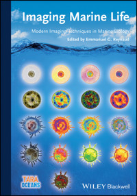
Plankton organisms (diatoms (rows 1–2), hydroid (row 3) and radiolarian (row 4) can be imaged using different techniques and different image processing schemes. The cover shows four different organisms: Planktoniella sol (Schütt, 1892; Marquesas Archipelago, Tara Oceans), Coscinodiscus sp. (Azores islands, Tara Oceans), Porpita porpita (Linnaeus, 1758; North Pacific Ocean; Luis Gutierrez-Heredia, Tara Oceans) and a radiolarian (Actinomma sp.; Mediterranean Sea, Tara Oceans). All original colour images were converted to 8-bit then processed using different Look up Tables (LUT) to highlight specific characteristics using NIH ImageJ. Background underwater scene © adimas, fotolia.com
Limit of Liability/Disclaimer of Warranty: While the publisher and author have used their best efforts in preparing this book, they make no representations or warranties with respect to the accuracy or completeness of the contents of this book and specifically disclaim any implied warranties of merchantability or fitness for a particular purpose. No warranty can be created or extended by sales representatives or written sales materials. The Advice and strategies contained herein may not be suitable for your situation. You should consult with a professional where appropriate. Neither the publisher nor authors shall be liable for any loss of profit or any other commercial damages, including but not limited to special, incidental, consequential, or other damages.
Library of Congress Card No.: applied for
British Library Cataloguing-in-Publication Data
A catalogue record for this book is available from the British Library.
Bibliographic information published by the Deutsche Nationalbibliothek
The Deutsche Nationalbibliothek lists this publication in the Deutsche Nationalbibliografie; detailed bibliographic data are available on the Internet at <http://dnb.d-nb.de>.
© 2014 Wiley-VCH Verlag GmbH & Co. KGaA, Boschstr. 12, 69469 Weinheim, Germany
Wiley-Blackwell is an imprint of John Wiley & Sons, formed by the merger of Wiley's global Scientific, Technical, and Medical business with Blackwell Publishing.
All rights reserved (including those of translation into other languages). No part of this book may be reproduced in any form – by photoprinting, microfilm, or any other means – nor transmitted or translated into a machine language without written permission from the publishers. Registered names, trademarks, etc. used in this book, even when not specifically marked as such, are not to be considered unprotected by law.
Print ISBN: 978-3-527-32744-7
ePDF ISBN: 978-3-527-66420-7
ePub ISBN: 978-3-527-67542-5
Mobi ISBN: 978-3-527-67543-2
oBook ISBN: 978-3-527-67541-8
Preface
The Earth, the Blue planet, is so called because its larger biotope is a vast and deep intertwined blue expanse of salted oceans and seas. The water is filled with billions of organisms which are mainly invisible to the naked eye. These life forms have allowed us to breathe and to conquer the lands. They are our ancestors, our stones, our oils, our food or the food of our food…and they may hold the key to our future in these troubled times of climate change.
The idea for this book germinated during an oceanographic survey in the Mediterranean Sea where, as a cell biologist coming out of a high ranked institute of fundamental research, I realized that many imaging techniques including 3D microscopy techniques were not easily available to marine biologists. I came back eager to share my knowledge with this large and amazing scientific community. Many of my friends responded positively and shared the burden of tracking any application of advanced imaging methods in marine biology and I would like to thank them all, in particular Renaud Boistel, who managed a huge crowd of co-authors to give us a wonderful chapter on X-ray related imaging techniques, Stephan Jericho and his colleagues for their patience, Gustaaf Hallegraeff who shared his long-standing experience of Electron Microscopy techniques applied to planktonic species, and finally my students who took some of the burden of collecting information and writing.
This book is not an encyclopaedia of all imaging techniques applied in marine biology. We made a choice and two main factors influenced our choices: three-dimension and promises.
First, water is always a three-dimensional medium that governs marine organisms: their shapes and forms but also, surely, their physiology. And so there is a crucial need to obtain three-dimensional views as this opens ways to deeper approaches: taxonomy, cell biology and in vivo biochemistry, fields well established in the medical world. Secondly, because there are so many imaging techniques out there, we had to make choices for this first edition, so we decided to drop the well-established techniques: light microscopy, cytometry…and promote less appreciated techniques or emerging ones that often give us a better three-dimensional view of the creatures of the oceans (Optical Projection Tomography, Light Sheet Microscopy etc.) and we made a big effort to make them available. We believe that these techniques can be highly beneficial to the marine biology community.
However, we provide some basic skills to enable the reader to photograph larger organisms (Chapter IX) and a historical perspective of ocean imagery (Chapter I) as a reminder of where we are coming from to enlighten the path of where we go…
This book is an introduction to marine biology imaging methods suitable not only for students wishing to pursue marine biology but also for established researchers eager to extend their knowledge of imaging methods that may improve their current or future research.
This first book, specifically-designed for the marine biology field, is work in progress. We hope you will find it informative and your feedback is welcome to improve our common knowledge.Swim long and image well!Emmanuel G. Reynaud
Kahi Kai (‘one sea’ in Hawaiian) is a project born in Hawai'i. Kahi Kai stands for a unique ocean representing all oceans of our planet, which contain an amazing and mostly unknown biodiversity. The mission of Kahi Kai is to raise awareness about our ocean legacy and to appeal to our shared responsibility to preserve the fascinating, mysterious, and highly endangered marine world (www.kahikai.org).
Some of the sea creature portraits featured as chapter openers were taken during the Tara Oceans expedition and are currently part of an itinerant exhibition that can be seen in 2013/2014 in French cultural centers and embassies throughout the world.
List of Contributors
Chapter 1
Under the Eye of Neptune: An Historical Perspective of Marine Creature Imagery
Emmanuel G. Reynaud
Abstract
The ocean may have been filled by meteorites bringing water to Earth but its creatures were drawn by men. Sea shells were eaten in caves, dolphins drawn on cave walls. For a long time, though, its surface was the only visible thing, a glittering mirror for the human soul where Gods and Monsters fought over submerged empires, pulling down Atlantis and raising volcanic spires with thunder and smoke. Once the smoke had cleared and man learned to fish then dive, the marvels of the sea appeared and glittered even thousands of meters deep, revealing a magnificent world beyond our imagination. From the jellyfish's ballet to intricate glass houses of radiolarians, this introductory chapter presents a concise history of how man images the invisible oceanic world. Copyright © 2013 John Wiley & Sons, Ltd.
1.1 Introduction
Black and dark was the Universe. Then, the Big Bang lit everything, making energy flow, and amongst the billions of particles thrown across space was our planet, a tiny ball of orange fire. No blue, no green, no life. With time and changes, perhaps meteorite showers, water appeared on Earth and built up the marine world, or at least a place like an ocean, 4.6 billion years ago.
Many theories have been proposed, discussed, discarded, and buried about the origin of life; often the ocean or marine environment is a component of the magical mix that gave rise to the chance for life on our planet. Cells appeared, close to deep sea volcanoes, or perhaps it was in rock pools; in a shallow bay stromatolites used the blazing sun's energy to create food sources and deposits that mark the origin and began the continents. Life expanded and thrived in these early oceans but there was no one to record the creatures that inhabited it. Only bones and stones give us a glimpse of these creatures and for many millions of years there were no eyes to see them, and no arm and digits to draw them or to build cameras to picture this massive world up to 12 km deep and spanning 70% of our Earth, from the frozen lands of Antartica to blooming coral reefs.
Lesen Sie weiter in der vollständigen Ausgabe!
Lesen Sie weiter in der vollständigen Ausgabe!
Lesen Sie weiter in der vollständigen Ausgabe!
Lesen Sie weiter in der vollständigen Ausgabe!
Lesen Sie weiter in der vollständigen Ausgabe!
Lesen Sie weiter in der vollständigen Ausgabe!
Lesen Sie weiter in der vollständigen Ausgabe!
Lesen Sie weiter in der vollständigen Ausgabe!
Lesen Sie weiter in der vollständigen Ausgabe!
Lesen Sie weiter in der vollständigen Ausgabe!


