Melanins and Melanosomes E-Book
142,99 €
Mehr erfahren.
- Herausgeber: Wiley-VCH
- Kategorie: Wissenschaft und neue Technologien
- Sprache: Englisch
The surface pigmentation of vertebrates is controlled by specialized cells able to synthesize a variety of pigments collectively known as melanins. Recent research has shown that melanins are produced not only in the skin but also in many other sites such as the eye, inner ear, muscles, etc., - where they are engaged in some unanticipated roles. The details of the synthetic pathway, the complexities of its regulation and biological significance that have been unravelled in recent research comprise a fascinating story and are of key importance in understanding the nature of diseases, including malignant melanoma one of the most rapidly spreading cancers.
Sie lesen das E-Book in den Legimi-Apps auf:
Seitenzahl: 832
Veröffentlichungsjahr: 2011
Ähnliche


Table of Contents
Cover
Related Titles
Title page
Copyright page
Dedication
Preface
List of Contributors
1 History of Melanosome Research
1.1 Introduction
1.2 Melanosome Research in the Pre-Seiji Era
1.3 Melanosome Research in the Seiji Era
1.4 Melanosome Research in the Post-Seiji Era
1.5 Other Historical Aspects
Acknowledgments
2 Classical and Nonclassical Melanocytes in Vertebrates
2.1 Definition of Melanogenic Cells
2.2 Distribution and Function of Melanogenic Cells
2.3 Embryonic Development of Melanogenic Cells
2.4 Transfer of Melanin from Classical and Nonclassical Melanocytes
3 Biological Chemistry of o-Quinones
3.1 General Biological Significance of o-Quinones
3.2 o-Quinone Reactivity
3.3 Role of o-Quinones in Melanogenesis
4 Biosynthesis of Melanins
4.1 Introduction
4.2 Raper–Mason Pathway
4.3 Structural and Functional Properties of the Melanogenic Enzymes
4.4 Regulation of the Melanogenic Pathway
4.5 Conclusions and Perspectives
Acknowledgments
5 Inhibitors and Enhancers of Melanogenesis
5.1 Introduction
5.2 Depigmenting Agents
5.3 Enhancers of Melanogenesis
6 Structure of Melanins
6.1 Introduction
6.2 Classification and General Properties of Melanins
6.3 Biosynthetic Studies
6.4 Degradative Studies
6.5 Analysis of Eumelanins and Pheomelanins
6.6 Conclusions
7 Properties and Functions of Ocular Melanins and Melanosomes
7.1 Introduction
7.2 Biogenesis of Ocular Melanosomes and Melanogenesis
7.3 Melanin Content in Pigmented Structures of the Eye
7.4 Structure of Ocular Melanosomes
7.5 Role of Ocular Melanin as a Broadband Optical Filter
7.6 Antioxidant Properties of Ocular Melanin
7.7 Pro-Oxidant Properties of Ocular Melanosomes
7.8 Other Properties of Ocular Melanosomes and Their Implications
7.9 Conclusions
8 Biological Role of Neuromelanin in the Human Brain and Its Importance in Parkinson’s Disease
8.1 What are Neuromelanins?
8.2 Phylogenetic Development of Neuromelanin
8.3 Development and Metabolism of Neuromelanin
8.4 Structure of Neuromelanin
8.5 Biological Role of Neuromelanin in the Human Brain
8.6 Is Neuromelanin Involved in Neurological Disease?
8.7 Effects of Neuromelanin In Vitro and In Vivo
8.8 Conclusions
Acknowledgments
9 Biogenesis of Melanosomes
9.1 Introduction
9.2 Melanosomes: Intracellular Organelles Specialized in Melanin Synthesis
9.3 Endocytic System and Formation of Melanosomes
9.4 Melanosome Maturation: Cargo Sorting to Mature Melanosomes
9.5 Conclusions
Acknowledgments
10 Transport and Distribution of Melanosomes
10.1 Introduction
10.2 Model Systems to Study Pigment Transport
10.3 Intracellular Melanosome Transport
10.4 Melanosome Motility in RPE: The Rab27a–Myrip–Myo7a Tripartite Complex
10.5 Melanosome Transfer
10.6 Fate of Melanin in the Keratinocyte
10.7 Conclusions
Acknowledgments
11 Genetics of Melanosome Structure and Function
11.1 Introduction
11.2 Genes Involved in Melanoblast Development, Migration, and Specification
11.3 Genes Involved in Melanocyte Differentiation, Survival, and Proliferation
11.4 Genes Involved in Regulating Melanocyte Function
11.5 Genes Involved in the Biogenesis of Melanosomes and Other Lysosome-Related Organelles
11.6 Genes Involved in Melanin Production
11.7 Genes Involved in Melanosome Movement, Transfer, and Distribution
11.8 Conclusions
12 Physiological and Pathological Functions of Melanosomes
12.1 Tissue Concentration of Melanosomes
12.2 Melanosome Properties and Functions Are Determined by Their Chemical Composition
12.3 Functional Microanatomy of the Melanosome
12.4 Melanosomes as Centers of Free Radical Activity
12.5 Melanosomes as Energy Transducers
12.6 Melanosomes and Metal Ions
12.7 Affinity of Melanosomes for Polycyclic and Other Compounds
12.8 Exploitation of Melanosomal Proteins and Melanin as Specific Targets in Melanoma Therapy
12.9 Conclusions
Acknowledgments
13 Dysplastic Nevi as Precursor Melanoma Lesions
13.1 Nevi as Risk Factors for Melanoma
13.2 Dysplastic Nevi as Precursor Lesions of Melanoma
13.3 Cytological Differences between Normal Skin Melanocytes and Dysplastic Nevus Cells: Melanosomal and Mitochondrial Aberrations
13.4 Metabolic Differences between Normal Skin Melanocytes and Dysplastic Nevus Cells: Preference for Pheomelanogenesis in Dysplastic Nevus Cells
13.5 Pheomelanogenesis as a Possible Cause of Intracellular Oxidative Imbalance
13.6 Dysplastic Nevus Cells as Senescent Cells
13.7 Are Dysplastic Nevus Cells a Class of Cells Exhibiting a Mutator Phenotype?
Index
Related Titles
Sobin, L. H., Gospodarowicz, M. K., Wittekind, C. (eds.)
TNM Classification of Malignant Tumours
2009
ISBN: 978-1-4443-3241-4
Rajpar, S., Marsden, J.
ABC of Skin Cancer
2008
ISBN: 978-1-4051-6219-7
Schwartz, R. A.
Skin Cancer
2008
ISBN: 978-1-4051-5961-6
Nordlund, J. J., Boissy, R. E., Hearing, V. J., King, R. A., Oetting, W. S., Ortonne, J.-P. (eds.)
The Pigmentary System
Physiology and Pathophysiology
2006
ISBN: 978-1-4051-2034-0
The Editors
Prof. Patrick A. Riley
Em. Prof. of Cell Pathology
University College London
Grange Avenue
London N20 8AB
United Kingdom
Prof. Jan Borovanský
Charles University
1st Faculty of Medicine
Institute of Biochemistry and Experimental Oncology
U Nemocnice 5
128 53 Praha 2
Czech Republic
Cover
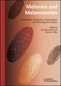
The cover design is intended to reflect the two principal elements of the book: melanins and melanosomes. The melanins are represented by the graduated background pigment spanning the range of colours from essentially black eumelanin, through reddish to paler shades, representing pheomelanin. The celluar organelles responsible for the production, containment and distribution of melanins, the melanosomes, are represented by symbolic images based on their electron microscopic appearance.
Limit of Liability/Disclaimer of Warranty: While the publisher and authors have used their best efforts in preparing this book, they make no representations or warranties with respect to the accuracy or completeness of the contents of this book and specifically disclaim any implied warranties of merchantability or fitness for a particular purpose. No warranty can be created or extended by sales representatives or written sales materials. The Advice and strategies contained herein may not be suitable for your situation. You should consult with a professional where appropriate. Neither the publisher nor authors shall be liable for any loss of profit or any other commercial damages, including but not limited to special, incidental, consequential, or other damages.
Library of Congress Card No.: applied for
British Library Cataloguing-in-Publication Data
A catalogue record for this book is available from the British Library.
Bibliographic information published by the Deutsche Nationalbibliothek
The Deutsche Nationalbibliothek lists this publication in the Deutsche Nationalbibliografie; detailed bibliographic data are available on the Internet at <http://dnb.d-nb.de>.
© 2011 Wiley-VCH Verlag & Co. KGaA, Boschstr. 12, 69469 Weinheim, Germany
Wiley-Blackwell is an imprint of John Wiley & Sons, formed by the merger of Wiley’s global Scientific, Technical, and Medical business with Blackwell Publishing.
All rights reserved (including those of translation into other languages). No part of this book may be reproduced in any form – by photoprinting, microfilm, or any other means – nor transmitted or translated into a machine language without written permission from the publishers. Registered names, trademarks, etc. used in this book, even when not specifically marked as such, are not to be considered unprotected by law.
ISBN: 978-3-527-32892-5
ISBN oBook: 978-3-527-63615-0
ISBN ePDF: 978-3-527-63614-3
ISBN ePub: 978-3-527-64455-1
ISBN mobi: 978-3-527-64454-4
Dedication
“All nature is but art, unknown to thee;
All chance, direction which thou canst not see”
Alexander Pope: An Essay on Man
Were we to ascribe to chance the existence of this volume we should have to begin with the moment in July 1952 when Professor A.F. Richter, Head of the Second Institute of Medical Chemistry at Charles University in Prague, opened a dust-covered cabinet from which he took, apparently at random, a bottle containing a dark powder and handed it to a young assistant with the words: “Young man, study the contents of this flask.” The assistant was Jiri Duchon and the label on the flask read: “Human melanosarcoma, prepared by H. Waelsch.”
Jiri Duchon was born on 27 July 1927, the only son of an eminent scientist. On graduating in medicine in 1952 he joined Richter’s laboratory and his careful analysis of the sample given to him set him upon the course of studies that were to occupy him for the rest of his life. He defended his PhD thesis in 1962 on the topic of “Urinary melanogens in melanoma” and he subsequently made many important contributions to quantitative analysis of the products of melanogenesis. In recognition of his early work, Jiri Duchon was awarded a Roosevelt Fellowship that enabled him to spend 15 months at Harvard in the laboratory of T.B. Fitzpatrick. This was in 1967–1968 when he met and established a friendship with Makoto Seiji who had just developed the methods for melanosome isolation. On his return to Prague, Jiri Duchon set about improving the isolation technique and analyzing these newly discovered organelles. Under his direction and inspiration the Prague laboratory became the leading European center for the detailed biochemical investigation of melanosomes. Jiri Duchon was Head of the Institute for 26 years and many of his collaborators have continued to contribute significantly to the field of study that he promoted.
Professor Duchon was an internationally recognized and highly respected member of the pigment cell fraternity, and was elected an Honorary Member of the European Society for Pigment Cell Research in 1998. It was partly in his honor that the scientific session on “Melanin and Melanosomes” was arranged at the Federation of European Biochemical Societies (FEBS) Congress in 2009, but tragically he was taken ill on the very day of the Symposium. He was full of encouragement for the project that grew out of the meeting, namely that of publishing a definitive volume devoted to the subject of his academic endeavors, but died on 2 November 2009, long before it was completed.
In recognition of his seminal role in the events that led to the production of this book we dedicate this volume to Jiri Duchon with affectionate remembrance of a fine scientist, an inspirational teacher, a kindly and cultivated companion, and a true friend.
Professor Jiri Duchon MD, PhD, DrSc (1927–2009)
(Photograph by K. Meister)
Preface
“To that small part of ignorance that we arrange and classify we give the name knowledge”
Ambrose Bierce
This book is entitled Melanin and Melanosomes, and is about pigment and pigmentation. It is important, however, that we bear in mind that, while the primary function of melanocytes is the production of pigment in melanosomes, these cells have other attributes and perform other significant functions. Some of these are well recognized, such as the involvement of the retinal pigment epithelium in photoreceptor physiology (detailed in Chapter 7). Another interesting possibility is that melanogenesis may be the source of some of the substrate for dopamine synthesis [1], and melanocytes may have other important neuroendocrine functions as pro-propriomelanocortin processing cells and a source of prostaglandin D synthase (reviewed by Takeda et al. [2]). Some of these actions may go some way to explaining the remarkable anatomical distribution of melanocytes, often in locations that are not illuminated, such as the leptomeninges.
However, this volume is devoted to melanin and melanosomes, and is primarily concerned with vertebrate, especially human, pigmentation. We include melanin that is formed by oxidative processes that are enzymatically catalyzed in specialized cells, both the neural crest-derived dendritic melanocytes (named “classical” melanocytes in Chapter 2) and optic cup-derived retinal pigment epithelial cells (“nonclassical” melanocytes), as well as melanin generated by other oxidative pathways, such as the neuromelanin of the midbrain. The importance of this latter pigment, particularly in relation to Parkinson’s disease, is set out in Chapter 8.
The enzymatically generated melanin in vertebrates is synthesized and deposited in specialized intracellular organelles, the melanosomes, and this book concentrates on the many aspects of the formation and functions of these organelles.
The melanosome is a highly specialized organelle, the history of which owes much to the early work at Charles University under the aegis of Jiri Duchon (1927–2009), to whom this book is dedicated.
Many of the important properties of melanosomes were established in Prague in the early 1970s by a series of investigations on isolated and purified preparations of this organelle, and investigators at Charles University have continued to contribute significantly to the advancement of this field, and to follow the developments that have taken place in elucidating the structure of melanosomes and the complex biological roles in which they are implicated.
This volume grew from a combination of auspicious factors. In 2009, the 34th Congress of the Federation of European Biochemical Societies (FEBS) was organized in Prague. Naturally, with one of us (J.B.) on the Organizing Committee, one of the scientific sessions was devoted to melanosomes and, in the wake of the discussions at this meeting, it was felt that there was a significant body of new data relating to melanosomes that could usefully be assembled in a volume devoted exclusively to this organelle.
It was hoped that such an overview, by integrating the diverse aspects of current knowledge, might help to generate a new understanding of the biological role of melanosomes and stimulate novel research effort in this interesting area of study.
We have been fortunate in our publisher, Wiley-VCH, who recognized the timely nature of the proposed volume, and we thank our commissioning editor, Gregor Cicchetti, and his team, especially Anne Chassin du Guerny, for their help and encouragement in bringing the project to fruition.
Of course, our main thanks go to our panel of distinguished international contributors who have generously given of their time and expertise in preparing the chapters that we hope form a coherent picture of the up-to-date knowledge in the field.
Last, but not least, this book celebrates a long and fruitful collaboration between the Editors involving many visits between Charles University and University College London. It is a pleasure to acknowledge the assistance of the British Council in enabling these exchanges.
We had hoped initially to have the opportunity to arrange the order of the chapters in the light of their ultimate content so that overlapping areas were most rationally ordered to enable the volume to be read more or less in sequence while allowing the, perforce abundant, cross-references to act as a secondary web in a cohesive network. However, pressure of time prevented us from completing this task and readers may find it more convenient to skip between the various contributions according to their interests and predilections. In principle, although the topics are inextricably intertwined, we have elected to place the contributions devoted to melanin – its biosynthesis, chemistry, and properties – at the front of the book, and those dealing with melanosomes – their structure, biogenesis, distribution, and properties – in the following chapters.
The topic is put into chronological context by a historical Introduction in Chapter 1, in which Jan Borovanský traces the steps in the discovery of the melanosome, illustrated by portraits of the important investigators that took part in these exciting early studies.
As this book is directed largely at aspects of human pigmentation, Chapter 2 consists of a detailed overview by Sophie Colombo, Irina Berlin, Véronique Delmas, and Lionel Larue of the specialized cells in vertebrates in which melanin production in melanosomes takes place. In their contribution a distinction is made between “classical” and “nonclassical” melanocytes. Chapter 3, by Patrick Riley, Christopher Ramsden, and Edward Land, emphasizes the central role of the generation and reactivity of o-quinones in melanogenesis, and is followed by Chapter 4 in which the biosynthesis of melanins is reviewed by José Carlos Garcia-Borrón and Conchita Olivares Sánchez. Chapter 5, by Alain Taïeb, Muriel Cario-André, Stefania Briganti, and Mauro Picardo, comprises an analysis of inhibitors and enhancers of melanogenesis. The current understanding of the structure of melanins is then reviewed in Chapter 6 by Shosuke Ito, Kasumasa Wakamatsu, Marco d’Ischia, Alessandra Napolitano, and Alessandro Pezzella, and this is followed in Chapter 7 by a description of the properties and functions of ocular melanins and melanosomes by Małgorzata Róanowska. Chapter 8, by Kay Double, Wakako Maruyama, Makako Naoi, Manfred Gerlach, and Peter Riederer, is devoted to the biological role of neuromelanin in the human brain and its importance in Parkinson’s disease. Chapter 9 consists of a detailed review of the biogenesis of melanosomes by Cédric Delevoye, Francesca Giordano, Michael Marks, and Graça Raposo. This is followed in Chapter 10, by Mireille Van Gele and Jo Lambert, by a description of the transport and distribution of melanosomes. The genetics of melanosome structure and function are skillfully summarized in Chapter 11 by Vincent Hearing. Chapter 12, by Jan Borovanský and Patrick Riley, is devoted to the properties and functions of melanosomes, and, in Chapter 13, the abnormalities of melanosomes and melanogenesis in melanoma precursor lesions are discussed by Stan Pavel, Nico Smit, and Karel Pizinger.
We firmly believe that this compilation of expertise embodies a significant work of scholarship, and we sincerely hope that the combined wisdom embraced by this volume conveys both the breadth of detailed and exciting knowledge that currently exists about melanin and melanosomes, and also reveals those shadowed areas of doubt and ignorance that await illumination in the future.
Patrick A. Riley
Jan Borovanský
March 2011
References
1 Eisenhofer, G., Tian, H., Holmes, C., Matsunaga, J., Roffler-Tarlov, S., and Hearing, V.J. (2003) Tyrosinase: a developmentally specific major determinant of peripheral dopamine. FASEB J., 17, 1248–1255.
2 Takeda, K., Takahashi, N.-H., and Shibahara, S. (2007) Neuroendocrine functions of melanocytes: beyond the skin-deep melanin maker. Tohoku J. Exp. Med., 211, 201–221.
List of Contributors
Irina Berlin
Institut Curie
Developmental Genetics of Melanocytes, INSERM U1021–CNRS UMR3347
Centre Universitaire
91405 Orsay
France
Jan Borovanský
Charles University
First Faculty of Medicine, Institute of Biochemistry and Experimental Oncology
U nemocnice 5
128 53 Prague 2
Czech Republic
Stefania Briganti
San Gallicano Dermatological Institute
Laboratory of Cutaneous Physiopathology
Elio Chianesi 53
00144 Rome
Italy
Muriel Cario-André
Université de Bordeaux
INSERM U1035
146 rue Léo Saignat
33076 Bordeaux
France
Sophie Colombo
Institut Curie
Developmental Genetics of Melanocytes, INSERM U1021–CNRS UMR3347
Centre Universitaire
91405 Orsay
France
Marco d’Ischia
University of Naples “Federico II”
Department of Organic Chemistry and Biochemistry
Via Cinthia 4
80126 Naples
Italy
Cédric Delevoye
Institut Curie
Centre de Recherche
Structure and Membrane Compartments
26 Rue d’Ulm
75248 Paris cedex 05
France
Véronique Delmas
Institut Curie
Developmental Genetics of Melanocytes, INSERM U1021–CNRS UMR3347
Centre Universitaire
91405 Orsay
France
Kay L. Double
University of New South Wales
Neuroscience Research Australia
Barker Street
Sydney, NSW 2031
Australia
José Carlos García-Borrón
University of Murcia
Department of Biochemistry and Molecular Biology
Campus de Espinardo
30100 Murcia
Spain
Manfred Gerlach
University of Würzburg
Clinical Neurobiology, Department of Child and Adolescence Psychiatry, Psychosomatics and Psychotherapy
Füchsleinstrasse 15
97080 Würzburg
Germany
Francesca Giordano
Institut Curie
Centre de Recherche
Structure and Membrane Compartments
26 Rue d’Ulm
75248 Paris cedex 05
France
Vincent J. Hearing
National Institutes of Health
Laboratory of Cell Biology
37 Convent Drive
Bethesda, MD 20892
USA
Shosuke Ito
Fujita Health University School of Health Sciences
Department of Chemistry
Toyoake
Aichi 470-1192
Japan
Jo Lambert
Ghent University Hospital
Department of Dermatology
De Pintelaan 185
9000 Ghent
Belgium
Edward J. Land
Keele University
School of Physical and Geographical Sciences, Lennard-Jones Laboratories
Keele Road
Keele ST5 5BG
UK
Lionel Larue
Institut Curie
Developmental Genetics of Melanocytes, INSERM U1021–CNRS UMR3347
Centre Universitaire, 91405 Orsay
France
Michael S. Marks
University of Pennsylvania
Departments of Pathology and Laboratory Medicine and Physiology, 513 Stellar-Chance Laboratories
422 Curie Boulevard
Philadelphia, PA 19104-6100
USA
Wakako Maruyama
National Research Center for Geriatrics and Gerontology
Department of Cognitive Brain Science
Obu
Aichi 474-8511
Japan
Makoko Naoi
Gifu International Institute of Biotechnology
Department of Neurosciences
Kakamigahara
Gifu 504-0838
Japan
Alessandra Napolitano
University of Naples “Federico II”
Department of Organic Chemistry and Biochemistry
Via Cinthia 4
80126 Naples
Italy
M. Concepción Olivares Sánchez
University of Murcia
Department of Biochemistry and Molecular Biology
Campus de Espinardo
30100 Murcia
Spain
Stanislav Pavel
Leiden University Medical Center
Department of Dermatology
PO Box 9600
2300 RC Leiden
The Netherlands
and
Charles University
Department of Dermatology, Faculty of Medicine
Husova 3
306 05 Pilsen
Czech Republic
Alessandro Pezzella
University of Naples “Federico II”
Department of Organic Chemistry and Biochemistry
Via Cinthia 4
80126 Naples
Italy
Mauro Picardo
San Gallicano Dermatological Institute
Laboratory of Cutaneous Physiopathology
Elio Chianesi 53
00144 Rome
Italy
Karel Pizinger
Charles University
Department of Dermatology Faculty of Medicine
Husova 3
306 05 Pilsen
Czech Republic
Christopher A. Ramsden
Keele University
School of Physical and Geographical Sciences, Lennard-Jones Laboratories
Keele Road
Keele ST5 5BG
UK
Graça Raposo
Institut Curie
Centre de Recherche
Structure and Membrane Compartments
26 Rue d’Ulm
75248 Paris cedex 05
France
Peter Riederer
University of Würzburg
Clinical Neurochemistry, Department of Psychiatry, Psychosomatics and Psychotherapy, and National Parkinson Foundation Centers of Excellence for Neurodegenerative
Diseases Research
Füchsleinstrasse 15
97080 Würzburg
Germany
Patrick A. Riley
Totteridge Institute for Advanced Studies
The Grange
Grange Avenue
London N20 8AB
UK
Małgorzata Róanowska
Cardiff University
School of Optometry and Vision Science
Maindy Road
Cardiff CF24 4LU
UK
Nico P.M. Smit
Leiden University Medical Center
Central Laboratory for Clinical Chemistry
Albinusdreef 2
2333 AC Leiden
The Netherlands
Alain Taïeb
Hôpital St André
Department of Dermatology and Pediatric Dermatology
CHU de Bordeaux
1 rue Jean Burguet
33077 Bordeaux
France
Mireille Van Gele
Ghent University Hospital
Department of Dermatology
De Pintelaan 185
9000 Ghent
Belgium
Kazumasa Wakamatsu
Fujita Health University School of Health Sciences
Department of Chemistry
Toyoake
Aichi 470-1192
Japan
1
History of Melanosome Research
Jan Borovanský
1.1 Introduction
Melanosomes were first proposed as specific organelles, unique to pigment cells, in a preliminary publication that appeared on 30 July 1960 [1]. An announcement had been made at the 21st Annual Meeting of the Society for Investigative Dermatology, at Miami Beach, Florida, USA on 13 June 1960 [2] and the news, that the chemical composition and enzyme activities in melanosomes and mitochondria are completely different, was considered to be of such significance that it appeared in a newspaper report (Figure 1.1). Similar data, with an emphasis on terminology, were published in 1963 [3].
Figure 1.1 Announcement of the independent status of melanosome in Medical News on 5 July 1960.
This advance was the result of collaborative work between M. Seiji (1926–1982), at that time working at the Department of Dermatology, Harvard Medical School in Boston under the leadership of T.B. Fitzpatrick (1919–2003) (Figure 1.2), and H. Blaschko and M.S.C. Birbeck, with whom Dr Fitzpatrick established scientific cooperation during his tenure of a Commonwealth Fellowship at the Department of Biochemistry, Radcliffe Infirmary in Oxford.
Figure 1.2 Professor Makoto Seiji (left) and Professor Thomas B. Fitzpatrick (right) in 1972.
The history of melanosome research can be formally divided into three parts: (i) the pre-Seiji era (prior to 1960), (i) the Seiji era (1960–1982), and (iii) the post-Seiji era (1983–).
1.2 Melanosome Research in the Pre-Seiji Era
The first description of mammalian pigment cells was published by Gustav Simon in 1841 [4] who observed round and stellate pigment cells in the hair bulbs of pig embryos. It was preceeded in 1838 by Purkyn’s description of pigment in the cells of the substantia nigra, which not only drew attention to pigment granules, but also noted the rise in their numbers with age [5]. We have to admire these early reports because their authors, armed only with primitive light microscopes, were able to ascertain that melanin was not diffusely distributed in the cytoplasm of pigmented cells, but was present in the form of discrete aggregates [5, 6] (Figures 1.3 and 1.4).
Figure 1.3 “Chromatophore” from donkey conjuctiva [7].
Figure 1.4 Cells of substantia nigra containing neuromelanin [5].
Deciphering the old literature is problematical as authors often fail to distinguish between melanin (the pigment itself), melanoprotein (the natural melanin–protein complex), and melanin granules (the subcellular organelle). If the method of separation is not adequately described, it is difficult to be certain what material was studied and any conclusions can be misleading [8]. The lack of electron microscopic identification of isolated material led to many misinterpretations; for example, the “melanopseudoglobulin” studied by Greenstein et al. [9] was later shown to be melanosomes [10] and Bolt’s “melanoprotein” [11], widely used in biophysical studies, turned out to consist of damaged melanosomes [12]. Mason et al. [10] posed the question of whether melanin granules were particles with a specific structure or consisted of random aggregates of precipitated metabolic products. The introduction of electron microscopy was able to resolve this matter and Laxer et al. [13] were able to discern an inner ultrastructure in isolated melanosomes. The first clear pictures were obtained only in 1956 [14].
An avalanche of papers in subsequent years brought with it enormous amounts of information on the ultrastructure of melanosomes and its changes during melanosome development (good examples are [15–17]). Other papers (reviewed in [18]) brought together ultrastructural and biochemical data that, in combination, laid the basis for the nomenclature of melanosomal ontogenesis.
By comparison with the morphological data, biochemical investigations of melanosomes were more modest, mainly due to the fact that ultrastructural data were derived from studies of intact cells or tissues, whereas biochemical research used samples prepared by relatively harsh preparative procedures. These samples sometimes consisted of melanins, or altered melanosomes, or their fragments, usually without any check of their nature or homogeneity [18].
Lesen Sie weiter in der vollständigen Ausgabe!
Lesen Sie weiter in der vollständigen Ausgabe!
Lesen Sie weiter in der vollständigen Ausgabe!
Lesen Sie weiter in der vollständigen Ausgabe!
Lesen Sie weiter in der vollständigen Ausgabe!
Lesen Sie weiter in der vollständigen Ausgabe!
Lesen Sie weiter in der vollständigen Ausgabe!
Lesen Sie weiter in der vollständigen Ausgabe!
Lesen Sie weiter in der vollständigen Ausgabe!
Lesen Sie weiter in der vollständigen Ausgabe!
Lesen Sie weiter in der vollständigen Ausgabe!
Lesen Sie weiter in der vollständigen Ausgabe!
Lesen Sie weiter in der vollständigen Ausgabe!


