Microcirculation Imaging E-Book
129,99 €
Mehr erfahren.
- Herausgeber: Wiley-VCH
- Kategorie: Wissenschaft und neue Technologien
- Sprache: Englisch
Adopting a multidisciplinary approach with input from physicists, researchers and medical professionals, this is the first book to introduce many different technical approaches for the visualization of microcirculation, including laser Doppler and laser speckle, optical coherence tomography and photo-acoustic tomography. It covers everything from basic research to medical applications, providing the technical details while also outlining the respective strengths and weaknesses of each imaging technique.
Edited by an international team of top experts, this is the ultimate handbook for every clinician and researcher relying on microcirculation imaging.
Sie lesen das E-Book in den Legimi-Apps auf:
Seitenzahl: 711
Veröffentlichungsjahr: 2012
Ähnliche


Table of Contents
Related Titles
Title Page
Copyright
Preface
List of Contributors
Chapter 1: A Historical Perspective of Imaging of the Skin and Its Gradual Uptake for Clinical Studies, Inclusive of Personal Reminiscences of Early Days of Microcirculation Societies
1.1 Early History
1.2 The Microcirculatory Societies
1.3 A Tour of Microcirculatory Centers in 1968
1.4 TV Video Projection
1.5 The Third World Congress of Microcirculation
1.6 Perfusion Monitoring and the Advent of the Laser Doppler
1.7 3D and 4D Tomographic Methods
1.8 Nonoptical Microcirculation Imaging
1.9 Panel Discussions and International Convergence
References
Chapter 2: Sidestream Dark-Field (SDF) Video Microscopy for Clinical Imaging of the Microcirculation
2.1 Introduction
2.2 Quantifying the Functional State of the Microcirculation
2.3 Microcirculatory Image Acquisition
2.4 Automated Microcirculation Image Analysis
2.5 International Consensus on Microcirculation Image Acquisition and Analysis
2.6 Future Prospects
2.7 Conclusion
References
Chapter 3: Clinical Applications of SDF Videomicroscopy
3.1 Introduction
3.2 Microcirculatory Alterations Visualized with OPS/SDF Imaging
3.3 Response of Microcirculatory Variables to Therapeutic Interventions
3.4 Perspective
References
Chapter 4: Laser Doppler Flowmetry
4.1 Theory
4.2 Conventional Measures
4.3 Hardware Realizations
4.4 Monte Carlo and LDF
References
Chapter 5: Toward Assessment of Speed Distribution of Red Blood Cells in Microcirculation
5.1 Introduction
5.2 Theory of Laser Doppler Spectrum Decomposition
5.3 Validation of the Spectrum Decomposition Method
5.4 In Vivo Measurements of Speed Distribution of Red Blood Cells in the Microvascular Network
5.5 Estimation of Anisotropy Factor
5.6 Conclusions and Future Developments
5.7 Biography
References
Chapter 6: Fast Full-Field Laser Doppler Perfusion Imaging
6.1 Introduction
6.2 Fast Full-Field Laser Doppler Perfusion Imaging: Why and How?
6.3 Characteristics of a CMOS-Based Full-Field Laser Doppler Perfusion Imager
6.4 Overview of ffLDPI Systems and Obtained Results
6.5 Comparative Behavior of CMOS-Based Systems: How Good Are They to Measure Perfusion?
6.6 Concluding Remarks
References
Chapter 7: Speckle Effects in Laser Doppler Perfusion Imaging
7.1 Introduction
7.2 What Are Speckles? Basic Properties
7.3 Significance of Speckles in LDPI
7.4 Further Analysis of the Consequence of Speckle in LDPI
7.5 Consequences and Concluding Remarks
References
Chapter 8: Laser Speckle Contrast Analysis (LASCA) for Measuring Blood Flow
8.1 Introduction: Fundamentals of Laser Speckle
8.2 Time-Varying Speckle
8.3 Full-Field Speckle Methods
8.4 Single-Exposure Speckle Photography
8.5 Laser Speckle Contrast Analysis (LASCA)
8.6 The Question of Speckle Size
8.7 Theory
8.8 Practical Considerations
8.9 Applications and Examples
8.10 Recent Developments
8.11 Conclusions
8.12 Acknowledgments
References
Chapter 9: Tissue Viability Imaging
9.1 Introduction
9.2 Operating Principle
9.3 Validation
9.4 Technology
9.5 Evaluation
9.6 Comparison with Other Instrumentation
9.7 Other Technology for Polarization Imaging
9.8 Discussion
References
Chapter 10: Optical Microangiography: Theory and Application
10.1 Introduction
10.2 Optical Coherence Tomography
10.3 Optical Microangiography (OMAG)
10.4 Applications of OMAG
10.5 Summary
10.6 Acknowledgments
References
Chapter 11: Photoacoustic Tomography of Microcirculation
11.1 Introduction
11.2 PAT Systems
11.3 Microcirculation Parameters Quantified by PAT
11.4 Biomedical Applications
11.5 Discussion and Perspectives
11.6 Animal Ethics
11.7 Acknowledgments
References
Chapter 12: Fluorescence and OCT Imaging of Microcirculation in Early Mammalian Embryos
12.1 Mouse Embryo Manipulations for Live Imaging
12.2 Imaging Vascular Development and Microcirculation Using Confocal Microscopy of Vital Fluorescent Markers
12.3 Live Imaging of Mammalian Embryonic Development and Circulation with OCT
12.4 Summary
References
Chapter 13: High Frequency Ultrasound for the Visualization and Quantification of the Microcirculation
13.1 Introduction
13.2 Ultrasound Fundamentals
13.3 Instrumentation
13.4 Applications of Micro-Ultrasound in Studies of the Microcirculation
13.5 Conclusions
Acknowledgments
References
Chapter 14: Studying Microcirculation with Micro-CT
14.1 Introduction
14.2 Micro-Computed Tomography
14.3 Specimen/Animal Preparation
14.4 Image Analysis
14.5 Summary and Future Developments
Acknowledgments
References
Chapter 15: Imaging Blood Circulation Using Nuclear Magnetic Resonance
15.1 Introduction
15.2 MR-Angiography
15.3 Perfusion Imaging
15.4 Blood Oxygenation Level-Dependent (BOLD) Contrast
References
Index
Related Titles
Wolfbeis, O. S. (ed.)
Fluorescence Methods and Applications
Spectroscopy, Imaging, and Probes
2008
ISBN: 978-1-57331-716-0
Ntziachristos, V., Leroy-Willig, A., Tavitian, B. (eds.)
Textbook of in vivo Imaging in Vertebrates
2007
ISBN: 978-0-470-01528-5
de Leon, M. J., Snider, D. A., Federoff, H. (eds.)
Imaging and the Aging Brain
2007
ISBN: 978-1-57331-659-0
The Editor
Prof. Dr. Martin J. Leahy
Tissue Optics and Microcirculation Imaging Facility
National Biophotonics and Imaging Platform Ireland (NBIPI)
School of Physics
NUI Galway
Ireland
Cover
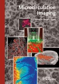
1. Live imaging of the developing vasculature in Tg(Flk1-myr::mCherry) X Tg(Flk1-H2B::EYFP) mice using confocal microscopy (for more details, see Fig. 2c in Chapter 12 by Irina V. Larina and Mary E. Dickinson). 2. The working principles of sidestream dark-field (SDF) imaging (for more details, see Fig. 2 in Chapter 2 by M.J. Milstein et al.). 3. Three-dimensional image of an experimental tumor (KHT) growing in a mouse (for more details, see Fig. 6 in Chapter 13 by Stuart Foster). 4. Blood flow changes during stroke: relative cerebral blood flow 10 min after occlusion of the middle cerebral artery in a rat (for more details, see Fig. 12 in Chapter 8 by Bryers et al., with kind permission by SPIE and A.K. Dunn, University of Texas). 5. Micro-CT image data (20 μm isotropic voxels) of the vascular bed of a rat heart, which was filled with a contrast agent (Microfil) and was imaged in situ. For more details, see Fig. 15 in Chapter 14 by Timothy L. Kline and Erik L. Ritman).
Limit of Liability/Disclaimer of Warranty: While the publisher and author have used their best efforts in preparing this book, they make no representations or warranties with respect to the accuracy or completeness of the contents of this book and specifically disclaim any implied warranties of merchantability or fitness for a particular purpose. No warranty can be created or extended by sales representatives or written sales materials. The Advice and strategies contained herein may not be suitable for your situation. You should consult with a professional where appropriate. Neither the publisher nor authors shall be liable for any loss of profit or any other commercial damages, including but not limited to special, incidental, consequential, or other damages.
Library of Congress Card No.: applied for
British Library Cataloguing-in-Publication Data
A catalogue record for this book is available from the British Library.
Bibliographic information published by the Deutsche Nationalbibliothek
The Deutsche Nationalbibliothek lists this publication in the Deutsche Nationalbibliografie; detailed bibliographic data are available on the Internet at <http://dnb.d-nb.de>.
© 2012 Wiley-VCH Verlag & Co. KGaA, Boschstr. 12, 69469 Weinheim, Germany
Wiley-Blackwell is an imprint of John Wiley & Sons, formed by the merger of Wiley's global Scientific, Technical, and Medical business with Blackwell Publishing.
All rights reserved (including those of translation into other languages). No part of this book may be reproduced in any form — by photoprinting, microfilm, or any other means — nor transmitted or translated into a machine language without written permission from the publishers. Registered names, trademarks, etc. used in this book, even when not specifically marked as such, are not to be considered unprotected by law.
Print ISBN: 978-3-527-32894-9
ePDF ISBN: 978-3-527-65122-1
ePub ISBN: 978-3-527-65121-4
mobi ISBN: 978-3-527-65120-7
oBook ISBN: 978-3-527-65123-8
Preface
This book brings together the main techniques used for imaging the small blood vessels, which supply nutritional oxygen and remove waste products from the cells of the body. The rapid development of new techniques to image the microcirculation (vessels <100 µm in diameter) in two and three dimensions was the main driver for publishing this book. The macrocirculation of the cardiovascular system enjoys a special place in medicine and is well catered for with imaging modalities that are ever present in our hospitals. X-ray CT, ultrasound, MRI, and PET all play an important role in diagnosis and treatment of the disorders of the large vessels. However, there is growing realization that some of the diseases that most threaten the quality and quantity of life in the developed world, such as diabetes and cancer, have their origins in the microcirculation. Therefore, new techniques with appropriate resolution were required to image these smaller vessels, and these largely depend on the rapid developments in photonics.
It is impossible to present all techniques that have been applied to microcirculation in this book. The editor is grateful to the (unknown) reviewers of the original proposal for their suggestion to supplement the biophotonics techniques well known to him with MRI and high-frequency ultrasound. The result is a more thorough covering of the field, although I am open to further suggestions for additions in future editions. Researchers, practitioners, and professionals in the fields of diabetes, cancer, wound healing, biomedical optics, and biophotonics, as well as professionals in other disciplines, such as laser physics and technology, fibre optics, spectroscopy, and biology, will find the book a useful resource. Graduate and undergraduate students studying biomedical physics and engineering, biomedical optics and biophotonics, and medical science would benefit greatly from consulting this reference.
Several Irish and international grants supported this project, particularly the National Biophotonics & Imaging Platform Ireland, funded by the Irish Government under the national development plan (NDP) 2007–2013 HEA PRTLI IV. I greatly appreciate the cooperation, contributions, patience, and support of all the contributors, my colleagues from the School of Physics at NUI Galway, the Department of Physics at the University of Limerick, the Royal College of Surgeons, and the National Biophotonics and Imaging Platform. Last, but not least, I would like to thank my family for their support and understanding during my work on this book.
NUI, Galway
March 2012
Martin J. Leahy
List of Contributors
1
A Historical Perspective of Imaging of the Skin and Its Gradual Uptake for Clinical Studies, Inclusive of Personal Reminiscences of Early Days of Microcirculation Societies
Terence J. Ryan and Martin J. Leahy
Modern microscopy of microcirculation was conceived by the observers of especially the seventeenth century, but gestation was in the hands of the national and continental Microcirculatory Societies that began in the decade 1954–1964. They were a meeting point between the laboratory investigator of microcirculation and the clinician. Zweifach [1] reviewing historical aspects of microcirculation research wrote “The usage of the term ‘microcirculation’ in an organic context is of comparatively recent vintage, first appearing consistently in the literature during the 1950s.” Global collaboration with a strong clinical input began with World Congresses of Microcirculation first held in Toronto in 1975, then in San Diego in 1979, next in Oxford in 1984, and every four to five years since then. As an Oxford clinician specializing in care of the skin, T. J. R. was roped in at the deep end of nonclinical pursuit of microcirculation on several occasions. However, this began by a chance observation that if one pushes tissue fluid away by indenting the skin with a steel probe, one gets much better visualization of the skin capillary bed [2, 3]. It has long been known that one sees at the surface of the skin only what its optical properties allow. Excised epidermis from white skin placed over a printed page is transparent enough to read through it. Melanin of pigmented skin prevents such visualization. The redness of blood provides in vivo pinkness, but blue blood in veins is a consequence of blue light being scattered more than red. This does not stop the practised clinician from easily recognizing the condition when black skin is flushed. Newton [4] discussed the decomposition of white light, and Doppler [5] made known that the effect of movement toward or away from the observer influenced the color observed. For several centuries, any observation of complex surfaces reflecting light was clarified by applying transparent oils to that surface.
1.1 Early History
George P. Fulton [6], Professor of Biology at the Boston University, writing on the historical perspective of the founding of the American Microcirculatory Society, lists Harvey and Lord Lister amongt the early influences on microcirculation, but it was interest in microscopy and improved microscopes in the seventeenth century that led to the first observations of blood flow by better imaging. The discoverers and forerunners of imaging were fascinated when they applied their new magnifying devices, and detecting transparency in some living tissues saw for the first time the movement of the content of the small capillaries. These early observations especially on red cells were well reviewed for the journal Blood Cells by Bessis and Delpechi [7]. As stated in that review, three men, Malpighi, Leeuwenhoek, and Swammerdam, made the most of the improvements in magnifying lenses in the early seventeenth century and noted red particles in blood capillaries in transparent tissues. Of these, van Leeuwenhoek of Delft (1632–1723) gained the most publicity by getting his observations published by the Royal Society of London [8]. Indeed, it was the tax collector, van Leeuwenhoek, who contributed one of the greatest innovations through his hobby by producing a very short focal length lens. This avoided chromatic aberrations, which plagued compound microscopes of the day, and yet produced sufficient magnification to reveal the structure of the blood cell (Figure 1.1) and its movement within organs and organisms if they were sufficiently transparent. It was this innovation that allowed Malpighi to confirm Harvey's theory that blood circulates from the arterial to the venous side via these small capillaries, and indeed, it can be considered the discovery of the microcirculation.
Figure 1.1 The discovery of the circulation of the blood, the shape of the red blood cell, and, most importantly, the microcirculation (, Charles Singer, G. Bell and Sons Ltd., London, Ref. [9]).
Lesen Sie weiter in der vollständigen Ausgabe!
Lesen Sie weiter in der vollständigen Ausgabe!
Lesen Sie weiter in der vollständigen Ausgabe!
Lesen Sie weiter in der vollständigen Ausgabe!
Lesen Sie weiter in der vollständigen Ausgabe!
Lesen Sie weiter in der vollständigen Ausgabe!
Lesen Sie weiter in der vollständigen Ausgabe!
Lesen Sie weiter in der vollständigen Ausgabe!
Lesen Sie weiter in der vollständigen Ausgabe!
Lesen Sie weiter in der vollständigen Ausgabe!
Lesen Sie weiter in der vollständigen Ausgabe!
Lesen Sie weiter in der vollständigen Ausgabe!
Lesen Sie weiter in der vollständigen Ausgabe!
Lesen Sie weiter in der vollständigen Ausgabe!
Lesen Sie weiter in der vollständigen Ausgabe!


