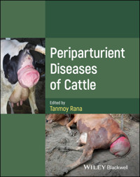
Periparturient Diseases of Cattle E-Book
153,99 €
Mehr erfahren.
- Herausgeber: John Wiley & Sons
- Kategorie: Fachliteratur
- Sprache: Englisch
Manage the health of cattle at a critical stage with this essential reference
Milk is one of the backbones of the global food economy, with its high vitamin content and key contribution to bone health. As a result, dairy farming is one of the most essential sectors of the global agricultural market, and the health of cattle is an issue of global importance. Periparturient diseases, those sustained in the period immediately before, during, and after giving birth, have a potentially devastating impact on the reproductive cycle of cattle, and an understanding of these conditions is a critical aspect of food production.
Periparturient Diseases of Cattle offers a comprehensive overview of these diseases, their pathogenesis, and their treatments. Summarizing all of the major periparturient disorders, their etiology, and their management, it is a critical resource for veterinary practitioners and others for whom cattle health is of fundamental importance. As a reference, a diagnostic aid, and a tool in farm management, this volume is indispensable.
Periparturient Diseases of Cattle readers will also find:
- In-depth description of disease advancement
- Detailed treatment of disorders including metritis, mastitis, ketosis, and many more
- Color figures and line drawings to illustrate key concepts
Periparturient Diseases of Cattle is ideal for student and working veterinarians, academicians, farm managers, industrialists, farm owners, and many more.
Sie lesen das E-Book in den Legimi-Apps auf:
Seitenzahl: 1269
Veröffentlichungsjahr: 2024
Ähnliche


Periparturient Diseases of Cattle
Edited by
Tanmoy Rana, M.V.Sc & PhD
Assistant Professor, Department of Veterinary Clinical ComplexWest Bengal University of Animal & Fishery SciencesKolkata, West Bengal, India
Copyright © 2025 by John Wiley & Sons, Inc. All rights reserved, including rights for text and data mining and training of artificial technologies or similar technologies.
Published by John Wiley & Sons, Inc., Hoboken, New Jersey.Published simultaneously in Canada.
No part of this publication may be reproduced, stored in a retrieval system, or transmitted in any form or by any means, electronic, mechanical, photocopying, recording, scanning, or otherwise, except as permitted under Section 107 or 108 of the 1976 United States Copyright Act, without either the prior written permission of the Publisher, or authorization through payment of the appropriate per‐copy fee to the Copyright Clearance Center, Inc., 222 Rosewood Drive, Danvers, MA 01923, (978) 750‐8400, fax (978) 750‐4470, or on the web at www.copyright.com. Requests to the Publisher for permission should be addressed to the Permissions Department, John Wiley & Sons, Inc., 111 River Street, Hoboken, NJ 07030, (201) 748‐6011, fax (201) 748‐6008, or online at http://www.wiley.com/go/permission.
Trademarks: Wiley and the Wiley logo are trademarks or registered trademarks of John Wiley & Sons, Inc. and/or its affiliates in the United States and other countries and may not be used without written permission. All other trademarks are the property of their respective owners. John Wiley & Sons, Inc. is not associated with any product or vendor mentioned in this book.
Limit of Liability/Disclaimer of Warranty: While the publisher and author have used their best efforts in preparing this book, they make no representations or warranties with respect to the accuracy or completeness of the contents of this book and specifically disclaim any implied warranties of merchantability or fitness for a particular purpose. No warranty may be created or extended by sales representatives or written sales materials. The advice and strategies contained herein may not be suitable for your situation. You should consult with a professional where appropriate. Further, readers should be aware that websites listed in this work may have changed or disappeared between when this work was written and when it is read. Neither the publisher nor authors shall be liable for any loss of profit or any other commercial damages, including but not limited to special, incidental, consequential, or other damages.
For general information on our other products and services or for technical support, please contact our Customer Care Department within the United States at (800) 762‐2974, outside the United States at (317) 572‐3993 or fax (317) 572‐4002.
Wiley also publishes its books in a variety of electronic formats. Some content that appears in print may not be available in electronic formats. For more information about Wiley products, visit our web site at www.wiley.com.
Library of Congress Cataloging‐in‐Publication DataNames: Rana, Tanmoy, editor.Title: Periparturient diseases of cattle / edited by Tanmoy Rana.Description: Hoboken, New Jersey : John Wiley & Sons, [2025] | Includes bibliographical references and index.Identifiers: LCCN 2024026582 (print) | LCCN 2024026583 (ebook) | ISBN 9781394203970 (hardback) | ISBN 9781394203987 (Adobe PDF) | ISBN 9781394203994 (epub)Subjects: MESH: Cattle Diseases | Pregnancy Complications–veterinary | Pregnancy, Animal–physiology | Parturition–physiology | CattleClassification: LCC SF961 (print) | LCC SF961 (ebook) | NLM SF 961 | DDC 636.2/0896–dc23/eng/20240719LC record available at https://lccn.loc.gov/2024026582LC ebook record available at https://lccn.loc.gov/2024026583
Cover Design: WileyCover Images: Courtesy of Tanmoy Rana
List of Contributors
Aakanksha Bhoker
Department of Veterinary Surgery and RadiologyBihar Veterinary CollegePatna, Bihar, India
Abdul Reshma
Department of Veterinary Gynaecology and ObstetricsVeterinary College and Research InstituteTamil Nadu Veterinary and Animal Sciences UniversitySalem, Tamil Nadu, India
Abhijeet Fernandes
Department of Veterinary Gynaecology and ObstetricsCollege of Veterinary Science, Rampura PhulGuru Angad Dev Veterinary and Animal Sciences UniversityBathinda, Punjab, India
Abhinav Suthar
Department of Veterinary MedicineCollege of Veterinary Science and Animal HusbandryKamdhenu UniversitySardarkrushinagar, Gujarat, India
Abhishek Bhardwaj
College of Veterinary ScienceGuru Angad Dev Veterinary and Animal Sciences UniversityLudhiana, Punjab, India
Abhishek Pathak
Apollo College of Veterinary MedicineRajasthan University of Veterinary and Animal SciencesBikaner, Jaipur, Rajasthan, India
Adeniyi Njideka
Department of Biological SciencesAlabama A&M UniversityNormal, Alabama, USA
Akhter Rasool
Department of Veterinary Gynaecology and ObstetricsMadras Veterinary CollegeChennai, Tamil Nadu, India
Allam Lukshiake
Veterinary MedicineAhmadu Bello UniversityZaria, Kaduna, Nigeria
Ambica Gadige
Department of Veterinary MedicineCollege of Veterinary Science, RajendranagarHyderabad, Telangana, India
Aminu M. Galadima
Usmanu Danfodiyo University SokotoSokoto, Nigeria
Amita Tiwari
Department of Veterinary MedicineCollege of Veterinary Science and Animal HusbandryNanaji Deshmukh Veterinary Science UniversityJabalpur, Madhya Pradesh, India
Amitava Roy
Livestock Farm ComplexWest Bengal University of Animal & Fishery SciencesKolkata, West Bengal, India
Ankesh Kumar
Veterinary Clinical ComplexBihar Veterinary CollegeBihar Animal Sciences UniversityPatna, Bihar, India
Anil Kumar
Department of Veterinary MedicineBihar Veterinary CollegePatna, Bihar, India
Ankit S. Prajapati
Department of Veterinary MedicineCollege of Veterinary Science and Animal HusbandryKamdhenu UniversitySardarkrushinagar, Gujarat, India
Annadurai Thangamani
Department of Veterinary Gynaecology and ObstetricsVeterinary College and Research InstituteTamil Nadu Veterinary and Animal Sciences UniversitySalem, Tamil Nadu, India
Apoorva Mishra
Department of Veterinary Surgery and RadiologyCollege of Veterinary Science and Animal HusbandryNanaji Deshmukh Veterinary Science UniversityJabalpur, Madhya Pradesh, India
Apra Shahi
Department of Veterinary Surgery and RadiologyCollege of Veterinary Science and Animal HusbandryNanaji Deshmukh Veterinary Science UniversityJabalpur, Madhya Pradesh, India
Arash Kheradmand
Department of Clinical SciencesSchool of Veterinary Medicine, Lorestan UniversityKhorram Abad, Lorestan, Iran
Arkaprabha Shee
Dhaanyaganga Krishi Vigyan Kendra, SargachiMurshidabad, West Bengal, India
Aruna Maramulla
Department of Veterinary Clinical ComplexCollege of Veterinary Science, MamnoorWarangal, Telangana, India
Arunmozhi Narayanaswamy
Veterinary Clinical Complex, Veterinary College andResearch Institute, OrathanaduTamil Nadu Veterinary and Animal Sciences UniversityThanjavur, Tamil Nadu, India
Ashok Ashwini
Department of Veterinary MedicineVeterinary CollegeBangalore, Karnataka, India
Ayyasamy Elango
Department of Veterinary Gynaecology and ObstetricsVeterinary College and Research InstituteTamil Nadu Veterinary and Animal Sciences UniversitySalem, Tamil Nadu, India
Babita Das
Department of Veterinary Surgery and RadiologyCollege of Veterinary Science and Animal HusbandryNanaji Deshmukh Veterinary Science UniversityJabalpur, Madhya Pradesh, India
Bagur K. Bhagya
Department of Veterinary Medicine, Veterinary CollegeBangalore, Karnataka, India
Bashir Maryam Darma
Veterinary Teaching HospitalFaculty of Veterinary MedicineBayero UniversityKano, Kano State, Nigeria
Bhavna Priya
Department of Veterinary GynaecologyBihar Veterinary CollegePatna, Bihar, India
Bhoopendra Singh
Department of Veterinary Gynaecology and ObstetricsCollege of Veterinary Science and Animal HusbandryAcharya Narendra Deva University of Agriculture and TechnologyAyodhya, Uttar Pradesh, India
Chhavi Gupta
Department of Veterinary Gynaecology and ObstetricsVeterinary College and Research Institute, TirunelveliTamil Nadu Veterinary and Animal Sciences UniversityTirunelveli, Tamil Nadu, India
Chiezey Ngozi Pauline
National Animal Production Research InstituteFaculty of Veterinary MedicineAhmadu Bello UniversityZaria, Kaduna, Nigeria
Chitra D. Chauhan
Department of Veterinary MedicineCollege of Veterinary Science and Animal HusbandryKamdhenu UniversitySardarkrushinagar, Gujarat, India
Deepak Ningwal
Department of Veterinary Gynaecology and ObstetricsCollege of Veterinary Science and Animal HusbandryNanaji Deshmukh Veterinary Science UniversityMadhya Pradesh, India
Gitesh Saini
Department of Veterinary Gynaecology and ObstetricsCollege of Veterinary Science, Rampura PhulGuru Angad Dev Veterinary and Animal Sciences University,Bathinda, Punjab, India
Gowdaiahnadoddi K. Chetan Kumar
Department of Veterinary MedicineVeterinary CollegeBangalore, Karnataka, India
Gurram S. Haritha
Department of Veterinary Clinical ComplexCollege of Veterinary Science (SVVU), GarividiVizianagaram District, Andhra Pradesh, India
Harshad H. Panchasara
Livestock Research StationCollege of Veterinary Science and Animal HusbandryKamdhenu UniversitySardarkrushinagar, Gujarat, India
Jitendra P. Singh
Department of Veterinary MedicineCollege of Veterinary Science and Animal HusbandryAcharya Narendra Deva University of Agriculture and TechnologyKumarganj, Ayodhya, Uttar Pradesh, India
Kabir Alam
Department of Veterinary Gynaecology and ObstetricsCollege of Veterinary Science and Animal HusbandryAcharya Narendra Deva University of Agriculture and TechnologyAyodhya, Uttar Pradesh, India
Kalyaan Udumalpet Sampathkumar
Department of Veterinary Gynaecology and ObstetricsMadras Veterinary CollegeTamil Nadu Veterinary and Animal Sciences UniversityChennai, Tamil Nadu, India
Kanika Tiwari
Department of Veterinary Pharmacy and ToxicologyCollege of Veterinary and Animal SciencesGovind Ballabh Pant University of Agriculture and TechnologyPantnagar, Uttarakhand, India
Manoj Kumar Ahirwar
Department of Veterinary Surgery and RadiologyCollege of Veterinary Science and Animal HusbandryNanaji Deshmukh Veterinary Science UniversityJabalpur, Madhya Pradesh, India
Mojetoluwa S. Afolabi
Department of Biological SciencesAlabama A&M UniversityNormal, Alabama, USA
Monica Gunasekar
Department of ClinicsMadras Veterinary CollegeChennai, Tamil Nadu, India
Morteza Taravat
Department of Clinical ScienceFaculty of Veterinary MedicineUniversity of TabrizTabriz, East Azerbaijan, Iran
Nasrul I. Shaikh
All Vet Diagnostic Solutions IndiaNew Delhi, Delhi, India
Nathaniel Ogunkunle
Department of Food and Animal ScienceAlabama A&M UniversityNormal, Alabama, USA
Naveen Swaroop Murikipudi
Department of Veterinary BiochemistrySri Venkateswara Veterinary UniversityTirupati, Andhrapradesh, India
Neeti Lakhani
Department of Animal NutritionCollege of Veterinary Science, Rampura PhulGuru Angad Dev Veterinary and Animal Sciences UniversityBathinda, Punjab, India
Pradeep Nag Beladakere Subramanyam
Department of Animal SciencesUniversity of MissouriColumbia, Missouri, USA
Prakash M. Chauhan
Department of Veterinary ClinicsCollege of Veterinary Science and Animal HusbandryKamdhenu UniversitySardarkrushinagar, Gujarat, India
Pramod Kumar
Department of Veterinary Physiology and BiochemistryCollege of Veterinary Science and Animal HusbandryAcharya Narendra Deva University of Agriculture andTechnology, Kumarganj, AyodhyaUttar Pradesh, India
Pranav Anjaria
College of Veterinary Science and Animal HusbandryKamdhenu UniversityAnand, Gujarat, India
Rajesh Kumar
Department of Veterinary Gynaecology and ObstetricsCollege of Veterinary Science and Animal HusbandryAcharya Narendra Deva University of Agriculture and TechnologyAyodhya, Uttar Pradesh, India
Rajesh Kumar
Department of Veterinary Surgery and RadiologyBihar Veterinary CollegePatna, Bihar, India
Ramasamy Rajkumar
Department of Veterinary Gynaecology and ObstetricsVeterinary College and Research InstituteTamil Nadu Veterinary and Animal Sciences UniversitySalem, Tamil Nadu, India
R.S. Sethi
College of Dairy Science and TechnologyGuru Angad Dev Veterinary and Animal Sciences UniversityLudhiana, Punjab, India
Randhir Singh
Department of Veterinary Surgery and RadiologyCollege of Veterinary Science and Animal HusbandryNanaji Deshmukh Veterinary Science UniversityJabalpur, Madhya Pradesh, India
Ravi Shankar Kumar Mandal
Department of Veterinary MedicineBihar Veterinary CollegeBihar Animal Sciences UniversityPatna, Bihar, India
Rayal S. Sagar
Department of Veterinary MedicineVeterinary CollegeBangalore, Karnataka, India
Reza Asadpour
Department of Clinical ScienceFaculty of Veterinary MedicineUniversity of TabrizTabriz, East Azerbaijan, Iran
Sabarinathan Akambaram
Veterinary Clinical ComplexVeterinary College and Research Institute, TirunelveliTamil Nadu Veterinary and Animal Sciences UniversityTirunelveli, Tamil Nadu, India
Sakhahari Daund Sushant
Department of Veterinary Gynaecology and ObstetricsCollege of Veterinary Science and Animal HusbandryAcharya Narendra Deva University of Agriculture and TechnologyAyodhya, Uttar Pradesh, India
Sakthivel Manokaran
Department of Veterinary Gynaecology and ObstetricsVeterinary College and Research InstituteTamil Nadu Veterinary and Animal Sciences UniversitySalem, Tamil Nadu, India
Samuel Uchenna Felix
National Animal Production Research InstituteFaculty of Veterinary MedicineAhmadu Bello UniversityZaria, Kaduna, Nigeria;Department of Food and Animal ScienceAlabama A&M UniversityNormal, Alabama, USA
Sanjeev Kumar
Department of Veterinary PathologyBihar Veterinary CollegePatna, Bihar, India
Sarath Thulasiraman
Department of Veterinary Gynaecology and ObstetricsVeterinary Clinical ComplexVeterinary College and Research InstituteTamil Nadu Veterinary and Animal Sciences UniversitySalem, Tamil Nadu, India
Sarita Devi
Department of Veterinary MedicineCollege of Veterinary Science and Animal HusbandryKamdhenu UniversitySardarkrushinagar, Gujarat, India
Satyavrat Singh
Department of Veterinary MedicineCollege of Veterinary Science and Animal HusbandryAcharya Narendra Deva University of Agriculture andTechnology, KumarganjAyodhya, Uttar Pradesh, India
Sengodan Raja
Department of Veterinary Gynecology and ObstetricsVeterinary College and Research Institute, OrathnaduTamil Nadu Veterinary and Animal Sciences UniversityThanjavur, Tamil Nadu, India
Shanmugasundaram Murugesan
Department of Veterinary Clinical MedicineVeterinary College and Research Institute, NamakkalTamil Nadu Veterinary and Animal Sciences UniversityNamakkal, Tamil Nadu, India
Shivangi Udainiya
Veterinary Polytechnic BhopalNanaji Deshmukh Veterinary Science UniversityBhopal, Madhya Pradesh, India
Shubhamitra Chaudhuri
Department of Veterinary Clinical ComplexWest Bengal University of Animal Fishery SciencesKolkata, West Bengal, India
Somu Yogeshpriya
Department of Veterinary MedicineVeterinary College and Research Institute, OrathanaduThanjavur, Tamil Nadu, India
Sonam Bhatt
Department of Veterinary MedicineBihar Veterinary College, Bihar Animal Sciences UniversityPatna, Bihar, India
Sowmiya Lakshmanan
Department of Veterinary Public Health and EpidemiologyVeterinary College and Research Institute, OrathanaduThanjavur, Tamil Nadu, India
Srujan Racharla
Department of Veterinary MedicineCollege of Veterinary and Animal ScienceGovind Ballabh Pant University of Agriculture andTechnology, Pantnagar, India
Subir Singh
Department of Medicine and Public HealthAgriculture and Forestry UniversityRampur, Nepal
Subramaniam Sivaraman
Department of ClinicsVeterinary College and Research Institute, NamakkalTamil Nadu Veterinary and Animal Sciences UniversityNamakkal, Tamil Nadu, India
Suman Biswas
Department of Avian ScienceWest Bengal University of Animal Fishery SciencesKolkata, West Bengal, India
Suman K. Singh
Department of Veterinary Surgery, MedicineEpidemiology and Public HealthPaklihawa CampusInstitute of Agriculture and Animal ScienceTribhuvan University, Kathmandu, Nepal
Suresh Kamineni
Veterinary Clinical MedicineSri Venkateswara Veterinary UniversityTirupati, Andhrapradesh, India
Suryakant S. Parikh
Veterinary Gynaecology and ObstetricsCattle Breeding FarmKamdhenu UniversityJunagadh, Gujarat, India
Tanmoy Rana
Department of Veterinary Clinical ComplexWest Bengal University of Animal & Fishery SciencesKolkata, West Bengal, India
Tapas K. Patbandha
Livestock Production and ManagementPolytechnic in Animal HusbandryCollege of Veterinary Science and Animal HusbandryKamdhenu UniversityJunagadh, Gujarat, India
Usmanu Danfodiyo
University SokotoSokoto, Nigeria
Varun Asediya
M.B. Veterinary CollegeRajasthan University of Veterinary and Animal SciencesBikaner, Dungarpur, Rajasthan, India
Vasantha Seshu Kumari Injarapu
Department of Veterinary PhysiologySri Venkateswara Veterinary UniversityTirupati, Andhrapradesh, India
Vivek Kumar Singh
Veterinary Clinical ComplexBihar Veterinary CollegeBihar Animal Sciences UniversityPatna, Bihar, India
Yakubu Mariam
Department of Food and Animal ScienceAlabama A&M UniversityNormal, Alabama, USA
Preface
With its huge annual milk production, dairy farming is one of the most important areas of the agricultural sector worldwide. A considerably large amount of milk production was procured from dairy cattle. Cattle farming is the most significant branch of the animal production industry as it produces a constant income. Milk has a considerably high nutrient content. It is enriched in key vitamins and minerals needed to keep the body healthy, such as those for bone health. Dairy is one of the most important sources of calcium that human beings have in their diet. A major challenge for dairy farmers, managers, and veterinarians is monitoring a dairy cow and its health during the periparturient period. Parturition is one of the most significant critical stages of the reproductive cycle and is a period with a significant death rate and the potential for severe debilitating injury to newborn calves. In contrast, the future efficiency of reproduction and milk yield can be affected during this time. Therefore, major efforts should be undertaken to minimize health problems that generally arise during the periparturient period. Keeping cows healthy is one of the most important steps in monitoring good fertility and an efficient milk yield. In addition, healthy cows produce more milk, rebreed sooner, and have lower culling rates than their unhealthy herd mates. The main value of this reference book is to describe disorders related to the periparturient period and the development of new prevention strategies. This book addresses the definition of metabolic diseases, metabolic disorders, production diseases, and transition cow diseases. This book aids in the pathogenesis of diseases, diagnosis, treatment, and management of periparturient disorders in cattle. This book enhances our understanding of the etiology and pathogenesis of periparturient diseases in transition cattle. The basic version is beneficial as a reference book for doctor of veterinary medicine (DVM) students as well as veterinary technicians, researchers, farm managers, and academicians in veterinary medicine and animal sciences to systematically develop their knowledge about these disorders. This book solves various aspects of periparturient disorders in the cow including evaluating the etiology and pathogenesis, diagnostic approach, and treatment of periparturient diseases. This book is especially intended for animal nutritionists, academics, industrialists, progressive farm owners, researchers, veterinarians, and DVM graduate students with a special interest in dairy cattle health, and management. The book is designed to be engaging by providing color figures, line figures, and tables. The book also provides useful information about the application of various biological systems involving genomics, transcriptomics, proteomics, and metabolomics for understanding the pathomechanisms and early diagnosis using specific biomarkers of periparturient disease in cattle. I hope that this book provides new paradigms to stimulate further research in clarifying the pathomechanisms and causality of periparturient diseases of cattle as well as developing new preventive strategies for reducing the incidence rates of these diseases in the future. I expect that the reader will find this book interesting, containing up‐to‐date information about periparturient diseases and will utilize this knowledge in their research and teaching the new generation.
Dr. Tanmoy Rana
Department of Veterinary Clinical ComplexWest Bengal University of Animal & Fishery SciencesKolkata, West Bengal, India
Acknowledgment
I would like to express my sincere gratitude to all those who contributed to this manuscript within the stipulated time. I am also extremely grateful to the Dean of the Faculty of Veterinary and Animal Sciences, and Hon'ble Vice Chancellor, West Bengal University of Animal and Fishery Sciences, Kolkata, India for providing me the opportunity to edit this book. I would also like to convey my warmest thanks to the departmental colleagues who energized my editing of the book. I want to thank Dr. Rituparna Bose, Editor, Health Sciences and Veterinary Medicines, Advanced Content Research and Learning; Bhavya Boopathi, Managing Editor, Health Professions and Veterinary Medicine; Susan Engelken, Editorial Program Coordinator; Selvakumar Gunakundru, Content Refinement Specialist; Baskaran; Keerthana, Support Service Administrator; Wiley; Dr. Ada Hagan, Alliance SciComm and Consulting, Owner; and others for their continual guidance and support during the preparation of this book. I also acknowledge the contributions of the many precious veterinary practitioners, researchers, and academicians whose works are cited profusely throughout the text of this book. This book is dedicated to veterinary students, researchers, academicians, farm managers, veterinary practitioners, and progressive farmers. I hope it will be a very useful resource. Lastly, I would like to convey my sincere thanks to my family who sacrificed a lot in giving me the time to edit this book.
Dr. Tanmoy Rana
Department of Veterinary Clinical ComplexWest Bengal University of Animal & Fishery SciencesKolkata, West Bengal, India
1Introduction to Periparturient Diseases of Cattle
Ambica Gadige1, Srujan Racharla2, Aruna Maramulla3, and Tanmoy Rana4
1 Department of Veterinary Medicine, College of Veterinary Science, Rajendranagar, Hyderabad, Telangana, India
2 Department of Veterinary Medicine, College of Veterinary and Animal Science, Govind Ballabh Pant University of Agriculture and Technology, Pantnagar, India
3 Department of Veterinary Clinical Complex, College of Veterinary Science, Mamnoor, Warangal, Telangana, India
4 Department of Veterinary Clinical Complex, West Bengal University of Animal & Fishery Sciences, Kolkata, West Bengal, India
1.1 Introduction
The term “periparturient” arises from the word “parturition” and the prefix “peri,” which literally but vaguely means “around.” The periparturient period in cattle refers to the 2 to 3 weeks pre‐ and postpartum and is a transitional period characterized by changes in endocrine status of the animal for lactogenesis and parturition. It is also characterized by changes in tissue metabolism, nutrient utilization, and disruptions in immune system functions (Kimura et al. 2006; Borsberry and Dobson, 1989).
The periparturient or transition period starts 4 weeks before and ends 4 weeks after calving and is marked by a significantly increased risk of disease. This duration primarily involves a series of adjustments to meet the demands of lactation, a process referred to as “homeorhetic”. Homeorhetic processes entail long‐term physiological adaptations to changing states, such as transitioning from a nonlactating to a lactating state or from a nonruminant to a ruminant state. These processes encompass a combined series of metabolic changes that enable animals to adapt to the challenges of their altered state.
Disruptions in homeorhetic changes often result in homeostasis disorders, which leads to conditions like hypocalcemia, downer‐cow syndrome, hypomagnesemia, ketosis, udder edema, abomasal displacement, metritis, and poor fertility, all of which are interconnected. The shifts in endocrine function and metabolism that accompany calving as well as the onset of lactation make the transition period inherently challenging for vets.
Despite these complexities, the potential to enhance subsequent production, health, and reproduction has spurred research into the nutritional management of periparturient cows. In broad terms, the goal for the transition cow is to minimize the risk of metabolic disorders related to macro‐mineral metabolism, such as calcium (Ca), magnesium (Mg), or phosphorus (P) deficiencies, and to avoid imbalances of sodium (Na) and potassium (K). The aim is also to prevent lipid metabolism issues that stem from insufficient energy intake during the dry period and early lactation as well as to maintain proper rumen function amid dietary changes and support an effective immune response (Akers et al. 1979).
Many cows affected by these periparturient diseases are over conditioned, which increases their vulnerability to conditions like milk fever, ketosis, digestive disorders, retained fetal membranes, metritis, mastitis, and foot problems. The morbidity (50–90%) and mortality (60%) rates can be high in affected herds (Grunes 1973).
Periparturient, production, and metabolic diseases are most important in dairy cows and pregnant ewes, compared to other domestic farm animals for whom a sporadic incidence is noticed. The higher susceptibility to disease during the periparturient period is attributed to the extremely high turnover of fluids, salts, and soluble organic materials during the early part of lactation. Because of this rapid rate of exchange in water, sodium, calcium, magnesium, chlorides, and phosphates, a sudden change in their excretion or secretion (in milk or by other routes) or a sudden variation in their intake because of changes in ingestion, digestion, or absorption, may cause abrupt, damaging changes in the internal environment of the animal. It is the volume of change in intake and secretion as well as the rapidity with which they can occur that affects the metabolic stability of the cow. In addition, if the continued nutritional demands of pregnancy are exacerbated by an inadequate diet in the dry period, the incidence of metabolic disease will increase.
One of the major challenges faced by dairy farmers and veterinarians is the maintenance of a cow's health during the periparturient period. The incidence of these diseases is an important source of economic loss to the dairy industry. Studies have verified that the incidence of metabolic and production‐related diseases including milk fever, mastitis, fatty liver disease, ketosis, metritis, hypomagnesemia, and abomasal displacements is highest during the periparturient period, and complications from dystocia and retained placentas commonly occur (Markusfeld 1987).
The initiating factor occurs before parturition, mainly during the dry period for most of these diseases. This is because pregnant heifers and cows in dry period are the most neglected animals on a farm due to poor housing and feeding conditions. This often results in decreased milk yield, poor reproductive efficiency, and increased periparturient disorders. Cows with high production are always at risk of problems in the early lactation period or periparturient period. As lactation is an extremely intensive metabolic process, high yielding cows may face severe metabolic disturbances around the period of parturition and lactation. The etiological factors can be hormone, metabolite, or nutrition based, which may lead to a metabolic imbalance in the animal. These imbalances, if prolonged, may result in extreme periparturient disorders that are detrimental to animal production and health.
Nutritional management in the early dry period is important for maintaining the health and productivity of cows in the transition period (Dann et al. 2006) as dry matter intake tends to decrease by more than 30% in the last 3 weeks of gestation (Hayirli et al. 2002). The incidence of metabolic diseases can be reduced by increasing dry matter intake and minimizing the period of a negative energy balance after calving (Roche et al. 2000).
Nutritional and sanitation requirements of the periparturient cow are understood better. Unfortunately, management and environmental conditions change constantly, and new problems develop as rapidly as old problems are solved. There is difficulty determining what a “normal” cow might be in a “normal” environment (Mather and Melancon 1981). Seasonal calving is often used in grazing dairy farms to maximize pasture nutrient utilization and avoid temporary environmental constraints, such as heat stress, during early lactation and the breeding period. In this type of dairy production system, reproductive efficiency is essential to obtain a concentrated calving season on a yearly basis. Cows must become pregnant in a short and preestablished period of time that in many cases uses a calendar day to begin and end it for all cows.
When concerned with breed susceptibility to periparturient diseases, between species and individuals, susceptibility varies according to the internal metabolism and quantity of milk production. Likewise, variation in the susceptibility between groups is due to variations in genetic (or) management factors. However, among all the breeds of dairy cattle, Jersey cows are high milk yielders and are found to be at high risk of developing periparturient diseases. Similarly, Guernsey's breed is reported to be more susceptible to ketosis (Radostits et al. 2006).
Several conditions such as milk fever, ketosis, injuries to the birth canal, nerve paralysis, mastitis, and metritis occur at a higher frequency during the periparturient period in cattle (Goff and Horst 1997). These conditions account for up to 8% of all diseases in dairy cattle (Markusfeld 1987) and cause substantial losses to the dairy industry in terms of increased generation interval, loss of genetic pool, increased deaths, reduced productivity of the animals, and the cost of treatment (Erb et al. 1985). To be successful and remain competitive, milk producers must minimize losses from infertility. Factors that affect fertility include both herd‐level management and individual cow factors. In this case, clinical reproductive diseases cause short‐term problems and milk yield losses as well as have long‐term, harmful effects on fertility (Roche et al. 2000). Moreover, cows that present with periparturient diseases are more susceptible to developing other diseases (Williams et al. 2007); thus, a healthy passage through the transition period is essential for proper reproductive efficiency in dairy cows. Assessing the impact of gynecological diseases on the overall level of reproductive processes has been well documented in European dairy cattle in temperate climates (Gilbert 2011). A great number of these studies were carried out in herds with two milkings per day, without the use of recombinant bovine somatotropin (rBST), throughout lactation and at latitudes where the heat load is not a serious hurdle for dairy operations. The dramatic increase in nutritional demands at the onset of lactation, coupled with the concurrent appetite suppression near calving, leads to extreme metabolic stress. Excessive mobilization of adipose tissue occurs to meet the nutritional demands of lactation and results in elevated concentrations of non‐esterified fatty acids (NEFA) and ketones, such as β‐hydroxybutyric acid (BHBA), in the circulation. Extensive evidence has shown that elevated NEFA and BHBA can alter immune functions, which contributes to immune suppression (Ingvartsen and Moyes 2015). During the periparturient period, cattle suffer from oxidative stress, which refers to the accumulation of reactive oxygen species (ROS) when their production exceeds the neutralizing capacity of antioxidant mechanisms, and it has been identified as a significant underlying factor of periparturient immunosuppression.
Metabolic diseases are associated with significant economic losses due to a significant decrease in milk yield and impaired reproductive performance in animals (Wang et al. 2021). Attention to detail and an understanding of reproduction, metabolism, digestive functions, milk secretion, and all aspects of husbandry are essential. To be successful, producers must minimize reproductive failure because reproductive performance affects the quantity of milk produced per cow per day of herd life, the number of potential replacements needed to maintain a constant herd size, and the longevity of the cow in the herd. In practice, this translates into well‐designed programs for herd health, milking, feeding, and reproductive management that minimize involuntary culling problems in cows by maintaining healthy, profitable cows.
The periparturient period is associated with multiple changes including hormonal changes, moving from a nonlactating to lactating state, and a major drop in feed intake. For these reasons, the serum levels of major macro minerals and glucose frequently change during this period (DeGaris and Lean 2008). Metabolic diseases are associated with low concentrations of these components in the blood. Moreover, subclinical forms of mineral deficiencies have been incriminated in decreasing production levels and decreased feed efficiency, which can cause huge losses to dairy farmers (Bacic et al. 2007). In this regard, metabolic profiling, which refers to the analysis of blood biochemical constituents, and monitoring parameters like milk production, urinalysis, and rectal temperature, are important tools for detecting metabolic disorders in dairy cattle before any clinical manifestations appear.
During the periparturient period, cattle are prone to disorders or diseases like milk fever, ketosis, post‐parturient hypomagnesemia, post‐parturient hypophosphatemia, ruminal acidosis, laminitis, fatty liver, fatty cow syndrome, hypokalemia, abomasal displacement (left side, right, or anterior), abomasal torsion, udder edema, mastitis, metritis, retention of the placenta, and pyometra.
Milk fever, or hypocalcemia, is the major periparturient disease in adult, lactating, dairy cattle during the third parity and older. It commonly occurs during the first 48 hours after parturition and less frequently, just before and even several weeks before parturition. It develops due to a decrease in the ionized calcium concentration in the body fluids and is characterized by generalized muscular weakness that results in sternal recumbency with a lateral deviation of the head and neck (Figure 1.1), anorexia, ruminal atony, scanty feces, and if not treated, it can lead to circulatory collapse and death (Radostits et al. 2006).
Ketosis in ruminants, particularly in cattle, is unique as it is as a primary metabolic disorder or syndrome distinct from any other recognized disease; it is characterized by abnormally high levels of acetone bodies in the blood and urine of cows displaying ketosis symptoms (Sjollema and Van Der Zande). Ketosis is commonly reported in fat, high producing cows 7–10 days after calving and is characterized by rapid weight loss, loss of appetite, dry feces, decreased milk production, and in many cases, nervous disturbances. Ketosis most commonly affects cows after their third to sixth calving, particularly when they are stabled. Repeated attacks could occur following subsequent calvings (Hutyra and Marek).
Post‐parturient hemoglobinuria (PPH) is a common issue in lactating dairy cattle that primarily affects highly productive animals, it is characterized by intravascular hemolysis, anemia, and hemoglobinuria. This condition is most commonly observed in cattle during their third to sixth lactation periods, and the highest risk periods are during late pregnancy through early lactation and from calving up to 1 month post parturition, most cases occur 2 to 4 weeks postpartum. Hemoglobinuria is more frequent in buffaloes compared to crossbred cows in their third lactation, with the highest incidence occurring during the winter season. While the exact cause of PPH remains unknown, it has been linked to potential factors such as phosphorus depletion or hypophosphatemia, copper deficiency, and certain hemolyzing substances found in specific feeds. The disease carries economic losses including treatment costs, decreased milk production, and high mortality rates.
Post‐parturient hypomagnesemia, or low blood magnesium levels, is a condition more common in cattle with a clinical presentation that ranges from acute with sudden death to subacute and chronic with varying symptoms. Occurrence is influenced by several factors including nutrition, lactation, and environmental conditions. The disease is characterized by hyperexcitability, incoordination, nervousness, seizures, and death. Prophylactic measures are important for prevention and involve soil and forage assessment as well as dietary supplementation with magnesium sources. Treatment options include intravenous, subcutaneous, or oral administration of magnesium‐containing solutions (Figure 1.2).
Figure 1.1 Milk fever in cattle.
Figure 1.2 Post‐parturient hypophosphatemia in cattle.
Another condition is “downer cow complex,” which encompasses various peripartum diseases in dairy cows that are often interconnected. Its incidence has risen due to drastic changes in management practices, in particular, the shift from individual cow care to group‐focused approaches. The group feeding of dry and lactating cows has led to the overfeeding of dry cows, which makes them more susceptible to metabolic and infectious diseases, calving issues, and reduced fertility. These conditions are referred to by several names, including “downers,” “alert downers,” “atypical milk fever,” “creeper cows,” and “fat cow syndrome” (FCS). Complications that result from parturient hypocalcemia (milk fever) often involve muscle, tendon, or nerve injuries.
Over conditioned, dry cows may experience the entire FCS, which includes foot abscesses, parturient hypocalcemia, retained placentas, metritis, ketosis, abomasal displacement, diarrhea, and mastitis. Such cases have a poor prognosis, even with extensive treatment. The prevention of FCS in a herd can be challenging and requires months of proper management to improve the physical condition of over conditioned cows. Factors like low milk production, extended calving intervals, feeding excessive concentrates after peak lactation or during the dry period, and free‐choice access to corn silage or high quality hay contribute to the development of FCS. Protein underfeeding or an early reduction in milk production combined with corn silage can also lead to energy over consumption and over conditioning in cows.
Hypokalemia in periparturient cattle is a commonly noticed disorder often associated with conditions such as left‐displaced abomasa (LDA), right‐displaced abomasa (RDA), abomasal volvulus (AV), abomasal impaction, clinical mastitis, retained placentas, and hepatic lipidosis. Severe hypokalemia in cows leads to decreased smooth muscle tone in the gastrointestinal tract, signs of depression, and profound skeletal muscle weakness that sometimes results in recumbency. The rise in severe hypokalemia cases may be attributed to factors like increased milk yield and the administration of multiple isoflupredone acetate doses to treat ketosis. The incidence of hypokalemia is common in lactating cows compared to beef cattle and is likely due to the higher incidence of LDA, RDA, and AV in dairy cows, conditions that cause alkalemia by sequestering chloride in the gastrointestinal tract, which decreases dietary chloride intake and causes potassium loss in the milk of lactating dairy cows. Hypokalemia can also occur in cattle with metabolic alkalosis or hyperglycemia due to a shift of potassium from extracellular to intracellular spaces. To develop effective treatment protocols for hypokalemic lactating dairy cows, a thorough understanding of the mechanisms that lead to hypokalemia is necessary.
Dairy cows experience moderate to severe fatty liver upon calving, which is associated with health issues that include mastitis, displaced abomasa, retained placentas, metritis, poor reproductive performance, immune suppression, ketosis, and production losses. Fatty liver occurs when the liver synthesizes triglycerides faster than they are removed through hydrolysis or secretion via very low‐density lipoproteins (VLDLs). Ruminants have an unusually slow rate of hepatic VLDL secretion, possibly due to limited essential nutrients for lipoprotein assembly. Attempts to supplement these nutrients have not been successful. Microsomal triglyceride transfer protein (MTP) is essential for VLDL assembly. It consists of a larger and a smaller subunit. Hepatic MTP may be a key factor in determining the rate of VLDL secretion and the severity of fatty liver in dairy cows. Nutritional manipulation, such as increasing energy consumption at calving, adjusting dietary carbohydrate content, or supplementing with propylene glycol, can reduce fat mobilization from adipose tissue and hepatic triglyceride concentration. In vitro studies have shown that increasing fatty acid concentrations in media or the presence of insulin and glucagon can impact bovine hepatocyte metabolism.
References
Akers, R.M., Bauman, D.E., Capuco, A.V., and Tucker, H.A. (1979). Inhibition of periparturient prolactin secretion effects on bovine mamlnary gland function.
Federation Proceedings
38 (3 Part 1): 1025.
Bacic, G., Karadjole, T., Macesic, N., and Karadjole, M. (2007). A brief review of etiology and nutritional prevention of metabolic disorders in dairy cattle.
Veterinary Archives
77 (6): 567–577.
Borsberry, S. and Dobson, H. (1989). Periparturient diseases and their effect on reproductive performance in five dairy herd.
The Veterinary Record
124 (9): 217–219.
https://doi.org/10.1136/vr.124.9.217
.
Dann, H., Litherland, N., Underwood, J. et al. (2006). Diets during far‐off and close‐up dry periods affect periparturient metabolism and lactation in multiparous cows.
Journal of Dairy Science
89 (9): 3563–3577.
DeGaris, P.J. and Lean, I.J. (2008). Milk fever in dairy cows: a review of pathophysiology and control principles.
Veterinary Journal
176 (1): 58–69.
Erb, H.N., Smith, R.D., Oltenacu, P.A. et al. (1985). Path model of reproductive disorders and performance, milk fever, mastitis, milk yield, and culling in Holstein cows.
Journal of Dairy Science
68: 3337–3349.
Gilbert, R.O. (2011). The effects of metritis on the establishment of pregnancy in cattle.
Reproduction, Fertility and Development
24: 252–257.
Goff, J.P. and Horst, R.L. (1997). Physiological changes at parturition and their relationship to metabolic disorders.
Journal of Dairy Science
80: 1260–1268.
Hayirli, A., Grummer, R., Nordheim, E., and Crump, P. (2002). Animal and dietary factors affecting feed intake during the prefresh transition period in Holsteins.
Journal of Dairy Science
85 (12): 3430–3443.
Ingvartsen, K.L. and Moyes, K.M. (2015). Factors contributing to immunosuppression in the dairy cow during the periparturient period.
The Japanese Journal of Veterinary Research
63 (1): S15–S24.
Kimura, K., Reinhardt, T.A., and Goff, J.P. (2006). Parturition and hypocalcaemia blunt calcium signals in immune cells of dairy cattle.
Journal of Dairy Science
89: 2588–2595.
Markusfeld, O. (1987). Periparturient traits in seven high yielding dairy herds. Incidence rates, association with parity and inter‐relationships among traits.
Journal of Dairy Science
70: 158–166.
Mather, E.C. and Melancon, J.J. (1981). The periparturient cow ‐ a pivotal entity in dairy production.
Journal of Dairy Science


