Post-Transcriptional Gene Regulation E-Book
108,99 €
Mehr erfahren.
- Herausgeber: Wiley-VCH
- Kategorie: Wissenschaft und neue Technologien
- Sprache: Englisch
Reflecting the rapid progress in the field, the book presents the current understanding of molecular mechanisms of post-transcriptional gene regulation thereby focusing on RNA processing mechanisms in eucaryotic cells. With chapters on mechanisms as RNA splicing, RNA interference, MicroRNAs, RNA editing and others, the book also discusses the critical role of RNA processing for the pathogenesis of a wide range of human diseases. The interdisciplinary importance of the topic makes the title a useful resource for a wide reader group in science, clinics as well as pharmaceutical industry.
Sie lesen das E-Book in den Legimi-Apps auf:
Seitenzahl: 601
Veröffentlichungsjahr: 2013
Ähnliche


Table of Contents
Related Titles
Title page
Copyright page
Foreword
List of Contributors
1 The Role of Cotranscriptional Recruitment of RNA-Binding Proteins in the Maintenance of Genomic Stability
1.1 Introduction
1.2 THO/TREX
1.3 Linking Transcription to Export of mRNP
1.4 Cotranscriptional R-loop Formation
1.5 R-loop-induced Double-Stranded (ds) DNA Breaks
1.6 Concluding Remarks
2 Transcription Termination by RNA Polymerase II
2.1 Messenger RNA Gene Termination
2.2 Small Nucleolar RNA Gene Termination Pathway
2.3 Choice between the Two Termination Pathways
2.4 Regulation of Transcription by Termination
2.5 Mechanisms of Termination by Other RNA Polymerases
2.6 Future Perspectives
Acknowledgments
3 Posttranscriptional Gene Regulation by an Editor: ADAR and its Role in RNA Editing
3.1 Introduction
3.2 The RNA Editing Kinship
3.3 The ADAR Gene Family
3.4 The Role of RNA in the A-to-I Editing Mechanism
3.5 Splice Site Alterations
3.6 A-to-I RNA Recoding Modifies Proteins Such As Neurotransmitters
3.7 Cellular Effects and in Vivo Phenotypes of ADAR Gene Inactivation
3.8 Noncoding RNA and Repetitive Sequences
3.9 Effects on the RNA Interference Silencing Pathway
3.10 Effects on MicroRNA Processing and Target Selection
3.11 RNA Editing Role as an Antiviral Mechanism
3.12 Conclusions
Acknowledgments
4 Posttranslational Modification of Sm Proteins: Diverse Roles in snRNP Assembly and Germ Line Specification
4.1 Introduction
4.2 Protein Methylation – Flavors and Functions
4.3 Sm Proteins Contain sDMA- and aDMA-Modified Arginines
4.4 SnRNP Assembly, the Survival Motor Neuron (SMN) Complex, and Spinal Muscular Atrophy (SMA)
4.5 PRMT5 and PRMT7 – The Sm Protein Methyltransferases
4.6 Sm Protein Methylation is Required for sn/RNP Assembly in Mammals
4.7 Sm Protein Methylation in Drosophila
4.8 Unresolved Questions: Sm Protein Methylation and snRNP Assembly
4.9 Conclusion – The Evolution of snRNP Assembly
4.10 Sm Proteins Are Required for Germ Cell Specification
4.11 Dart5, Valois, Sm Proteins, and Tudor Anchoring
4.12 Unresolved Questions: Sm Proteins and Germ Cell Specification
4.13 The Transcriptional Functions of PRMT5
4.14 Arginine Methylation – No Longer in the Shadow of Phosphorylation
4.15 Sm Proteins – Doughnut-Shaped Relics of Our RNA Past
5 Structure, Function, and Biogenesis of Small Nucleolar Ribonucleoprotein Particles
5.1 Introduction
5.2 The Guide RNA
5.3 The Core sno/sRNP Proteins
5.4 Assembly and Structural Organization of sno/sRNPs
5.5 Asymmetric Assembly, Structure, and Activity of the Box C/D
5.6 The Box H/ACA RNP Structure and Assembly of the Eukaryotic RNP
5.7 Summary
6 Mechanistic Insights into Mammalian Pre-mRNA Splicing
6.1 Introduction
6.2 Chemistry of Splicing
6.3 Composition and Assembly of the Spliceosome
6.4 Control of Spliceosome Assembly and Activation
6.5 Spliceosome Structure and Dynamics
6.6 The Structure of the Spliceosomal Active Site and the Mechanism of Catalysis
6.7 Fidelity in Splicing
6.8 Concluding Remarks
Acknowledgments
7 Splicing Decisions Shape Neuronal Protein Function across the Transcriptome
7.1 Introduction
7.2 A Diversity of RNA-Binding Protein Regulators
7.3 Gene-Specific and Global Experimental Approaches to Splicing Mechanisms
7.4 Alternative Splicing of Dscam Pre-mRNA: Mechanism and Significance for the Development of Neuronal Circuits
7.5 The NMDA R1 Receptor: Brain-Region-Specific and Activity-Dependent Splicing
7.6 Alternative Splicing Response to Neuronal Excitation
7.7 Splicing Silencing by PTB: Versatility of Mechanism and Cross-Regulation of nPTB
7.8 Upstream Regulation of Splicing Factors by MicroRNAs
7.9 Conclusions and Prospects
Acknowledgments
8 Noncoding RNA: The Major Output of Gene Expression
8.1 Introduction
8.2 What is ncRNA?
8.3 Discovery of ncRNAs
8.4 ncRNA Families
8.5 Examples for ncRNA Functions
8.6 Perspectives
8.7 Note Added in Proof
Acknowledgments
9 Noncoding RNAs, Neurodevelopment, and Neurodegeneration
9.1 Introduction
9.2 Expression of ncRNAs in the Nervous System
9.3 ncRNAs in Neurodevelopment
9.4 ncRNAs in Neurodevelopmental and Neuropsychiatric Diseases
9.5 ncRNAs in Neurodegeneration
9.6 Concluding Remarks and Perspectives
Acknowledgments
10 The Evolution of the Modern RNA World
10.1 Evolution of Noncoding RNA Gene
10.2 Evolution of Transcriptional Regulation
10.3 Evolution of Posttranscriptional Gene Regulation
10.4 Phenotypic Evolution by the Origination of New ncRNA Genes and Perspectives between Protein-Coding and ncRNA Genes
Index
Related Titles
Meyers, R. A. (ed.)
Epigenetic Regulation and Epigenomics
2012
ISBN: 978-3-527-32682-2
Stamm, S., Smith, C., Lührmann, R. (eds.)
Alternative pre-mRNA Splicing
Theory and Protocols
2012
ISBN: 978-3-527-32606-8
Harbers, M., Kahl, G. (eds.)
Tag-based Next Generation Sequencing
2012
ISBN: 978-3-527-32819-2
Snustad, D. P., Simmons, M. J.
Genetics International Student Version
2011
ISBN: 1-118-09242-2
Caetano-Anollés, G.
Evolutionary Genomics and Systems Biology
2010
ISBN: 978-0-470-19514-7
The Editor
Dr. Jane Wu
Northwestern University
Center for Genetic Medicine
303 E. Superior St
Chicago, IL 60611
USA
Cover
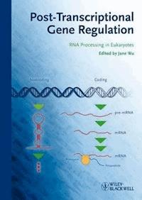
Illustrated on the cover is a simplified summary of our current understanding of eukaryotic gene regulation. Most of our knowledge about gene expression and regulation has come from studies of protein coding genes (“Coding”), although nonprotein coding genes (“Noncoding”) may account for the vast majority of eukaryotic genomes. Following transcription from DNA (depicted as the double-stranded helix), precursor noncoding RNA gene products (diagrammed as the stem-looped structure) or messenger RNA precursors (pre-mRNAs) undergo multistep posttranscriptional RNA processing before they become functionally active in engaging their target genes or serving as messengers (mRNAs) to direct polypeptide synthesis in the protein synthesis machinery (Ribosomes). Each step of these complex gene expression processes (depicted as green triangles) is under intricate regulation to meet the cell’s needs in adapting to its constantly changing environment. (Contributed by Mengxue Yang and Jane Y. Wu.)
Limit of Liability/Disclaimer of Warranty: While the publisher and author have used their best efforts in preparing this book, they make no representations or warranties with respect to the accuracy or completeness of the contents of this book and specifically disclaim any implied warranties of merchantability or fitness for a particular purpose. No warranty can be created or extended by sales representatives or written sales materials. The Advice and strategies contained herein may not be suitable for your situation. You should consult with a professional where appropriate. Neither the publisher nor authors shall be liable for any loss of profit or any other commercial damages, including but not limited to special, incidental, consequential, or other damages.
Library of Congress Card No.: applied for
British Library Cataloguing-in-Publication Data
A catalogue record for this book is available from the British Library.
Bibliographic information published by the Deutsche Nationalbibliothek
The Deutsche Nationalbibliothek lists this publication in the Deutsche Nationalbibliografie; detailed bibliographic data are available on the Internet at <http://dnb.d-nb.de>.
© 2013 Wiley-VCH Verlag GmbH & Co. KGaA, Boschstr. 12, 69469 Weinheim, Germany
Wiley-Blackwell is an imprint of John Wiley & Sons, formed by the merger of Wiley’s global Scientific, Technical, and Medical business with Blackwell Publishing.
All rights reserved (including those of translation into other languages). No part of this book may be reproduced in any form – by photoprinting, microfilm, or any other means – nor transmitted or translated into a machine language without written permission from the publishers. Registered names, trademarks, etc. used in this book, even when not specifically marked as such, are not to be considered unprotected by law.
Print ISBN: 978-3-527-32202-2
ePDF ISBN: 978-3-527-66542-6
ePub ISBN: 978-3-527-66541-9
mobi ISBN: 978-3-527-66540-2
oBook ISBN: 978-3-527-66543-3
Cover Design Formgeber, Eppelheim
Typesetting Toppan Best-set Premedia Limited, Hong Kong
Foreword
RNA is the “working substance” of genetics, a multipart actor playing almost every part in the expression of genetic information. The central dogma of molecular biology places RNA centrally between the cell’s genetic library of DNA, and its catalytic engines the proteins, underscoring RNA’s key importance in the flux of information. Yet the deceptive simplicity of the original scheme as set out by Crick greatly underrepresents the myriad functions of RNA, and its critical role in orchestrating the almost impossibly complex regulatory networks of the cell.
There are many reasons why RNA plays such a big part in the genetic life of the cell. The chemical nature of DNA has been selected to be relatively immutable to change; it has a rather staid, stay-at-home character. RNA by contrast is its rather racy cousin that is into everything. The 2′-hydroxyl group of RNA adds immeasurably to its chemical reactivity and structural repertoire. Moreover, as a formally single-stranded molecule, RNA can fold up into complex structures that bind ligands with huge selectivity (and there is a whole dimension of genetic regulation that is based on riboswitch function) and even exhibit catalytic activity. These properties were most probably the key to the early evolution of life, and the strongest indicator of this is telegraphed in the catalysis of peptidyl transfer by RNA in the modern ribosome. While most catalysis in the cell is now performed by proteins with their greater chemical flexibility, RNA has taken up most of the remaining genetic functions and run with them. The ancient role of RNA may be one of the reasons why it occupies such a central role in contemporary biology.
These additional functions have revealed themselves over a long period. I am old enough to remember the tremendous surprise we all felt when it transpired that eukaryotic genes were interrupted by introns, requiring splicing processes accurate to the nucleotide and a hugely complex apparatus to achieve that. This machine, the spliceosome, is a dynamic assembly of functional RNA and proteins, yet at its heart is probably at least in part a ribozyme. Unsurprisingly, splicing is precisely and dynamically regulated in a coordinated way in the cell, and alternative splicing events can increase genetic coding power by enabling exon skipping that is important in development.
Another unexpected development was the discovery that specific nucleotides could be chemically changed to “edit” the genetic content of the RNA. The classic example is the deamination of adenosine to generate inosine by the enzyme adenosine deaminases acting on RNA (ADAR). Of course, the high level of nucleotide modification in the transfer RNAs (tRNAs) should indicate that the chemical alteration of “plain vanilla” RNA is likely to be considerable. In archaea and eukaryotes, RNA is subject to site-specific methylation and pseudouridylation by relatively small RNA–protein machines, the box C/D and H/ACA small nucleolar ribonucleoprotein particles (snoRNPs). These exhibit an ordered assembly that begins with the binding of a protein that stabilizes a ubiquitous structural motif in RNA called the kink turn, and ends when the methylase or uridine isomerase binds to the complex.
Transcription of RNA is tightly regulated but does not have the same checks on accuracy that DNA replication requires. There is no major requirement for repair processes for RNA; it lives by a more throwaway philosophy. And that is just what happens. In nonsense-mediated decay, RNA containing a premature termination codon is selectively degraded. This process is also used to regulate the synthesis of RNA-binding proteins. At the other end of the central dogma, translation is also regulated, and remarkably this can be involved in learning and memory.
Nobody can be unaware of the revolution that has swept through RNA biology with the discovery of gene regulation mediated by small RNA species. While the fraction of the genome that encodes proteins is really quite small, it transpires that much more of the genome encodes regulatory RNAs that are transcribed from untranslated regions and introns. Presumably for decades, RNA investigators were running these species off the ends of their gels, yet these noncoding RNA species have now assumed a vast importance and have become an industry both as a regulatory mechanism and a powerful laboratory tool. They could even yet be the source of therapeutic agents, although as ever the big problem there remains delivery. Regulatory RNA networks of great complexity will ultimately require the methods of systems biology and mathematical modeling to be fully understood.
Given the central importance of RNA in the life of the cell, it is obvious that defects will occur with serious consequences for function. These can lead to important diseases, exemplified by the role of noncoding RNA species in neurodevelopment and neurodegeneration. The role of RNA in disease is a significant issue in human health that we neglect at our peril. I suspect we have only begun to scratch the surface of this topic and await exciting developments.
This book provides a guided tour through the key functions of posttranscriptional gene regulation, that is, pretty much an overview of RNA biology, written by the experts in each area. Importantly, it also covers some aspects of the pathology of RNA when things go wrong. I congratulate Jane Wu in putting this book together – it is a highly valuable resource for many of us who are interested in eukaryotic gene regulation.
David M.J. Lilley, PhD, FRSBeijing, May 2012 University of Dundee
List of Contributors
Jennifer A. AokiColumbia UniversityDepartment of Biological Sciences1117 Fairchild CenterNew York, NY 11740USA
Stephen BuratowskiHarvard Medical SchoolDepartment of Biological Chemistry and Molecular Pharmacology240 Longwood AvenueBoston, MA 02115USA
Piero CarninciOmics Science CenterLSA Technology Development GroupFunctional Genomics Technology Team1-7-22 Suehiro-choTsurumi-kuYokohamaKanagawa 230-0045Japan
and
Omics Science CenterLSA Technology Development GroupOmics Resource Development Unit1-7-22 Suehiro-choTsurumi-kuYokohamaKanagawa 230-0045Japan
Mengmeng ChenChinese Academy of SciencesInstitute of BiophysicsState Laboratory of Brain and CognitionBeijing 100101China
Ying ChenThe University of ChicagoDepartment of Ecology and EvolutionCommittee on Genetics1101 E 57 StreetChicago, IL 60637USA
Hongzheng DaiThe University of ChicagoDepartment of Ecology and EvolutionCommittee on Genetics1101 E 57 StreetChicago, IL 60637USA
Jill A. DembowskiUniversity of PittsburghDepartment of Biological Sciences4249 Fifth AvenuePittsburgh, PA 15260USA
Jianwen DengChinese Academy of SciencesInstitute of BiophysicsState Laboratory of Brain and CognitionBeijing 100101China
Sebastian M. FicaUniversity of ChicagoDepartment of Molecular Genetics & Cell BiologyCLSC 821A920 East 58th StChicago, IL 60637USA
Katherine S. GodinMedical Research Council Laboratory of Molecular BiologyStructural Studies DivisionHills RoadCambridge CB1 2QWUK
and
University of WashingtonDepartment of ChemistryBox 351700Seattle, WA 98195USA
Graydon B. GonsalvezGeorgia Health Sciences UniversityDepartment of Cellular Biology and Anatomy1459 Laney Walker BlvdAugusta, GA 30912USA
Paula J. GrabowskiUniversity of PittsburghDepartment of Biological Sciences4249 Fifth AvenuePittsburgh, PA 15260USA
Matthias HarbersDNAFORM Inc.Leading Venture Plaza 275-1 Ono-choTsurumi-kuYokohamaKanagawa 230-0046Japan
Hisashi IizasaThe Wistar InstituteDepartment of Gene Expression & Regulation3601 Spruce StreetPhiladelphia, PA 19104USA
Yukio KawaharaThe Wistar InstituteDepartment of Gene Expression & Regulation3601 Spruce StreetPhiladelphia, PA 19104USA
Minkyu KimSeoul National UniversityWCU Department of Biophysics and Chemical Biology1 Gwanak-Ro, Gwanka-Gu, Seoul 151-747Korea
Jianghong LiuChinese Academy of SciencesInstitute of BiophysicsState Laboratory of Brain and CognitionBeijing 100101China
Manyuan LongThe University of ChicagoDepartment of Ecology and EvolutionCommittee on Genetics1101 E 57 StreetChicago, IL 60637USA
James L. ManleyColumbia UniversityDepartment of Biological Sciences1117 Fairchild CenterNew York, NY 11740USA
A. Gregory MateraUniversity of North CarolinaDepartment of BiologyProgram in Molecular Biology & Biotechnology and Lineberger Comprehensive Cancer Center3352 Genome Science BuildingChapel Hill, NC 27599-3280USA
Melissa MeffordUniversity of ChicagoDepartment of Molecular Genetics & Cell BiologyCLSC 821A920 East 58th StChicago, IL 60637USA
Kazuko NishikuraThe Wistar InstituteDepartment of Gene Expression & Regulation3601 Spruce StreetPhiladelphia, PA 19104USA
Eliza C. SmallUniversity of ChicagoDepartment of Molecular Genetics & Cell BiologyCLSC 821A920 East 58th StChicago, IL 60637USA
Jonathan P. StaleyUniversity of ChicagoDepartment of Molecular Genetics & Cell BiologyCLSC 821A920 East 58th StChicago, IL 60637USA
Louis ValenteThe Wistar InstituteDepartment of Gene Expression & Regulation3601 Spruce StreetPhiladelphia, PA 19104USA
Gabriele VaraniUniversity of WashingtonDepartment of ChemistryBox 351700Seattle, WA 98195USA
and
University of WashingtonDepartment of BiochemistrySeattle, WA 98195USA
Jane Y. WuNorthwestern University Feinberg School of MedicineCenter for Genetic MedicineLurie Comprehensive Cancer CenterDepartment of Neurology303 E. SuperiorChicago, IL 60611USA
Mengxue YangChinese Academy of SciencesInstitute of BiophysicsState Laboratory of Brain and CognitionBeijing 100101China
and
Northwestern University Feinberg School of MedicineCenter for Genetic MedicineLurie Comprehensive Cancer CenterDepartment of Neurology303 E. SuperiorChicago, IL 60611USA
Kun ZhuChinese Academy of SciencesInstitute of BiophysicsState Laboratory of Brain and CognitionBeijing 100101China
Li ZhuChinese Academy of SciencesInstitute of BiophysicsState Laboratory of Brain and CognitionBeijing 100101China
Boris ZinshteynThe Wistar InstituteDepartment of Gene Expression & Regulation3601 Spruce StreetPhiladelphia, PA 19104USA
1
The Role of Cotranscriptional Recruitment of RNA-Binding Proteins in the Maintenance of Genomic Stability
Jennifer A. Aoki and James L. Manley
1.1 Introduction
All steps in transcription and pre-mRNA processing are extensively coordinated. The carboxy-terminal domain (CTD) of the large subunit of RNA polymerase (RNAP) II plays an important role in cotranscriptionally recruiting factors necessary for capping, splicing, polyadenylation, and other mRNA processing events [1–4]. The CTD acts as a platform for these factors to bind, and this process is coordinated by phosphorylation changes that occur during transcription [5]. Although transcription and RNA processing steps are not obligatorily coupled, as seen by the fact that these processes have been studied for many years as individual steps, some posttranscriptional modifications have been shown to be functionally coupled in vitro such as transcription capping [6] and transcription 3′ end processing [7]. There has also been recent evidence for “reverse coupling,” where a proximal 5′ splice site enhances the recruitment of basal transcription initiation factors to the promoter [8]. While it is still unclear if transcription and splicing are functionally coupled in the cell [9, 10], there is evidence that cotranscriptional recruitment of serine-arginine (SR) proteins onto pre-mRNA is vital in maintaining genomic stability [11, 12]. Indeed, recent work shows that ASF/SF2, an SR protein first discovered for its role in constitutive and alternative splicing [13, 14], is a component of a high-molecular-weight (HMW) complex formed on pre-mRNA during cotranscriptional splicing assays [15], reflecting the early recruitment of ASF/SF2 and other SR proteins to nascent RNA during transcription.
All the steps in transcription and RNA processing appear to function together to produce an export-competent and translatable mRNP. In yeast, where splicing is less frequent, transcription is coupled to loading of export factors and mRNP formation through the THO/TREX complex [16]. Mutation of factors in the THO/TREX complex also results in genomic instability [16]. In metazoans, transcription is linked to mRNP formation through splicing [17], formation of the exon junction complex (EJC) [18, 19], and THO/TREX recruitment [16].
Below we will discuss the coordination of transcription and pre-mRNA processes that inherently protects the genome from invasion of nascent RNA into DNA of the transcribing locus. The invading RNA can then hybridize to the template DNA, producing an aberrant R-loop structure, leaving the coding strand of the DNA single stranded and subject to DNA damage and strand breakage (Figure 1.1). We will describe other examples of cotranscriptionally formed R-loops and speculate on mechanisms that cause such structures to lead to genomic instability.
Figure 1.1 Schematic of a cotranscriptionally formed R-loop structure. Nascent RNA hybridizes with template DNA, leaving coding DNA single stranded.
1.2 THO/TREX
1.2.1 THO/TREX in Saccharomyces cerevisiae
The THO complex proteins were first discovered in genetic screens for their role in transcription elongation of GC-rich genes in S. cerevisiae [20]. The complex consists of Hpr1, Tho2, Mft2, and Thp2, which are recruited to elongating RNAP II complexes. In addition to impairment of transcriptional elongation, THO mutants cause reduced efficiency of gene expression and an increase in hyper-recombination between direct repeats [21]. Mutations in hpr1, tho2, and mft1 can also produce export defects and retention of transcripts at sites of transcription [16]. This reflects the association of THO with export factors Yra1 and Sub1 to form the TREX complex (transcription/export). TREX is recruited early to actively transcribing genes and travels entire length of genes with RNAP II [16]. Interestingly, mutants of the export machinery, Sub2, Yra1, Mex67, and Mtr2 also have THO-like phenotypes of defective transcription and hyper-recombination [16]. Further investigation of 40 selected mutants representing various steps in biogenesis and export of mRNP showed a weak but significant effect on recombination and transcript accumulation [22]. In particular, mutants of the nuclear exosome and 3′ end processing machinery showed inefficient transcription elongation and genetic interactions with THO. The TREX complex exemplifies the importance of the link between transcription and export-competent mRNP formation in yeast with the maintenance of the genomic integrity.
Lesen Sie weiter in der vollständigen Ausgabe!
Lesen Sie weiter in der vollständigen Ausgabe!
Lesen Sie weiter in der vollständigen Ausgabe!
Lesen Sie weiter in der vollständigen Ausgabe!
Lesen Sie weiter in der vollständigen Ausgabe!
Lesen Sie weiter in der vollständigen Ausgabe!
Lesen Sie weiter in der vollständigen Ausgabe!
Lesen Sie weiter in der vollständigen Ausgabe!
Lesen Sie weiter in der vollständigen Ausgabe!
Lesen Sie weiter in der vollständigen Ausgabe!


