The Craniotomy Atlas E-Book
164,99 €
Mehr erfahren.
- Herausgeber: Thieme
- Kategorie: Fachliteratur
- Sprache: Englisch
"… the neurosurgical primer that every resident will own and study" - Robert Spetzler
Given that the great majority of brain surgeries are preceded by a craniotomy, mastering the procedure is essential for junior residents. Choosing the appropriate craniotomy and executing it safely is the difference between a straightforward case with good access to the target and a procedure where access to the target is needlessly traumatic and may even be impossible.
Professor Raabe's The Craniotomy Atlas provides precise instructions for performing all common neurosurgical cranial exposures, including: convexity approaches, midline approaches, skull base approaches, transsphenoidal approaches and more. Instructions for each craniotomy include positioning, head fixation, aesthetic considerations, and protecting the dura mater.
Special Features:
- More than 600 high-quality operative photographs and brilliant illustrations support the step-by-step descriptions, with all the precision and attention to detail that neurosurgeons have come to expect from the editor Professor Raabe, and the associate editors Professors Meyer, Schaller, Vajkoczy, and Winkler.
- Full coverage of complications and risk factors
- Checklist with summaries of the critical steps
All residents and trainees in neurosurgery will treasure this essential resource, which will help build confidence when performing these critical neurosurgical procedures.
This book includes complimentary access to a digital copy on https://medone.thieme.com.
Das E-Book können Sie in Legimi-Apps oder einer beliebigen App lesen, die das folgende Format unterstützen:
Seitenzahl: 351
Veröffentlichungsjahr: 2019
Ähnliche


The Craniotomy Atlas
EditorAndreas Raabe, MDProfessor, Chairman, and DirectorDepartment of NeurosurgeryInselspital, Bern University HospitalBern, Switzerland
Associate EditorsBernhard Meyer, MDProfessor, Chairman, and DirectorDepartment of NeurosurgeryUniversity Hospital rechts der IsarTechnical University of MunichMunich, Germany
Karl Schaller, MDProfessor, Chairman, and DirectorDivision of NeurosurgeryDepartment of Clinical NeurosciencesUniversity Hospital of GenevaGeneva, Switzerland
Peter Vajkoczy, MDProfessor, Chairman, and DirectorDepartment of NeurosurgeryCharité - Universitätsmedizin BerlinBerlin, Germany
Peter A. Winkler, MDProfessor, Chairman, and DirectorDepartment of NeurosurgeryUniversity Hospital – Salzburg UniversitySalzburg, Austria
926 illustrations
ThiemeStuttgart • New York • Delhi • Rio de Janeiro
Library of Congress Cataloging-in-Publication Data is available from the publisher
© 2019. Thieme. All rights reserved.
Inselspital holds copyright to all photographs and illustrations used in this work unless otherwise stated. Used with permission from Inselspital, Bern University Hospital, Bern, Switzerland.
Thieme Publishers StuttgartRüdigerstrasse 14, 70469 Stuttgart, Germany+49 [0]711 8931 421, [email protected]
Thieme Publishers New York333 Seventh Avenue, New York, NY 10001 USA+1 800 782 3488, [email protected]
Thieme Publishers DelhiA-12, Second Floor, Sector-2, Noida-201301Uttar Pradesh, India+91 120 45 566 00, [email protected]
Thieme Publishers Rio de Janeiro,Thieme Publicações Ltda.Edifício Rodolpho de Paoli, 25° andarAv. Nilo Peçanha, 50 – Sala 2508Rio de Janeiro 20020-906 Brasil+55 21 3172 2297
Cover design: Thieme Publishing GroupTypesetting by DiTech Process Solutions, India
Printed in Germany by CPI Books 5 4 3 2 1
ISBN 978-3-13-205791-3
Also available as an e-book:eISBN 978-3-13-205801-9
Important note: Medicine is an ever-changing science undergoing continual development. Research and clinical experience are continually expanding ourknowledge, in particular ourknowledge of proper treatment and drug therapy. Insofar as this book mentions any dosage or application, readers may rest assured that the authors, editors, and publishers have made every effort to ensure that such references are in accordance with the state of knowledge at the time of production of the book.
Nevertheless, this does not involve, imply, or express any guarantee or responsibility on the part of the publishers in respect to any dosage instructions and forms of applications stated in the book. Every user is requested to examine carefully the manufacturers’ leaflets accompanying each drug and to check, if necessary in consultation with a physician or specialist, whether the dosage schedules mentioned therein or the contraindications stated by the manufacturers differ from the statements made in the present book. Such examination is particularly important with drugs that are either rarely used or have been newly released on the market. Every dosage schedule or every form of application used is entirely at the user’s own risk and responsibility. The authors and publishers request every user to report to the publishers any discrepancies or inaccuracies noticed. If errors in thiswork are foundafter publication, errata will be posted at www.thieme.com on the product description page.
Some of the product names, patents, and registered designs referred to in this book are in fact registered trademarks or proprietary names even though specific reference to this fact is not always made in the text. Therefore, the appearance of a name without designation as proprietary is not to be construed as a representation by the publisher that it is in the public domain.
This book, including all parts thereof, is legally protected by copyright. Any use, exploitation, or commercialization outside the narrow limits set by copyright legislation without the publisher’s consent is illegal and liable to prosecution. This applies in particular to photostat reproduction, copying, mimeographing or duplication of any kind, translating, preparation of microfilms, and electronic data processing and storage.
To my wonderful wife, Katrin; my children, Tanja, Max, and Clemens; my parents; my family, who are my life. To my residents, who always inspire me.
To my colleagues, who are mentors, teachers, and friends.
To those who help to take care of my patients, making me feel grateful for their efforts.
To my patients, who trusted me and who were my reason to strive for excellence.
Andreas Raabe
Contents
Foreword
Robert F. Spetzler
Foreword
Volker Seifert
Preface
Acknowledgments
Call for Submissions
Contributors
1Basics
1.1 Craniotomies Overview
Andreas Raabe and Peter A. Winkler
1.2 Difference between Approach and Craniotomy
Andreas Raabe
1.3 Craniotomies We Have Omitted from This Book and Why
Andreas Raabe, Bernhard Meyer, Peter Vajkoczy, and Karl Schaller
1.4 Positioning
1.4.1 Basic Rules
Andreas Raabe and Janine Abu-Isa
1.4.2 Supine
Philippe Schucht
1.4.3 Supine Lateral
Christian F. Freyschlag and Claudius Thomé
1.4.4 Lateral
Philippe Schucht
1.4.5 Lateral Oblique or Park Bench
Daniel Hänggi
1.4.6 Park Bench
David Bervini and Janine Abu-Isa
1.4.7 Prone/Concorde
Christian Fung
1.4.8 Semisitting
Andreas Raabe
1.5 Rigid Head Fixation
Christian Fung
1.6 Esthetic Considerations in Neurosurgical Procedures
Mihai A. Constantinescu, Irena Zubak, and Andreas Raabe
1.6.1 Skin Incision
1.6.2 Burr Holes
1.6.3 Mini-plates or Craniotomy Fixation Caps
1.6.4 Craniotomy Caps
1.6.5 The Temporalis Muscle
1.6.6 Secondary Procedures for Restoration of Contour after Temporal Muscle Atrophy
1.7 Protection of the Dura Mater
Andreas Raabe and David Bervini
1.7.1 Potential Problems Arising from a Laceration of the Dura Mater
1.7.2 Measures to Protect the Integrity of the Dura Mater
1.8 Sinus Laceration
Sandro Krieg and Bernhard Meyer
1.8.1 Introduction
1.8.2 Prevention Strategies
1.8.3 Management of Sinus Laceration
1.8.4 Special Considerations
1.9 Frontal Sinus Breach and Repair
Andreas Raabe and Marco Caversaccio
1.9.1 Landmarks for the Frontal Sinus
1.9.2 Principles of Repair
1.9.3 Surgical Technique for Repairing in the Case of a Frontal Sinus Breach
2Landmarks
2.1 Schematic Cortical Anatomy
Andreas Raabe and Peter A. Winkler
2.2 Craniocerebral Topography
Irena Zubak, Andreas Raabe, and Karl Schaller
2.2.1 Introduction
2.2.2 Craniometric Points and Lines and Their Reference to Intracranial Structures
2.2.3 Skull Base Points
2.2.4 Other Cranial Landmarks
2.3 Identifying Cortical Landmarks and Fiber Tracts in MRI
J. Goldberg, M. Murek, L. Häni, K. Schaller, and A. Raabe
2.3.1 Introduction
2.3.2 Cortical Landmarks—Primary Motor and Sensory Cortex
2.3.3 Cortical Landmarks—Language Areas
2.3.4 Cortical Landmarks—Primary Visual Cortex
2.3.5 Determining the Position of Important Fiber Tracts on MRI—Corticospinal Tract (CST)
2.3.6 Determining the Position of Important Fiber Tracts on MRI—Arcuate Fascicle
2.3.7 Determining the Position of Important Fiber Tracts on MRI—Optical Tract
3Convexity Craniotomies
3.1 Convexity Craniotomy Planning
Andreas Raabe and Jens Fichtner
3.2 Planning of Craniotomies at the Skull Convexity without the Use of Navigation
Florian Ringel and Andreas Kramer
3.3 Supratentorial Convexity Craniotomy
Philippe Schucht
4Midline Craniotomies
Ulrich Sure and Philipp Dammann
4.1 Sinus-Crossing Craniotomies—Basic Principles
Ulrich Sure and Philipp Dammann
4.2 Supratentorial Midline Craniotomy
4.2.1 Frontal
Ulrich Sure and Philipp Dammann
4.2.2 Frontoparietal
Ulrich Sure and Philipp Dammann
4.2.3 Parieto-occipital
Ulrich Sure and Philipp Dammann
4.3 Infratentorial Midline Craniotomy
4.3.1 Infratentorial Supracerebellar
Ulrich Sure and Philipp Dammann
4.3.2 Median Suboccipital (Involving Foramen Magnum)
Ulrich Sure and Philipp Dammann
5Skull Base Craniotomies
5.1 Frontal Craniotomies
5.1.1 Bifrontal
Torstein R. Meling and Marton König
5.2 Frontotemporal Craniotomies
5.2.1 Facial Nerve Anatomy and Protection
Andreas Raabe and Peter A. Winkler
5.2.2 Superficial Temporal Artery Preservation during Frontolateral Approaches
Andreas Raabe and Peter Vajkoczy
5.2.3 Supraorbital
Nikolai Hopf and Robert Reisch
5.2.4 Frontolateral
Andreas Raabe
5.2.5 Helsinki Lateral Supraorbital
Juha Hernesniemi and Hugo Andrade-Barazarte
5.2.6 Pterional
Peter Vajkoczy and Andreas Raabe
5.3 Temporal Craniotomies
5.3.1 Temporobasal Craniotomy
Bernhard Meyer
5.4 Posterior Fossa
5.4.1 Retrosigmoid Craniotomy
Marcos Tatagiba, Florian H. Ebner, and Georgios Naros
6Skull Base Extensions
6.1 Orbitozygomatic Craniotomy
Andreas Raabe
6.2 Orbitocraniotomy
Daniel Hänggi
6.3 Intradural Anterior Clinoidectomy
Andreas Raabe and Karl Schaller
6.4 Far (Enough) Lateral Approach
Andreas Raabe, Johannes Goldberg, and David Bervini
7Transsphenoidal Approach
7.1 Microsurgical Endonasal Approach
Christian F. Freyschlag and Claudius Thomé
7.2 Endoscopic Approach
Henry Schroeder and Jörg Baldauf
8Decompressive Hemicraniectomy
Jürgen Beck
9Approaches to the Orbita
9.1 Frontolateral Approach to the Orbit
Torstein R. Meling
9.2 Lateral Orbitotomy
Torstein R. Meling
Index
Foreword
The Craniotomy Atlas, edited by Professor Raabe, is intended to be a resource for residents and new neurosurgeons with the goal of providing precise instructions for performing common neurosurgical exposures. Professor Raabe and his coauthors have used high-quality operative photographs accompanied by excellent illustrations to compile an atlas that far exceeds expectations. The beautiful step-by-step compilation for each approach will make this volume an essential companion for every neurosurgical resident and a useful reference for the new neurosurgeon. The precision and attention to detail that we have come to expect from Raabe has reached a new high in this book. With the introduction of intraoperative indocyanine green angiography to the neurosurgical community, Andreas Raabe had already cemented his place as a foremost contributor to our specialty—with this book, he will have created the neurosurgical primer that every resident will own and study.
As with any neurosurgical procedure, there are differences among neurosurgeons based on experience and training. For example, with the exception of the sigmoid sinus, I routinely cross all other sinuses by just using the footplate of the drill rather than multiple burr holes. After washing out the bone dust with irrigation, one can look right down the bone cut and verify that the footplate is extradural, and the dura can be separated from the bone by placing sufficient pressure on the underside of the bone while crossing the sinus as readily as with multiple burr holes and any other instrument. Although many roads lead to Rome, I find that this volume, except for a few inconsequential differences, provides the best highway to get there. I congratulate the authors for this detailed, beautifully illustrated, step-by-step guide to performing the routine craniotomies that all residents and neurosurgeons need to master.
Robert F. Spetzler, MDPhoenix, ArizonaUnited States
Foreword
There is no doubt among neurosurgeons that a correct and tailored craniotomy, apart from the detailed preoperative planning, represents the decisive first step toward a successful intracranial operation. The Craniotomy Atlas, edited by Professor Andreas Raabe and compiled with contributions of a large number of experienced neurosurgeons, is primarily aimed at the neurosurgical resident and younger neurosurgeon. However, as a seasoned and experienced neurosurgeon, who has selected over the years his own armamentarium of favorite craniotomies and surgical variations, I have found it highly interesting to wander through the abundance of beautiful and detailed illustrations as well as the exact and informative step-by-step descriptions of the various craniotomies presented in this atlas. Although there exists a large number of neurosurgical textbooks with detailed descriptions of surgical approaches, these are mostly presented within the context of the underlying intracranial target, mainly a tumorous or vascular lesion.
I am not aware of a comparable and up-to-date compilation of craniotomies, covering all aspects—basic considerations such as positioning and attention to surgical landmarks, routine craniotomies, and elaborate skull base craniotomies and its extensions. The outstanding attention to details presented in this atlas reflects the meticulous way of preparation and performance of every craniotomy by Professor Raabe, as I have seen over the many years during which we have both worked together. Within this context, it is a pleasure and an honor to applaud the editor and his co-authors for this excellent contribution to the art of craniotomy, which will surely stand as a surgical reference for many years to come.
Volker Seifert, MD, PhDFrankfurtGermany
Preface
Craniotomies are an essential part of brain surgery. They are regarded as important but rather basic procedures that are the prelude to the intradural neurosurgical operation proper. An optimally placed craniotomy provides the basis for a simple or sophisticated intradural approach and a straightforward case. Wrongly placed, it completely changes the operation, making access to the neurosurgical target traumatic or impossible.
Neurosurgeons start with simple craniotomies early on in their training. Junior residents learn how to perform a specific craniotomy from senior residents or attendings. Many textbooks and journal articles describe the various craniotomies in detail and serve as excellent reference sources.
Despite being “mainstream” knowledge, for the first “Frankfurt craniotomy course” that Bernhard Meyer, Peter Vajkoczy, Peter Winkler, and I organized in 2004, there was an overwhelming number of applications for only 20 course seats. The applicants were searching for a systematic collection and teaching of information related to craniotomies. We learned from the course participants that craniotomies are far from being standardized, with numerous variations even within the same department. Since then, yearly courses have been held in Frankfurt and, since 2008, also in Bern and Geneva with an equally high number of applicants for the restricted number of available course seats.
This book is a logical effort to continue this teaching and extend the systematic collection of knowledge about standard and some extended craniotomies and related aspects. I hope that it contributes to a better understanding of the underlying concept and anatomy, greater standardization of the operations, and an improved technique when performing the planned craniotomy.
Andreas Raabe, MD
Acknowledgments
I would like to express my deep gratitude to Anja Giger and Alain Blank, who provided the superb illustrations for this book. Over a period of 3 years, it was always a pleasure to sit together and discuss the details of the authors’ photographs and how these should be depicted in the illustrations. Without their artistic skills and their invaluable contribution, this book would not have been possible.
I am specifically grateful to Luisa Tonarelli, who accompanied the development of this book from the very first chapter to the final printed version. Her help, advice, expertise, and hard work were indispensable in bringing this volume to publication.
Finally, I would like to thank Susan Kaplan, Irena Zubak, Janine Abu-Isa, Katharina Lutz, Michael Murek, David Bervini, Johannes Goldberg, Levin Häni, and Jonathan Rychen for their time and advice during the review of the chapters of this book.
Andreas Raabe
Call for Submissions
The techniques and knowledge described in this book reflect the personal views, teaching, and experience of its authors. We know that the content of this book is far from comprehensive. We are also aware that skilled surgeons around the world have their own tricks and modifications of craniotomies, usually derived from personal experience and for good reasons.
Therefore, we invite authors to submit their modification, nuance, or technique in the form of a step-by-step series of photographs with a text description, like the chapters in this book. The topic may range from a craniotomy not yet included in this book to a technical note or a nuance of an already described craniotomy; however, it should be recognized as useful, reproducible, and potentially suitable for routine use. These submissions will undergo peer review by experienced neurosurgeons as well as young residents. If accepted, illustrations will be produced to complement the photographs, and a corresponding chapter will be added to the book.
We are aware that only a limited number of carefully selected additional chapters on a craniotomy or a nuance can be included in this collection. But, despite having arrived in the digital age, we still believe in the educational value of a book, in which a compilation of the most important craniotomies and the related knowledge can be found. All contributions have been peer-reviewed and selected as pearls of wisdom for neurosurgical residents.
Before submitting a manuscript, authors should contact the Editorial Office to request for the technical specifications and to have the topic checked for potential duplication and suitability.
Inquiries and submissions should be sent to:
Editorial Office Craniotomy Book
Department of Neurosurgery
Inselspital, University of Bern
3010 Bern
Switzerland
Email: [email protected]
Contributors
Janine Abu-Isa, MD
Department of Neurosurgery
Inselspital, Bern University Hospital
Bern, Switzerland
Hugo Andrade-Barazarte, PhD
Juha Hernesniemi International Center for Neurosurgery
Henan People's Provincial Hospital
Zhengzhou, China
Jörg Baldauf, MD, PD
Department of Neurosurgery
University Medicine Greifswald
Greifswald, Germany
Jürgen Beck, MD
Professor and Medical Director
Department of Neurosurgery
Neurocenter
University of Freiburg
Freiburg, Germany
David Bervini, MD
Department of Neurosurgery
Inselspital, Bern University Hospital
Bern, Switzerland
Marco Caversaccio, MD
Professor, Chairman, and Director
Department of ENT, Head and Neck Surgery
Inselspital, Bern University Hospital
Bern, Switzerland
Mihai A. Constantinescu, MD
Professor, Chairman, and Director
Department of Plastic and Hand Surgery
Inselspital, Bern University Hospital
Bern, Switzerland
Philipp Dammann, MD
Department of Neurosurgery
University Hospital of Essen
Essen, Germany
Florian H. Ebner, MD
Professor
Department of Neurosurgery
University of Tübingen
Tübingen, Germany
Jens Fichtner, MD
Department of Neurosurgery
Inselspital, Bern University Hospital
Bern, Switzerland
Christian F. Freyschlag, MD
Department of Neurosurgery
University Hospital Innsbruck
Medical University of Innsbruck
Innsbruck, Austria
Christian Fung, MD
Department of Neurosurgery
Neurocenter
University of Freiburg
Freiburg, Germany
Johannes Goldberg, MD
Department of Neurosurgery
Inselspital, Bern University Hospital
Bern, Switzerland
Daniel Hänggi, MD
Professor, Chairman, and Director
Department of Neurosurgery
University Hospital Mannheim
University of Heidelberg
Mannheim, Germany
Levin Häni, MD
Department of Neurosurgery
Inselspital, Bern University Hospital
Bern, Switzerland
Juha Hernesniemi
Professor Emeritus, Former Chairman, and Director
Department of Neurosurgery
Helsinki University Hospital
University of Helsinki
Helsinki, Finland
Nikolai Hopf, MD
Professor and Director
NeuroChirurgicum
Center for Endoscopic and Minimally Invasive Neurosurgery
Stuttgart, Germany
Marton König
Department of Neurosurgery
Oslo University Hospital
University of Oslo
Oslo, Norway
Andreas Kramer, MD
Department of Neurosurgery
University of Mainz
Mainz, Germany
Sandro Krieg, MD, PD
Department of Neurosurgery
and TUM-Neuroimaging Center
University Hospital rechts der Isar
Technical University of Munich
Munich, Germany
Torstein R. Meling, MD
Professor
Division of Neurosurgery
Department of Clinical Neurosciences
University Hospital of Geneva
Geneva, Switzerland
Michael Murek, MD
Department of Neurosurgery
Inselspital, Bern University Hospital
Bern, Switzerland
Georgios Naros, MD
Department of Neurosurgery
University of Tübingen
Tübingen, Germany
Robert Reisch, MD
Professor
Endomin Center for Endoscopic and Minimally Invasive Neurosurgery
Hirslanden Clinic
Zurich, Switzerland
Florian Ringel, MD
Professor, Chairman, and Director
Department of Neurosurgery
University of Mainz
Mainz, Germany
Henry Schroeder, MD
Professor, Chairman, and Director
Department of Neurosurgery
University Medicine Greifswald
Greifswald, Germany
Philippe Schucht, MD
Department of Neurosurgery
Inselspital, Bern University Hospital
Bern, Switzerland
Ulrich Sure, MD
Professor, Chairman, and Director
Department of Neurosurgery
University Hospital of Essen
Essen, Germany
Marcos Tatagiba, MD
Professor, Chairman, and Director
Department of Neurosurgery
University of Tübingen
Tübingen, Germany
Claudius Thomé, MD
Professor, Chairman, and Director
Department of Neurosurgery
Medical University of Innsbruck
Innsbruck, Austria
Irena Zubak, MD
Department of Neurosurgery
Inselspital, Bern University Hospital
Bern, Switzerland
1 Basics
1.1 Craniotomies Overview
Andreas Raabe and Peter A. Winkler
There are four basic categories of supratentorial and infratentorial craniotomy:
1. Convexity craniotomies may be performed anywhere according to the surgical target and goal of the operation. They range from burr holes and mini-craniotomy to decompressive hemicraniectomy, which is the most extensive variant.
2. Midline craniotomies are used for midline approaches that take advantage of subdural anatomical corridors to reach superficial, deep, or contralateral targets. The supratentorial suboccipital craniotomy with an intradural approach along the falx and the tentorium or an infratentorial suboccipital craniotomy with a supracerebellar approach are possible variants.
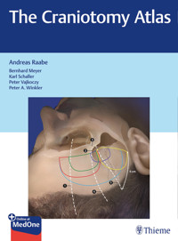
3. Skull base craniotomies range from the frontal midline to the foramen magnum, covering the entire skull base. ▶Fig. 1.1 and ▶Fig. 1.2 demonstrate the continuum of approaches which are often overlapping and are named according to their location at the skull base.
4. Skull base extensions are added to standard skull base craniotomies. They allow access with angles of approach or to structures that cannot be easily reached with standard skull base craniotomies. Typical skull base extensions are anterior clinoidectomy, removal of the orbital rim or zygoma (orbitozygomatic), transpetrosal approaches, the suprameatal extension after retrosigmoid craniotomy or the far-(enough) lateral extension to the foramen magnum (see Chapter 6, Skull Base Extensions).
Supratentorial skull base craniotomies can be divided according to their location, their frontal and temporal extension (size), and their relation to the sylvian fissure. There is no uniform classification, but the following general rules may serve as a guide to the terminology (see ▶Table 1.1).
Table 1.1 Systematics of skull base craniotomies—supratentorial
Fig. 1.1 Systematics of skull base craniotomies—supratentorial. Supratentorial frontotemporal skull base craniotomies, 45° view (a) and lateral view (b). 1, frontolateral; 2, supraorbital; 3, standard pterional; 4, mini-pterional; 5, frontotemporal; 6, anterior temporal; 7a–c anterior, middle, posterior temporobasal; 8, sylvian fissure/sphenoid wing.
Infratentorial skull base craniotomies are performed along the sigmoid sinus or the foramen magnum (see ▶Table 1.2 for further details).
Table 1.2 Systematics of skull base craniotomies—infratentorial
Location
Description
Suboccipital median infra-transverse-sinus
Midline craniotomy for supracerebellar median or paramedian approaches, e.g., for access to the pineal region or tentorial dural fistulas.
Suboccipital lateral infra-transverse-sinus
These are craniotomies based on the same principle as the midline craniotomies for an intradural approach along the subdural space parallel to the tentorium. Typically, they are used for supracerebellar lateral approaches to the midbrain or other regions. They are horizontally oriented compared to the retrosigmoid craniotomy, with more exposure along the transverse sinus and less along the sigmoid sinus. A modification is the suboccipital far-lateral infra-transverse-sinus craniotomy.
Retrosigmoid
Typically ranges from the transverse sinus to the base of the posterior fossa along the sigmoid sinus to gain access to the cerebellopontine angle. May vary in size and be centered more superiorly or inferiorly: vertically oriented.
Suboccipital median periforaminal craniotomy with opening of the foramen magnum
Typically bilateral, there is a mini-version, for example, in Chiari-decompression surgery.
Suboccipital lateral periforaminal craniotomy with opening of the foramen magnum
The lateral suboccipital craniotomy with opening of the foramen magnum is the basic craniotomy for the far lateral approach which can be regarded as a skull base extension of the basal suboccipital craniotomy.
Fig. 1.2 Systematics of skull base craniotomies—infratentorial. Craniotomies of the posterior fossa. 9, suboccipital median infra-transverse-sinus; 10, suboccipital lateral infra-transverse-sinus; 11, suboccipital far-lateral infra-transverse-sinus; 12, retrosigmoid; 13, suboccipital median periforaminal (with opening of the foramen magnum); 14, mini-suboccipital median periforaminal (with opening of the foramen magnum); 15, suboccipital lateral periforaminal (with opening of the foramen magnum); 16, far-lateral extension.
1.2 Difference between Approach and Craniotomy
Andreas Raabe
Although often used synonymously, there is a difference between a craniotomy and an approach. Approach is the broader term and is often used for craniotomy and intradural preparation. In this book, we discuss only the steps of the craniotomy, i.e., to reach bony exposure. With a few exceptions, we stay outside the dura. We will therefore mostly use the term craniotomy instead of approach, and generally reserve the latter to describe the dissection and exposure after opening the dura mater. Craniotomy and approach may be different as in the examples given below. However, as already mentioned, the term “approach” often overlaps with craniotomy and intradural preparation.
Examples:
• Supraorbital craniotomy and subfrontal approach.
• Pterional craniotomy and transsylvian approach.
• Temporobasal craniotomy and subtemporal approach.
• Suboccipital lateral craniotomy and supracerebellar lateral approach.
• Median suboccipital craniotomy and telovelar approach.
1.3 Craniotomies We Have Omitted from This Book and Why
Andreas Raabe, Bernhard Meyer, Peter Vajkoczy, and Karl Schaller
This book is intended primarily for young residents, to serve as a guide to understanding the various craniotomies. It describes the most often used craniotomies, but we decided not to include those that are used only very rarely. Therefore, it does not cover highly specialized skull base craniotomies and their extension, such as posterior transpetrosal, translabyrinthine, transcochlear, or combined approaches, nor is it our aim to provide a complete atlas of approaches and extensions.
We acknowledge that these specialized skull base approaches had their place in the heyday of skull base surgery. However, nowadays they are often replaced by a staged procedure or a combination of simpler craniotomies that provide a less invasive strategy with lower morbidity than a technically demanding and more invasive approach. Moreover, radiosurgery and endovascular treatment often complete a less invasive treatment for many patients.
We are also aware that the nomenclature for the craniotomies varies around the world and that experienced surgeons use their own tricks and modifications when performing craniotomies.
1.4 Positioning
1.4.1 Basic Rules
Andreas Raabe and Janine Abu-Isa
Time spent on careful positioning is time well spent. Mistakes in positioning may render any surgical plan, even if it is conceptually perfectly elaborated, impossible. Positioning is the first strategic step for the operation; it is the first digit of the code number to unlock the door to the target of brain surgery. Correct positioning can open the surgical field, achieve gravity retraction, reduce bleeding, and provide the most relaxing position for the surgeon.
Positioning should be highly standardized in each department to improve communication, to save time, and to achieve the goal of the surgery. Use of photographs, step-by-step instructions, and a checklist is recommended.
Fig. 1.3 Craniotomy-to-lesion trajectory. This is the first and most important factor determining the position of the head.
The position of the head depends on the following factors (also see ▶Fig. 1.3, ▶Fig. 1.4, ▶Fig. 1.5, and ▶Fig. 1.6):
1. Planned Surgical Trajectory
The surgical trajectory is the line between the craniotomy and the surgical target, i.e., the midline craniotomy and the tumor in the third ventricle, or the subtemporal craniotomy and the midbrain cavernoma, or the convexity craniotomy and the underlying meningioma (▶Fig. 1.3).
2. Position of the Surgeon
The same surgical trajectory can vary according to the preferred position of the surgeon (see below).
3. Gravity Retraction or Drainage
When gravity retraction is a major part of the surgery, it may become the dominant principle, for instance, in contralateral or midline approaches via the dependent hemisphere or when the semisitting position is preferred in some cases for posterior fossa surgery for pineal or cerebellopontine targets.
4. Measures for Avoiding Potential Position-Related Complications
Such measures include positioning to minimize intracranial pressure, venous congestion, and air embolism, as well as improved orientation if only standardized head positions are allowed.
Every head position can be achieved by combining head rotation (▶Fig. 1.4a) with patient’s body positioning (▶Fig. 1.4b):
• Rotation of the head from 0° to 60° (this can be tested in the awake patient before surgery: in younger patients a rotation up to 90° may be possible, whereas in elderly patients head rotation may be limited to 30°), with the desired degree of head flexion and tilting.
• Selection of one of five supplemental positions of the patient’s body to achieve the final desired head position. These five body positions should be standardized.
Fig. 1.4 Combining positioning of the head and the body of the patient. Head rotation (a) combined with five body positions (b) allows the surgeon to gain access to every trajectory. Special positions are also possible (e.g., semisitting).
Except for special positions (e.g., semisitting), one of the following five basic positions are applied (▶Fig. 1.4b):
• Supine: quick and easy.
• Supine oblique (45°) upper body rotation with the pelvis and legs supine: still quick.
• Lateral recumbent: more complicated, takes more time.
• Lateral oblique or park bench (135°): more complicated, takes more time.
• Prone: more complicated, takes more time and should be avoided if possible because of increased venous congestion.
For instance, a horizontal head position can be achieved by combining:
• 90° head rotation and supine body position or
• 45° head rotation and 45° upper body rotation or
• 0° head rotation and lateral recumbent position.
Fig. 1.5 Positioning of the patient’s body. Typical positioning for different locations of craniotomies.
Fig. 1.6 Position of the surgeon. There are two basic positions for the surgeon: the first is more upright, closer to the surgical field and short instruments, and the hands or fingers are supported (a). The second is a somewhat more oblique position with slightly longer instruments, and forearms or elbows supported (b). Both can achieve the goals of a relaxed surgeon, excellent stability, minimized trembling, and soft instrument movements with maximum haptic feedback about resistance of structures and tactile information. Normally, the positioning of the microscope and the patient’s head follows the position of the surgeon. Make yourself comfortable and then adjust the microscope and the patient, unless otherwise required by the planned surgical trajectory and the specific goals. (▶Fig. 1.6a is reproduced courtesy of Volker Seifert and ▶Fig. 1.6b courtesy of Robert F. Spetzler.)
1.4.2 Supine
Philippe Schucht
See ▶Fig. 1.7 and ▶Fig. 1.8.
Fig. 1.7 Body position. View from the top (a) and the side (b). The supine position is the simplest position. The body and the legs lie straight and the right arm lies parallel to the body. The left arm lies at an angle on a separate armrest to allow insertion of arterial and venous lines and should be loosely fixed.
Attention should be paid to making sure that the body is well cushioned and that the sheets beneath the patient have no wrinkles. In particular in long procedures, incorrect patient positioning may result in decubiti.
Rotating the patient during surgery may give you a better angle of view. For rotating during surgery, prop the patient with additional side pads.
Fig. 1.8 (a, b) Head position. The shoulders should reach the edge of the table. The head is elevated by approximately 5–10 cm in order to facilitate venous drainage
Checklist
• Use side pads to prop the patient if you consider rotating the patient during surgery.
• Cushion the patient well and avoid wrinkles in the sheets to prevent decubiti.
• Elevate the head by approximately 5 to 10 cm to facilitate venous drainage.
1.4.3 Supine Lateral
Christian F. Freyschlag and Claudius Thomé
Supine lateral positioning (45°) of the patient’s upper body is used to enable access to the posterior fossa and the cerebellopontine angle, but may also be used for other approaches where the head is placed with the midline horizontally. In our experience, a sitting or semisitting position offers no advantage over lateral positioning. Although this has long been debated, we do not consider that gravitation-facilitated dissection outweighs disadvantages such as the complexity of positioning, need for exclusion of a persistent foramen ovale, and the risk of venous air embolism.1, 2 Furthermore, operating on a patient in the sitting or semisitting position is less ergonomic and more exhausting for most surgeons. Refer to ▶Fig. 1.9, ▶Fig. 1.10, ▶Fig. 1.11, and ▶Fig. 1.12.
Fig. 1.9 Equipment. The devices needed for supine lateral positioning are found in the basic neurosurgical OR: a three-pin Mayfield clamp, two wedge-shaped cushions, and a support for the pelvis that can be attached to the table.
Fig. 1.10 Positioning of the patient. Supporting the pelvis helps maintain the position during rotation of the table.
Fig. 1.11 Body positioning. It is possible to place the patient flat on the operating table.3 However, the positioning is easier and rotation of the cervical spine and vascular structures is minimized if the shoulder is elevated by a wedge-shaped cushion. In older patients, this support is mandatory due to the patients’ limited neck movement.
Fig. 1.12 Positioning. To obtain optimal conditions and exposure for a retrosigmoid approach, it is necessary to rotate the head (without creating excessive tension on the neck) away from the surgical site. The head is moved in three directions4: (1) contralateral rotation toward a position parallel to the floor, (2) bending of the vertex toward the floor (retroflexion), and (3) inclination of the head to open the cervical-suboccipital angle. This maneuver raises the mastoid process so that it becomes the highest point, while creating space between shoulder and head, which can be increased by retracting the upper shoulder caudally and securing it with adhesive tape.
If the patient has limited neck mobility, the table can be easily rotated to compensate for this limitation. The use of a support prevents the patient from sliding.
Finally, pressure points of the upper and lower extremities are meticulously padded to avoid any injury during surgery.
Checklist
• Make sure the patient’s body is moved sufficiently toward the top end of the operating table (when operating on a patient in a supine lateral position, you tend to sit at 90° to the patient’s longitudinal axis).
• Always support the patient’s position—you might want to rotate the table for better exposure.
• Positioning needs three crucial head movements: rotation, inclination, and lateral flexion.
References
[1] Nozaki K. Selection of semisitting position in neurosurgery: essential or preference? World Neurosurg 2014;81(1):62–63
[2] Feigl GC, Decker K, Wurms M, et al. Neurosurgical procedures in the semisitting position: evaluation of the risk of paradoxical venous air embolism in patients with a patent foramen ovale. World Neurosurg 2014;81(1):159–164
[3] Wait SD, Gazzeri R, Galarza M, Teo C. Simple, effective, supine positioning for the retrosigmoid approach. Minim Invasive Neurosurg 2011;54(4):196–198
[4] Elhammady MS, Telischi FF, Morcos JJ. Retrosigmoid approach: indications, techniques, and results. Otolaryngol Clin North Am 2012;45(2):375–397, ix
1.4.4 Lateral
Philippe Schucht
See ▶Fig. 1.13, ▶Fig. 1.14, ▶Fig. 1.15, ▶Fig. 1.16, and ▶Fig. 1.17 for reference on lateral positioning.
Fig. 1.13 View of the lateral position from above. Both arms lie secured and tension-free on separate armrests. The legs are slightly flexed and fixed. The position of the arms and legs, as well as the side pad just below the abdomen, increases the body’s stability and allows rotation of the patient to the surgeon’s right side if necessary during surgery.
Fig. 1.14 Side view of the lateral position. The head lies aligned with the body’s axis and can be tilted and flexed as required. The ventral supporting side pad is positioned below the abdomen to avoid an increase of the abdominal pressure. The lower arm lies aligned with the top edge of the table at a 90° angle to the body. A cushion is placed between the legs to prevent pressure sores on the knees and ankles. Both arms and legs are secured by belts.
Fig. 1.15 Lateral position from posterior. Two posteriorly supporting side pads, positioned just beneath the scapulae and over the buttocks, provide lateral stability and allow the patient to be rotated to the left if necessary during surgery. The patient’s back is aligned with the left edge of the table at an angle of 90°.
Fig. 1.16 Relieving pressure from the shoulder by a supporting cushion below the axilla. It is important to prevent putting pressure on the lower shoulder. The arm and shoulder are both positioned exactly in line with the upper edge of the table, but still on the table. To relieve pressure on the shoulder, a special supporting cushion should be placed beneath the thorax and immediately below the lower arm. We use a pneumatic cushion (yellow arrows), which lifts the thorax slightly while relieving the pressure from the shoulder (blue double arrow).
Fig. 1.17 Lateral position from above. The shoulder should reach the edge of the table but still entirely lie on it. Special care should be taken to cushion the shoulder and the hips well in order to avoid decubiti. The head can now be adjusted to the surgical position. Maximal flexion of the head should be limited to a chin–manubrium distance of two fingers.
Checklist
• Use separate armrests for both arms; use a cushion between the legs.
• Flexing the legs, correct positioning of the arms, and using a subabdominal side pad stabilize the patient ventrally.
• Propping with pads on the patient’s buttock and scapulae stabilizes the patient dorsally.
• Cushion the shoulder and the hips well to avoid decubiti.
1.4.5 Lateral Oblique or Park Bench
Daniel Hänggi
The lateral oblique (135°) or park bench position with the arm on the table is suitable for posterior fossa or occipital midline and lateral craniotomies as well as for subtemporal and temporal approaches. It allows rapid positioning of the patient and is less often associated with serious complications—including venous air embolism, hypotension, pneumocephalus, and laryngeal trauma—than the sitting position. In addition, in comparison to the prone position, it offers considerable advantages to the neurosurgeon in terms of gravity-assisted drainage and reduced cerebellar retraction.
The patient should be positioned on the left or right side to achieve a surgical corridor without using a spatula-assisted retraction. See ▶Fig. 1.18, ▶Fig. 1.19, ▶Fig. 1.20, ▶Fig. 1.21, ▶Fig. 1.22, and ▶Fig. 1.23.
Fig. 1.18 Body positioning and support. The patient is placed in a lateral recumbent position with the contralateral (lower) arm stretched out on top of the table. A support is mounted at the lumbar spine and opposite at the pelvis to make sure that the patient can be rotated up to 30° during surgery. Cushions or soft padding of the lower arm, below and between the knees, and below and between the ankles is important to avoid pressure marks and neural or vascular complications.
Fig. 1.19 Ipsilateral shoulder and arm. The shoulders should be rotated about 45° away from the surgeon so that they are out of the line of access. An additional option is to tape the ipsilateral shoulder inferiorly to provide more room for the surgeon to work. Traction during taping should be applied carefully to avoid neural injury. The ipsilateral (upper arm) is positioned and fixed on a cushion positioned anteriorly at the level of the patient’s abdomen.
Fig. 1.20 Head flexion. The head should be inclined as much as possible, but two fingers’ distance should be left between the chin and the jugulum.
Fig. 1.21 Head rotation. Head rotation varies depending on the point of access required for posterior fossa midline or lateral craniotomies as well as for subtemporal and temporal approaches. The Mayfield pins must be placed in a plane perpendicular to the planned line of access, and the paired pins should be placed on the underside of the head. This is important for later positioning of the retraction system.
Fig. 1.22 Body tilt. Finally, the operating table is turned into reverse Trendelenburg position to achieve maximal gravity-assisted drainage and to minimize venous congestion.
Fig. 1.23 Final head position. The craniotomy site is at or near the highest point. In summary, this position with the shoulder rotated anteriorly and the head flexed and tilted toward the floor provides an excellent position for the surgeon with ample space for both hands.
Checklist
• The patient is placed in a lateral recumbent position with the contralateral (lower) arm stretched out on top of the table.
• The shoulders should be rotated about 45° away from the surgeon.
• The head should be inclined as much as possible.
• Head rotation depending on the point of access required for posterior fossa midline or lateral craniotomies.
• Finally, the operating table is turned into reverse Trendelenburg position.
1.4.6 Park Bench
David Bervini and Janine Abu-Isa
The park bench position is suitable for approaches to the posterior fossa, including retrosigmoid and occipital midline, and for lateral infratentorial craniotomies in general. Unlike in the lateral recumbent position, the patient’s shoulder and lower arm hang over the edge of the table. This may allow greater contralateral head flexion and tilt, and hence a wider opening of the lateral craniocervical corridor. See ▶Fig. 1.24, ▶Fig. 1.25, ▶Fig. 1.26, ▶Fig. 1.27, ▶Fig. 1.28, ▶Fig. 1.29, and ▶Fig. 1.30.
Fig. 1.24 Attachment of the Mayfield clamp. Ideally, the base of the Mayfield clamp is attached to the operating table with a special connection piece, which moves the vertical main base of the clamp underneath the edge of the table and away from the down-hanging arm.
Fig. 1.25 Positioning of the patient’s body. The intubated patient is turned into a lateral recumbent position to rest on his/her lateral chest. The axilla is positioned 5 cm beyond the edge of the table. The axilla and the upper arm are carefully padded.
Fig. 1.26 Support of the lower body. The patient’s lower body is secured by a support at the level of the lumbosacral spine and pelvis (see also ▶Fig. 1.25).
Fig. 1.27 Positioning of the patient’s head. The head is secured in the Mayfield pin holder, one pin anteriorly and two pins posteriorly. Alternatively, the Mayfield clamp can be placed vertically with two pins pointing down and one pin up. The head is rotated as far as needed, depending on the laterality of the surgical approach. After rotation, the head is flexed and slightly tilted downward, making sure to preserve a distance of two finger widths between the chin and the manubrium. This helps to prevent venous congestion and/or kinking of the tracheal tube. The craniotomy site should be at or near the highest point. Finally, the operating table is tilted in the reverse Trendelenburg position to minimize venous congestion. If needed, additional lateral tilting can be performed after verifying that the body is properly supported.
Fig. 1.28 Positioning of the lower arm. The lower arm rests on a soft arm board or a sling inserted above the flexible attachment arm of the Mayfield clamp, between the operating table and the clamp itself. Special care should be taken to avoid compression of the ulnar nerve, the axilla, and elbow.
Fig. 1.29 Fixation of the upper arm and shoulder.


