78,99 €
Mehr erfahren.
- Herausgeber: John Wiley & Sons
- Kategorie: Wissenschaft und neue Technologien
- Sprache: Englisch
An authoritative, updated text that offers an introduction to crystals and crystal structure with coverage of crystallography, and microscopy of materials
Written in a friendly, non-mathematical style, the updated second edition of Crystals and Crystal Structures offers a comprehensive exploration of the key elements of crystals and crystal structures. Starting with the basics, it includes information on multiple areas of crystallography, including modulated structures, quasicrystals and protein crystallography, and interdisciplinary applications as diverse as the relationship between physical properties and symmetry.
To enhance comprehension of the material presented, the book contains a variety of problems and exercises. The revised second edition offers new material and updates in the field including:
- An introduction to the use of high intensity X-ray analysis of protein structures
- Advances in imaging, scanning electron microscopy, and cryo-electron microscopy
- The relationship between symmetry and physical properties highlighting new findings and an introduction to tensor notation in describing these relationships in a concise fashion
- Nanoparticles as well as crystallographic aspects, defects, surface defects and the impact of these crystallographic features on properties
- Perovskite structures and their variations and the inclusion of their wide-ranging properties
Written for students ofcrystallography, chemistry, physics, materials science, biosciences and geology, Crystals and Crystal Structures, Second Edition provides an understanding of the subject and enables students to read scientific papers and articles describing a crystal structure or use crystallographic databases.
Sie lesen das E-Book in den Legimi-Apps auf:
Seitenzahl: 562
Veröffentlichungsjahr: 2020
Ähnliche


Table of Contents
Cover
Preface
Chapter 1: Crystals and Crystal Structures
1.1 Crystal Families and Crystal Systems
1.2 Unit Cells and Miller Indices
1.3 The Determination of Crystal Structures
1.4 The Description of Crystal Structures
1.5 Crystal Structures: Metals
1.6 Crystal Structures: Binary Compounds
1.7 The Cubic
Perovskite
Structure
1.8 The Structure of Urea
1.9 The Density of a Crystal
Answers to Introductory Questions
Problems and Exercises
Chapter 2: Lattices, Planes and Directions
2.1 Two‐dimensional Lattices
2.2 Unit Cells
2.3 The Reciprocal Lattice in Two Dimensions
2.4 Three‐dimensional Lattices
2.5 Rhombohedral, Hexagonal and Cubic Lattices
2.6 Alternative Unit Cells
2.7 The Reciprocal Lattice in Three Dimensions
2.8 Lattice Planes and Miller Indices
2.9 Hexagonal Lattices and Miller‐Bravais Indices
2.10 Miller Indices and Planes in Crystals
2.11 Directions
2.12 Lattice Geometry
Answers to Introductory Questions
Problems and Exercises
Chapter 3: Two‐dimensional Patterns and Tiling
3.1 The Symmetry of an Isolated Shape: Point Symmetry
3.2 Rotation Symmetry of a Plane Lattice
3.3 The Symmetry of the Plane Lattices
3.4 The Ten Plane Crystallographic Point Symmetry Groups
3.5 The Symmetry of Patterns: The 17 Plane Groups
3.6 Two‐dimensional ‘crystal structures’
3.7 General and Special Positions
3.8 Tesselations
Answers to Introductory Questions
Problems and Exercises
Chapter 4: Symmetry in Three Dimensions
4.1 The Mirror Plane and Axes of Rotation
4.2 Axes of Inversion: Rotoinversion
4.3 Axes of Inversion: Rotoreflection
4.4 The Hermann‐Mauguin Symbols for Point Groups
4.5 The Symmetry of the Bravais Lattices
4.6 The Crystallographic Point Groups
Answers to Introductory Questions
Problems and Exercises
Chapter 5: Symmetry and Physical Properties
5.1 Properties and Symmetry
5.2 Point Groups and Physical Properties
5.3 Specification of Physical Properties
5.4 Refractive Index
5.5 Optical Activity
5.6 The Pyroelectric Effect
5.7 Dielectric Properties
5.8 Magnetic Point Groups and Colour Symmetry
Answers to Introductory Questions
Problems and Exercises
Chapter 6: Building Crystal Structures from Lattices and Space Groups
6.1 Symmetry of Three‐dimensional Patterns: Space Groups
6.2 The Crystallographic Space Groups
6.3 Space Group Symmetry Symbols
6.4 The Graphical Representation of the Space Groups
6.5 Building a Structure from a Space Group: Cs
3
P
7
6.6 The Structure of Diopside, MgCaSi
2
O
6
6.7 The Structure of Alanine, C
3
H
7
NO
2
Answers to Introductory Questions
Problems and Exercises
Chapter 7: Diffraction and Crystal Structure Determination
7.1 The Occurrence of Diffracted Beams: Bragg’s Law
7.2 The Geometry of the Diffraction Pattern
7.3 Particle Size
7.4 The Intensities of Diffracted Beams
7.5 The Atomic Scattering Factor
7.6 The Structure Factor
7.7 Structure Factors and Intensities
7.8 Numerical Evaluation of Structure Factors
7.9 Symmetry and Reflection Intensities
7.10 The Temperature Factor
7.11 Powder X‐ray Diffraction
7.12 Neutron Diffraction
7.13 Structure Determination Using X‐ray Diffraction
7.14 Solving the Phase Problem
7.15 Electron Microscopy
7.16 Protein Crystallography
Answers to Introductory Questions
Problems and Exercises
Chapter 8: The Depiction of Crystal Structures
8.1 The Size of Atoms
8.2 Sphere Packing
8.3 Metallic Radii
8.4 Ionic Radii
8.5 Covalent Radii
8.6 Van der Waals Radii
8.7 Ionic Structures and Structure Building Rules
8.8 The Bond Valence Model
8.9 Structures in Terms of Non‐metal (Anion) Packing
8.10 Structures in Terms of Metal (Cation) Packing
8.11 Cation‐Centred Polyhedral Representations of Crystals
8.12 Polyhedral Representations of Crystals and Diffusion Paths
8.13 Structures as Nets
8.14 Organic Structures
8.15 Protein Structures
Answers to Introductory Questions
Problems and Exercises
Chapter 9: Defects, Modulated Structures and Quasicrystals
9.1 Defects and Occupancy Factors
9.2 Defects and Unit Cell Parameters
9.3 Defects and Density
9.4 Modular Structures
9.5 Polytypes
9.6 Crystallographic Shear (CS) Phases
9.7 Planar Intergrowths and Polysomes
9.8 Incommensurately Modulated Structures
9.9 Quasicrystals
Answers to Introductory Questions
Problems and Exercises
Appendix A: Vector Addition and Subtraction
Appendix B: Crystallographic Data for Some Inorganic Crystal Structures
Appendix C: Schoenflies Symbols
Appendix D: The 230 Space Groups
Appendix E: Complex Numbers
Appendix F: Complex Amplitudes
Answers to Problems and Exercises
Chapter 1
Chapter 2
Chapter 3
Chapter 4
Chapter 5
Chapter 6
Chapter 7
Chapter 8
Chapter 9
Bibliography
General
Atomic Radii and Size
Bond‐valence
Crystallography, Crystal Chemistry and Physics
SFX and XFELs – Femtosecond Crystallography
Cryo‐EM
References to Ba
x
Fe
2
S
4
References to Sr
x
TiS
3
Modular structures:
Nets:
Proteins
Quasicrystals:
Tiling:
Index
End User License Agreement
List of Tables
Chapter 1
Table 1.1 The seven crystal systems
Table 1.2
Strukturbericht
symbols and names for simple structure types
Chapter 2
Table 2.1 The five plane lattices
Table 2.2 Bravais lattices
Table 2.3 Reciprocal and real space cells
Table 2.4 Interplanar spacing,
d
hkl
Table 2.5 Unit cell volume,
V
Table 2.6 Interplanar angle, φ.
Chapter 3
Table 3.1 Rotation axes in a plane lattice
Table 3.2 The order of two‐dimensional point group and plane group symbols
Table 3.3 Plane (two‐dimensional) symmetry groups
Table 3.4 General equivalent atomic coordinates in plane group
p
4.
Table 3.5 Positions in the plane group
p
2
Table 3.6 Positions in the plane group
p
4
Chapter 4
Table 4.1 Correspondence of rotoreflection and rotoinversion axes
Table 4.2 The order of the Hermann‐Mauguin symbols in point groups
Table 4.3 The symmetries of the Bravais lattices
Table 4.4 Crystallographic point groups
Chapter 5
Table 5.1 Non‐centrosymmetric crystal classes and physical properties
Table 5.2 The Laue classes
Table 5.3 Refractive indices of some mineral crystals
a
Table 5.4 Pyroelectric directions and coefficients
Table 5.5 Coefficients for dielectric susceptibility
Chapter 6
Table 6.1 Rotation, inversion, and screw axes allowed in crystals
Table 6.2 The crystallographic space group letter symbols
Table 6.3 Symmetry elements in space group symbols
Table 6.4 Order of the Hermann‐Mauguin symbols in space group symbols
Table 6.5 The meaning of some space group symbols
Table 6.6 Point groups and space groups
Table 6.7 Graphical symbols used in space group diagrams
Table 6.8 Positions in the space group
P
4
Table 6.9 Positions in the space group
P
4
1
Table 6.10 Crystallographic data for Cs
3
P
7
in the space group
P
4
1
Table 6.11 The positions of the Cs atoms in a unit cell of Cs
3
P
7
Table 6.12 Crystallographic data for diopside, CaMgSi
2
O
6
Table 6.13 Positions in the space group
C
1 2/
c
1
Table 6.14 Crystallographic data for (
S
)‐alanine [L‐alanine, (
S
)‐(+)‐2‐aminop...
Table 6.15 Positions in the space group
P
2
1
2
1
2
1
Table 6.16 Crystallographic data for (
R
,
S
)‐alanine [DL‐alanine, (
R
,
S
)‐2‐ami...
Table 6.17 Positions in the space group
P n a
2
1
, No. 33
Chapter 7
Table 7.1 The Cromer‐Mann coefficients for sodium, firstly with λ in Å, and s...
Table 7.2 Calculation of
F
(200) for TiO
2
, rutile
Table 7.3 Calculation of
F
(100) for TiO
2
, rutile
Table 7.4 Reflection conditions for Bravais lattice cells
Table 7.5 Multiplicity of equivalent reflections for powder diffraction patte...
Table 7.6 Powder diffraction data for the rutile forms of SnO
2
and PbO
2
, Cu r...
Chapter 8
Table 8.1 Bond valence data for TiZn
2
O
4
Table 8.2 Structures in terms of non‐metal (anion) packing
Chapter 9
Table 9.1 Some polytypes of SiC and ZnS
Appendix C
Table C.1 Symmetry operations and symmetry elements
Table C.2 Schoenflies symbols for point groups
Table C.3 Combinations of symmetry elements and group symbols.
Table C.4 Crystallographic point group symbols
List of Illustrations
Chapter 1
Figure 1.1 (a) Quartz (SiO
2
) crystals, showing hexagonal morphology; (b) pyr...
Figure 1.2 Reference axes used to characterise the seven crystal systems.
Figure 1.3 Rhombohedral (trigonal) and hexagonal axes: (a, b) axes in equiva...
Figure 1.4 Irrespective of how they are arranged, regular pentagons cannot f...
Figure 1.5 (a) schematic depiction of a crystal built of rectangular (orthor...
Figure 1.6 The position of an atom,
x
,
y
and
z
, in a unit cell.
Figure 1.7 Atoms at positions 0, 0, 0; 0, ½ 0; ½, ½, 0; and ½, ½, ½ in a uni...
Figure 1.8 The cubic unit cell of the A1, copper, structure.
Figure 1.9 The cubic unit cell of the A2, tungsten, structure.
Figure 1.10 The hexagonal unit cell of the A3, magnesium, structure: (a) per...
Figure 1.11 The cubic unit cell of the B1 (halite) structure of NaCl: (a) pe...
Figure 1.12 The tetragonal unit cell of the rutile structure TiO
2
: (a) persp...
Figure 1.13 The cubic unit cell of the fluorite structure CaF
2
: (a) perspect...
Figure 1.14 The cubic unit cell of the perovskite structure SrTiO
3
.
Figure 1.15 The crystal structure of urea: (a) perspective view of the tetra...
Chapter 2
Figure 2.1 Part of an infinite lattice: the numbers are the indices,
u
,
v
, o...
Figure 2.2 The unit cells of the five plane lattices: (a) oblique (
mp
); (b) ...
Figure 2.3 The relationship between (a) the rectangular (
oc
) lattice and (b)...
Figure 2.4 (a) A fragment of a ‘lattice’ with pentagonal symmetry; (b, c) di...
Figure 2.5 The construction of a Wigner‐Seitz cell or Dirichlet region: the ...
Figure 2.6 The construction of a reciprocal lattice (see text for details)....
Figure 2.7 The first Brillouin zone of a reciprocal lattice: (a) the real la...
Figure 2.8 The 14 Bravais lattices. Note that the lattice points are exagger...
Figure 2.9 The relationship between a cubic lattice and a rhombohedral latti...
Figure 2.10 Wigner‐Seitz cells: (a) the body‐centred cubic lattice; (b) the ...
Figure 2.11 The construction of a monoclinic reciprocal lattice; see text fo...
Figure 2.12 Miller indices of lattice planes: (a, b) (100); (c, d) (010); (e...
Figure 2.13 Part of the set of (122) lattice planes.
Figure 2.14 Determination of Miller indices: count the spaces crossed on pas...
Figure 2.15 Negative Miller indices: (a) (110); (b) (
10); (c) (1
0); (d) (
Figure 2.16 Miller‐Bravais indices in hexagonal lattices.
Figure 2.17 Planes in a crystal of fluorite, CaF
2
: (a) (100); (b) (200); (c)...
Figure 2.18 Directions in a lattice: (a) [230]; (b) [110].
Figure 2.19 Directions in a lattice.
Figure 2.20 Vectors: (a) vectors in a lattice; (b) the displacement of part ...
Figure 2.21 Directions in a hexagonal lattice.
Chapter 3
Figure 3.1 Mirror symmetry of letters. The symmetry element in each case is ...
Figure 3.2 Symmetry of triangles: (a) mirrors in an equilateral triangle; (b...
Figure 3.3 Mirror (a) and rotation axes: (b) monad (no symbol); (c) diad, tw...
Figure 3.4 Lattice rotation: (a) a lattice row, A–O–B; (b) the same row rota...
Figure 3.5 Symmetry of the plane lattices.
Figure 3.6 The ten crystallographic point groups.
Figure 3.7 The plane groups
p
1 and
p
2: (a) the motif of point group 1 added ...
Figure 3.8 The plane groups
pm
and
cm
: (a) the plane lattice
op
; (b) the pat...
Figure 3.9 The glide operation: (a) reflection across a mirror line; (b) tra...
Figure 3.10 The locations of the symmetry elements within the unit cells of ...
Figure 3.11 Space group diagrams for planar group
p
1: (a) symmetry elements;...
Figure 3.12 Space group diagrams for planar group
p
2: (a) symmetry elements;...
Figure 3.13 Space group diagrams for planar groups
pm
and
pg
: (a) symmetry e...
Figure 3.14 Space group diagrams for planar group
p
4: (a) symmetry elements;...
Figure 3.15 The regular tesselations: (a) equilateral triangles; (b) squares...
Figure 3.16 A periodic tiling using equilateral (not regular) pentagons.
Figure 3.17 Kites and darts: (a) a rhombus with each edge equal to the golde...
Figure 3.18 The ‘infinite sun’ pattern of Penrose tiles.
Chapter 4
Figure 4.1 The enantiomorphic (mirror image) forms of the molecule alanine, ...
Figure 4.2 Rotation axes in nature: (a) approximately fivefold, but really o...
Figure 4.3 The centre of symmetry operator (see text).
Figure 4.4 The operation of a twofold improper rotation axis
, equivalent t...
Figure 4.5 The five Platonic solids, together with the point group symbols: ...
Figure 4.6 Symmetry elements present in a regular tetrahedron: (a) the three...
Figure 4.7 Symmetry elements present in a regular octahedron: (a) the three ...
Figure 4.8 The operation of a twofold rotoreflection improper rotation axis
Figure 4.9 Symmetry elements present in a hexagonal crystal: (a) directions ...
Chapter 5
Figure 5.1 (a) Mirror transformations of a polar vector,
p
; (b) mirror trans...
Figure 5.2 The electric field of a light wave: (a) perspective view; (b) loo...
Figure 5.3 Schematic diagram of the rotation of the plane of linearly polari...
Figure 5.4 (a) The structure of right‐handed high quartz; the
c
‐axis height ...
Figure 5.5 The structures of tartaric acid: (a) (
d
)‐, (+)‐ or (2R,3R)‐tartar...
Figure 5.6 Schematic of a pyroelectric or ferroelectric crystal: (a) unit ce...
Figure 5.7 The wurtzite structure of ZnS: (a) the crystal structure of wurtz...
Figure 5.8 The ideal cubic perovskite structure: (a) atom locations; (b) the...
Figure 5.9 BaTiO
3
: (a) cubic form (T > 398 K); (b) the tetragonal unit cell ...
Figure 5.10 An applied external electric field,
E
, leads to an observable po...
Figure 5.11 Polarisation: (a) electronic; (b) ionic; (c) orientational (see ...
Figure 5.12 Magnetic ordering: (a) ferromagnetic; (b) antiferromagnetic; (c)...
Figure 5.13 Magnetic structures of iron: (a) the structure of non‐magnetic b...
Chapter 6
Figure 6.1 (a)–(e) The operation of a 4
2
screw axis parallel to the
z
direct...
Figure 6.2 Elm twigs with the leaves arranged following a 2
1
screw symmetry....
Figure 6.3 Alternative settings for space group No. 62,
Pnma
.
Figure 6.4 Axial transformations in space group No. 62.
Figure 6.5 Space group diagrams for space group No. 75,
P
4: (a) symmetry ele...
Figure 6.6 Space group diagrams for space group No. 76,
P
4
1
: (a) symmetry el...
Figure 6.7 The structure of Cs
3
P
7
: (a) unit cell projected down the
c
‐axis, ...
Figure 6.8 Space group diagrams for space group No. 15,
C
1 2/
c
1: (a) symme...
Figure 6.9 The structure of diopside, CaMgSi
2
O
6
: (a) unit cell projected dow...
Figure 6.10 Space group diagrams for space group No. 19,
P
2
1
2
1
2
1
: (a) sym...
Figure 6.11 The structure of optically active (
S
)‐alanine projected down the...
Figure 6.12 Space group diagrams for space group No. 33,
P n a
2
1
: (a) symme...
Figure 6.13 The structure of (
R
,
S
)‐alanine projected down the
b
‐axis. All h...
Chapter 7
Figure 7.1 The geometry of Bragg’s law for the diffraction of X‐rays from a ...
Figure 7.2 The Ewald construction: (a) the reciprocal lattice; (b) a vector ...
Figure 7.3 The geometry of the Ewald construction.
Figure 7.4 Electron diffraction patterns: (a) schematic diagram showing the ...
Figure 7.5 Comparison of real space and reciprocal space formation of diffra...
Figure 7.6 The effect of crystallite shape on the form of a diffraction spot...
Figure 7.7 The Ewald sphere intersects fewer reciprocal lattice points in a ...
Figure 7.8 Atomic scattering factors for titanium (Ti), silicon (Si) and oxy...
Figure 7.9 The phase difference between waves: (a) 0; (b) π/2; (c) π; (d) 3π...
Figure 7.10 The scattering of waves from a unit cell: (a) (100) planes; (b) ...
Figure 7.11 The representation of scattered waves as vectors: (a) a scattere...
Figure 7.12 Representation of
f
A
as a complex number on an Argand diagram.
Figure 7.13 The vector addition of scattered waves contributing to the 200 r...
Figure 7.14 Diffraction from a body‐centred unit cell: (a) a body‐centred un...
Figure 7.15 The
hk
0 section of the weighted reciprocal lattice of rutile, Ti...
Figure 7.16 The effect of the temperature factor on the scattering power of ...
Figure 7.17 ORTEP diagram of the molecule Ru
3
(CO)
10
(PMe
2
napth)
2
. All atoms a...
Figure 7.18 (a) X‐ray diffraction from a powder sample; (b) the diffraction ...
Figure 7.19 Powder diffraction patterns: (a) a single crystal diffraction pa...
Figure 7.20 Powder diffraction patterns of
rutile
structure: (a) tin dioxide...
Figure 7.21 The Patterson function: (a) a molecule, containing atoms A, B, C...
Figure 7.22 The formation of a magnified diffraction pattern in an electron ...
Figure 7.23 Formation of (a) a diffraction pattern, and (b) an image, in an ...
Figure 7.24 Schematic of the formation of an image in an electron microscope...
Figure 7.25 Electron micrographs: (a) a crystal of a niobium oxide close in ...
Figure 7.26 Representation in the Gaussian plane of the phase relationships ...
Figure 7.27 Representation in the Gaussian plane of the phase relationships ...
Figure 7.28 Representation in the Gaussian plane of the phase relationships ...
Figure 7.29 Representation in the Gaussian plane of the phase relationships ...
Figure 7.30 Representation in the Gaussian plane of the phase relationships ...
Figure 7.31 Schematic of serial femtosecond crystallography (SFX).
Figure 7.32 Schematic of single particle cryo‐EM.
Figure 7.33 Cryo‐EM image of DNA origami constructed by inducing hundreds of...
Chapter 8
Figure 8.1 A single close‐packed layer of spheres.
Figure 8.2 Hexagonal (…ABAB…) close‐packing of spheres.
Figure 8.3 Hexagonal unit cell of hexagonal close‐packing of spheres.
Figure 8.4 Cubic close‐packing of spheres.
Figure 8.5 Cubic unit cell of cubic close‐packing of spheres.
Figure 8.6 Metallic radii for coordination number 12 [CN12].
Figure 8.7 Ionic radii. Values marked * are for high‐spin states, which have...
Figure 8.8 Covalent radii.
Figure 8.9 Van der Waals (non‐bonded) radii.
Figure 8.10 The relationship between bond valence,
v
, and bond length (nm) f...
Figure 8.11 TiO
6
octahedra: (a) an ideal TiO
6
octahedron, with equal Ti–O bo...
Figure 8.12 Tetrahedral and octahedral sites in close‐packed arrays of spher...
Figure 8.13 The fluorite (CaF
2
) structure as an array of edge‐shared CsF
8
cu...
Figure 8.14 Representations of octahedra: (a) ‘ball and stick’, in which a s...
Figure 8.15 (a) Perspective view of the cubic structure of ReO
3
, drawn as co...
Figure 8.16 Representations of tetrahedra: (a) ‘ball and stick’, in which a ...
Figure 8.17 The structure of diopside: (a) projection down the monoclinic
b
‐...
Figure 8.18 The anion‐centred polyhedron (rhombic dodecahedron) found in the...
Figure 8.19 The anion‐centred polyhedron found in the hexagonal closest‐pack...
Figure 8.20 Perspective view of the structure of α‐AgI: (a) the body‐centred...
Figure 8.21 The structures of graphite and boron nitride: (a) a single layer...
Figure 8.22 Three‐dimensional nets: (a) the cubic diamond structure; (b) the...
Figure 8.23 Depiction of urea: (a) chemical structural formula; (b) using co...
Scheme 8.1 The formation of a peptide bond.
Scheme 8.2 A primary sequence of amino acid residues in a protein; (a) singl...
Figure 8.24 The α‐helix: (a) the succession of carbon and nitrogen atoms alo...
Figure 8.25 The structure of the protein myoglobin. This is composed of a co...
Figure 8.26 The β‐sheet: (a) the succession of carbon and nitrogen atoms alo...
Figure 8.27 Organisational levels in protein crystallography: (a) secondary ...
Chapter 9
Figure 9.1 The structure of the disordered alloy CuAu. The Cu and Au atoms a...
Figure 9.2 Vegard’s law: (a) ideal Vegard’s law behaviour; (b) negative devi...
Figure 9.3 Planar faults: (a) unit cell of the parent structure; (b) crystal...
Figure 9.4 (a) Electron micrograph showing a flake of SrTiO
3
containing diso...
Figure 9.5 Structures derived from that of the parent phase by stacking alte...
Figure 9.6 Schematic representation of the structures of the Sr
n
(Nb,Ti)
n
O
3n
...
Figure 9.7 The structure of ordered copper–gold alloys: (a) CuAu II; (b) CuA...
Figure 9.8 (a). The structure of the cubic form of zinc sulfide (zincblende,...
Figure 9.9 Simplified representations of polytypes: (a) wurtzite, 2H, (11); ...
Figure 9.10 Crystallographic shear in WO
3
: (a) perspective view of idealised...
Figure 9.11 Electron micrograph of slightly reduced WO
3
showing disordered {...
Figure 9.12 (a) The idealised structure of W
11
O
32
; (b) the monoclinic unit c...
Figure 9.13 The idealised structure of (130) CS planes. The squares represen...
Figure 9.14 The evolution of diffraction patterns of materials containing CS...
Figure 9.15 Electron diffraction patterns from: (a) a crystal containing dis...
Figure 9.16 The stacking of slabs with two different compositions, A and B: ...
Figure 9.17 Idealised structures of the Ruddlesden–Popper phases Sr
n + 1
...
Figure 9.18 The idealised structure of the Aurivillius phases viewed along [...
Figure 9.19 The idealised structures of some high‐temperature superconductor...
Figure 9.20 Superlattice reflections on diffraction patterns: (a) strong ref...
Figure 9.21 Schematic representations of normal and modulated crystal struct...
Figure 9.22 The idealised structure of the Ba
x
FeS
2
incommensurate phases: (a...
Figure 9.23 The idealised structure of the Sr
x
TiO
3
incommensurate phases: (a...
Figure 9.24 Electron diffraction pattern from the quasicrystalline alloy Al
7
...
Figure 9.25 Icosahedra: (a) icosahedral geometry; (b) part of the As
3
Co skut...
Figure 9.26 Icosahedra, arranged with the same orientation, but not on a lat...
Figure 9.27 A Penrose tiling: (a) the decagons are all oriented in the same ...
Appendix A
Figure A.1 Vector notation.
Appendix E
Figure E.1 Representation of a complex number by an Argand diagram.
Figure E.2 Representation of a vector as a complex number.
Appendix F
Figure F.1 Representation of an atomic scattering factor as a complex amplit...
Figure F.2 The addition of two complex amplitude vectors.
Guide
Cover
Table of Contents
Begin Reading
Pages
iii
iv
ix
x
1
2
3
4
5
6
7
8
9
10
11
12
13
14
15
16
17
19
20
21
22
23
24
25
26
27
28
29
30
31
32
33
34
35
36
37
38
39
40
41
42
43
44
45
46
47
49
50
51
52
53
54
55
56
57
58
59
60
61
62
63
64
65
66
67
68
69
70
71
72
73
74
75
76
77
78
79
80
81
82
83
84
85
86
87
88
89
90
91
93
94
95
96
97
98
99
100
101
102
103
104
105
106
107
108
109
110
111
112
113
114
115
116
117
118
119
120
121
122
123
124
125
126
127
128
129
130
131
132
133
134
135
136
137
138
139
140
141
143
144
145
146
147
148
149
150
151
152
153
154
155
156
157
158
159
160
161
162
163
164
165
166
167
168
169
170
171
172
173
174
175
176
177
178
179
180
181
182
183
184
185
186
187
188
189
190
191
192
193
194
195
196
197
198
199
200
201
202
203
204
205
206
207
208
209
210
211
212
213
214
215
216
217
218
219
220
221
223
224
225
226
227
228
229
230
231
232
233
234
235
236
237
238
239
240
241
242
243
244
245
246
247
248
249
250
251
252
253
254
255
256
257
258
259
260
261
263
264
267
268
269
270
271
272
273
274
275
276
277
278
279
280
281
282
283
284
285
286
287
289
290
291
292
293
294
295
296
297
CRYSTALS AND CRYSTAL STRUCTURES
Second Edition
RICHARD J. D. TILLEY
Emeritus Professor,Cardiff University, UK
This edition first published 2020© 2020 John Wiley & Sons Ltd
“John Wiley & Sons Inc. (1e, 2006)”.
All rights reserved. No part of this publication may be reproduced, stored in a retrieval system, or transmitted, in any form or by any means, electronic, mechanical, photocopying, recording or otherwise, except as permitted by law. Advice on how to obtain permission to reuse material from this title is available at http://www.wiley.com/go/permissions.
The right of Richard J.D. Tilley to be identified as the author of this work has been asserted in accordance with law.
Registered OfficesJohn Wiley & Sons, Inc., 111 River Street, Hoboken, NJ 07030, USAJohn Wiley & Sons Ltd, The Atrium, Southern Gate, Chichester, West Sussex, PO19 8SQ, UK
Editorial OfficeJohn Wiley & Sons Ltd, The Atrium, Southern Gate, Chichester, West Sussex, PO19 8SQ, UK
For details of our global editorial offices, customer services, and more information about Wiley products visit us at www.wiley.com.
Wiley also publishes its books in a variety of electronic formats and by print‐on‐demand. Some content that appears in standard print versions of this book may not be available in other formats.
Limit of Liability/Disclaimer of WarrantyIn view of ongoing research, equipment modifications, changes in governmental regulations, and the constant flow of information relating to the use of experimental reagents, equipment, and devices, the reader is urged to review and evaluate the information provided in the package insert or instructions for each chemical, piece of equipment, reagent, or device for, among other things, any changes in the instructions or indication of usage and for added warnings and precautions. While the publisher and authors have used their best efforts in preparing this work, they make no representations or warranties with respect to the accuracy or completeness of the contents of this work and specifically disclaim all warranties, including without limitation any implied warranties of merchantability or fitness for a particular purpose. No warranty may be created or extended by sales representatives, written sales materials or promotional statements for this work. The fact that an organization, website, or product is referred to in this work as a citation and/or potential source of further information does not mean that the publisher and authors endorse the information or services the organization, website, or product may provide or recommendations it may make. This work is sold with the understanding that the publisher is not engaged in rendering professional services. The advice and strategies contained herein may not be suitable for your situation. You should consult with a specialist where appropriate. Further, readers should be aware that websites listed in this work may have changed or disappeared between when this work was written and when it is read. Neither the publisher nor authors shall be liable for any loss of profit or any other commercial damages, including but not limited to special, incidental, consequential, or other damages.
Library of Congress Cataloging‐in‐Publication DataNames: Tilley, R. J. D., author.Title: Crystals and crystal structures / Richard J.D. Tilley, Emeritus Professor, University of Cardiff.Description: Second edition. | Hoboken, NJ : Wiley, 2020. | Includes bibliographical references and index.Identifiers: LCCN 2020000435 (print) | LCCN 2020000436 (ebook) | ISBN 9781119548386 (paperback) | ISBN 9781119548614 (adobe pdf) | ISBN 9781119548591 (epub)Subjects: LCSH: Crystals. | Crystallography.Classification: LCC QD905.2 .T56 2020 (print) | LCC QD905.2 (ebook) | DDC 548–dc23LC record available at https://lccn.loc.gov/2020000435LC ebook record available at https://lccn.loc.gov/2020000436
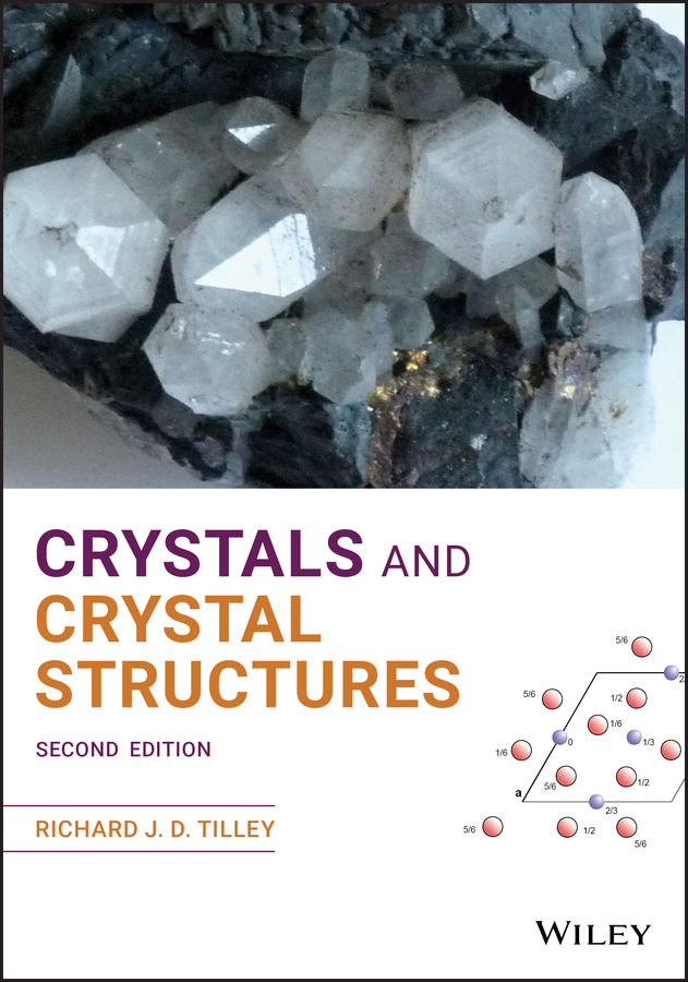
Cover Design: WileyCover Image: Hexagonal quartz Courtesy Richard Tilley5.4 High Quartz crystal Courtesy of Richard Tilley
Preface
Crystallography – the study of crystals, their structures and properties – plays an important role in a wide range of disciplines, including biology, chemistry, materials science and technology, mineralogy and physics, as well as engineering. The scientific breakthroughs in the first half of the twentieth century, leading to applications as diverse as nuclear power and semiconductor technology, were built, to a considerable extent, upon detailed understanding of metallic and inorganic crystal structures. In the latter half of the twentieth century, molecular biology, founded upon the determination of molecular crystal structures, has led to a deep‐rooted change in medicine, with the structures of biologically important molecules such as insulin, the epoch‐making determination of the crystal structure of DNA, and recent studies of protein structures being pivotal.
With these applications in mind, this book is designed as an introductory text for students and others who need to understand crystals and crystallography without necessarily becoming crystallographers. The aim is to explain how crystal structures are described for anyone coming to this area of study for the first time. At the end of it, a student should be able to read scientific papers and articles describing a crystal structure, or use crystallographic databases, with complete confidence and understanding. In addition, the book contains an introduction to areas of crystallography, such as modulated structures, quasicrystals and protein crystallography, that are currently the subject of active research.
The book is organised into nine chapters. The first of these provides an introduction to the subject. Chapters 2, 3 and 4 are concerned with fundamental crystallographic concepts: symmetry, lattices and lattice geometry, while the newly‐added Chapter 5 outlines the close relationship between crystal symmetry and physical properties. Chapter 6 is focused upon how to build or display a crystal structure, given the information normally available in databases. Chapter 7 outlines the relationship between diffraction and structure, including information on X‐ray, electron and neutron diffraction. Chapter 8 describes the principal ways in which structures are depicted in order to reveal chemical and physical relationships and biochemical reactivity. The final chapter, Chapter 9, is concerned with defects in crystals and recent developments in crystallography, such as the recognition of incommensurately modulated crystals and quasicrystals.
Compared with the first edition, all chapters have been rewritten and Chapter 5 is a new addition. Many figures have been redrawn and clarified and additional problems have been added in most chapters. However, the overall format of the book is carried over from the first edition. Each chapter is prefaced by three ‘Introductory Questions’. These are questions that have been asked by students in the past, and to some extent provide a focus for the student at the beginning of each chapter. The answers are given at the end of the chapter. Within each chapter, new crystallographic concepts, invariably linked to a definition of the idea, are given in bold type when encountered for the first time. All chapters are provided with a set of problems and exercises, designed to reinforce the concepts introduced. These are of two types. A multiple choice ‘Quick Quiz’ is meant to be tackled rapidly, and aims to reveal gaps in the reader’s grasp of the preceding material. The ‘Calculations and Questions’ sections are more traditional, and contain numerical exercises that reinforce concepts that are often described in terms of mathematical relationships or equations. A revised and updated bibliography provides students with sources for further exploration of the subject.
It is a pleasure to acknowledge the considerable assistance received in the preparation of this edition. The staff of the Trevithick Library, Cardiff University, were of great help in obtaining access to both historical literature and the rapidly expanding current scientific literature in the areas dealt with here. In addition, the staff at John Wiley, notably Emma Strickland, gave encouragement and assistance at all times during the production of this book. Last, but by no means least, I thank my wife Anne, without whose continual support this volume would not have been possible.
Richard J. D. Tilley
February 2020


