61,99 €
Mehr erfahren.
- Herausgeber: John Wiley & Sons
- Kategorie: Wissenschaft und neue Technologien
- Sprache: Englisch
The derivation of structural information from spectroscopic data is now an integral part of organic chemistry courses at all Universities. A critical part of any such course is a suitable set of problems to develop the students’ understanding of how organic structures are determined from spectra. The book builds on the very successful teaching philosophy of learning by hands-on problem solving; carefully graded examples build confidence and develop and consolidate a student’s understanding of organic spectroscopy.
Organic Structures from Spectra, 6th Edition is a carefully chosen set of about 250 structural problems employing the major modern spectroscopic techniques, including Mass Spectrometry, 1D and 2D 13C and 1H NMR Spectroscopy and Infrared Spectroscopy. There are 25 problems specifically dealing with the interpretation of spin–spin coupling in proton NMR spectra and 10 problems based on the quantitative analysis of mixtures using proton and carbon NMR spectroscopy. The accompanying text is descriptive and only explains the underlying theory at a level that is sufficient to tackle the problems. The text includes condensed tables of characteristic spectral properties covering the frequently encountered functional groups.
The examples themselves have been selected to include all important structural features and to emphasise connectivity arguments and stereochemistry. Many of the compounds were synthesised specifically for this book. In this collection, there are many additional easy problems designed to build confidence and to demonstrate basic principles.
The Sixth Edition of this popular textbook:
- now incorporates many new problems using 2D NMR spectra (C–H Correlation spectroscopy, HMBC, COSY, NOESY and TOCSY);
- has been expanded and updated to reflect the new developments in NMR spectroscopy;
- has an additional 40 carefully selected basic problems;
- provides a set of problems dealing specifically with the quantitative analysis of mixtures using NMR spectroscopy;
- features proton NMR spectra obtained at 200, 400 and 600 MHz and 13C NMR spectra including routine 2D C–H correlation, HMBC spectra and DEPT spectra;
- contains a selection of problems in the style of the experimental section of a research paper;
- includes examples of fully worked solutions in the appendix;
- has a complete set of solutions available to instructors and teachers from the authors.
Organic Structures from Spectra, Sixth Edition will prove invaluable for students of Chemistry, Pharmacy and Biochemistry taking a first course in Organic Chemistry.
Sie lesen das E-Book in den Legimi-Apps auf:
Seitenzahl: 213
Veröffentlichungsjahr: 2020
Ähnliche


Table of Contents
COVER
PREFACE
LIST OF TABLES
LIST OF FIGURES
1 INTRODUCTION
1.1 GENERAL PRINCIPLES OF ABSORPTION SPECTROSCOPY
1.2 CHROMOPHORES
1.3 DEGREE OF UNSATURATION
1.4 CONNECTIVITY
1.5 SENSITIVITY
1.6 PRACTICAL CONSIDERATIONS
2 ULTRAVIOLET (UV) SPECTROSCOPY
2.1 THE NATURE OF ULTRAVIOLET SPECTROSCOPY
2.2 BASIC INSTRUMENTATION
2.3 QUANTITATIVE ASPECTS OF ULTRAVIOLET SPECTROSCOPY
2.4 CLASSIFICATION OF UV ABSORPTION BANDS
2.5 SPECIAL TERMS IN UV SPECTROSCOPY
2.6 IMPORTANT UV CHROMOPHORES
2.7 THE EFFECT OF SOLVENTS
3 INFRARED (IR) SPECTROSCOPY
3.1 ABSORPTION RANGE AND THE NATURE OF IR ABSORPTION
3.4 IMPORTANT IR CHROMOPHORES
4 MASS SPECTROMETRY
4.1 IONISATION PROCESSES
4.2 INSTRUMENTATION
4.3 MASS SPECTRAL DATA
4.4 REPRESENTATION OF FRAGMENTATION PROCESSES
4.5 FACTORS GOVERNING FRAGMENTATION PROCESSES
4.6 EXAMPLES OF COMMON TYPES OF FRAGMENTATION
5
1
H NUCLEAR MAGNETIC RESONANCE (NMR) SPECTROSCOPY
5.1 THE PHYSICS OF NUCLEAR SPINS AND NMR INSTRUMENTS
5.2 BASIC NMR INSTRUMENTATION AND THE NMR EXPERIMENT
5.3 CHEMICAL SHIFTS IN 1H NMR SPECTROSCOPY
5.4 SPIN–SPIN COUPLING IN 1H NMR SPECTROSCOPY
5.5 ANALYSIS OF 1H NMR SPECTRA
5.6 CORRELATION OF 1H–1H COUPLING WITH STRUCTURE
5.7 THE NUCLEAR OVERHAUSER EFFECT (NOE) IN 1H NMR SPECTROSCOPY
5.8 LABILE AND EXCHANGEABLE PROTONS
REFERENCES
6
13
C NMR SPECTROSCOPY
6.1 COUPLING AND DECOUPLING IN
13
C NMR SPECTRA
6.2 THE NUCLEAR OVERHAUSER EFFECT (NOE) IN 13C NMR SPECTROSCOPY
6.3 DETERMINING 13C SIGNAL MULTIPLICITY USING DEPT
6.4 SHIELDING AND CHARACTERISTIC CHEMICAL SHIFTS IN 13C NMR SPECTRA
7 2-DIMENSIONAL NMR SPECTROSCOPY
7.1 PROTON–PROTON INTERACTIONS BY 2D NMR
7.2 PROTON–CARBON INTERACTIONS BY 2D NMR
8 MISCELLANEOUS TOPICS
8.1 SOLVENTS FOR NMR SPECTROSCOPY
8.2 SOLVENT-INDUCED SHIFTS
8.3 DYNAMIC NMR SPECTROSCOPY – THE NMR TIME-SCALE
8.4 THE EFFECT OF CHIRALITY
8.5 THE NMR SPECTRA OF “OTHER NUCLEI”
9 DETERMINING THE STRUCTURE OF ORGANIC COMPOUNDS FROM SPECTRA
Collection of Spectra
9.1 SOLVING PROBLEMS
9.2 WORKED EXAMPLES
10 PROBLEMS
Problem 1
Problem 2
Problem 3
Problem 4
Problem 5
Problem 6
Problem 7
Problem 8
Problem 9
Problem 10
Problem 11
Problem 12
Problem 13
Problem 14
Problem 15
Problem 16
Problem 17
Problem 18
Problem 19
Problem 20
Problem 21
Problem 22
Problem 23
Problem 24
Problem 25
Problem 26
Problem 27
Problem 28
Problem 29
Problem 30
Problem 31
Problem 32
Problem 33
Problem 34
Problem 35
Problem 36
Problem 37
Problem 38
Problem 39
Problem 40
Problem 41
Problem 42
Problem 43
Problem 44
Problem 45
Problem 46
Problem 47
Problem 48
Problem 49
Problem 50
Problem 51
Problem 52
Problem 53
Problem 54
Problem 55
Problem 56
Problem 57
Problem 58
Problem 59
Problem 60
Problem 61
Problem 62
Problem 63
Problem 64
Problem 65
Problem 66
Problem 67
Problem 68
Problem 69
Problem 70
Problem 71
Problem 72
Problem 73
Problem 74
Problem 75
Problem 76
Problem 77
Problem 78
Problem 79
Problem 80
Problem 81
Problem 82
Problem 83
Problem 84
Problem 85
Problem 86
Problem 87
Problem 88
Problem 89
Problem 90
Problem 91
Problem 92
Problem 93
Problem 94
Problem 95
Problem 96
Problem 97
Problem 98
Problem 99
Problem 100
Problem 101
Problem 102
Problem 103
Problem 104
Problem 105
Problem 106
Problem 107
Problem 108
Problem 109
Problem 110
Problem 111
Problem 112
Problem 113
Problem 114
Problem 115
Problem 116
Problem 117
Problem 118
Problem 119
Problem 120
Problem 121
Problem 122
Problem 123
Problem 124
Problem 125
Problem 126
Problem 127
Problem 128
Problem 129
Problem 130
Problem 131
Problem 132
Problem 133
Problem 134
Problem 135
Problem 136
Problem 137
Problem 138
Problem 139
Problem 140
Problem 141
Problem 142
Problem 143
Problem 144
Problem 145
Problem 146
Problem 147
Problem 148
Problem 149
Problem 150
Problem 151
Problem 152
Problem 153
Problem 154
Problem 155
Problem 156
Problem 157
Problem 158
Problem 159
Problem 160
Problem 161
Problem 162
Problem 163
Problem 164
Problem 165
Problem 166
Problem 167
Problem 168
Problem 169
Problem 170
Problem 171
Problem 172
Problem 173
Problem 174
Problem 175
Problem 176
Problem 177
Problem 178
Problem 179
Problem 180
Problem 181
Problem 182
Problem 183
Problem 184
Problem 185
Problem 186
Problem 187
Problem 188
Problem 189
Problem 190
Problem 191
Problem 192
Problem 193
Problem 194
Problem 195
Problem 196
Problem 197
Problem 198
Problem 199
Problem 200
Problem 201
Problem 202
Problem 203
Problem 204
Problem 205
Problem 206
Problem 207
Problem 208
Problem 209
Problem 210
Problem 211
Problem 212
Problem 213
Problem 214
Problem 215
Problem 216
Problem 217
Problem 218
Problem 219
Problem 220
Problem 221
Problem 222
Problem 223
Problem 224
Problem 225
Problem 226
Problem 227
Problem 228
Problem 229
Problem 230
Problem 231
Problem 232
Problem 233
Problem 234
Problem 235
Problem 236
Problem 237
Problem 238
Problem 239
Problem 240
Problem 241
Problem 242
Problem 243
Problem 244
Problem 245
Problem 246
Problem 247
Problem 248
Problem 249
Problem 250
Problem 251
Problem 252
Problem 253
Problem 254
Problem 255
Problem 256
Problem 257
Problem 258
Problem 259
Problem 260
Problem 261
Problem 262
Problem 263
Problem 264
Problem 265
Problem 266
Problem 267
Problem 268
Problem 269
Problem 270
Problem 271
Problem 272
Problem 273
Problem 274
Problem 275
Problem 276
Problem 277
Problem 278
Problem 279
Problem 280
Problem 281
Problem 282
Problem 283
Problem 284
Problem 285
Problem 286
Problem 287
Problem 288
Problem 289
Problem 290
Problem 291
Problem 292
Problem 293
Problem 294
Problem 295
Problem 296
Problem 297
Problem 298
Problem 299
Problem 300
Problem 301
Problem 302
Problem 303
Problem 304
Problem 305
Problem 306
Problem 307
Problem 308
Problem 309
INDEX
END USER LICENSE AGREEMENT
List of Tables
Chapter 2
Table 2.1 Observable UV Absorption Bands for Acetophenone
Table 2.2 The Effect of Extended Conjugation on UV Absorption
Table 2.3 UV Absorption Bands in Common Carbonyl Compounds
Table 2.4 UV Absorption Bands in Common Benzene Derivatives
Chapter 3
Table 3.2 C–H IR Absorption Frequencies in Common Functional Groups
Table 3.3 C≡N and C≡C Absorption Frequencies in Common Functional Groups...
Table 3.4 C=O IR Absorption Frequencies in Common Functional Groups
Table 3.5 Characteristic IR Absorption Frequencies for Functional Groups
Chapter 4
Table 4.1 Accurate Masses of Selected Isotopes
Table 4.2 Common Fragments and their Masses
Chapter 5
Table 5.1 Nuclear Spins and Magnetogyric Ratios for Common NMR-Active Nuclei...
Table 5.2 Resonance Frequencies of 1H and 13C Nuclei in Magnetic Fields of Dif...
Table 5.3 Typical 1H Chemical Shift Values (δ) in Selected Organic Comp...
Table 5.4 Typical
1
H Chemical Shift Values (δ) of Selected P...
Table 5.5 1H Chemical Shift Values (δ) for Protons in Common Alkyl Deri...
Table 5.7 Approximate
1
H Chemical Shifts (δ) for Olefinic Protons
Table 5.8 Approximate 1H Chemical Shifts (δ) for Aromatic Protons in Be...
Table 5.9 1H Chemical Shifts (δ) for Protons in some Polynuclear Aromat...
Table 5.10 Typical
1
H–
1
H Coupling Constants
Table 5.11 Relative Line Intensities for Simple Multiplets
Table 5.12 Proton–Proton Coupling Constants in Aromatic and Heteroaromatic Rin...
Chapter 6
Table 6.1 The Number of Aromatic 13C Resonances in Benzenes with Different Sub...
Table 6.2 Typical 13C Chemical Shift Values in Selected Organic Compounds...
Table 6.3 Typical
13
C Chemical Shift Ranges in Organic Compounds
Table 6.5 13C Chemical Shifts (δ) for sp3-hybridised Carbons in Alkyl D...
Table 6.6 13C Chemical Shifts (δ) for sp2-hybridised Carbons in Vinyl D...
Table 6.7 13C Chemical Shifts (δ) for sp-hybridised Carbons in Alkynes ...
Table 6.8 Approximate 13C Chemical Shifts (δ) for Aromatic Carbons in ...
Table 6.9 Characteristic 13C Chemical Shifts (δ) in some Polynuclear A ...
Chapter 8
Table 8.1 1H and 13C Chemical Shifts for Common NMR solvents
List of Illustrations
Chapter 1
Figure 1.1 Schematic Absorption Spectrum
Figure 1.2 Definition of a Spectroscopic Transition
Chapter 2
Figure 2.1 Schematic Representation of an IR or UV Spectrometer
Figure 2.2 Schematic Representation of a Double-Beam Absorption Spectrometer...
Figure 2.3 Definition of Absorbance (A)
Chapter 4
Figure 4.1 Schematic Mass Spectrum
Figure 4.2 Schematic Diagram of an Electron-Impact Magnetic Sector Mass Spect...
Figure 4.3 Relative Intensities of the Cluster of Molecular Ions for Molecule...
Chapter 5
Figure 5.1 A Spinning Positive Charge Generates a Magnetic Field and Behaves l...
Figure 5.2 Schematic Representation of a CW NMR Spectrometer
Figure 5.3 Schematic Representation of a Pulsed NMR Spectrometer
Figure 5.4 1H NMR Spectra: (a) Time Domain Spectrum (FID); (b) Frequency Domai...
Figure 5.5
1
H NMR Spectrum of Bromoethane (400 MHz, CDCl
3
)
Figure 5.6 Shielding/deshielding Zones for Common Non-aromatic Functional Grou...
Figure 5.7 1H NMR Spectrum of Bromoethane (400 MHz, CDCl3) Showing the Multip...
Figure 5.8 Characteristic Multiplet Patterns for Common Organic Fragments...
Figure 5.9 Aromatic Region of the 1H NMR Spectrum of 2-Bromotoluene (acetone-...
Figure 5.10 Simulated 1H NMR Spectra of a 2-Spin System as the Ratio ...
Figure 5.11 A Portion of the 1H NMR Spectrum of Styrene Epoxide (100 ...
Figure 5.12 The 60 MHz
1
H NMR Spectrum of a 4-Spin AMX...
Figure 5.13 Selective Decoupling in the
1
H NMR Spectrum of Bromoethane...
Figure 5.14 Selective Decoupling in a Simple 4-Spin System
Figure 5.15 Characteristic Aromatic Splitting Patterns in the 1H NMR Spectra ...
Figure 5.16 Characteristic Aromatic Splitting Patterns in the 1H NMR Spectra ...
Figure 5.17 1H NMR Spectrum of p-Nitrophenylacetylene (200 MHz as a 10% solut...
Figure 5.18 Aromatic Region of the 1H NMR Spectrum of 2,4-Dinitrotoluene. (i)...
Figure 5.19 D2O Exchange in the 1H NMR Spectrum of 1-Propanol (300 MHz, CDCl3...
Chapter 6
Figure 6.1 13C NMR Spectra of Methyl Cyclopropyl Ketone (CDCl3 solvent, 100 M...
Chapter 7
Figure 7.1 Acquisition of a 2D NMR spectrum: a series of individual FIDs are ...
Figure 7.2 Representations of 2D NMR Spectra: (a) Stacked Plot; (b) Contour P...
Figure 7.3 Representations of Phase-sensitive 2D NMR Spectra: (a) Stacked Plo...
Figure 7.4
1
H COSY Spectrum of 1-Iodobutane (CDCl
3
solvent, 298K, 400 MHz)
Figure 7.5 1H TOCSY Spectrum of Butyl Ethyl Ether (CDCl3 solvent, 298K, 400 MH...
Figure 7.6 1H NOESY Spectrum of β-Butyrolactone (CDCl3 solvent, 298K, 600 MHz)...
Figure 7.7 1H–13C me-HSQC Spectrum of 1-Iodobutane (CDCl>3 solvent, 298K, 1H 4...
Figure 7.8 1H–13C HMBC Spectrum of 1-Iodobutane (CDCl3 solvent, 298K, 1H 400 ...
Figure 7.9 1H–13C HMBC Spectrum of 2-Bromophenol (CDCl3 solvent, 298K, 1H 400...
Chapter 8
Figure 8.1 Schematic NMR Spectra of Two Exchanging Nuclei
Figure 8.2
1
H NMR Spectrum of the Aliphatic Region of Cysteine
Guide
Cover
Table of Contents
Begin Reading
Pages
iii
iv
ix
x
xi
xiii
xiv
xv
xvi
1
2
3
4
5
6
7
8
9
10
11
12
13
14
15
16
17
18
19
20
21
22
23
24
25
26
27
28
29
30
31
32
33
34
35
36
37
38
39
40
41
42
43
44
45
46
47
48
49
50
51
52
53
54
55
56
57
58
59
60
61
62
63
64
65
66
67
68
69
70
71
72
73
74
75
76
77
78
79
80
81
82
83
84
85
86
87
88
89
90
91
92
93
94
95
96
97
98
99
100
101
102
103
104
105
106
107
108
109
110
111
112
113
115
116
117
118
119
120
121
122
123
124
125
126
127
128
129
130
131
132
133
134
135
136
137
138
139
140
141
142
143
144
145
146
147
148
149
150
151
152
153
154
155
156
157
158
159
160
161
162
163
164
165
166
167
168
169
170
171
172
173
174
175
176
177
178
179
180
181
182
183
184
185
186
187
188
189
190
191
192
193
194
195
196
197
198
199
200
201
202
203
204
205
206
207
208
209
210
211
212
213
214
215
216
217
218
219
220
221
222
223
224
225
226
227
228
229
230
231
232
233
234
235
236
237
238
239
240
241
242
243
244
245
246
247
248
249
250
251
252
253
254
255
256
257
258
259
260
261
262
263
264
265
266
267
268
269
270
271
272
273
274
275
276
277
278
279
280
281
282
283
284
285
286
287
288
289
290
291
292
293
294
295
296
297
298
299
300
301
302
303
304
305
306
307
308
309
310
311
312
313
314
315
316
317
318
319
320
321
322
323
324
325
326
327
328
329
330
331
332
333
334
335
336
337
338
339
340
341
342
343
344
345
346
347
348
349
350
351
352
353
354
355
356
357
358
359
360
361
362
363
364
365
366
367
368
369
370
371
372
373
374
375
376
377
378
379
380
381
382
383
384
385
386
387
388
389
390
391
392
393
394
395
396
397
398
399
400
401
402
403
404
405
406
407
408
409
410
411
412
413
414
415
416
417
418
419
420
421
422
423
424
425
426
427
428
429
430
431
432
433
434
435
436
437
438
439
440
441
442
443
444
445
446
447
448
449
450
451
452
453
454
455
456
457
458
459
460
461
462
463
464
465
466
467
468
469
470
471
472
473
474
475
476
477
478
479
480
481
482
483
484
485
486
487
488
489
490
491
492
493
494
495
496
497
498
499
500
501
502
503
504
505
506
507
508
509
510
511
512
513
514
515
516
517
518
519
520
521
522
523
524
525
526
527
528
529
530
531
532
533
534
535
536
537
538
539
540
541
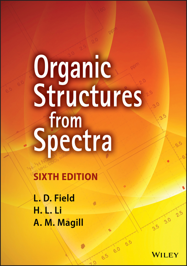
Organic Structures from Spectra
Sixth Edition
L. D. Field
Professor of Chemistry
School of Chemistry
University of New South Wales, Australia
H. L. Li
Senior Research Associate
School of Chemistry
University of New South Wales, Australia
A. M. Magill
Honorary Research Associate
School of Chemistry
University of New South Wales, Australia
This edition first published 2020
© 2020 John Wiley & Sons Ltd
Edition History
John Wiley & Sons (4e, 2008)
John Wiley & Sons (5e, 2013)
All rights reserved. No part of this publication may be reproduced, stored in a retrieval system, or transmitted, in any form or by any means, electronic, mechanical, photocopying, recording or otherwise, except as permitted by law. Advice on how to obtain permission to reuse material from this title is available at http://www.wiley.com/go/permissions.
The right of L. D. Field, H. L. Li and A. M. Magill to be identified as the authors of this work has been asserted in accordance with law.
Registered Offices
John Wiley & Sons, Inc., 111 River Street, Hoboken, NJ 07030, USA
John Wiley & Sons Ltd, The Atrium, Southern Gate, Chichester, West Sussex, PO19 8SQ, UK
Editorial Office
John Wiley & Sons Ltd, The Atrium, Southern Gate, Chichester, West Sussex, PO19 8SQ, UK
For details of our global editorial offices, customer services, and more information about Wiley products visit us at www.wiley.com.
Wiley also publishes its books in a variety of electronic formats and by print-on-demand. Some content that appears in standard print versions of this book may not be available in other formats.
Limit of Liability/Disclaimer of Warranty
In view of ongoing research, equipment modifications, changes in governmental regulations, and the constant flow of information relating to the use of experimental reagents, equipment, and devices, the reader is urged to review and evaluate the information provided in the package insert or instructions for each chemical, piece of equipment, reagent, or device for, among other things, any changes in the instructions or indication of usage and for added warnings and precautions. While the publisher and authors have used their best efforts in preparing this work, they make no representations or warranties with respect to the accuracy or completeness of the contents of this work and specifically disclaim all warranties, including without limitation any implied warranties of merchantability or fitness for a particular purpose. No warranty may be created or extended by sales representatives, written sales materials or promotional statements for this work. The fact that an organization, website, or product is referred to in this work as a citation and/or potential source of further information does not mean that the publisher and authors endorse the information or services the organization, website, or product may provide or recommendations it may make. This work is sold with the understanding that the publisher is not engaged in rendering professional services. The advice and strategies contained herein may not be suitable for your situation. You should consult with a specialist where appropriate. Further, readers should be aware that websites listed in this work may have changed or disappeared between when this work was written and when it is read. Neither the publisher nor authors shall be liable for any loss of profit or any other commercial damages, including but not limited to special, incidental, consequential, or other damages.
Library of Congress Cataloging-in-Publication Data
Names: Field, L. D., author.
Title: Organic structures from spectra / L.D. Field, Professor of
Chemistry, School of Chemistry, University of New South Wales, H.L.
Li, Senior Research Fellow, School of Chemistry, University of New South
Wales, A.M. Magill, Honorary Research Fellow, School of Chemistry, University of New South Wales.
Description: Sixth edition. | Hoboken, NJ : Wiley, 2020. | Includes bibliographical references and index.
Identifiers: LCCN 2020004972 (print) | LCCN 2020004973 (ebook) | ISBN 9781119524809 (paperback) | ISBN 9781119524793 (adobe pdf) | ISBN 9781119524847 (epub)
Subjects: LCSH: Spectrum analysis–Problems, exercises, etc. | Organic compounds–Structure–Problems, exercises, etc.
Classification: LCC QD272.S6 S74 2020 (print) | LCC QD272.S6 (ebook) | DDC 543/.17—dc23
LC record available at https://lccn.loc.gov/2020004972
LC ebook record available at https://lccn.loc.gov/2020004973
Cover Design: Wiley
Cover Image: Courtesy of Professor L. D. Field, Dr. Hsiu Lin Li and Dr. Alison Magill; © Chernookaya/Shutterstock
PREFACE
This is the Sixth Edition of the text “Organic Structures from Spectra”. The original text, published in 1986 by J R Kalman and S Sternhell, was a remarkable instructive text at a time where spectroscopic analysis, particularly NMR spectroscopy, was becoming widespread and routinely available in many chemical laboratories. The original text was founded on the premise that the best way to learn to obtain “structures from spectra” is to build up skills by practising on simple problems. Editions two through five of the text have been published at about five-yearly intervals and each revision has taken account of new developments in spectroscopy as well as dropping out techniques that have become less important or obsolete over time. The collection has grown substantially and we are deeply indebted to Dr John Kalman and to Emeritus Professor Sev Sternhell for their commitment and contribution to all of the previous editions of “Organic Structures from Spectra”.
Edition Six of the text has been expanded to include a new selection of problems and many of the problems now incorporate 2D NMR spectra (COSY, TOCSY, NOESY, C–H Correlation spectroscopy or HMBC).
The overarching philosophy remains the same as in previous editions of the text:
Theoretical exposition is kept to a minimum, consistent with gaining an understanding of those aspects of the various spectroscopic techniques which are actually used in solving problems. Experience tells us that both mathematical detail and in-depth theoretical description of advanced techniques merely confuse or overwhelm the average student.


