133,99 €
Mehr erfahren.
- Herausgeber: Wiley-VCH GmbH
- Kategorie: Wissenschaft und neue Technologien
- Sprache: Englisch
Discover this timely, comprehensive, and up-to-date exploration of crucial aspects of the use of nanomaterials in analytical chemistry
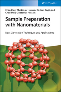
Sample Preparation with Nanomaterials: Next Generation Techniques for Sample Preparation delivers insightful and complete overview of recent progress in the use of nanomaterials in sample preparation. The book begins with an overview of special features of nanomaterials and their applications in analytical sciences. Important types of nanomaterials, like carbon nanotubes and magnetic particles, are reviewed and biological sample preparation and lab-on-a-chip systems are presented.
The distinguished author places special emphasis on approaches that tend to green and reduce the cost of sample treatment processes. He also discusses the legal, economical, and toxicity aspects of nanomaterial samples. This book includes extensive reference material, like a complete list of manufacturers, that makes it invaluable for professionals in analytical chemistry.
Sample Preparation with Nanomaterials offers considerations of the economic aspects of nanomaterials, as well as the assessment of their toxicity and risk. Readers will also benefit from the inclusion of:
- A thorough introduction to nanomaterials in the analytical sciences and special properties of nanomaterials for sample preparation
- An exploration of the mechanism of adsorption and desorption on nanomaterials, including carbon nanomaterials used as adsorbents
- Discussions of membrane applications of nanomaterials, surface enhanced raman spectroscopy, and the use of nanomaterials for biological sample preparation
- A treatment of magnetic nanomaterials, lab-on-a-chip nanomaterials, and toxicity and risk assessment of nanomaterials
Perfect for analytical chemists, materials scientists, and process engineers, Sample Preparation with Nanomaterials: Next Generation Techniques for Sample Preparation will also earn a place in the libraries of analytical laboratories, universities, and companies who conduct research into nanomaterials and seek a one-stop resource for sample preparation.
Sie lesen das E-Book in den Legimi-Apps auf:
Seitenzahl: 586
Veröffentlichungsjahr: 2021
Ähnliche


Table of Contents
Cover
Title Page
Copyright
1 Nanomaterials (NMs) in Analytical Sciences
1.1 Introduction
1.2 Types of NMs
1.3 Applications of NMs
1.4 Conclusions
References
2 Special Properties of Nanomaterials (NMs) for Sample Preparation
2.1 Introduction
2.2 Mechanical Properties of NMs
2.3 Thermal Properties of NMs
2.4 Electrical Properties of NMs
2.5 Optical Properties of NMs
2.6 Magnetic Properties of NMs
2.7 Adsorption Properties of NMs
2.8 Conclusions
References
3 Adsorption Mechanism on Nanomaterials (NMs)
3.1 Introduction
3.2 Adsorption Process
3.3 Conclusions and Future Perspective
References
4 Carbon Nanomaterials (CNMs) as Adsorbents for Sample Preparation
4.1 Introduction
4.2 Carbon Nanomaterials (CNMs)
4.3 Adsorption on CNMs
4.4 Applications of CNMs
4.5 Conclusions
References
5 Membrane Applications of Nanomaterials (NMs)
5.1 Introduction
5.2 Traditional Membranes
5.3 Carbon Nanomaterial-based Membranes
5.4 Nanoparticle-based Membranes
5.5 Molecularly Imprinted Polymer (MIP)-based Membranes
5.6 Conclusions
References
6 Surface-Enhanced Raman Spectroscopy (SERS) with Nanomaterials (NMs)
6.1 Introduction
6.2 Theory of SERS
6.3 SERS Mechanisms
6.4 Determination of SERS Enhancement Factor
6.5 Selection Rules
6.6 Fabrications of SERS Substrates
6.7 Applications of SERS
6.8 Conclusions
References
7 Nanomaterials (NMs) for Biological Sample Preparations
7.1 Introduction
7.2 The Use of NMs in Diagnostic Platforms
7.3 NMs-based Lab-on-a-chip (LOC) Platforms
7.4 Biomedical Applications of NMs
7.5 Sensor Applications of NMs
7.6 Conclusions
References
8 Magnetic Nanomaterials for Sample Preparation
8.1 Introduction
8.2 Synthesis of Magnetic Nanoparticles
8.3 Solid-Phase Extraction (SPE)
8.4 Magnetic Solid-Phase Extraction (MSPE)
8.5 Conclusions and Future Trends
References
9 Lab-on-a-Chip with Nanomaterials (NMs)
9.1 Introduction
9.2 Lab-on-a-Chip (LOC) Concept
9.3 NM-Based LOC Platforms
9.4 Conclusions and Future Perspectives
References
10 Toxicity and Risk Assessment of Nanomaterials
10.1 Introduction
10.2 Hazard Assessment of Nanomaterials
10.3 Toxicity Mechanism of Nanomaterials
10.4 The Traditional Risk Assessment Paradigm
10.5 Strategies for Improving Specific Risk Assessment
10.6 Conclusions
References
11 Economic Aspects of Nanomaterials (NMs) for Sample Preparation
11.1 Introduction
11.2 Toxicity Concerns of NMs
11.3 Global Market for NM-Based Products
11.4 Conclusions
References
12 Legal Aspects of Nanomaterials (NMs) for Sample Preparation
12.1 Introduction
12.2 Safety Issues of NMs
12.3 Regulatory Aspects of NMs
12.4 Conclusions
References
13 Monitoring of Nanomaterials (NMs) in the Environment
13.1 Introduction
13.2 Toxicity and Safety Concerns of NMs
13.3 Main Sources and Transport Routes of Nanopollutants
13.4 Requirements of Analytical Approaches
13.5 Sampling of NMs in Environmental Samples
13.6 Separation of NMs in Environmental Samples
13.7 Detection Techniques for the Characterization of NMs
13.8 Conclusions
References
14 Future Prospect of Sampling
14.1 Introduction
14.2 Sampling
14.3 Sample Preparation
14.4 Green Chemistry
14.5 Miniaturization of Analytical Systems
14.6 Conclusions
References
Index
End User License Agreement
List of Tables
Chapter 2
Table 2.1 Comparison of Young's modulus values for the carbon-based nanomater...
Chapter 3
Table 3.1 Comparison between physical adsorption and chemical adsorption.
Chapter 5
Table 5.1 Some latest researches on the application of composite membranes ba...
Chapter 10
Table 10.1 A brief description of various features of nanomaterials relevant ...
List of Illustrations
Chapter 1
Figure 1.1 The schematic depiction of the formation of SWCNTs and MWCNTs by ...
Figure 1.2 The historical development of fullerenes and their derivatives si...
Figure 1.3 Color dependence of various gold-based nanomaterials on their sha...
Figure 1.4 Nanotechnology-based tools for personalized medicine.
Figure 1.5 The schematic representation of the (a) functionalization procedu...
Figure 1.6 (a) The scheme of the preparation of molecular imprinting-based f...
Figure 1.7 The scheme of the preparation of molecular imprinting-based fluor...
Figure 1.8 The illustration of the fabrication of molecular imprinting-based...
Chapter 3
Figure 3.1 Types of adsorption isotherms according to the IUPAC classificati...
Figure 3.2 The schematic depiction of the preparation of magnetic Fe
3
O
4
@SiO
2
Figure 3.3 The schematic representation of the preparation of magnetic CNTs ...
Figure 3.4 The schematic demonstration of the preparation of polyamine-modif...
Figure 3.5 The schematic representation of the prepared graphene oxide modif...
Chapter 4
Figure 4.1 The schematic representation of the types of CNTs. (a) single-wal...
Figure 4.2 The schematic depiction of the preparation of graphene through re...
Figure 4.3 The schematic demonstration of the different types of fullerenes....
Figure 4.4 The schematic demonstration of the fabrication of GO-based nanoco...
Figure 4.5 The schematic depiction of the (a) preparation of polydopamine-co...
Chapter 5
Figure 5.1 The schematic depiction of the preparation of GO-based nanocompos...
Figure 5.2 The schematic demonstration of bare and PI-MWCNT electrode and th...
Figure 5.3 The schematic demonstration of the preparation of CNT-based Au NP...
Figure 5.4 The schematic demonstration of the fabrication of CNTs–PS nanocom...
Figure 5.5 The schematic demonstration of the removal of target dye compound...
Figure 5.6 The schematic depiction of the molecular imprinting approach.
Figure 5.7 The preparation of MIP-based polydopamine/GO nanocomposite membra...
Figure 5.8 The schematic demonstration of the preparation of MWCNT-based eno...
Figure 5.9 The fabrication of MIP/MWCNT nanocomposite membrane-based electro...
Chapter 6
Figure 6.1 The schematic demonstration of SERS technique.
Figure 6.2 The schematic depiction of the environmental applications of SERS...
Figure 6.3 (a) The schematic depiction of the magnetic separation and SERS-b...
Figure 6.4 The schematic depiction of the simultaneous detection of f-PSA an...
Figure 6.5 (a) The schematic representation of the detection of H7N9 virus b...
Chapter 7
Figure 7.1 The schematic depiction of an LOC platform.
Figure 7.2 The schematic depiction of an LOC platform developed by Dimov et ...
Figure 7.3 The schematic demonstration of the drug delivery and photothermal...
Figure 7.4 The schematic depiction of the preparation of magnetic nanocompos...
Figure 7.5 The schematic demonstration of the preparation of GO-chitosan nan...
Figure 7.6 The schematic demonstration of fluorescence-based detection of gl...
Figure 7.7 The schematic demonstration of molecular imprinting technique....
Figure 7.8 The illustration of the fabrication of molecular imprinting-based...
Chapter 8
Figure 8.1 The schematic demonstration of the preparation of magnetic Fe
3
O
4
/...
Figure 8.2 The schematic depiction of preparation of magnetic IIPs toward Cr...
Figure 8.3 The schematic depiction of the preparation of magnetic Fe
3
O
4
/SiO
2
Figure 8.4 The schematic depiction of the preparation of magnetic CoFe
2
O
4
@Si...
Figure 8.5 The schematic demonstration of the dispersive extraction process ...
Figure 8.6 The schematic demonstration of the preparation of magnetic Fe
3
O
4
@...
Chapter 9
Figure 9.1 The schematic demonstration of an LOC system.
Figure 9.2 The schematic depiction of the preparation of a graphene-based th...
Figure 9.3 The schematic demonstration of the gold nIDA on polymer substrate...
Figure 9.4 (a) The schematic depiction of the preparation of the microchanne...
Figure 9.5 The schematic demonstration of the solid-phase approach for the t...
Figure 9.6 The schematic demonstration of the prepared interfacing nanomecha...
Chapter 11
Figure 11.1 The schematic depiction of the interactions of NMs with the endo...
Figure 11.2 Number of products using NMs in the period from 2012 to 2020....
Figure 11.3 Number of products and types of NMs produced in the years betwee...
Chapter 13
Figure 13.1 The schematic depiction of various routes and crucial transforma...
Chapter 14
Figure 14.1 The schematic depiction of the percentage share of the steps con...
Figure 14.2 The schematic depiction of the aspects of sustainability.
Figure 14.3 The aspects of green chemistry.
Guide
Cover Page
Table of Contents
Begin Reading
Pages
iii
iv
1
2
3
4
5
6
7
8
9
10
11
12
13
14
15
16
17
18
19
20
21
22
23
24
25
26
27
28
29
30
31
32
33
34
35
36
37
38
39
40
41
42
43
44
45
46
47
48
49
50
51
52
53
54
55
56
57
58
59
60
61
62
63
64
65
66
67
68
69
71
72
73
74
75
76
77
78
79
80
81
82
83
84
85
86
87
88
89
90
91
93
94
95
96
97
98
99
100
101
102
103
104
105
106
107
108
109
110
111
112
113
114
115
117
118
119
120
121
122
123
124
125
126
127
128
129
130
131
132
133
134
135
136
137
138
139
140
141
142
143
144
145
147
148
149
150
151
152
153
154
155
156
157
158
159
160
161
162
163
164
165
166
167
168
169
170
171
173
174
175
176
177
178
179
180
181
182
183
184
185
186
187
188
189
190
191
192
193
195
196
197
198
199
200
201
202
203
204
205
206
207
208
209
210
211
212
213
214
215
216
217
219
220
221
222
223
224
225
226
227
228
229
230
231
232
233
234
235
236
237
238
239
240
241
242
243
244
245
246
247
248
249
251
252
253
254
255
256
257
258
259
260
261
262
263
264
265
266
267
268
269
270
271
272
273
274
275
276
277
278
279
280
281
282
283
284
285
286
287
289
290
291
292
293
294
295
Sample Preparation with Nanomaterials
Next Generation Techniques and Applications
Dr. Chaudhery Mustansar Hussain
Dr. Rüstem Keçili
Dr. Chaudhery Ghazanfar Hussain
Authors
Dr. Chaudhery Mustansar HussainNew Jersey Institute of TechnologyDepartment of Chemistry & Environmental SciencesUniversity Heights, NewarkNew JerseyUnited States of America
Dr. Rüstem KeçiliAnadolu UniversityYunus Emre Vocational School of Health ServicesYeşiltepe, Çamlıdere Sk. No. 626210 Tepebaşı/EskişehirTurkey
Dr. Chaudhery Ghazanfar HussainDepartment of Computer Science and Information TechnologyUniversity of LahoreDefence RoadLahorePakistan
Cover Courtesy of Chaudhery Mustansar Hussain
All books published by Wiley-VCH are carefully produced. Nevertheless, authors, editors, and publisher do not warrant the information contained in these books, including this book, to be free of errors. Readers are advised to keep in mind that statements, data, illustrations, procedural details or other items may inadvertently be inaccurate.
Library of Congress Card No.: applied for
British Library Cataloguing-in-Publication Data A catalogue record for this book is available from the British Library.
Bibliographic information published by the Deutsche Nationalbibliothek The Deutsche Nationalbibliothek lists this publication in the Deutsche Nationalbibliografie; detailed bibliographic data are available on the Internet at <http://dnb.d-nb.de>.
© 2021 WILEY-VCH GmbH, Boschstr. 12, 69469 Weinheim, Germany
All rights reserved (including those of translation into other languages). No part of this book may be reproduced in any form – by photoprinting, microfilm, or any other means – nor transmitted or translated into a machine language without written permission from the publishers. Registered names, trademarks, etc. used in this book, even when not specifically marked as such, are not to be considered unprotected by law.
Print ISBN: 978-3-527-33817-7ePDF ISBN: 978-3-527-68560-8ePub ISBN: 978-3-527-68561-5oBook ISBN: 978-3-527-68562-2
1Nanomaterials (NMs) in Analytical Sciences
1.1 Introduction
Analytical science is the branch of science that finds solutions and improvement of classical methods by fulfilling the demands for analytical information modeled by modern economic and scientific society [1]. Analytical information is about describing chemical systems and determination of their components. Technically, analytical information consists of two major components, i.e. analytical capabilities and analytical properties of the system to be analyzed. The measurement of upgrading of an analytical method, characterization, and its results is called analytical capabilities, while analytical properties can be of three different types, i.e. basic, principal, and socio-environmental. In the past, most of the developments are generally focused on techniques and solving instrumental problems, and there was no scope of socio-environmental aspects.
The rapid progresses of the microelectronic industry since 1960s established semiconductor preparation techniques to enhance the density of transistors in integrated circuits. In 1980s, these techniques led to the first design and fabrication of the microelectromechanical systems (MEMSs) [2]. With the introduction of microelectronics and computer technologies, analytical science has undergone remarkable expansions in terms of automation, miniaturization, higher sensitivities, and greater precision and complete analytical effectiveness.
Nanotechnology is defined as the technology that appreciates and controls the matter at dimensions between 1 and 100 nm, called nanomaterials (NMs). NMs have received much attention in the past decade due to the novel physical and chemical properties associated with their size and shape [3–12]. Widespread applications and outstanding performance of NMs not only have accelerated the development of materials science but also offer many opportunities in other related disciplines and have a significant impact on many fields of science, including chemistry, electronics, optics, medicine, and bioanalysis. NMs possess different properties compared to the same material in their coarser or bulk form. NM structures can be in the form of particles, pores, wires, or tubes or combination of these. Unusual properties such as high conductivity, greater heat transfer, higher melting temperature, exceptional optical and magnetic properties, and super adsorbent, etc., provide NMs for a wide range of novel applications [13–20]. The main objective of nanotechnology is to use these exceptional properties of NMs and develop new products, tools, and methods. Nanotechnology can play an important role in analytical sciences. Principal design and fabrication of NMs with the incorporation of interfacial elements would be of paramount importance for the whole process of molecular analysis at present times and near future. The objective of this chapter is to focus on recent developments with different types of NMs in analytical sciences, i.e. sample preparation, separation, extraction, and identification techniques.
NMs can be highly selective materials when it comes to purification techniques. During last few years, these have been intensively researched in all types of chromatographic techniques such as gas chromatography [21], liquid chromatography [22], capillary chromatography [23], and membrane technology [24]. Overall, NMs are proven extremely important and exhibited effective improvements over present systems. However, obstacles to overcome them in separation sciences are the aggregations, stability, safety, and economic issues.
This chapter provides an overview of NMs in analytical sciences. The chapter starts with the description of the types of NMs including carbon nanotubes (CNTs), fullerenes (FULs), graphene, inorganic nanoparticles, and magnetic nanoparticles. Then, various examples on the application of NMs are demonstrated.
1.2 Types of NMs
Unique properties of NMs and new methods for the analysis of NMs are strong areas for research these days. NMs have initiated the development of new ways of performing target concentration and detection and new analytical methods and instrumentation for measuring the properties of NMs. The optical and electronic properties of NMs are often dominated by their surface chemistry, and this makes the task of analyzing NMs immensely more challenging than bulk materials or homogeneous solutions. A particularly intriguing incentive for developing new approaches to NMs fabrication is the potential for creating new constructs with nanoscale order and unique functional properties. NMs for analysis can be constructed with the manipulation of one or more components.
1.2.1 Graphene
Graphene was discovered in 2004 by Professor Andre Geim and Professor Kostya Novoselov from the University of Manchester [25]. Since then, many efforts were put into the design and development of new graphene-based functional materials. In addition, the Nobel Prize for Physics in 2010 was awarded to Geim and Novoselov in the University of Manchester for their research on graphene [26]. The excellent features of graphene make it the starting point of new technologies in many application areas. Although graphene has a very low thickness, it is more robust than the diamond and 300 times stronger than steel. It conducts electricity better than copper, passes light, and bends without any deformation and can be put into any desired shape. It also exhibits great features such as large surface area, high stability, and layered structure. These superior features of graphene make it as an excellent NM in many applications such as separation processes, sensors, bioimaging, and drug delivery [27–30].
Due to the large requirement for research and applications, there has been much investment put into the development of methods to prepare the high-quality graphene in bulk. The preparation methods can be classified into two categories, one is “top-down” and the other is “bottom-up.” The former one is to break graphite into graphene through approaches like mechanical cleavage, liquid exfoliation, thermal expansion, and electrochemical exfoliation. The latter one is to synthesize graphene by the techniques such as chemical vapor deposition (CVD), arc discharge, epitaxial growth on SiC, and unzipping CNTs [31–34].
1.2.2 Carbon Nanotubes (CNTs)
CNTs were first discovered by Ijima [35] in 1991 and were successfully employed for different purposes in analytical sciences due to their mechanical, electric, optical, and magnetic properties as well as their extremely large surface area [36, 37]. CNTs are hollow graphitic materials composed of one or multiple layers of graphene sheets: single-walled carbon nanotubes (SWCNTs) and multiwalled carbon nanotubes (MWCNTs), respectively. The schematic depiction of the formation of SWCNTs and MWCNTs by rolling of graphite layer is shown in Figure 1.1.
The synthesis of CNTs can be carried out by means of three main techniques: CVD [39], laser ablation (LA) [40], and catalytic arc discharge (CAD) [41]. CVD seems to be the most efficient approach for the synthesis of CNTs for analytical applications due to high purity and desirable tuning at low temperature. However, for all the synthesis methods, the presence of different undesired by-products (such as carbonaceous residues, amorphous carbon, metal impurities, and others) makes it necessary to purify CNTs. To purify the synthesized nanotubes, different strategies including chemical oxidation, physical separation, or combination of both chemical and physical techniques have been employed so far. Chemical oxidation is a purification system based on the fact that carbonaceous impurity residues are oxidized sooner than CNTs. The main advantage is its easy use, but it should be noted that the oxidation process affects the structure of the nanotube introducing functional groups (hydroxyl, carbonyl, and carboxyl) and defects in the side walls. Physical purification procedures are based on the different physical properties (such as size, ratio, weight, electrical and magnetic characteristics, etc.) between impurities and CNTs. Filtration, centrifugation, chromatography, and electrophoresis are the commonly employed techniques. The disadvantages of these procedures are: first, the elimination of certain impurities is inefficient; second, a high dispersion of CNTs is required; and third, only a low quantity of CNT can be purified.
Figure 1.1 The schematic depiction of the formation of SWCNTs and MWCNTs by rolling of graphite layer.
Source: Khan et al. [38]. © 2017, Elsevier.
The adsorption sites on CNTs are on the wall and in the interstitial spaces between tubes. These sites are easily accessed for both adsorption and rapid desorption. The impurity coverage on the CNT reduces their availability because the sorbate has to diffuse through these impurities to reach the CNT. Moreover, the porous structure of impurities introduces mass transfer limitations, slowing both adsorption and desorption. Understanding these characteristics is important for their application separation media. The excellent features of CNTs, along with their nanoscale features, make them ideal candidates for microscale devices for gas as well as liquid phase analysis. In gas phase analysis, one can implement a micro-concentrator or a micro-sorbent trap, which has been used in a variety of online chromatography and sensing applications. The purpose of such devices is usually to act as a fast pre-concentrator or to modulate the concentration of a stream for real-time monitoring. They have been fabricated in small capillary tubing and also by micromachining silicon and other substrates. The interesting liquid phase pre-concentrating applications are microscale solid-phase extraction (μ-SPE) and solid-phase microextraction (SPME).
Functionalization of CNTs offers a unique opportunity of altering the physical and chemical characteristics of the CNTs [42, 43]. The presence of a covalently attached functional group can alter the retention/affinity of the CNT surface and important properties such as polarity, hydrophilicity, and other specific interactions. The functional groups may also alter diffusional resistance and reduce the accessibility and affinity of CNT surfaces for certain analytes. Functionalization also enhances interaction with polymers and other materials, thus facilitating the formation of composites that can be used as pre-concentrating substrates. These include polymers and sol–gel-type immobilization.
1.2.3 Fullerenes (FULs)
FULs have attracted considerable attention in different fields of science since their discovery in 1985 [44]. Physical, chemical, and biological properties of fullerenes have shown their promising applications for future. It is inferred that size, hydrophobicity, three-dimensionality, and electronic configurations make them an appealing subject in chemistry. The historical development of fullerenes and their derivatives since their discovery is illustrated in Figure 1.2.
Figure 1.2 The historical development of fullerenes and their derivatives since their discovery.
Source: Pan et al. [45]. © 2020, Elsevier.
FULs are often accompanied with large amounts of soot; therefore, purification is a necessary step before their real applications are realized. Purification of FULs normally involves extraction from soot and then separation by liquid chromatography. Additionally, other purification techniques, like elution on molecular sieves or activated charcoal, gel permeation, and supercritical fluids purification, have also been used.
FULs are practically insoluble in water and generally tend to form aggregates in aqueous media [45]. To disperse these in water, prolonged stirring for days is needed. Addition of additives, e.g. surfactants, cyclodextrins, or sugar polymers, offer a possibility to disperse these in water more efficiently. FULs can also be dispersed in water by first dissolving it in an organic solvent, then adding the solution to water and evaporating-off the organic solvent.
Incorporation of FULs into polymeric materials to improve their processability has become a common practice in applications. The unique properties of FULs, such as their spherical shape, conjugated three-dimensional π-electronic system, and the ability to exhibit donor–acceptor or π–π interactions, make them attractive candidates for analytical applications [46]. As a result, these unique NMs can be successfully employed as chromatographic stationary nanophases in a wide temperature range (80–360 °C), with good thermal stability and high selectivity for aromatic compounds. Chemical functionalization of FULs is often carried out to alter their chemical properties and/or improve miscibility with host polymers.
1.2.4 Inorganic Nanoparticles
Inorganic NMs have received interest in analytical sciences because of the large specific surface area, lower weight, excellent mechanical strength, and magnetism [47–49]. However, in the field of sample preparation, only a few are prominent, while others remain unexplored yet. The prominent ones are gold, silver, titanium, and silica nanoparticles.
1.2.4.1 Gold and Silver Nanoparticles
Recent interest in the development of novel strategies for the generation of gold nanoparticles stems from their potential applications in the fields of physics, chemistry, biology, medicine, and materials science. Gold nanoparticles coupled with tunable surface properties in the form of inorganic or inorganic–organic hybrid are excellent platform for developing a broad range of analytical methods. Therefore, depending on their size, shape, degree of aggregation, and nature of the protecting organic shells on their surface, these can be engineered for individual and multifold applications, including chemical sensing and imaging [50, 51]. First, the gold core is essentially inert, and monodispersed nanoparticles can be synthesized with core sizes ranging from 1 to 150 nm. Further versatility is imparted by relatively simple functionalization via thiol linkages. Additionally, gold nanoparticles have been shown to exhibit size-dependent optical properties, which make them candidates for spectroscopic techniques such as surface-enhanced Raman spectroscopy (SERS). Gold nanoparticle-based fluorescent probes have also been used in various applications [52, 53]. The advantage of using gold nanoparticles in biological labeling is that visible light can be used to observe a color shift from red to blue when it forms aggregates. The ability to synthesize Au nanoparticles of different size is a great asset, for example, very small nanoparticles (4–6 nm) are essential for biological labeling, while SERS is typically carried out using relatively larger (around 35 nm) particles. Because of their unique optical features, gold nanoparticles find applications in many research fields. The coating of materials with gold nanoparticles is extensively applied for the sampling of scanning electron microscope (SEM) to enhance the electronic stream, which helps to achieve great SEM images with high quality.
Silver nanoparticles have attracted a great deal of attention in recent years due to their optical, physical, and chemical properties that differentiate them from corresponding bulk properties [54]. Hence, they have found many applications in catalysis, photonics, optoelectronics, information storage, and antibacterial materials. Silver powders, having ultrafine and uniformly distributed particle size, are of considerable use in the electronics industry as thick film conductors in integrated circuits due to their unique properties such as high electrical and thermal conductivity and high resistance to oxidation. Silver nanoparticles are also important SERS-active substrates that significantly enhance Raman intensities [55]. Currently, there are many kinds of silver-based SERS substrates, such as charged silver colloidal solution, acid-etching silver foil, metal island film formed through thermal evaporation, laser-ablated silver plate, etc.
1.2.4.2 Titanium Nanoparticles
Titanium dioxide (TiO2) is one of the most widely used of the NMs and has found wide ranging applications such as heterogeneous photocatalysis and energy conversion, etc. [56, 57]. TiO2naoparticles (NPs) are normally synthesized by sol–gel process, hydrothermal methods, and also high-energy methods such as laser pyrolysis/ablation, CVD, and plasma methods. Interest in Titania in analytical chemistry arises from its high chemical stability and cost-effectiveness. Strong oxidizing power of the holes, redox selectivity, high photo-stability, and easy preparation make TiO2 a powerful candidate as an efficient adsorbent.
1.2.4.3 Silica Nanoparticles
Mesoporous silicas are composed of a honeycomb-like porous structure with hundreds of empty channels (mesopores) that are able to absorb/encapsulate relatively large amounts of analytes. Unique properties, such as high surface area (>900 m2 g−1), large pore volume (>0.9 cm3 g−1), tunable pore size with a narrow distribution (2–10 nm), and good chemical and thermal stability, make them attractive for various sample preparation applications. Silica nanoparticles have gained great interest for use as sorbents, separation media in chromatography, and membrane separation [58, 59]. For mesoporous silica materials, it is difficult to separate the small particles from liquid. Therefore, these normally incorporate some functionalization, which provides nanosilica immobility, mechanical stability, and water insolubility.
1.2.5 Magnetic Nanoparticles
Magnetic nanoparticles are interesting advanced materials and have gained great attention among researchers from a wide range of disciplines in science [60–63]. Magnetic nanoparticles were successfully applied for different applications such as magnetic fluids [64], catalysis [65], magnetic resonance imaging [66], and environmental remediation [67, 68]. Magnetic nanoparticles exhibit superparamagnetic feature which can be attracted to a magnetic field, but retain no residual magnetism after the field is removed. Thus, it is very easy to separate these magnetic nanoparticles adhered with target compound(s) from complex matrices such as environmental, food, and biological samples by using an external magnetic field. There is no centrifugation or filtration step needed during this process. However, an unavoidable drawback associated with these nanoparticles in this range of their size is instability of these magnetic nanoparticles that tends to cause formation of agglomerates in the sample solution. Moreover, naked metallic nanoparticles are highly chemically active and can easily be oxidized in air that leads to loss of their dispersibility and magnetism feature. Therefore, it is important to design and develop protection approaches for the chemical stabilization of the naked magnetic nanoparticles. These approaches for the modification of the surfaces of magnetic nanoparticle comprise grafting technique or coating with an inorganic layer such as carbon or silica or coating with organic compounds including polymers and surfactants [69].
1.3 Applications of NMs
1.3.1 NMs in Separation Processes
NMs can be successfully employed in separation processes. For example, gold nanoparticles were successfully applied in the capillary electrophoresis (CE)-based analysis due to the improved separation resolutions [20]. These nanoparticles also behave as a pseudo-stationary phase in the existence of poly(ethylene oxide) (PEO) for the efficient separation of double-stranded DNA. The interaction between PEO and DNA molecule is increased in the existence of nanoparticles thereby enhancing the sieving ability of PEO without any change in its viscosity. The separation of acidic and basic proteins using a capillary filled with surfactant-capped gold nanoparticles was also reported [70] along with great separation efficiencies toward various target compounds [71]. A facile approach was developed and demonstrated for the fabrication of highly effective separation columns coated with octadecylamine-capped gold nanoparticles for open tubular capillary gas chromatography [72].
Monodispersed silica nanotubes having desired size and shape were designed and successfully prepared by using template-assisted synthesis approach. These developed nanotubes have inner voids that can be filled with various compounds varying from large protein molecules to small molecules, and the inner and outer surfaces of the nanotube can be efficiently functionalized. One of the interesting applications of the nanotubes is the nanophase extractor for the efficient removal of small compounds from the solution. Because the outer surface of the nanotubes exhibits hydrophilic feature and inner surface shows hydrophobic behavior, these NMs are promising candidates for the efficient extraction of the lipophilic compounds from the aqueous samples. The nanotube functionalized with antibody enables an ultimate extraction selectivity. The antibody produced against the drug, 4-[3-(4-fluorophenyl)-2-hydroxy-1-[1,2,4]-triazol-1-yl-propyl]-benzonitrile (FTB), selectively binds to the RS enantiomer and the Fab fragments of the antibody are immobilized on the inner and outer surfaces of the silica nanotubes [73] (Figure 1.3).
1.3.2 NMs in Biomedical Applications
Nanotechnology addresses these issues by designing and applying NM-based drug delivery systems that are capable of achieving enhanced biopharmaceutical features by altering the biopharmaceutical and pharmacokinetics features of the target pharmaceutical compound. Various NMs such as polymeric nanoparticles, liposomes, nanoemulsions, micelles, and dendrimers can be successfully used as nanodrug delivery systems (Figure 1.4). With the innovative nanoscience- and nanotechnology-based approaches, NMs having different functionalities, shapes, and distinct physical, chemical, and biological features were designed and developed for the successful applications in the pharma industry.
The functionalized NMs exhibit biomimetic features since they can change their unique features that enable them to be applied in drug delivery and self-healing materials [76]. The examples include the application of polymeric NMs as artificial muscles that can contract and return to original shape when short-circuited. These can replicate muscular action and can have strong visual effects.
Figure 1.3 Color dependence of various gold-based nanomaterials on their shape and size. (a) Gold nanorods, (b) silica–gold core–shell nanoparticles, and (c) gold nanocages. The intense color of these nanoparticles arises from the collective excitation of their conduction electrons or surface plasmon resonance modes. (d) Optical dark-field scattering micrograph of gold nanorods.
Source: Dreaden et al. [74]. © 2020, Royal Society of Chemistry.
Figure 1.4 Nanotechnology-based tools for personalized medicine.
Source: Sharma and Hussain [75]. © 2020, Elsevier.
One of the interesting and promising research fields is the designing and development of NMs whose structure and chemical features are responsive to the biocatalytic action of natural enzymes. Enzymes play a crucial role in all metabolic and biological processes, and their dysregulation is a feature of many diseases. Therefore, the NMs can efficiently sense these enzymes in the physiological environment and behave as an effective and promising tool for therapeutic and diagnostic applications. Various examples of enzyme-responsive NMs are polymeric NMs [77], phospholipids [78], and inorganic NMs [79]. Functional mesoporous silica nanoparticles (MSNs) were successfully designed and prepared by using alkoxysilane tether, α-cyclodextrin, and multifunctional peptides to target tumor cells and decrease the side effects of doxorubicin (DOX), which is an antitumor drug. The multifunctional peptides were composed of the cell-penetrating peptide of seven arginine (R7) sequences, an enzyme-cleavable peptide of Gly-Phe-Leu-Gly (GFLG peptide), and a tumor-targeting peptide of Arg-Gly-Asp-Ser (RGDS peptide). When MSNs loaded with DOX are incubated with tumor cells and normal cells, the nanoparticles could efficiently target tumor cells through the specific interactions between RGDS and integrin receptor αvβ3 overexpressed on tumor cells, followed by penetrating cell membrane with the aid of R7 sequence. Once DOX-loaded silica nanoparticles penetrate into the cellular membrane, the antitumor drug DOX is quickly released because of the breakage of GFLG peptide cleaved by cathepsin B, resulting in increased antitumor activity [80]. This effective enzyme-responsive nanoparticle-based drug delivery system exhibits an excellent potential in the area of nanomedicine (Figure 1.5).
Among various applications of NMs, the most promising applications are in the area of biomedical sciences. These interesting applications have crucial effects on the human health since these NMs are targeted toward the diagnostic techniques for disease which are fast and low cost. To decrease the side effects and increase the therapeutic effects, the pharmaceutical compounds should be selectively targeted at the disease site and accumulated for a prolonged time in the body. Drug release refers to the design and development of efficient approaches, techniques, and formulations required to transport any pharmaceutical compound safely within the body to obtain the efficient therapeutic effects [81]. In this regard, NM-based drug release systems have opened new opportunities in the area of medicine. NM-based drug release systems accumulate and bind specifically to the disease target with controlled release behavior. The careful design and preparation of these NMs as effective drug carriers should address various key issues [82] such as biodegradability and biocompatibility, high stability at physiological pH values, excellent drug-loading capacity without any toxicity, and industrial production of these NMs in large scale for clinical applications. The stimuli-responsive drug release systems deliver the drug at specific tissues in the systematic administration. These do not freely extravasate during the blood circulation and are released at the targets where the nanocarriers accumulate by active or passive targeting strategy [83].
Figure 1.5 The schematic representation of the (a) functionalization procedure of the MSNs, (b) MSNs loaded with DOX under physiological conditions, (c) RGDS targeted to the tumor cell, (d) endocytosis into a specific tumor cell, (e) Cathepsin Benzyme-triggered drug release in the cytoplasm, (f) apoptosis of the tumor cell.
Source: Cheng et al. [80]. © 2015, American Chemical Society.
An immunoassay is a biochemical technique that is efficiently applied in the detection of the concentration of a small molecule or a macromolecule in a sample solution by using an antigen or an antibody. In this approach, the key issue is to obtain a measurable signal in response to a binding effect. Screen-printed electrodes have attracted great attention as immunosensors because of their unique features such as miniaturized dimension, low-cost and facile fabrication, and large-scale production. A single drop of sample, i.e. only a few microliters of a sample solution, is used to conduct all the immunological stages, thereby decreasing the consumption of the reagents. Screen-printed electrodes can be successfully coated with nanoparticles that increase the sensitivity of these electrodes [84]. In an interesting study, screen-printed electrode was modified with silver nanoparticles and effectively applied for the preliminary screening of cystic fibrosis that is a common genetic disease caused by the autosomal recessive gene known as the cystic fibrosis transmembrane conductance regulator gene [85]. It affects multiple organs such as lungs and intestines, which causes the irregular transport of chloride and sodium ions across the epithelial cells [86]. It has been reported that the oxidation of silver is sensitive to concentration of chloride ions [87]. Sweat chloride levels are important for the effective diagnosis of cystic fibrosis. A layer of silver nanoparticles is deposited on the working electrode and anodically stripped off to generate silver cations. The anodic stripping voltammetry of silver nanoparticles yields a single silver oxidation peak in the absence of chloride ions, whereas two voltammetry stripping peaks (one for AgCl and other for the oxidation of Ag to Ag(I) ions) are obtained observed in the existence of chloride ions. The silver chloride peak is used for the quantitative determination of chloride concentration [88].
On the other hand, Parkinson's disease and Alzheimer's disease are the most common neurodegenerative diseases that affected 1.6% of the world population and around 26 million people, respectively [89, 90]. This has posed a growing challenge for the patients, clinicians, care givers, and society. The current treatment approaches rely on the clinical symptoms of the diseases, and therefore, the efficient diagnostic techniques depend on the proficiency of the treating physician. Nanoparticle-based sensor systems are successfully applied to distinguish groups of Parkinson's disease and Alzheimer's disease patients from a healthy control group. The diagnostic approach relies on the identification of patterns of volatile organic compounds in the exhaled breath [91]. The disease-related changes in the blood chemistry may be transmitted to the alveolar exhaled breath through the lungs even in the initial stages of the disease. The nanoparticle-based sensor undergoes fast and reversible changes in electrical resistance on being exposed to characteristic volatile organic compounds. Thus, breath prints can be the basis for the design and fabrication of facile, low-cost, non-invasive biomarker using nanoparticle-based sensor systems [92].
1.3.3 NMs in Sensor Platforms
Graphene and graphene-based nanocomposite materials were successfully applied in sensor applications. For example, Kong et al. reported the preparation of a nanocomposite material composed of reduced GO and Au/Ag bimetallic nanoparticles for the sensitive detection of daunorubicin in human serum samples 93. In this study, the prepared nanosensor showed high recognition behavior toward the target compound daunorubicin in a linear concentration range between 0.01 and 15 μg ml−1. The detection limit was obtained as 3 ng ml−1.
In another important study conducted by Arumugam and Kim [94], the sensitive recognition of ascorbic acid in aqueous samples was successfully carried out by using the prepared GO modified with CdSe and ZnS quantum dots. In their study, the prepared GO-based nanosensor displayed great affinity and sensitivity for the ascorbic acid, and the detection limit was achieved as 568 pM.
GO and GO-based NMs were also successfully applied for the selective recognition and removal of various pollutants from environmental samples. For example, Ghorbani et al. prepared GO functionalized with ethylene diamine for the removal of Cd(II) and Pb(II) ions from wastewater samples [95]. The effects of various factors such as the amount of the adsorbent, shaking rate, pH, and time on the extraction behavior of the prepared ethylene diamine-functionalized GO toward the target ions were investigated. The obtained results confirmed that the prepared ethylene diamine-functionalized GO successfully removed the target Cd(II) and Pb(II) ions from wastewater samples. The extraction percentages for Cd(II) and Pb(II) ions were achieved as 99.4% and 99.6%, respectively.
In another important study conducted by Keshvardoostchokami et al. [96], GO/Ag nanocomposite was developed for the efficient removal of an insecticide pollutant imidacloprid from water samples. The obtained results showed that approximately 63% of the target insecticide imidacloprid was removed from 50 ml water sample containing 10 mg l−1 by using 30 mg GO/Ag nanocomposite.
On the other hand, molecularly imprinted polymers (MIPs) are highly selective polymeric materials that mimic the shape-specific binding mechanism which is found naturally occurring in the biological structures (i.e. 3-D binding site of an enzyme or the glycoprotein recognition sites in antibodies). However, since they are designed and prepared from artificial polymers, MIPs display the same robustness and adaptability found in sensors constructed from artificial materials. MIPs as biomimetic recognition components efficiently combine the accuracy and highly specific binding affinity of a sensor system toward the target compound(s), with the great reusability and robustness [97–105].
The chemiluminescence technique is extensively applied in a number of areas because of its great sensitivity toward the target compound(s). However, it has some drawbacks such as low selectivity. Thus, one of the most crucial and urgent fields of the researches is to design and develop new systems having improved selectivity. Many efforts were put to overcome this significant problem. The combination of the chemiluminescence technique and molecular imprinting technology is a powerful approach that can be successfully applied for the development of molecular imprinting-based spectroscopic sensor systems toward pharmaceuticals. For example, Zor et al. prepared a photoluminescent sensor system composed of magnetic silica, photoluminescent graphene quantum dots (GQDs) and molecularly imprinted polypyrrole for the sensitive recognition of pesticide tributyltin that exhibits genotoxic feature and lead to increase in endocrine disruptions [106]. In their research, tributyltin was chosen as the template compound for the synthesis of selective MIP. The developed photoluminescent composite nanosensor based on molecular imprinting was effectively employed for the selective and sensitive recognition of tributyltin in sea water and water samples. The achieved limit of detection values for tributyltin in sea water and water samples were 42.56 and 12.78, respectively.
On the other hand, in the fluorescence-based detection of the target compound(s) by using molecular imprinting-based nanosensors, appropriate functional monomers having fluorescence feature are chosen for the development of molecular imprinting-based fluorescence sensor systems [107]. Once the target compound(s) selectively binds to recognition sites of the molecular imprinting-based sensor, intensity of the fluorescence rises or reduces depending on the design of the imprinted nanosensor.
In another interesting research carried out by Zhang et al. [108], a fluorescence-based nanosensor composed of ZnS/CdSe quantum dots has a selective MIP layer toward carbaryl which is an insecticide. The researchers successfully applied this nanosensor for the effective recognition of carbaryl in food samples including rice and cabbage. The achieved outputs from the experiments for the fluorescence-based detection of the target insecticide carbaryl displayed that the fluorescence-based imprinted nanosensor showed excellent recognition behavior and selectivity for carbaryl in the existence of its anologues such as isocarb and metolcarb.
In a research reported by Masteri-Farahani et al. [109], a molecular imprinting-based fluorescence nanosensor was developed for the efficient detection of methamphetamine, which is a quite addictive and illegal psychostimulant drug. For this purpose, they embedded GQDs into the MIP particles as schematically illustrated in Figure 1.3a. The obtained outputs showed that the developed molecular imprinting-based fluorescence nanosensor can sensitively and selectively recognize methamphetamine even in the existence of other similar compounds (i.e. ibuprofen, amphetamine, morphine, and codeine). The detection limit was achieved as 1.7 μg l−1.
Liu et al. prepared a sensitive molecular imprinting-based fluorescence nanosensor for the recognition of metronidazole in serum samples [110]. In this research, first, carbon dots (CDs) were synthesized using longan peels by applying high-pressure microwave approach. Then, the preparation of CDs-embedded MIPs was carried out by using the functional monomers acrylamide (AM) and glycidyl methacrylate (GMA), cross-linker ethylene glycol dimethylacrylate (EGDMA), and initiator 2,2′-azobisisobutyronitrile (AIBN) as schematically illustrated in Figure 1.4.
The developed molecular imprinting-based fluorescence nanosensor was employed for the sensitive and selective recognition of metronidazole in serum samples. The achieved sensor response toward metronidazole was linear in the range from 50 to 1200 ng ml−1, and the limit of detection was obtained as 17.4 ng ml−1.
In a work performed by Jalili et al. [111], penicillin G in milk samples was successfully detected by using the molecular imprinting-based fluorescence nanosensor having CDs. In their work, first, the synthesis of silica nanoparticles embedded with blue-emissive CDs was performed. Then, MIP film was formed on the surface of the prepared nanocomposite (CDs@silica nanoparticles) by using 3-aminopropyltriethoxysilane (APTES) and tetraethoxysilanz (TEOS) as functional monomers. The prepared molecular imprinting-based fluorescence nanosensor having CDs displayed excellent sensitivity toward penicillin G. The obtained sensor response was linear in the range between 1 and 32 nM. The limit of detection was found as 0.34 nM (Figure 1.6).
Figure 1.6 (a) The scheme of the preparation of molecular imprinting-based fluorescence nanosensor for methamphetamine. (b) Fluorescence quenching of molecular imprinting-based nanosensor in the existence of methamphetamine.
Source: Masteri-Farahani et al. [109]. © 2020, Elsevier.
Figure 1.7 The scheme of the preparation of molecular imprinting-based fluorescence nanosensor for the recognition of metronidazole.
Source: Liu et al. [110]. © 2019, Elsevier.
Stoian et al. developed a molecular imprinting-based electrochemical sensor for the selective and sensitive recognition of an antibiotic azithromycin in biological samples including tears, plasma, and urine samples [112]. For this purpose, the electrode position of the selective MIP layer was carried out on the surface of a glassy carbon electrode as schematically demonstrated in Figure 1.7. The functional monomer 3-thienyl boronic acid and cross-linker 2,2′-bithiophene were used for the formation of MIP layer on the electrode surface. The fabricated electrochemical sensor displayed excellent response toward the target antibiotic azithromycin in a linear range from 13.33 nM to 66.67 μM. The limit of detection was obtained as 0.85 nM (Figure 1.8).
1.4 Conclusions
NMs offer unique opportunities for designing ultrasensitive (bio)sensors and analytical assays. The broad potential of multifunctional nanoparticles for (bio)molecular recognition events and separations are required.
The remarkable sensitivity of the new multifunctional nanoparticle-based sensing protocols opens up the possibility for detecting disease markers, biothreat agents, chemicals, or infectious agents that cannot be measured by conventional methods. The use of multifunctional nanoparticles for detecting proteins is still in its infancy (most of the work has been focused on separations), but the experience gained in ultrasensitive DNA detection may provide a useful starting point. Despite analytical research in the field is active, there is still much space to explore and to explain tremendous opportunities and significant challenges to be afforded. For example, whereas present work is more concentrated on multifunctional magnetic nanoparticles, very few studies on multifunctional gold nanoparticles or quantum dots (QDs) are still appearing. Probably, the most prominent advantage of multifunctional magnetic nanoparticles over others lies in the fact that the particles can be easily functionalized and magnetically manipulated using permanent magnets independent of normal chemical or biological analysis.
Figure 1.8 The illustration of the fabrication of molecular imprinting-based electrochemical sensor for azithromycin.
Source: Stoian et al. [112]. © 2020, Elsevier.
The challenges of processing nanoparticles easily and their multifunctionalization with (bio) molecules or other nanoparticles in a biological and chemical friendly manner are just beginning to be addressed. Just starting with the base nanoparticle, one of the most significant barriers along the road is the poor reproducibility and the wide range of size distribution of nanoparticle product synthesized from wet chemical methods. Improved synthetic methodologies that can lead to true monodispersity and satisfactory reproducibility of nanoparticles with well-controlled shape remain to be developed. On the other hand, nanoparticle surface modification is also a critical step for protecting the nanoparticle core, improving aqueous dispersibility and obtaining appropriate surface multifunctionality for the development of highly sensitive and selective “nanosensors.” Even, from a physicochemical point of view, clear and quantitative characterizations of the surface chemistry of the nanoparticles should be provided in terms of the number of surface reactive functions that are effectively present at the surface of the particles.
The rapid advancement of imaging technology over the past decade has opened opportunities for the design of specific and multifunctional nanoprobes for medical and pharmaceutical applications such as molecular imaging, drug delivery, targeting, and cancer research, some of which can be considered analytical. The use of multifunctional nanoparticles would have advantage for preoperative tumor assessment and intraoperative surgical guidance for the tumor tissue resection. Whereas successful experiments were conducted in vitro, progress in practical applications of these multifunctional nanoparticles may be slow, particularly due to the unknown effects of nanoparticles in human health.
The development of novel multifunctional NMs and the exact investigation of their performance characteristics open the way to new analytical devices in the future. It is a highly multidisciplinary area, requiring a range of scientific knowledge, from inorganic/organic chemistry involved in the preparation of different types of nanoparticles, through chemistry and biological sciences to allow for their functionalization and, of course, the basic physics of optics and magnetic materials. It is now generally recognized that nanotechnologies and biosciences will be one of the leading and most promising areas of research and development in the twenty-first century. We believe that multifunctional nanoparticles could play a very important role in these developments.
A wide number of novel applications of carbon-based NMs have been recently reported. Besides chemical composition, size and shape are probably the most important variables identified. In addition, the hydrophobicity of the surface (affected by the number of functional groups) plays a key role in the optical, electrochemical, and adsorptive properties of carbon-based NMs and should be carefully evaluated. Typically, hydrophobic particles can be used to enhance nonspecific interactions (via increases in surface area) with organic molecules. On the other hand, highly derivatized carbon-based surfaces provide excellent platforms to develop applications based on electrostatic and specific interactions.
Although there is an incredible volume of literature supporting the use of carbon-based NMs, researchers should carefully assess the properties of such materials before claiming exceptional behavior. Additionally, while the main focus of reports published in past years has been the use of CNTs, this trend may change as researchers develop new types of carbon-based materials. Among them, graphene was identified as one emerging allotrope that could play a fundamental role in the preparation of future sensors. All things considered, the use of carbon-based nanoparticles in analytical chemistry has been obviously advantageous and has enabled the integration of analytical chemistry with a large number of fields. Although today the use of nanotechnology in analytical chemistry has a fairly young approach that mixes art, intuition, and science, many researchers around the world have recognized the utility of NMs. We believe that the analytical applications of carbon-based NMs will continue to rise and will soon develop into a mature and independent field. Materials science has always been an exciting research area. There are two capabilities of advanced materials science and technology that we found fascinating always. The physico-chemical properties of materials can be controlled via structural designs or manipulation of physical dimensions, as well as through incorporation of suitable components within the materials or modification of their surfaces. When incorporated appropriately into analytical chemistry, advanced materials ought to enhance performance levels of many existing analytical techniques or create new exciting techniques derived from novel applications and exploitation of unique properties of these advanced materials. Analytical chemistry is progressing toward analysis at the molecular level, whereby interfacing materials that interact directly with analytes will play increasingly important roles. In the same way, extensive research that interfaces between nanomaterials and analytical techniques will be very important in near future.
References
1
Adams, F. and Barbante, C. (2015). Nanotechnology and analytical chemistry,
Chapter 4
. In:
Comprehensive Analytical Chemistry
, vol. 69 (eds. F. Adams and C. Barbante), 125–157. Elsevier.
2
Bao, M. and Wang, W. (1996). Future of microelectromechanical systems (MEMS).
Sensor. Actuat. A
56: 135–141.
3
Hussain, C.M. and Keçili, R. (2020).
Modern Environmental Analysis Techniques for Pollutants
. The Netherlands: Elsevier.
4
Büyüktiryaki, S., Keçili, R., and Hussain, C.M. (2020). Functionalized nanomaterials in dispersive solid phase extraction: advances and prospects.
Trends Anal. Chem.
https://doi.org/10.1016/j.trac.2020.115893
.
5
Keçili, R., Büyüktiryaki, S., and Hussain, C.M. (2019). Advancement in bioanalytical science through nanotechnology: past, present and future.
Trends Anal. Chem.
110: 259–276.
6
Hussain, C.M. and Kharisov, B. (2017).
Advanced Environmental Analysis – Application of Nanomaterials
. The Royal Society of Chemistry.
7
Hussain, C.M. (2017). Magnetic nanomaterials for environmental analysis. In:
Advanced Environmental Analysis-Application of Nanomaterials
(eds. C.M. Hussain and B. Kharisov). The Royal Society of Chemistry.
8
Hussain, C.M. (2019).
Handbook of Nanomaterials in Analytical Chemistry: Modern Trends in Analysis
. Elsevier.
9
Büyüktiryaki, S., Sümbelli, Y., Keçili, R., and Hussain, C.M. (2019). Lab-on-chip platforms for environmental analysis. In:
Encyclopedia of Analytical Science
, 3e (eds. P. Worsfold, C. Poole, A. Townshend and M. Miró), 267–273. Academic Press.
10
Keçili, R. and Hussain, C.M. (2018). Recent progress of imprinted nanomaterials in analytical chemistry.
Int J. Anal. Chem.
: 8503853.
11
Keçili, R. and Hussain, C.M. (2018). Mechanism of adsorption on nanomaterials,
Chapter 4
. In:
Nanomaterials Chromatography
(ed. C.M. Hussain), 89–115. Elsevier.
12
Keçili, R., Büyüktiryaki, S., and Hussain, C.M. (2018). Engineered nanosensors based on molecular imprinting technology, Chapter 57. In:
Micro and Nano Technologies, Handbook of Nanomaterials for Industrial Applications
(ed. C.M. Hussain), 1031–1046. Elsevier.
13
Zhi, D., Yang, T., Yang, J. et al. (2020). Targeting strategies for superparamagnetic iron oxide nanoparticles in cancer therapy.
Acta Biomater.
102: 13–34.
14
Alphandéry, E. (2020). Iron oxide nanoparticles for therapeutic applications.
Drug Discovery Today
25 (1): 141–149.
15
Anzar, N., Hasan, R., Tyagi, M. et al. (2020). Carbon nanotube – a review on synthesis, properties and plethora of applications in the field of biomedical science.
Sensor. Int.
1: 100003.
16
Verma, B. and Balomajumder, C. (2020). Surface modification of one-dimensional carbon nanotubes: a review for the management of heavy metals in wastewater.
Environ. Technol. Inno.
17: 100596.
17
Tiwari, S., Purabgola, A., and Kandasubramanian, B. (2020). Functionalised graphene as flexible electrodes for polymer photovoltaics.
J. Alloys Compd.
825: 153954.
18
Nandanapalli, K.R., Mudusu, D., and Lee, S. (2019). Functionalization of graphene layers and advancements in device applications.
Carbon
152: 954–985.
19
Korkmaz, S. and Kariper, İ.A. (2020). Graphene and graphene oxide based aerogels: synthesis, characteristics and supercapacitor applications.
J. Energy Storage
27: 101038.
20
Xu, T. and Lee, J.-K. (2014). Nanomaterials: electrical, magnetic, and photonic applications.
JOM
66 (4): 654–654.
21
Talebpour, Z., Haghighi, F., and Sanati-Nezhad, A. (2018). On-a-chip gas chromatography with nanomaterials (NMs),
Chapter 12
. In:
Nanomaterials in Chromatography
(ed. C.M. Hussain), 343–358. Elsevier.
22
Zhang, M. and Qiu, H. (2015). Progress in stationary phases modified with carbonaceous nanomaterials for high-performance liquid chromatography.
TrAC Trends Anal. Chem.
65: 107–121.
23
Hu, W., Hong, T., Gao, X., and Ji, Y. (2014). Applications of nanoparticle-modified stationary phases in capillary electrochromatography.
TrAC Trends Anal. Chem.
61: 29–39.
24
Keçili, R., Büyüktiryaki, S., and Hussain, C.M. (2020). Membrane applications of nanomaterials,
Chapter 7
. In:
Handbook of Nanomaterials in Analytical Chemistry
(ed. C.M. Hussain), 159–182. Elsevier.
25
Geim, A.K. and Novoselov, K.S. (2007). The rise of graphene.
Nat. Mater.
6 (3): 183–191.
26
Gerstner, E.D. (2010). Nobel Prize 2010: Andre Geim and Konstantin Novoselov.
Nat. Phys.
6: 836–837.
27
Liu, Z., Wang, W., Ju, X. et al. (2017). Graphene-based membranes for molecular and ionic separations in aqueous environments.
Chin. J. Chem. Eng.
25: 1598–1605.
28
Nag, A., Mitra, A., and Mukhopadhyay, S.C. (2018). Graphene and its sensor-based applications: a review.
Sensor. Actuat. A Phys.
270: 177–194.
29
Lin, J., Chen, X., and Huang, P. (2016). Graphene-based nanomaterials for bioimaging.
Adv. Drug Delivery Rev.
105: 242–254.
30
Liu, J., Dong, J., Zhang, T., and Peng, Q. (2018). Graphene-based nanomaterials and their potentials in advanced drug delivery and cancer therapy.
J. Controlled Release
286: 64–73.
31
Kim, H., Abdala, A.A., and Macosko, C.W. (2010). Graphene/polymer nanocomposites.
Macromolecules
43 (16): 6515–6530.
32
Young, R.J., Kinloch, I.A., Gong, L., and Novoselov, K.S. (2012). The mechanics of graphene nanocomposites: a review.
Compos. Sci. Technol.
72 (12): 1459–1476.
33
Sutter, P.W., Flege, J.I., and Sutter, E.A. (2008). Epitaxial graphene on ruthenium.
Nat. Mater.
7 (5): 406–411.
34
Sutter, P., Hybertsen, M.S., Sadowski, J.T., and Sutter, E. (2009). Electronic structure of few-layer epitaxial graphene on Ru(0001).
Nano Lett.
9 (7): 2654–2660.
35
Iijima, S. (1991). Helical microtubules of graphitic carbon.
Lett. Nat.
354: 56–58.
36
Mohamed, A.E.-M.A. and Mohamed, M.A. (2020). Carbon nanotubes: synthesis, characterization, and applications,
Chapter 2
. In:
Micro and Nano Technologies, Carbon Nanomaterials for Agri-Food and Environmental Applications
(ed. K.A. Abd-Elsalam), 21–32. Elsevier.
37
Indarto, A., Ikhsan, N.A., and Wibowo, I. (2020). Applications of carbon nanotubes for controlling waterborne pathogens, Chapter 20. In:
Waterborne Pathogens
(eds. M.N.V. Prasad and A. Grobelak), 433–461. Butterworth-Heinemann.
38
Khan, I., Saeed, K., and Khan, I. (2019). Nanoparticles: properties, applications and toxicities.
Arabian J. Chem.
12: 908–931.
39
Sharma, A., Patwardhan, A., Dasgupta, K., and Joshi, J.B. (2019). Kinetic study of boron doped carbon nanotubes synthesized using chemical vapour deposition.
Chem. Eng. Sci.
207: 1341–1352.
40
Mwafy, E.A. and Mostafa, A.M. (2019). Multi walled carbon nanotube decorated cadmium oxide nanoparticles via pulsed laser ablation in liquid media.
Opt. Laser Technol.
111: 249–254.
41
Mohammad, M.I., Moosa, A.A., Potgieter, H., and Ismael, M.K. (2013). Carbon nanotubes synthesis via arc discharge with a Yttria catalyst.
ISRN Nanomater.
: 785160.
42
Abousalman-Rezvani, Z., Eskandari, P., Roghani-Mamaqani, H., and Salami-Kalajahi, M. (2020). Functionalization of carbon nanotubes by combination of controlled radical polymerization and “grafting to” method.
Adv. Colloid Interface Sci.
278: 102126.
43
Merum, S., Veluru, J.B., and Seeram, R. (2017). Functionalized carbon nanotubes in bio-world: applications, limitations and future directions.
Mater. Sci. Eng., B
223: 43–63.
44
Kroto, H.W., Heath, J.R., O'brien, S.C. et al. (1985). C60: buckminsterfullerene.
Nature
318: 162–163.
45
Pan, Y., Liu, X., Zhang, W. et al. (2020). Advances in photocatalysis based on fullerene C60 and its derivatives: properties, mechanism, synthesis, and applications.
Appl. Catal., B
265: 118579.
46
Yan, L., Zhao, F., Li, S. et al. (2011). Low-toxic and safe nanomaterials by surface-chemical design, carbon nanotubes, fullerenes, metallofullerenes, and graphenes.
Nanoscale
3 (2): 362–382.
47
Malmsten, M. (2013). Inorganic nanomaterials as delivery systems for proteins, peptides, DNA, and siRNA.
Curr. Opin. Colloid Interface Sci.
18
(5): 468–480.
48
Ariga, K., Ji, Q., McShane, M.J. et al. (2012). Inorganic nanoarchitectonics for biological applications.
Chem. Mater.
24
(5): 728–737.
49
Zinchenko, A. (2012). Templating of inorganic nanomaterials by biomacromolecules and their assemblies.
Polym. Sci. Ser. C
54
(1): 80–87.
50
Peixoto de Almeida, M., Pereira, E., Baptista, P. et al. Gold nanoparticles as (bio)chemical sensors,
Chapter 13
. In:
Comprehensive Analytical Chemistry
, vol. 66 (eds. M. Valcárcel and Á.I. López-Lorente), 529–567. Elsevier.
51
McMahon, S.J. and Currell, F.J. (2013). Gold nanoparticles for imaging and radiotherapy,
Chapter 3
. In:
Frontiers of Nanoscience
, vol. 5 (ed. H. Summers), 65–93. Elsevier.
52
Xiao, Q., Gao, H., Lu, C., and Yuan, Q. (2012). Gold nanoparticle-based optical probes for sensing aminothiols.
TrAC Trends Anal. Chem.
40: 64–76.
53
Chen, L. and Liang, J. (2020). An overview of functional nanoparticles as novel emerging antiviral therapeutic agents.
Mater. Sci. Eng., C
112: 110924.
54
Deshmukh, S.P., Patil, S.M., Mullani, S.B., and Delekar, S.D. (2019). Silver nanoparticles as an effective disinfectant: a review.
Mater. Sci. Eng., C
97: 954–965.
55
Tong, Q., Wang, W., Fan, Y., and Dong, L. (2018). Recent progressive preparations and applications of silver-based SERS substrates.
TrAC Trends Anal. Chem.
106: 246–258.
56
Lan, Y., Lu, Y., and Ren, Z. (2013). Mini review on photocatalysis of titanium dioxide nanoparticles and their solar applications.
Nano Energy
2: 1031–1045.
57
Mahmoud, W.M.M., Rastogi, T., and Kümmerer, K. (2017). Application of titanium dioxide nanoparticles as a photocatalyst for the removal of micropollutants such as pharmaceuticals from water.
Curr. Opin. Green Sustain. Chem.
6: 1–10.
58
Sierra, I., Pérez-Quintanilla, D., Morante, S., and Gañán, J. (2014). Novel supports in chiral stationary phase development for liquid chromatography. Preparation, characterization and application of ordered mesoporous silica particles.
J. Chromatogr. A
1363: 27–40.
59


