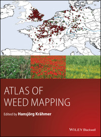
Atlas of Weed Mapping E-Book
209,99 €
Mehr erfahren.
- Herausgeber: John Wiley & Sons
- Kategorie: Fachliteratur
- Sprache: Englisch
Weeds are variously defined as plants growing where they are not wanted, plants that interfere with human activity. Weeds affect everyone in the world by reducing crop yield and quality, delaying or interfering with harvesting, interfering with animal feeding, reducing animal health, preventing water flow, as plant parasites, etc. It is estimated that those problems cause $ billions worth of crop losses annually and the global cost of controlling weeds also runs into many $ billions every year.
Atlas of Weed Mapping presents an introductory overview on the occurrence of the most common weeds of the world. The book notably includes:
- Description of cropping practices and explanations for the global distribution of weeds
- Invasive plant mapping
- Aquatics and wetland plants with histological plant details
- Theoretical and practical aspects of weed mapping
- Aspects on the documentation of herbicide resistance
- Biodiversity, rare weeds and the dominance of the most common weeds
Fully illustrated with more than 800 coloured figures and a number of tables, this new characterisation of anthropogenic vegetation will be interesting for readers of a great number of disciplines such as agriculture, botany, ecology, geobotany and plant community research. More than a hundred experts have contributed data to this unique compilation.
Sie lesen das E-Book in den Legimi-Apps auf:
Seitenzahl: 783
Veröffentlichungsjahr: 2016
Ähnliche


Table of Contents
Cover
Title Page
Copyright
Contributors
Acknowledgements
Introduction
References
Part I: Continental views of weed infestation maps
Chapter 1: Europe
Wheat
Maize
Oilseed rape
References
Chapter 2: Asia
Introduction
Rice
Wheat
Maize
References
General
China
India
Iran
Nepal
Pakistan
Russia and Kazakhstan
Turkey
Chapter 3: North America
Maize
Soybean
Wheat
Canola
References
Chapter 4: South America
Soybeans
Maize
Sugar cane
Wheat
References
Chapter 5: Africa
Growing conditions
Statistics
Wheat
Maize
Cassava
References
Chapter 6: Australia
Wheat
References
Part II: Special crop view and mapping of cotton weeds
Chapter 7: Cotton cultivation
Origin and use of cotton
Cropping areas
Cotton types
References
Chapter 8: Global cotton weed distribution
India and Pakistan
China
The USA
Uzbekistan
Australia
Brazil
Africa
The Mediterranean countries
References
Chapter 9: Farming practices and weed infestation
Planting date and weed infestation
Crop management and weed control
References
Chapter 10: Summary of global cotton weed distribution
References
Part III: Invasive weed species
Chapter 11: Overview of selected problems
References
Part IV: Global zones with similar weed infestation
Chapter 12: Introduction to global zones with similar weed infestation
References
Chapter 13: Cereal weed belts
References
Chapter 14: Maize weed belts and areas of similar weed infestation
References
Chapter 15: Soybean weed zones and areas
Chapter 16: Rice weed belts
References
Part V: General observations on all infested sites
Chapter 17: Ranks and number of weed species in a defined crop
Chapter 18: Specialization of weeds and biodiversity
Introduction
Global players
Typical weeds of crops in temperate climates
Typical weeds of continental climates
Typical weeds of tropical crops
Typical weeds of subtropical crops
References
Part VI: Answers to key questions: What makes which weed grow where and when?
Chapter 19: Weeds as crop companions
Migration of crops and weeds in history
Can we associate weeds with specific crops?
References
Chapter 20: Can we associate weeds with specific environmental conditions?
The establishment of weed communities under varying environmental factors
Can mathematical weed population models contribute to the question why weeds occur where and when?
Life-cycles and lifespan of weeds
References
Chapter 21: What makes weeds grow in monocultures, what makes them compete with the crop and with other weeds?
References
Part VII: Aesthetics, rare weeds and production objectives in agriculture
Chapter 22: Rare weeds in arable crops and aesthetics: harmony or hunger?
References
Part VIII: Weeds in meadows, pastures and rangeland
Chapter 23: Overview of grassland
References
PART IX: Aquatic and wetland weeds
Chapter 24: Introduction
References
Chapter 25: Morphological adaptation to water
References
Chapter 26: Aerenchyma within the stem
References
Chapter 27: Stem and vascular bundle modifications
References
Chapter 28: The root
References
Chapter 29: The leaf
References
Chapter 30: Vegetative propagation
References
Chapter 31: Aesthetics, species attractiveness and rare aquatic species
References
Chapter 32: Growing conditions of aquatic plants
References
Chapter 33: Dominance and noxious effects of selected aquatic and wetland species
References
Chapter 34: Adaptation of terrestrial weeds to water stress: Waterlogging and temporary hypoxia
References
Chapter 35: Weeds in rice
References
PART X: Which ecological rules described in textbooks will help us to understand the unevenness of weed species distribution?
Chapter 36: Asymmetric competition within arable crops
References
Chapter 37: Comparison of closely related species and their ability to grow as weeds in crops
References
PART XI: Factors contributing to the temporal and spatial distribution of weed resistance: a map–based analysis
Chapter 38: How has Alopecurus myosuroides resistance changed over the years?
Introduction
Temporal and spatial weed resistance development: a case study in southern Germany
Discussion
References
Chapter 39: Weeds to watch
PART XII: Conflict between the dominance of some weeds and the intention to preserve rare species
Chapter 40: Can we shape nature into what we want it to be?
References
PART XIII: Weed data collection, analysis and presentation of results
Chapter 41: Introduction to weed mapping methodology
Introduction
Chapter 42: Data collection
Choice of data collection method
Continuous data collection
Sample data collection
Description of vegetation attributes
References
Chapter 43: Approaches to the analysis of weed distribution
Introduction to data analysis
Basic statistics
Distance-based approach
Geostatistical approach to weed mapping
Remote sensing approach
The phytosociological approach
The GIS approach
References
Chapter 44: Presentation of weed mapping results
Data representation
Choropleth maps
Grid map (or raster map)
Binary map (or indicator map)
Point raster maps
Point map
Symbol map
Chart maps
Range maps (or sometimes called distribution maps)
Contour map (also called isoline or isopleth map)
Filled contour map
References
Appendix
Colour codes for monocot weeds in arable crops
Colour codes for dicot weeds in arable crops (a)
Colour codes for dicot weeds in arable crops (b)
Colour codes for aquatic and wetland weeds
Colour codes for rice weeds
Colour codes for weeds in meadows, pastures and rangeland (a)
Colour codes for weeds in meadows, pastures and rangeland (b)
Index
End User License Agreement
Pages
vii
ix
1
2
3
5
7
8
9
10
11
12
13
14
15
16
17
18
19
20
21
22
23
24
25
26
27
28
29
30
31
32
33
34
35
36
37
38
39
40
41
42
43
44
45
46
47
48
49
50
51
52
53
54
55
56
57
58
59
60
61
62
63
64
65
66
67
68
69
70
71
72
73
74
75
76
77
78
79
80
81
82
83
85
87
88
89
90
91
92
93
94
95
96
97
98
99
100
101
102
103
105
106
107
108
109
110
111
113
115
116
117
118
119
120
121
122
123
124
125
126
127
129
130
131
132
133
135
136
137
138
139
140
141
142
143
144
145
146
147
148
149
150
151
152
153
154
155
156
157
158
159
160
161
162
163
164
165
167
169
170
171
172
173
174
175
177
178
179
180
181
183
185
186
187
188
189
190
191
192
193
194
195
196
197
198
199
200
201
202
203
204
205
206
207
208
209
210
211
212
213
214
215
216
217
218
219
220
221
222
223
224
225
226
227
228
229
230
231
232
233
234
235
236
237
238
239
240
241
242
243
244
245
246
247
248
249
250
251
252
253
254
255
256
257
258
259
260
261
262
263
264
265
266
267
268
269
270
271
272
273
274
275
276
277
278
279
280
281
282
283
284
285
286
287
288
289
290
291
292
293
294
295
296
297
298
299
300
301
302
303
304
305
306
307
308
309
310
311
312
313
314
315
316
317
318
319
320
321
322
323
324
325
326
327
328
329
330
331
332
333
334
335
336
337
338
339
340
341
342
343
344
345
346
347
348
349
350
351
352
353
354
355
356
357
358
359
360
361
362
363
364
365
366
367
368
369
370
371
372
373
374
375
376
377
378
379
380
381
382
383
384
385
386
387
388
389
390
391
392
393
394
395
396
397
398
399
401
402
403
404
405
406
407
409
410
411
412
413
414
415
416
417
418
419
420
421
423
425
426
427
429
430
431
432
433
434
435
436
437
438
439
440
441
442
443
444
445
446
447
448
449
450
451
452
453
454
455
456
457
458
459
460
461
462
463
464
465
467
468
469
470
471
472
Guide
Cover
Table of Contents
Introduction
Part I: Continental views of weed infestation maps
Begin Reading
List of Illustrations
Chapter 1: Europe
Figure 1.1 Average weed infestation in cereals, most frequent grasses.
Figure 1.2 Cleavers and blackgrass in a wheat field near Stuttgart, Germany, 10 June 2009.
Figure 1.3 Average weed infestation in wheat, the second most frequent grasses.
Figure 1.4 Average weed infestation in wheat, the third most frequent grasses. Note: The
Alopecurus
species in Scandinavia and Finland is
A. geniculatus
.
Figure 1.5 Average weed infestation in cereals, most frequent dicots.
Figure 1.6 Average weed infestation in cereals, second most frequent dicots.
Figure 1.7 Average weed infestation in cereals, third most frequent dicots.
Figure 1.8 Average weed infestation in maize, most frequent grasses.
Figure 1.9 Average weed infestation in maize, second most frequent grasses.
Figure 1.10 Average weed infestation in maize, third most frequent grasses.
Figure 1.11 Average weed infestation in maize, most frequent dicots.
Figure 1.12 Average weed infestation in maize, second most frequent dicots.
Figure 1.13 Average weed infestation in maize, third most frequent dicots.
Figure 1.14 Flowering oilseed rape near Frankfurt, Germany, 11 May 2012.
Figure 1.15 Volunteer cereals in oilseed rape near Frankfurt, Germany; 1 December 2012 – both suffering from frost.
Figure 1.17 Average weed infestation in oilseed rape, most frequent monocots.
Figure 1.18 Average weed infestation in oilseed rape, second most frequent monocots.
Figure 1.19 Average weed infestation in oilseed rape, third most frequent monocots.
Figure 1.16
Tripleurospermum maritimum
in oilseed rape near Frankfurt, Germany, 7 June 2009.
Figure 1.20 Average weed infestation in oilseed rape, most frequent dicots.
Figure 1.21 Average weed infestation in oilseed rape, second most frequent dicots.
Figure 1.22 Average weed infestation in oilseed rape, third most frequent dicots.
Figure 1.23
Cirsium arvense
in an oilseed rape field in southern Germany, 21 June 2012.
Chapter 2: Asia
Figure 2.1 Transplanted paddy rice near Yuki, Japan.
Figure 2.3 Rice research at the IRRI near Manila, the Philippines.
Figure 2.2 Rice near Yuki, Japan before harvest.
Figure 2.4 Average weed infestation in wheat of the Russian Federation, most frequent grasses.
Figure 2.5 Average weed infestation in wheat of the Russian Federation, second most frequent grasses.
Figure 2.6 Average weed infestation in wheat of the Russian Federation, distribution of
Sonchus arvensis
as the most frequent cereal weed in the Russian Federation.
Figure 2.7 Average weed infestation in wheat of the Russian Federation, second most frequent dicots.
Figure 2.8 Distribution of spring and winter wheat in China.
Figure 2.9 Average weed infestation in Chinese wheat, most frequent grasses.
Figure 2.10 Average weed infestation in Chinese wheat, second most frequent grasses.
Figure 2.11 Average weed infestation in Chinese wheat, most frequent dicots.
Figure 2.12 Average weed infestation in Chinese wheat, second most frequent dicots.
Figure 2.13 Average weed infestation in Indian, Nepalese and Pakistani wheat, most frequent grasses.
Figure 2.14 Average weed infestation in Indian, Nepalese and Pakistani in wheat, second most frequent grasses.
Figure 2.15 Average weed infestation in Indian, Nepalese and Pakistani in wheat, most frequent dicots.
Figure 2.16 Average weed infestation in Indian, Nepalese and Pakistani in wheat, second most frequent dicots.
Figure 2.17 Major cereal-growing (wheat and barley) areas in Iran. Source: Adapted from a map by the U. S. Central Intelligence Agency, via Wikimedia Commons.
Figure 2.18 Most frequent monocot weeds in Iranian cereals.
Figure 2.19 Second most frequent monocot weeds in Iranian cereals.
Figure 2.20 Most frequent dicot weeds in Iranian cereals.
Figure 2.21 Second most frequent dicot weeds in Iranian cereals.
Figure 2.22 Most frequent monocot weeds in Turkish cereals.
Figure 2.23 Second most frequent monocot weeds in Turkish cereals.
Figure 2.24 Third most frequent monocot weeds in Turkish cereals.
Figure 2.25 Most frequent dicot weeds in Turkish cereals.
Figure 2.26 Second most frequent dicot weeds in Turkish cereals.
Figure 2.27 Third most frequent dicot weeds in Turkish cereals.
Figure 2.28 Most frequent grass weeds in maize in China.
Figure 2.29 Second most frequent grass weeds in maize in China.
Figure 2.30 Most frequent dicot weeds in maize in China.
Figure 2.31 Second most frequent dicot weeds in maize in China.
Figure 2.32 Most frequent monocot weeds in maize in India and Pakistan.
Figure 2.33 Second most frequent monocot weeds in maize in India and Pakistan.
Figure 2.34 Third most frequent monocot weeds in maize in India and Pakistan.
Figure 2.35 Most frequent dicot weeds in maize in India and Pakistan.
Figure 2.36 Second most frequent dicot weeds in maize in India and Pakistan.
Figure 2.37 Third most frequent dicot weeds in maize in India and Pakistan.
Chapter 3: North America
Figure 3.1 Maize near Sabin, Minnesota, USA, 13 July 2008.
Figure 3.2 Most frequent monocot weeds in US corn.
Figure 3.3 Second most frequent monocot weeds in US maize.
Figure 3.4 Third most frequent monocot weeds in US maize.
Figure 3.5 Most frequent dicot weeds in US maize.
Figure 3.6 Second most frequent dicot weeds in US maize.
Figure 3.7 Third most frequent dicot weeds in US maize.
Figure 3.8
Setaria faberi
in soybeans near Brownsburg, Indiana, USA, 20 July 2009.
Figure 3.9 Volunteer RR maize in RR soybeans near Sabin, Minnesota, USA, 13 July 2008.
Figure 3.10 Spring wheat near Innisfail, Alberta, Canada, 14 July 2008.
Figure 3.11
Avena fatua
in North Dakota spring wheat, 17 July 2008.
Figure 3.12 North American wheat varieties. Source: Derived from USDA ARS data.
Figure 3.13 North American wheat varieties (summarizing and more naturalistic view); winter wheat: blue, spring wheat: orange. Source: Data from www.usda.gov/oce/weather/pubs/Other/MWCACP/Graphs/USA/US_WheatWinter.pdf and webarchives.cdlib.org/sw1bc3ts3z/http://ers.usda.gov/Briefing/Wheat/maps.htm
Figure 3.14 Most frequent grasses in North American wheat.
Figure 3.15 Most frequent dicots plus
Allium vineale
(monocot) in North American wheat.
Figure 3.16 Canola near Innisfail, Alberta, Canada, 17 July 2008.
Chapter 4: South America
Figure 4.1 Soybean field near Sidrolandia, Mato Grosso do Sul, Brazil, 10 Dec 2006.
Figure 4.2
Bidens pilosa
,
Euphorbia heterophylla
and
Rottboellia cochinchinensis
in soybean field near Sidrolandia, Mato Grosso do Sul, Brazil, 10 Dec. 2006.
Figure 4.3
Eleusine indica
in soybean field near Rondonopolis, Mato Grosso, Brazil, 8 Dec. 2006.
Figure 4.4 Most frequent monocot weeds in South American soybeans; two different
Digitaria
species:
D. horizontalis
in Brazil and
D. sanguinalis
in Argentina.
Figure 4.5 Second most frequent monocot weeds in South American soybeans; two different
Digitaria
species:
D. horizontalis
in Brazil and
D. sanguinalis
in Argentina.
Figure 4.6 Third most frequent monocot weeds in South American soybeans.
Figure 4.7
Euphorbia heterophylla
in soybean field near Dourados, Mato Grosso do Sul, Brazil, 29 November 2005.
Figure 4.8 Most frequent dicot weeds in South American soybeans.
Figure 4.9 Second most frequent dicot weeds in South American soybeans.
Figure 4.10 Third most frequent dicot weeds in South American soybeans.
Figure 4.11
Borreria latifolia
syn.
Spermacoce latifolia
in South American soybeans.
Figure 4.12 Most frequent monocot weeds in South American maize.
Figure 4.13 Second most frequent monocot weeds in South American maize; two different
Digitaria
species:
D. horizontalis
in Brazil and
D. sanguinalis
in Argentina.
Figure 4.14 Third most frequent monocot weeds in South American maize;
D. horizontalis
in Brazil and
D. sanguinalis
in Argentina.
Figure 4.15
Chenopodium album
in maize field near Chacabuco, Argentina, 5 Dec. 2005.
Figure 4.16 Most frequent dicot weeds in South American maize.
Figure 4.17 Second most frequent dicot weeds in South American maize.
Figure 4.18 Third most frequent dicot weeds in South American maize.
Figure 4.19
Sida rhombifolia
and
Senna obtusifolia
in a maize field near Rondonopolis, Mato Grosso, Brazil, 29 Nov. 2005.
Figure 4.20
Conyza canadensis
on fallow field near Chacabuco, Argentina, 5 Dec. 2012.
Figure 4.21 Two sugar cane plantations in different stages of development, São Paolo State, January 2003.
Figure 4.22 Sugar cane near Hangzhou, China.
Figure 4.23 Sugar cane near Cali, Colombia. Source: Photograph by A. Laiblin, Schering AG.
Figure 4.24 Most frequent monocot weeds in Brazilian sugar cane.
Figure 4.25 View into young sugar cane plantation with
Ipomoea purpurea
infestation, São Paolo State, January 2003.
Figure 4.26 View into young sugar cane plantation with weeds removed by herbicides, São Paolo State, January 2003.
Figure 4.27 Most frequent monocot weeds in South American wheat.
Figure 4.28 Second most frequent monocot weeds in South American wheat; Avena:
A. fatua
in Argentina,
A. strigosa
in Brazil.
Figure 4.29 Third most frequent monocot weeds in South American wheat;
Digitaria sanguinalis
is the depicted species in Argentinian cereals.
Figure 4.30 Most frequent dicot weeds in South American wheat.
Figure 4.31 Second most frequent dicot weeds in South American wheat.
Figure 4.32 Third most frequent dicot weeds in South American wheat.
Chapter 5: Africa
Figure 5.1 Most frequent monocot weeds in African wheat.
Figure 5.2 Second most frequent monocot weeds in African wheat.
Figure 5.3 Third most frequent monocot weeds in African wheat;
Lolium temulentum
in Ethiopia,
Lolium multiflorum
and
Lolium rigidum
in North Africa.
Figure 5.4 Most frequent dicot weeds in African wheat.
Figure 5.5 Second most frequent dicot weeds in African wheat.
Figure 5.6 Third most frequent dicot weeds in African wheat.
Figure 5.7 Most frequent monocot weeds in African maize.
Figure 5.8 Second most frequent monocot weeds in African maize.
Figure 5.9 Third most frequent monocot weeds in African maize.
Figure 5.10 Most frequent dicot weeds in African maize.
Figure 5.11 Second most frequent dicot weeds in African maize.
Figure 5.12 Third most frequent dicot weeds in African maize.
Figure 5.13 Most frequent monocot weeds in African cassava.
Figure 5.14 Most frequent dicot weeds in African cassava.
Chapter 6: Australia
Figure 6.1
Raphanus raphanistrum
and
Lolium rigidum
in a wheat field near Adelaide, Australia, 25 Sept. 2006.
Figure 6.2 Most frequent monocot weeds in Australian wheat:
Avena
.
Figure 6.3 Second most frequent monocot weeds in Australian wheat:
Lolium
.
Figure 6.4 Most frequent dicot weeds in Australian wheat:
Raphanus
.
Chapter 7: Cotton cultivation
Figure 7.1 American upland cotton in September 2007. Source: Photograph courtesy of Bayer CropScience AG. Reproduced with permission.
Figure 7.2 Major cotton growing areas of the world.
Figure 7.3 Cotton harvest near Lubbock, Texas, September 2009. Source: Photograph courtesy of Bayer CropScience AG. Reproduced with permission.
Chapter 8: Global cotton weed distribution
Figure 8.1 Most frequent monocot cotton weeds in India and Pakistan. Source: Photograph courtesy of Bayer CropScience AG.
Figure 8.2 Second most frequent monocot cotton weeds in India and Pakistan.
Figure 8.3 Most frequent dicot cotton weeds in India and Pakistan.
Figure 8.4 Second most frequent dicot cotton weeds in India and Pakistan.
Figure 8.5 Most frequent monocot cotton weeds in China.
Figure 8.6 Most frequent dicot cotton weeds in China.
Figure 8.7 Most frequent monocot weeds in US cotton.
Figure 8.8 Most frequent dicot weeds in US cotton.
Figure 8.9 Surveyed areas in Greece, according to Economou et al. (2005).
Figure 8.10 Relative abundance of weeds in Pthiotida. Source: Data from Economou et al. (2005).
Figure 8.12 Relative abundance of weeds in Rodopi. Source: Data from Economou et al. (2005).
Figure 8.13 Cotton growing areas in Turkey.
Chapter 11: Overview of selected problems
Figure 11.1 Global distribution of Japanese knotweed. Source: Adapted from NOBANIS, DAISIE and other organizations.
Figure 11.2
Fallopia sachalinensis
along the River Kelvin in Glasgow, 12 June 2009.
Figure 11.3
Fallopia japonica
in a forest near Wiesbaden, 13 October 2010.
Figure 11.4 Dry remains of
Fallopia japonica
in a recreation area near Frankfurt, 8 April 2012.
Figure 11.5 Newly emerging shoots of
Fallopia japonica
on the ground of area presented in Figure 11.4.
Figure 11.6 Same site as Figure 11.5, regrown
Fallopia japonica
; plants were between 3m and 4 m tall on 4 September 2013.
Figure 11.7 Global distribution of Himalayan balsam. Source: Adapted from NOBANIS, DAISIE and other organizations.
Figure 11.8
Impatiens glandulifera
in a forest near Wiesbaden, 13 October 2010.
Figure 11.9 Inflorescence of
Impatiens glandulifera
in a forest near Frankfurt, 31 July 2011.
Figure 11.10 Global distribution of giant hogweed. Source: Adapted from NOBANIS, DAISIE and other organizations.
Figure 11.11
Heracleum mantegazzianum
plants at the River Kelvin in Glasgow, 14 June 2009.
Figure 11.12 Young
Heracleum mantegazzianum
plant on a public footpath in Glasgow, 14 June 2009.
Figure 11.13 Blister caused by a small droplet of
Heracleum mantegazzianum
sap, 15 June 2010.
Figure 11.14
Pteridium aquilinum
in a forest near Frankfurt, 30 October 2010 – not an invasive species, according to the IUCN definition.
Chapter 13: Cereal weed belts
Figure 13.1 Global cereal weed belts: Triticum-Avena-Lolium (TRAVLO), Triticum-Avena-Setaria (TRAVSE), Triticum-Avena-Phalaris (TRAVPH); the equator is marked by a red line, 35° North and South are each marked by a blue line.
Figure 13.2 Global cereal weed belts: Triticum-Alopecurus-Galium (TRALGA).
Figure 13.3 Global cereal weed belts: Triticum-Avena-Descurainia (TRAVDE).
Chapter 14: Maize weed belts and areas of similar weed infestation
Figure 14.1 Global maize weed belts: Zea-Digitaria-Sorghum (ZEDISO).
Figure 14.2 Global maize weed areas: Zea-Brachiaria-Commelina (ZEBRCO); the equator is marked by a red line, 20° North and South are each marked by a green line.
Figure 14.3 Global maize weed areas: Zea-Cynodon-Cyperus (ZECYCY).
Figure 14.4 Global maize weed zones: Zea-Chenopodium-Echinochloa (ZECHEC).
Chapter 16: Rice weed belts
Figure 16.1 Global rice-growing areas. Sources: IRRI, USDA, FAO, personal communication with country representatives.
Figure 16.2 Global rice weed belts, most frequent monocot weeds.
Figure 16.3 Global rice weed belts, second most frequent monocot weeds.
Chapter 19: Weeds as crop companions
Figure 19.1 Number of horses on the Great Plains between 1880 and 1960. Source: Adopted from Cunfer (2005).
Chapter 20: Can we associate weeds with specific environmental conditions?
Figure 20.1
Galium aparine
near Hofheim, Germany, 18 December 2012.
Figure 20.2 Same field as in Figure 20.1, 2 February 2013.
Figure 20.3 Same field as in Figure 20.1 and 20.2, 19 February 2013.
Figure 20.4
Veronica persica
tolerating frost and snow, Hofheim, 11 February 2013.
Figure 20.5
Veronica persica
flowering despite frost and snow, Hofheim, 19 February 2013.
Figure 20.6
Equisetum arvense
in maize, indicating wet soil, near Crailsheim, Germany, 21 June 2012.
Figure 20.7
Chenopodium album
in maize suffering from drought stress, south of France, July 2001.
Figure 20.8
Polygonum lapathifolium
in maize suffering from drought stress, Trebur, Germany, July 2009.
Figure 20.9
Descurainia sophia
at the edge of a rye field near Potsdam, Germany.
Figure 20.10 Do size and numbers count? Seeds of
Amaranthus palmeri
from Mississippi, 9 September 2008. Source: Photograph by Martin Hess.
Figure 20.11 Early developmental stage of winter wheat without weed infestation, near Frankfurt, Germany, 2 November 2008.
Figure 20.12 Early developmental stage of maize and emerging weeds (
Chenopodium album, Amaranthus retroflexus, Polygonum lapathifolium, Echinochloa crus-galli
), Frankfurt, Germany, 15 May 2012.
Figure 20.13 Winter wheat in spring,
Galium aparine
and
Tripleurospermum maritimum
covering the ground, near Ulm, Germany, 2 April 2010.
Figure 20.14 Heavy infestation of a late drilled maize field near Frankfurt with a great number of
Chenopodium album
and with a few
Amaranthus retroflexus
plants, 27 July 2011. The ground is mainly covered by these two weed species.
Figure 20.15 The application of a post-emergence grass herbicide in this plot resulted in the simultaneous germination of
Tripleurospermum maritimum
, 10 May 2012.
Figure 20.16 One single wheat field in the middle of the picture is heavily infested whereas the others are almost clear, near Frankfurt, 19 July 2012.
Figure 20.17 Winter wheat on both sites of the road; left field infested with silky bentgrass and blackgrass, right field almost clear, 19 July 2012.
Figure 20.18 Blackgrass in maize near Crailsheim, 21 June 2012.
Figure 20.19 Blackgrass in sugar beet near Öhringen, 6 August 2012.
Figure 20.20 High density of blackgrass ears in winter barley field near Frankfurt, 18 June 2012.
Figure 20.21
Chenopodium album
and
Amaranthus retroflexus
in a potato field near Frankfurt.
Figure 20.22
Chenopodium album
in another potato field near Frankfurt.
Figure 20.23 Uneven distribution of
Tripleurospermum maritimum
in a wheat field near Crailsheim, Germany, 21 June 2012.
Figure 20.24 Uneven distribution of
Alopecurus myosuroides
in a spring barley field near Crailsheim, Germany, 21 June 2012.
Figure 20.25 Single blackgrass plant with more than forty tillers.
Figure 20.26 Simultaneously germinating
Senecio vulgaris
near Frankfurt, Germany, 10 October 2012.
Figure 20.27 Wheat field near Heidelberg, Germany, supposedly without any weed infestation, 21 June 2012.
Figure 20.28 A closer look at the field in Figure 20.27: hidden weeds.
Figure 20.29 Young lamb's quarters plant in well-developed oilseed rape near Frankfurt, Germany, 24 May 2012.
Figure 20.30
Veronica persica
on ground of well-developed wheat field near Stuttgart, Germany, 27 May 2012.
Figure 20.31
Tripleurospermum maritimum
in oilseed rape field near Frankfurt, Germany, 7 June 2009.
Figure 20.32 Differences between 5-week-old plants grown under different conditions. Left: outdoor conditions in Frankfurt, middle of March to end of April 2011. Right: greenhouse conditions (220 μE m
-2
, day/night temperature regime of 22°C and 14°C). Source: Krähmer and Baur (2013). Reproduced by permission of John Wiley & Sons.
Figure 20.33 Fast-growing weeds after cereal harvest near Frankfurt, 19 July 2012.
Figure 20.34 Enlarged view of Figure 20.33, showing flowering
Myosotis arvensis
,
Fallopia convolvulus
and
Polygonum lapathifolium
.
Figure 20.35 A similar situation in a freshly harvested oilseed rape field, 19 July 2012.
Figure 20.36 Same field as shown in Figure 20.35 with winter wheat 6 months later, carpet of
Myosotis arvensis
, 2 February 2013.
Figure 20.37 Same field as in Figure 20.35 and 20.36, end of April 2013.
Figure 20.38 After herbicide treatment, 16 May 2013,
Figure 20.39 After cereal harvest;
Myosotis arvensis
produces seed again on 19 August 2013.
Figure 20.40 Cereal stubble near Frankfurt, 19 August 2013:
Chenopodium album
.
Figure 20.41 Cereal stubble near Frankfurt, 19 August 2013: small
Chenopodium album
setting seed.
Figure 20.42 Volunteer oilseed rape near Frankfurt, 19 August 2013.
Figure 20.43 Volunteer oilseed rape and flowering
Convolvulus arvensis
in oilseed rape stubble near Frankfurt, 24 August 2013.
Figure 20.44 Volunteer barley near Frankfurt, 24 August 2013.
Figure 20.45
Setaria glauca
in oilseed rape stubble near Frankfurt, 24 August 2013.
Figure 20.46
Echinochloa crus-galli
in cereal stubble near Frankfurt, 19 August 2013.
Figure 20.47
Setaria glauca
in maize near Frankfurt, 19 August 2013.
Figure 20.48
Setaria glauca
in sugar beet near Frankfurt, 3 October 2008.
Figure 20.49
Echinochloa crus-galli
in maize near Frankfurt, 24 August 2013.
Figure 20.50 Weeds on field borders can be a huge source of seeds for the next season.
Papaver rhoeas
and
Tripleurospermum maritimum
near Frankfurt, Germany, 19 June 2009.
Figure 20.51 Mechanical weed control in maize near Heidelberg; weeds left in crop rows, 21 June 2012.
Figure 20.52 Strips of
Apera spica-venti
left as a result of a missing overlap of spray zones, Liederbach, 26 June 2012.
Figure 20.53 Strips of tractor tracks full of
Tripleurospermum maritimum
, near Saarbrücken, Germany, 27 June 2012.
Figure 20.54 Abandoned soybean field heavily infested with glyphosate-resistant
Amaranthus palmeri
in Mississippi, September 2008. Source: Photograph provided by Martin Hess.
Chapter 21: What makes weeds grow in monocultures, what makes them compete with the crop and with other weeds?
Figure 21.1 Monoculture of poppies on a rural site near cereal and oilseed rape fields, Frankfurt, 12 June 2012.
Figure 21.2 Monopolistic blackgrass infestation of wheat field near Crailsheim, 21 June 2012.
Figure 21.3 Competition caused by foxtails, maize is severely stunted in the untreated plot, on the right, pre-emergence application of metolachlor, Delaware, 24 June 1998.
Figure 21.4 Fertilizers and irrigation can help maize to survive despite heavy weed infestation, Champaign, Illinois, July 2002.
Figure 21.5 Oilseed rape, closed canopy, near Frankfurt, 23 November 2012.
Figure 21.6
Vicia hirsuta
in wheat field near Heidelberg, 21 June 2012.
Figure 21.7
Convolvulus arvensis
in wheat field near Frankfurt, 17 June 2012.
Figure 21.8
Fallopia convolvulus
‘strangling’ wheat near Frankfurt, 24 June 2012.
Figure 21.9
Galium aparine
atop winter barley field near Frankfurt, 18 June 2012. This field was also heavily infested with
Alopecurus myosuroides
.
Figure 21.10
Convolvulus arvensis
growing atop wheat near Frankfurt, 13 July 2012.
Figure 21.11
Apera spica-venti
in wheat field near Frankfurt, 18 June 2012.
Figure 21.12
Cirsium arvense
growing much higher than oilseed rape near Frankfurt, 18 June 2012.
Figure 21.13 Wild oats outgrowing spring barley near Frankfurt, 18 June 2012.
Figure 21.14
Elytrigia repens
outgrowing wheat near Frankfurt, 18 June 2012.
Figure 21.15
Atriplex micrantha
growing on strip separating motorway lanes near Stuttgart, Germany, 1 July 2012.
Chapter 22: Rare weeds in arable crops and aesthetics: harmony or hunger?
Figure 22.1 Blue cornflower with bumble bee and mayweed – symbols of harmony? Kelkheim, Germany, 19 June 2013.
Figure 22.2 Poppy in oilseed rape near Frankfurt, Germany.
Figure 22.3 Poppy and cornflower – a motif for many artists; near Saarbrücken, Germany, 27 June 2012.
Figure 22.4
Lathyrus tuberosus
in a wheat field border near Frankfurt, 4 July 2010.
Figure 22.5 Field border strip near Friedberg, Germany, with mustard and
Phacelia tanacetifolia
, 23 July 2012.
Figure 22.6
Melampyrum arvense
in wheat field near Saarbrücken, Germany, 27 June 2012.
Figure 22.7 Simultaneous occurrence of blackgrass and silky bentgrass in a wheat field near Frankfurt; no longer a rare sight, 14 June 2012.
Figure 22.8
Adonis annua
, Frankfurt, 25 Sept. 2009, a greenhouse plant.
Figure 22.9
Anagallis foemina
, JKI, Braunschweig, 29 June 2009.
Figure 22.10
Anagallis foemina
, Crete, 3 April 2007.
Figure 22.11
Anagallis arvensis
, ruderal site near Frankfurt, Germany, 17 June 2012.
Figure 22.12
Anagallis arvensis
f.
azurea
, in oilseed rape near Frankfurt, Germany, 27 Sept. 2008.
Figure 22.13
Caucalis platycarpos
, flowering greenhouse plant, in Frankfurt, 20 August 2009.
Figure 22.14
Caucalis platycarpos
, fruits of greenhouse plant, in Frankfurt, 26 August 2009.
Figure 22.15
Legousia speculum-veneris
, Frankfurt, Germany, 20 August 2009, a greenhouse plant.
Figure 22.16
Nigella damascena
, ornamental, in Bad Nauheim, Germany, 17 July 2010.
Figure 22.17
Consolida regalis
in wheat near Heidelberg, Germany, 21 June 2012.
Figure 22.18
Consolida regalis
in wheat near Prague, Czech Republic, 13 May 2009.
Chapter 23: Overview of grassland
Figure 23.1 Meadow near Prags in Northern Italy with orchids and lilies, 7 July 2012.
Figure 23.2 Meadow near Bruneck, Northern Italy, 5 July 2012.
Figure 23.3 Hay production near Friedberg, Germany, 23 July 2012.
Figure 23.4 Pasture near Prags in Northern Italy, 2 July 2012,
Figure 23.5 Meadow near Frankfurt, Germany, infested with
Rumex obtusifolius
, 19 July 2012.
Figure 23.6 Pasture near Frankfurt with
Rumex obtusifolius
avoided by horses, 5 August 2012.
Figure 23.7 Rush pasture (
Juncus spec
) near Loch Lomond, 17 October 2012.
Figure 23.8
Colchicum autumnale
in meado`w near Frankfurt, 11 September 2010.
Figure 23.9
Colchicum autumnale
in meadow near Frankfurt, enlarged view of Figure 23.8 11 September 2010.
Figure 23.10
Senecio jacobaea
on border of field, Frankfurt, 24 June 2012.
Figure 23.11
Veratrum album
ssp
lobelianum
in pasture near Prags, Northern Italy.
Figure 23.12
Veratrum album
ssp
lobelianum
inflorescence.
Figure 23.13 Meadow near Olang in Northern Italy with dominating
Heracleum sphondylium
due to high soil nitrogen content, 4 July 2012.
Figure 23.14 Most common monocot weeds in hay, pastures and rangeland of the southern USA (WSSA data).
Figure 23.15 Most common dicot weeds in hay, pastures and rangeland of the southern USA (WSSA data).
Chapter 24: Introduction
Figure 24.1 Most frequent monocot aquatic weeds in southern US States, based on WSSA data.
Figure 24.2 Most frequent dicot aquatic weeds in southern US States, based on WSSA data.
Figure 24.3 Floating aquatic plants on a side channel of the River Rhine near Riedstadt-Erfelden, 30 July 2012.
Figure 24.4
Elodea nuttallii
from the River Rhine near Riedstadt-Erfelden, 30 July 2012.
Figure 24.5
Elodea nuttallii
, enlarged view of Figure 24.4.
Figure 24.6 Water canal near Trebur, Germany, with floating eutrophic algae and various aquatic macrophytes, 27 July 2012.
Figure 24.7
Lemna
species on canal near Trebur, 27 July 2012.
Figure 24.8
Polygonum amphibium
with large leaves at the bottom of the picture,
Ceratophyllum demersum
in the middle and
Spirogyra
spec. as filamentous algae.
Figure 24.9
Ceratophyllum demersum
in the middle, surrounded by
Spirogyra
spec.
Figure 24.10 Flowering
Potamogeton nodosus
on the River Rhine near Riedstadt-Erfelden, not regarded as a weed in many parts of Europe but in other parts of the world, 30 July 2012.
Figure 24.11
Potamogeton nodosus
on the River Jagst, 6 August 2012.
Figure 24.12
Spirogyra
spec., microscopic view, 100x.
Figure 24.13
Ceratophyllum demersum
.
Figure 24.14
Ceratophyllum demersum
, enlarged view.
Figure 24.15
Eichhornia crassipes
covering the surface of an artificial pond, Frankfurt Botanical Garden, 29 July 2012.
Figure 24.16 Flowering
Eichhornia crassipes
, Frankfurt Botanical Garden, 29 July 2012.
Figure 24.17 Thickened petiole of
Eichhornia crassipes
(arrows), Frankfurt Botanical Garden, 29 July 2012.
Figure 24.18
Vallisneria gigantea
in a garden centre near Frankfurt, 30 July 2012.
Figure 24.19
Myriophyllum aquaticum
, botanical garden in Darmstadt, 4 August 2012.
Figure 24.20
Trapa natans
, botanical garden in Mainz, 15 August 2012.
Figure 24.21
Phragmites australis
, Trebur, 7 October 2012.
Figure 24.22
Phragmites australis
, Lake Constance, 25 May 2013.
Chapter 25: Morphological adaptation to water
Figure 25.1 Stoma on the upper side of a
Hottonia palustris
leaf.
Figure 25.2 Stoma on the upper side of a
Callitriche
leaf.
Chapter 26: Aerenchyma within the stem
Figure 26.1 Radial arrangement of lacunae in the stem of
Ceratophyllum demersum
.
Figure 26.2 Radial arrangement of lacunae in the stem of
Myriophyllum aquaticum
.
Figure 26.3 Longitudinal section through stem of
Myriophyllum aquaticum
showing cells forming the lacunae. They are arranged perpendicular to the parenchymatic cortex cells of the stem surrounding the lacunae.
Figure 26.4 Radial arrangement of lacunae in the stem of
Bacopa caroliniana
.
Figure 26.5 Radial arrangement of lacunae in the stem of
Utricularia australis
.
Figure 26.6 Two rows of lacunae in the stem of
Elodea nuttallii
.
Figure 26.7 Even arrangement of lacunae around stele within the stem of
Potamogeton natans
.
Figure 26.8 Lacunae in the stem of
Hottonia palustris
.
Figure 26.9 Lacunae in the stem of
Hippuris vulgaris
.
Figure 26.10 Honeycomb lacunae in a young rhizome of
Ludwigia grandiflora
.
Figure 26.11 Honeycomb lacunae in a young rhizome of
Ludwigia grandiflora
.
Figure 26.12 Lacunae in a young shoot of
Mimulus rigens
.
Figure 26.13 Transverse section through stolon of
Gratiola officinalis
stolon.
Figure 26.14
Gratiola officinalis
in an artificial pond.
Figure 26.15 ‘Leafy aerenchyma’ in a young shoot of
Typha minima
.
Figure 26.16 Honeycomb lacunae in the shoot of
Glyceria maxima
.
Figure 26.17 Honeycomb lacunae in the shoot of
Glyceria maxima
, enlarged view of Figure 22.16.
Figure 26.18 ‘Hollow and leafy aerenchyma’ in a young shoot of
Phragmites australis
, macro-view.
Figure 26.19 Aerenchyma in a young shoot of
Phragmites australis
.
Figure 26.20 Aerenchyma in a young shoot of
Phalaris arundinacea
.
Figure 26.21 Aerenchyma in a young shoot of
P. arundinacea
, enlarged view.
Figure 26.22 Aerenchyma in a young shoot of
P. arundinacea
, lacuna between two sclerenchyma layers.
Figure 26.23 Aerenchyma in a young shoot of
Eleocharis palustris
.
Figure 26.24 Stoma in a young shoot of
Eleocharis palustris
.
Figure 26.25
Juncus effusus
near Loch Lomond, Scotland.
Figure 26.26 Stellate parenchyma in a culm of
Juncus effusus
(arrows).
Figure 26.27 Transverse section through culm of
Juncus effusus
.
Figure 26.28 Stellate parenchyma within culm of
Juncus effusus
.
Figure 26.29 Inflorescence of
Juncus effusus
breaking through culm laterally.
Figure 26.30 Inflorescence of
Juncus effusus
breaking through culm laterally; culm cut in halves with a razer blade.
Figure 26.31 Node-like interruption of central air canal in
Juncus effusus
culm.
Figure 26.32 Flowering
Menyanthes trifoliata
.
Figure 26.33 Floating plants of
Menyanthes trifoliata
with long and strong rhizomes.
Figure 26.34 Transverse section through rhizome of
Menyanthes trifoliata,
macro-view.
Figure 26.35 Transverse section through rhizome of
Menyanthes trifoliata,
micro-view.
Figure 26.36 Longitudinal section through rhizome of
Menyanthes trifoliata
.
Figure 26.37 Transverse section through rhizome of
Menyanthes trifoliata
, cortical and medullary aerenchyma.
Figure 26.38 Aerenchyma in a rhizome of
Equisetum hyemale
, macro-view.
Figure 26.39 Aerenchyma in a rhizome of
Equisetum hyemale
.
Figure 26.40 Aerenchyma in a stem of
Equisetum hyemale,
macro-view.
Figure 26.41 Aerenchyma in a stem of
Equisetum hyemale
.
Figure 26.42 Aerenchyma in a stem of
Schoenoplectus lacustris
, vascular bundles and lacunae are evenly distributed over the section.
Figure 26.43 Stellate cells in the aerenchyma of a
Schoenoplectus lacustris
stem (arrows).
Chapter 27: Stem and vascular bundle modifications
Figure 27.1 Ten vascular bundles within the stele of
Potamogeton natans
– enlarged view of Figure 26.7. t2,T2,t2 = traces of the second higher leaf (trio-bundle); t1,T1,t1= traces of next higher leaf (according to Arber 1920); c = cauline or axillary bundles.
Figure 27.2 Vascular bundle within the stele of
Potamogeton natans
, enlarged view of Figure 27.1, different staining procedure.
Figure 27.3 Vascular bundle within the stele of
Potamogeton perfoliatus
showing the O-shaped endodermoid cells.
Figure 27.4 U-shaped endodermoid of
Potamogeton pectinatus
.
Figure 27.5 Vessel with secondary wall thickening next to a sieve tube in
Potamogeton natans
, longitudinal section.
Figure 27.6 Xylem elements within the stele of
Hottonia palustris
, longitudinal section.
Figure 27.7 Typical xylem elements within the stele of
Myriophyllum aquaticum
, longitudinal section.
Figure 27.8 Sieve tube in
Potamogeton natans
, longitudinal section.
Figure 27.9 Transverse section through a stolon of
Hydrocharis morsus-ranae
.
Figure 27.10 Transverse section through a stolon of
Hydrocharis morsus-ranae
with a cortical vascular bundle, enlarged view of Figure 27.9.
Figure 27.11 Transverse section through a stolon of
Hydrocharis morsus-ranae
, enlarged view of Figure 27.10 showing the central stele.
Figure 27.12 Transverse section through the stele of
Ceratophyllum demersum
.
Figure 27.13 Transverse section through the ‘cortex’ of
Heteranthera reniformis
.
Figure 27.14 Transverse section through the stele of
Typha minima
.
Figure 27.15 Transverse section through a central vascular bundles within the stele of a
Typha minima
rhizome.
Figure 27.16 Transverse section through a peripheral vascular bundle within the stele of a
Typha minima
rhizome.
Figure 27.17 Fibrous vascular bundle in the ‘cortex’ of a
Typha minima
rhizome.
Figure 27.18 Female and male inflorescences on stalk of
Typha latifolia
.
Figure 27.19 Transverse section of
Typha latifolia
stalk.
Figure 27.20 Transverse section of
Typha latifolia
stalk, enlarged view.
Figure 27.21 Transverse section of
Typha latifolia
stalk, single vascular bundle.
Figure 27.22 Distribution of vascular tissue in old rhizome of
Ludwigia grandiflora
.
Figure 27.23 Vascular cylinder of young
Ludwigia grandiflora
rhizome with cambium.
Figure 27.24 Vascular cylinder in advanced
Ludwigia grandiflora
rhizome.
Figure 27.25 Vascular cylinder in advanced
Ludwigia grandiflora
rhizome.
Figure 27.26 Distribution of vascular tissue in stem of
Ludwigia grandiflora
.
Figure 27.27 Bicollateral vascular tissue in stem of
Ludwigia grandiflora
.
Figure 27.28 Internal phloem in stem of
Ludwigia grandiflora
– enlarged view.
Figure 27.29 Internal phloem in stem of
Ludwigia grandiflora
, longitudinal section.
Figure 27.30 Medullary phloem in stem of
Ludwigia grandiflora
, transverse section.
Figure 27.31 Medullary phloem in stem of
Ludwigia grandiflora
, transverse section.
Figure 27.32 Medullary phloem in stem of
Ludwigia grandiflora
, transverse section, enlarged view and different staining technology.
Figure 27.33 Medullary phloem in stem of
Ludwigia grandiflora
, longitudinal section, macro-view.
Figure 27.34 Medullary phloem in stem of
Ludwigia grandiflora
, longitudinal section, microscopic view.
Figure 27.35
Lagarosiphon major
, from a garden centre near Frankfurt, Germany.
Figure 27.36
Lagarosiphon major
, transverse section through the stem.
Figure 27.37
Lagarosiphon major
, transverse section through the stele.
Figure 27.38
Vallisneria spiralis
, transverse section through the stolon.
Figure 27.39
Vallisneria spiralis
, transverse section through the stolon, enlarged view.
Figure 27.40
Vallisneria spiralis
, transverse section through a single bundle.
Figure 27.41 Transverse section through the stem of
Veronica persica
(left, from Krähmer & Baur 2013) and a stolon of
Veronica beccabunga
(right).
Figure 27.42 Transverse section through the cortex of
Veronica beccabunga
.
Figure 27.43 Transverse section through the cortex of
Veronica persica
.
Figure 27.44
Gratiola officinalis
stolon, transverse section.
Figure 27.45
Hippuris vulgaris
in a private pond near Frankfurt.
Figure 27.46 Stem of
Hippuris vulgaris
with leaves in whorls.
Figure 27.47 Stele of
Hippuris vulgaris
, transverse section.
Figure 27.48 Transverse section through a stolon of
Eichhornia crassipes
.
Figure 27.49 Transverse section through a stolon of
Eichhornia crassipes
, enlarged view.
Figure 27.50 Transverse section through a vascular bundle in a stolon of
Eichhornia crassipes
.
Figure 27.51 Transverse section through a stolon of
Pistia stratiotes
.
Figure 27.52 Transverse section through a vascular bundle in a stolon of
Pistia stratiotes
.
Figure 27.53 Transverse section through a stolon of
Trapa natans
.
Figure 27.54 Transverse section through a stolon of
Trapa natans
, vascular bundle ring.
Figure 27.55 Transverse section through a vascular bundle in the stolon of
Trapa natans
, enlarged view.
Figure 27.56 Transverse section through a stolon of
Hydrocleys nymphoides
.
Figure 27.57 Transverse section through a stolon of
Hydrocleys nymphoides
, central stele.
Figure 27.58 Transverse section through a stolon of
Hydrocleys nymphoides
, central stele, enlarged view.
Figure 27.59 Transverse section through rhizome of
Carex acuta
.
Figure 27.60 Transverse section through rhizome bundle of
Carex acuta
.
Figure 27.61 Transverse section through a rhizome of
Juncus effusus
showing amphivasal vascular bundles.
Figure 27.62 Transverse section through an amphivasal vascular bundle within a rhizome of
Juncus effusus
.
Figure 27.63 Transverse section through rhizome of
Acorus gramineus
, macro view.
Figure 27.66 Transverse section through rhizome of
Acorus gramineus
, intercellular space within rhizome.
Figure 27.64 Transverse section through rhizome of
Acorus gramineus
, endodermoid separating central bundles and peripheral bundles.
Figure 27.65 Transverse section through rhizome of
Acorus gramineus
, amphivasal bundles.
Figure 27.66 Transverse section through rhizome of
Acorus gramineus
, intercellular space within rhizome.
Figure 27.67 Transverse section through rhizome of
Eriophorum angustifolium
.
Figure 27.68 Transverse section through rhizome of
Eriophorum angustifolium
, collateral bundles.
Figure 27.69 Transverse section through culm of
Eleocharis palustris
.
Figure 27.70 Transverse section through culm of
Eleocharis palustris
.
Figure 27.71 Transverse section through a rhizome of
Scirpus sylvaticus
.
Figure 27.72 Transverse section through a rhizome of
Scirpus sylvaticus
, enlarged view.
Figure 27.73 Triangular stem of
Carex cespitosa
carrying one terminal spike with staminate flowers and three spikes with pistillate flowers below.
Figure 27.74 Transverse section through a stem of
Carex cespitosa
.
Figure 27.75 Longitudinal section through the stem of
Carex cespitosa
.
Figure 27.76 Transverse section through a stem of
Carex remota
.
Figure 27.77 Transverse section through a stem of
Carex remota
, enlarged view.
Figure 27.78 Transverse section through a stem of
Carex pseudocyperus
.
Figure 27.79 Transverse section through a single vascular bundle of
Carex pseudocyperus
.
Figure 27.80
Schoenoplectus lacustris
with rhizome.
Figure 27.81 Transverse section of
Schoenoplectus lacustris
rhizome, macro-view.
Figure 27.82 Transverse section of
Schoenoplectus lacustris
rhizome, stereo-zoom view.
Figure 27.83 Transverse section of
S. lacustris
rhizome, enlarged, different staining method.
Figure 27.84 Dual sheath and vascular bundles in
S. lacustris
rhizome.
Figure 27.85 Single collateral vascular bundle in
S. lacustris
rhizome, xylem in U-shape.
Figure 27.86 Single vascular bundle in
S. lacustris
rhizome, only two tracheids visible.
Figure 27.87 Single vascular bundle in
S. lacustris
rhizome, almost amphivasal structure.
Figure 27.88
Juncus effusus
, rhizome indicated by blue arrow.
Figure 27.89
S. lacustris
near Frankfurt, Germany.
Figure 27.90 Single, round stem
S. lacustris
with inflorescence.
Figure 27.91 Transverse section through stem of
S. lacustris
, stereo-zoom view.
Figure 27.92 Transverse section through stem of
S. lacustris
, enlarged view.
Figure 27.93 Transverse section through stem of
S. lacustris
, sub-epidermal layers.
Figure 27.94 Transverse section through peduncle of
Caltha palustris
, macro-view.
Figure 27.95 Transverse section through peduncle of
Caltha palustris
, single ridge.
Figure 27.96 Transverse section through stolon of
Ranunculus lingua
.
Figure 27.97 Transverse section through a stem of
Ranunculus lingua
.
Figure 27.98 Transverse section through a stem of
Ranunculus flammula
.
Figure 27.99 Transverse section through a vascular bundle of
Ranunculus flammula
.
Figure 27.100 Transverse section through a stolon of
Ranunculus reperns
– no lacunae.
Figure 27.101 Transverse section through a stolon of
Ranunculus reperns
, enlarged view.
Figure 27.102 Transverse section through rhizome of
Phragmites australis
.
Figure 27.103 Transverse section through rhizome of
Phragmites australis
.
Figure 27.104 Transverse section through rhizome of
Phalaris arundinacea
.
Figure 27.105 Transverse section through a peduncle of
Sparganium natans
.
Figure 27.106 Transverse section through a peduncle of
Sparganium natans
.
Figure 27.107 Transverse section through a vascular bundle within the peduncle of
Sparganium natans
.
Figure 27.108 Transverse section through a peduncle of
Calla palustris
, reflected light.
Figure 27.109 Two inflorescences of
Alisma plantago-aquatica
, ruler with 30cm length.
Figure 27.110 Two flowers of
Alisma plantago-aquatica
.
Figure 27.111 Transverse section through peduncle of
Alisma plantago-aquatica
.
Figure 27.112 Transverse section through peduncle of
Alisma plantago-aquatica
, enlarged view.
Figure 27.113 Transverse section through peduncle of
Alisma plantago-aquatica
showing secretory canals within chlorenchyma.
Figure 27.114 Flowering pickerel weed,
Pontederia cordata
.
Figure 27.115 Newly forming inflorescence of pickerel weed within leaf sheath of
Pontederia cordata
.
Figure 27.116 Inflorescence of pickerel weed,
Pontederia cordata
.
Figure 27.117 Transverse section through petiole of
Pontederia cordata
.
Figure 27.118 Transverse section through upper part of
Pontederia cordata
peduncle.
Figure 27.119 Transverse section through vascular bundle in a peduncle.
Figure 27.120 Longitudinal section through ‘node’ at transition zone to
Pontederia cordata
peduncle, macro-view.
Figure 27.121 Longitudinal section through “node” at transition zone to
Pontederia cordata
peduncle, microscopic view.
Figure 27.122 Transverse section through stem below node.
Figure 27.123 Transverse section through peduncle base of
Pontederia cordata
.
Chapter 28: The root
Figure 28.1 Adventitious roots of
Ranunculus lingua.
Figure 28.2 Adventitious roots of
Heteranthera reniformis
.
Figure 28.3 Adventitious roots of
Ranunculus flammula
.
Figure 28.4 Transverse section through stem of
Heteranthera reniformis
showing adventitious roots.
Figure 28.5 Transverse section through stem of
Heteranthera reniformis
with xylem entering adventitious root (in red).
Figure 28.6 Root of
Hydrocharis morsus-ranae
with conspicuous root hairs.
Figure 28.7 Root of
Hydrocleys nymphoides
with long root hairs.
Figure 28.8 Adventitious roots of
Potamogeton natans
(arrows).
Figure 28.9 Transverse section through adventitious root of
Potamogeton natans
, overview.
Figure 28.10 Transverse section through adventitious root of
Potamogeton natans
, enlarged overview of central stele.
Figure 28.11 Transverse section through adventitious root of
Potamogeton natans
, details, roundish endodermis cells.
Figure 28.12 Transverse section through root of
Echinodorus
Oriental, radially stretched endodermis cells.
Figure 28.13 Adventitious roots of
Myriophyllum spicatum
(arrows).
Figure 28.14 Transverse section through tetrarch root of
Myriophyllum spicatum
.
Figure 28.15 Transverse section through tetrarch xylem of
Myriophyllum aquaticum
.
Figure 28.16 Transverse section through triarch root of
Myriophyllum aquaticum
.
Figure 28.17 Transverse section through advanced root stage of
Myriophyllum aquaticum
, stele with central triangular xylem.
Figure 28.18 Transverse section through stem of
Myriophyllum aquaticum
, stele with circular vascular cylinder and central pith in contrast to the root pattern (Figure 28.17).
Figure 28.19 Transverse section through rhizodermis and multilayered exodermis of
Myriophyllum aquaticum
.
Figure 28.20 Transverse section through root lacunae of
Myriophyllum aquaticum
.
Figure 28.21 Longitudinal section through root of
Echinodorus
Oriental.
Figure 28.22 Longitudinal section through root of
Echinodorus
Oriental, enlarged view and different staining method compared to Figure 28.21.
Figure 28.23 Longitudinal section through diaphragm of
Echinodorus
Oriental.
Figure 28.24 Transverse view of
Echinodorus
Oriental diaphragm.
Figure 28.25 Transverse section through rhizodermis and hypodermis of ‘normal’
Ludwigia grandiflora
root.
Figure 28.26 Adventitious root of
Elodea nuttallii
(arrow).
Figure 28.27 Transverse section of adventitious root of
Elodea nuttallii.
Figure 28.29 Transverse section of central stele in an adventitious root of
Elodea nuttallii
, different staining technology.
Figure 28.28 Transverse section of central stele in an adventitious root of
Elodea nuttallii
Figure 28.30 Transverse section through root of
Elodea nuttallii
compared with root section of
Apera spica-venti
, low magnification. Source: From Krähmer and Baur (2013). Reproduced by permission of John Wiley & Sons, Ltd.
Figure 28.31 Transverse section through root of
Apera spica-venti
, higher magnification than in Figure 28.30.
Figure 28.32 Transverse section through root of
Butomus umbellatus
.
Figure 28.33 Transverse section through root of
Butomus umbellatus
, enlarged view.
Figure 28.34 Newly forming lacuna in root of
Calla palustris
.
Figure 28.35 Transverse section through root of
Cyperus serotinus
showing tangential lysigeny. Source: From Krähmer and Baur (2013). Reproduced by permission of John Wiley & Sons, Ltd.
Figure 28.36 Transverse section through root of
Carex acuta
.
Figure 28.37 Transverse section through root of
Carex acuta
demonstrating air cavities.
Figure 28.38 Stele in root of
Carex acuta.
Figure 28.39 Transverse section through root of
Carex pseudocyperus
.
Figure 28.40 Transverse section through root of
Eichhornia crassipes
.
Figure 28.41 Transverse section through stele in root of
Eichhornia crassipes
.
Figure 28.42 Transverse section through root of
Pistia stratiotes
.
Figure 28.43 Transverse section through root of
Pistia stratiotes
.
Figure 28.44 Transverse section through root of
Pistia stratiotes
.
Figure 28.45 Transverse section through tetrarch root of
Trapa natans
a honeycomb-like aerenchyma pattern.
Figure 28.46 Root of
Acorus gramineus
intercellular space within central cortex.
Figure 28.47 Stele of hexarch
Acorus gramineus
root.
Figure 28.48 Transverse section of
Glyceria maxima
root.
Figure 28.49 Enlarged view of
Glyceria maxima
root.
Figure 28.50 Stele of
Glyceria maxima
root.
Figure 28.51 Adventitious root on
Phragmites australis
culm with many small, lateral roots.
Figure 28.52 Fully differentiated root of
Phragmites australis
, transverse section.
Figure 28.53 Differentiating root of
Phragmites australis
, transverse section.
Figure 28.54 Transverse section of a
Phragmites australis
root
,
cell remnants forming lacuna walls.
Figure 28.55 Transverse section of a
Phragmites australis
root, centripetally collapsing cells within the cortex.
Figure 28.56 Transverse section of a
Phragmites australis
root, centrifugally collapsing cells within the cortex.
Figure 28.57 Transverse section of a
Phragmites australis
root, cellulose in cell wall remnants stained blue.
Figure 28.58 Transverse section through the outer protective layers of a
Phragmites
root.
Figure 28.59 Fully developed pentarch root of
Mimulus rigens
with cortical cavities.
Figure 28.60 Hexarch root of
Gratiola officinalis.
Figure 28.61 Two different root types in the ‘water form’ of
Ludwigia grandiflora
.
Figure 28.62 Pentarch root of
Ludwigia grandiflora
.
Figure 28.63 Beginning separation of cells in the cortex of
Ludwigia grandiflora
(arrows). Each cell is surrounded by four other cells.
Figure 28.64 Elongating intercellular spaces (arrows) of cells in the cortex of
Ludwigia grandiflora
, enlarged view.
Figure 28.65 Radially arranged cell rows in the cortex of
Ludwigia grandiflora
.
Figure 28.66 Hexagonal and tetragonal intercellular spaces between cells in the cortex of
Ludwigia grandiflora
.
Figure 28.67 Transverse section through root of
Ludwigia grandiflora
water form with fully developed central stele.
Figure 28.68 Transverse section through stele of
Ludwigia grandiflora
(enlarged view of Figure 28.67).
Figure 28.69 Transverse section through phloem of
Ludwigia grandiflora
: Sieve tubes and companion cells are arranged around a central purple-stained parenchymatic cell.
Figure 28.70 Longitudinal section through stele of
Ludwigia grandiflora
.
Figure 28.71 Aerenchymatous root of
Ludwigia grandiflora
. The white appearance of the surface indicates that aerenchyma has already developed a few millimetres behind the calyptra.
Figure 28.72 Young aerenchymatous root of
Ludwigia grandiflora
, longitudinal section, a few millimetres behind the root tip.
Figure 28.73 Cell protuberances in longitudinal section through the aerenchymatous roots of
Ludwigia grandiflora
.
Figure 28.74 Cell protuberances in longitudinal section through the aerenchymatous roots of
Ludwigia grandiflora
; different staining technology.
Figure 28.75 Primary aerenchyma formation in the outer cortex zone of
L. grandiflora
root; no aerenchyma meristem visible yet, transverse section.
Figure 28.76 Prolonged cells in outer cortex layers; transverse section.
Figure 28.77 Primary aerenchyma formation in outer cortex layers, transverse section.
Figure 28.78 Secondary aerenchyma producing meristem originating within the stele; transverse section.
Figure 28.79 Secondary aerenchyma produced by a meristem originating within the stele, transverse section.
Figure 28.80 Aerenchyma mesh in longitudinal view.
Figure 28.81 Enlarged view of aerenchyma mesh; enlarged longitudinal view.
Figure 28.82 Aerenchyma mesh; transverse view.
Figure 28.83 Aerenchyma mesh; longitudinal macro-view.
Figure 28.84 Transverse section through a root of
Ludwigia grandiflora
‘land form’: almost no intercellular space, regular cork formation by a phellogen.
Figure 28.85 Transverse section through a root of
Ludwigia grandiflora
‘land form’: almost no intercellular space, regular cork formation by a phellogen and massive starch storage within secondary phloem and parenchyma cells.
Chapter 29: The leaf
Figure 29.1 Leaf of
Potamogeton nodosus
in Goslawickie Lake, Poland.
Figure 29.2 Leaf of
Potamogeton natans
in a pond near Ulm, Germany.
Figure 29.3 Leaf of
Potamogeton natans
.
Figure 29.4 Leaf of
Potamogeton natans
, stoma.
Figure 29.5 Leaves of
Potamogeton perfoliatus
, Lednica Lake, Poland.
Figure 29.6 Leaf of
Potamogeton perfoliatus
, transverse section.
Figure 29.7 Leaf blade of
Potamogeton perfoliatus
with three cell layers, transverse section.
Figure 29.8 Midvein of
P. perfoliatus
leaf, transverse section.
Figure 29.9 Leaf of
Ceratophyllum demersum
.
Figure 29.10 Transverse section through a single leaf branch of
Ceratophyllum demersum
(blue arrow in Figure 29.9).
Figure 29.11 Transverse section through a leaf of
Ceratophyllum demersum
below two merged branches (red arrow in Figure 29.9). Two lacunae are the result of the merger.
Figure 29.12 Transverse section through a vascular bundle in
Ceratophyllum demersum
leaf.
Figure 29.13
Potamogeton pectinatus
in Goslawickie Lake, Poland.
Figure 29.14
Potamogeton pectinatus
, enlarged view.
Figure 29.15 Leaf of
Potamogeton pectinatus
, transverse section.
Figure 29.16 Vascular bundle in a
Potamogeton pectinatus
leaf between lacunae, transverse section; enlarged view of Figure 29.15.
Figure 29.17 Diaphragm in a petiole lacuna of
Heteranthera reniformis
.
Figure 29.18 Diaphragms in petiole lacunae of
Echinodorus
Oriental.
Figure 29.19 Diaphragm in petiole lacunae of
Echinodorus
Oriental, three-dimensional impression of stellate diaphragm cells.
Figure 29.20 Thin section of diaphragm cells in petiole lacunae of
Echinodorus
Oriental.
Figure 29.21 Transverse section of a
Pontederia cordata
petiole.
Figure 29.22 Diaphragm in a
Pontederia cordata
petiole.
Figure 29.23 Diaphragm in a
Pontederia cordata
petiole with idioblast.
Figure 29.24
Pontederia cordata
petiole with diaphragm idioblast containing raphides.
Figure 29.25
Pontederia cordata
, lacuna in a petiole, longitudinal section.
Figure 29.26
Pontederia cordata
, diaphragm in a petiole with stiletto-like raphide, longitudinal section
Figure 29.27
Pontederia cordata
, vascular bundle in a petiole, transverse section.
Figure 29.28
Pontederia cordata
, vascular bundle in a petiole, longitudinal section.
Figure 29.29 Transverse section through a leaf of
Vallisneria spiralis
.
Figure 29.30 Sprouting
Potamogeton natans
with phyllodial leaves in April.
Figure 29.31 Phyllodial leaf of
Potamogeton natans
.
Figure 29.32 Phyllodial leaf of
Potamogeton natans
, transverse section.
Figure 29.33 Vascular bundle in a phyllodial leaf of
Potamogeton natans
, transverse section.
Figure 29.34 Minor vascular bundle in a phyllodial leaf of
Potamogeton natans
, transverse section.
Figure 29.35 Minor vascular bundle in a phyllodial leaf of
Potamogeton natans
, longitudinal section.
Figure 29.36 Lacunae in a phyllodial leaf of
Potamogeton natans
, longitudinal section.
Figure 29.37 Lacunae in a phyllodial leaf of
Potamogeton natans
, longitudinal section.
Figure 29.38 Adjacent lacunae in a phyllodial leaf of
Potamogeton natans
, longitudinal section.
Figure 29.39 Phyllodial leaf of
Potamogeton natans
broadening at the tip beginning of June 2013.
Figure 29.40 Two different leaf forms of
Potamogeton nodosus
; Lichenskie Lake, Poland, 2 September 2012.
Figure 29.41 Two different leaf forms of
Ranunculus lingua
.
Figure 29.42 Two different leaf forms of
Ludwigia grandiflora
; left – water form, right- land form.
Figure 29.43 Three different leaf forms of
Ranunculus aquatilis
: lobed floating leaves, dissected flattened leaves and filiform submerged leaves.
Figure 29.44 Two different floating leaf forms of
Ranunculus aquatilis
.
Figure 29.45 Two different leaf forms of
Ranunculus aquatilis
above water.
Figure 29.46 Transverse section through a filiform submerged leaf of
Ranunculus aquatilis
.
Figure 29.47 Transverse section through a dissected flattened leaf of
Ranunculus aquatilis
.
Figure 29.48 Transverse section through a plane floating leaf of
Ranunculus aquatilis
.
Figure 29.49
Ranunculus fluitans
in the River Danube (Inzigkofen, May 2013).
Figure 29.50
Ranunculus fluitans
starting to flower in the River Danube (Inzigkofen, May 2013).
Figure 29.51 Capillary
Ranunculus fluitans
leaf cut before first fork.
Figure 29.52 Round, capillary and forked
Ranunculus fluitans
leaves from the River Danube (Inzigkofen, May 2013).
Figure 29.53 Capillary
Ranunculus fluitans
leaf cut behind first fork.
Figure 29.54 Capillary
Ranunculus fluitans
leaf cut behind second fork.
Figure 29.55 Capillary
Ranunculus fluitans
leaf cut behind second fork, enlarged view.
Figure 29.56 Dissected leaf of
Ranunculus circinatus
.
Figure 29.57 Heterophylly in
Sagittaria sagittifolia
.
Figure 29.58 Transverse section of a
Lagarosiphon major
leaf.
Figure 29.59 Transverse section of the midvein in a
Lagarosiphon major
leaf.
Figure 29.60 Transverse section of
Lagarosiphon major
leaf blade.
Figure 29.61
Cabomba caroliniana
.
Figure 29.62 Leaf of
Cabomba caroliniana
, transverse section.
Figure 29.63
Cabomba caroliniana
, surface view.
Figure 29.64 Leaf whorls of
Myriophyllum aquaticum
with dissected leaves.
Figure 29.65 Transverse section of the rachis of a
Myriophyllum aquaticum
leaf.
Figure 29.66 Transverse section of a
Myriophyllum aquaticum
rachis, enlarged.
Figure 29.67 Transverse section of a
Myriophyllum aquaticum
leaflet.
Figure 29.68
Callitriche
spec. floating on the River Kelvin, Scotland.
Figure 29.69
Callitriche
leaves.
Figure 29.70 Transverse section of a
Callitriche
leaf.
Figure 29.71 Transverse section of a
Callitriche
leaf; 8-celled glandular trichome on the lower leaf side.
Figure 29.72 Surface view of a
Callitriche
leaf; 8-celled glandular trichome on lower leaf side.
Figure 29.73 Surface view of a
Callitriche
leaf; separating leaf veins.
Figure 29.74 Surface view of a
Callitriche
leaf; leaf veins merging at leaf tip.
Figure 29.75 Leaves of
Echinodorus
Oriental, an ornamental.
Figure 29.76 Venation in leaf of
Echinodorus
Oriental.
Figure 29.77 Transverse section of an
Echinodorus
Oriental leaf.
Figure 29.78 Transverse section through a petiole of
Echinodorus major
; arrows point to vascular bundles.
Figure 29.79 Transverse section of an
Echinodorus
Oriental petiole; arrows point to vascular bundles.
Figure 29.80 Transverse section of the central vascular bundle in a
Echinodorus
Oriental petiole.
Figure 29.81 Transverse macro-section of a
Butomus umbellatus
leaf, arrows point to large central vascular bundles.
Figure 29.82 Transverse micro-section of a
Butomus umbellatus
leaf.
Figure 29.83 Transverse section of a
Butomus umbellatus
leaf with central vascular bundle.
Figure 29.84 Transverse section through peripheral layers of a
Butomus umbellatus
leaf.
Figure 29.85
Stratiotes aloides
, Botanical Garden of Darmstadt.
Figure 29.86 Transverse section of
Stratiotes aloides
leaf.
Figure 29.87 Transverse section of
Stratiotes aloides
leaf, enlarged view.
Figure 29.88 Transverse section of
Stratiotes aloides
leaf, higher magnification.
Figure 29.89 Transverse section of
Stratiotes aloides
leaf, higher magnification.
Figure 29.90 Longitudinal section through a lacuna in a
Stratiotes aloides
leaf.
Figure 29.91 Transverse macro-section of a
Typha minima
leaf, air chambers marked by arrows.
Figure 29.92 Transverse section of a
Typha minima
leaf, air chambers are separated by septa with vascular bundles.
Figure 29.93 Transverse section of a
Typha minima
leaf, ‘right corner’.
Figure 29.94 Transverse section of
Typha minima
leaf, middle part, different staining method.
Figure 29.95 Transverse section of a
Typha minima
leaf, median fibrous septum bundle with phloem only.
Figure 29.96 Transverse section of a
Typha minima
leaf, peripheral septum bundle with xylem elements in a row.
Figure 29.97 Transverse section of a
Phalaris arundinacea
leaf.
Figure 29.98 Transverse section of a vascular bundle in
Phalaris arundinacea
leaf.
Figure 29.99
Glyceria maxima
in a ditch near Podwidzkie Lake, Poland, September 2012.
Figure 29.100 Transverse section of a
Glyceria maxima
leaf blade.
Figure 29.101 Stellate cells in the cavity of a
Glyceria maxima
leaf.
Figure 29.102 Transverse section of the midrib of a
Glyceria maxima
leaf.
Figure 29.103 Transverse section of an
Eriophorum angustifolium
leaf.
Figure 29.104 Enlarged view of a transverse
Eriophorum angustifolium
leaf blade section.
Figure 29.105 Enlarged view of
Eriophorum angustifolium
leaf midrib area.
Figure 29.106 Transverse section through
Carex remota
leaf.
Figure 29.107
Sparganium natans
, Botanical Garden, Mainz, Germany.
Figure 29.108
Sparganium natans
, transverse leaf section.
Figure 29.109
Sparganium natans
, transverse leaf section.
Figure 29.110 Surface view of of
Pistia stratiotes
leaf.
Figure 29.111 Surface view of cut
Pistia stratiotes
leaf.
Figure 29.112 Hairs of
Pistia stratiotes
leaf.
Figure 29.113 Transverse section of
Pistia stratiotes
leaf with air chambers (arrows).
Figure 29.114 Section of
Pistia stratiotes
leaf parallel to surface showing air chambers (arrows).
Figure 29.115 Transverse section through assimilating layers in
Pistia stratiotes
leaf.
Figure 29.116 Transverse section of
Pistia stratiotes
, upper assimilating leaf layer.
Figure 29.117 Thickened petiole of
Trapa natans
(arrow).
Figure 29.118 Transverse section of thickened
Trapa natans
petiole; air cavities marked by arrows.
Figure 29.119 Transverse section of thickened
Trapa natans
petiole in reflected light.
Figure 29.120 Macroscopically visible air cavities on the abaxial leaf side of
Trapa natans
.
Figure 29.121 Transverse section through a leaf of
Trapa natans
.
Figure 29.122 Transverse section through a petiole of
Eichhornia crassipes
, macro-view.
Figure 29.123 Longitudinal section through a petiole of
Eichhornia crassipes
, macro-view.
Figure 29.124 Flowering
Hydrocleys nymphoides
, Botanical Garden, Mainz.
Figure 29.125 Air cavities on the abaxial side of a
Hydrocleys nymphoides
leaf marked with arrows.
Figure 29.126 Air cavities on the abaxial side of a
Hydrocleys nymphoides
leaf marked with arrows.
Figure 29.127 Transverse section through right half of ensiform leaf of
Acorus gramineus
.
Figure 29.129 Transverse section through edge of
Acorus gramineus
leaf.
Figure 29.130 Transverse section through vascular bundle in
Acorus gramineus
leaf.
Figure 29.131 Transverse section through oil cells in
Acorus gramineus
leaf.
Figure 29.132 Undersurface of a
Hydrocharis morsus-ranae
leaf.
Figure 29.133 Transverse section through a petiole of
Hydrocharis morsus-ranae
, vascular bundles arranged on an arc (red arrows).
Figure 29.134 Transverse section through a petiole of
Hydrocharis morsus-ranae
, enlarged view.
Figure 29.135 Transverse section through a leaf blade of
Hydrocharis morsus-ranae
.
Figure 29.136 Stoma in a leaf blade of
Hydrocharis morsus-ranae
.
Figure 29.137 Transverse section through a petiole of a
Nymphaea
hybrid.
Figure 29.138 Transverse section through a petiole of a
Nymphaea
hybrid with idioblasts in aerenchyma (red arrows).
Figure 29.139 Transverse section through a petiole of a
Nymphaea
hybrid with idioblasts within the aerenchyma (black arrows).
Figure 29.140 Transverse section through a petiole of a
Nymphaea
hybrid with two vascular bundles and lacuna in between.
Figure 29.141 Transverse section through a petiole of a
Nymphaea
hybrid with two vascular bundles and an air canal in between; metaxylem is located in both bundles next to the lacuna.
Figure 29.142 Longitudinal section through a petiole of a
Nymphaea
hybrid showing metaxylem elements of two separate vascular bundles.
Figure 29.143 Transverse section through a leaf blade of
Gratiola officinalis
.
Figure 29.144 Transverse section through a leaf blade of
Gratiola officinalis
, enlarged view.
Figure 29.145 Stoma in hypostomatic leaf blade of
Gratiola officinalis
.
Figure 29.146 Transverse section through leaf blade of
Ludwigia grandiflora
.
Chapter 30: Vegetative propagation
Figure 30.1
Vallisneria spiralis
from Lichenskie Lake in Poland with stolons (arrows), 2 September 2012.
Figure 30.2 Stolon of
Pistia stratiotes
(arrow), 23 August 2012.
Figure 30.3 Stolon of
Eichhornia crassipes
(arrow).
Figure 30.4 Rhizomes and new shoots of
Hippuris vulgaris
.
Figure 30.5 Turions of
Utricularia australis
(arrows) from Czarne Lake, Poland, 4 September 2012.
Figure 30.6 Turion of
Utricularia australis
longitudinally cut in half.
Figure 30.7 Longitudinal section through turion of
Utricularia australis
with air chambers.
Figure 30.8
Utricularia australis
germinated from turion in spring (entangled by filiform algae).
Figure 30.9 Turion of
Hydrocharis morsus-ranae
next to root with root hairs, 20 September 2012.
Figure 30.10 Longitudinal section through turion of
Hydrocharis morsus-ranae
, macro-view.
Figure 30.11 Longitudinal section through turion of
Hydrocharis morsus-ranae
.
Figure 30.12 Longitudinal section through turion of
Hydrocharis morsus-ranae
with lateral shoot primordia.
Figure 30.13 Longitudinal section through leaflet in turion of
Hydrocharis morsus-ranae
with starch grains.
Figure 30.14
Hydrocharis morsus-ranae
germinated from turion in Spring.
Figure 30.15 Winter buds (arrows) of
Elodea canadensis
.
Figure 30.16 Tuber of
Potamogeton pectinatus
.


