Clinical Maxillary Sinus Elevation Surgery E-Book
89,99 €
Mehr erfahren.
- Herausgeber: John Wiley & Sons
- Kategorie: Fachliteratur
- Sprache: Englisch
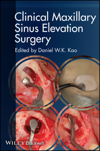
Maxillary sinus elevation, followed by placement of a wide variety of grafting materials, has been the generally accepted surgical protocol for the development of bone in the sinus cavity. Over the years, various techniques have been proposed for maxillary sinus elevation, which differ in surgical approach, bone graft materials, and advanced technology application for hard tissue and soft tissue management.
Dr. Kao and a team of experts begin by discussing anatomy, radiographic image applications and limitations, and then provide step-by-step clinical procedures for the lateral window technique, including piezosurgery, and the trans-alveolar methods, including balloon and controlled hydostatic sinus elevation. Also included are chapters on post-operative care and complication management.
Sie lesen das E-Book in den Legimi-Apps auf:
Seitenzahl: 303
Veröffentlichungsjahr: 2014
Ähnliche


CONTENTS
Cover
Title page
Copyright
Contributors
Acknowledgments
Introduction
References
Chapter 1: Anatomy and Physiology of the Maxillary Sinus
Anatomy of the maxillary sinus
Pathophysiology of the maxillary sinus
References
Chapter 2: The Applications and Limitations of Conventional Radiographic Imaging Techniques
Introduction
Periapical radiography
Panoramic radiography
Conclusion
References
Chapter 3: The Applications and Limitations of Advanced (3-Dimensional) Radiographic Imaging Techniques
Introduction
Conventional cross-sectional tomography
Medical-grade CT and CBCT
Conclusion
References
Chapter 4: Conventional Instruments Preparation and Preclinical Training of the Lateral Window Technique
Introduction to the lateral window technique
Suture materials and technique
Preclinical training on a hen's egg and sinus model
References
Chapter 5: Clinical Procedures of the Lateral Window Technique
Anesthesia
Incision (flap design)
Hard tissue management
Bone graft materials and barrier membrane
References
Chapter 6: Avoiding and Managing Complications for the Lateral Window Technique
Introduction
Problems to be avoided
The medical history
Radiographic examination
Septa
Prevention of hemorrhage
Summary, presurgical prevention of complications
Complications during the surgical procedure
Postoperative instructions to help avoid postoperative complications
References
Chapter 7: Advanced Techniques of the Lateral Window Technique
Background of piezosurgical techonology (piezoelectric bone surgery)
The applications of piezosurgical technology in lateral window procedures
Surgical steps
References
Chapter 8: Basic Instruments and Materials of the Transalveolar Approach
Background
Instrument preparation for the transalveolar approach: osteotome technique
References
Chapter 9: Clinical Procedures of the Transalveolar OsteotomeApproach
Incision design
References
Chapter 10: Postoperation Management of the Transalveolar Osteotome Approach
Postoperative instruction
Complications in the sinus elevation surgery
Appendix A: preoperative instructions for the transalveolar osteotome approach
Appendix B: postoperative instructions for the transalveolar osteotome approach
References
Chapter 11: Advanced Techniques of the Transalveolar Approach
Clinical procedures
Other surgical techniques
Surgical protocol
Clinical case
References
Chapter 12: Decision Tree for Maxillary Sinus Elevation Options
References
Chapter 13: Choices of Bone Graft Materials
Bone grafting materials overview
Tissue engineering materials
References
Chapter 14: Review of Dental Implant Success and Survival Rates
Success and survival criteria
Success and survival rates
References
Additional relevant works
Index
Eula
List of Tables
Introduction
Table I.1 Surgical and restorative treatment options for vertical interarch discrepancy.
Chapter 04
Table 4.1 Basic surgery instruments list.
Table 4.2 Lateral window sinus lift instruments list.
Table 4.3 Suture materials and other instruments.
Chapter 05
Table 5.1 Advantages and disadvantages of handpieces.
Table 5.2 Advantages and disadvantages of techniques for bone island handling.
Table 5.3 Properties of various types of bone grafting materials.
Chapter 06
Table 6.1 Postoperative complications.
Chapter 08
Table 8.1 Osteotome instrument kit.
Chapter 14
Table 14.1 Criteria for selecting between the delayed placement and the simultaneous placement techniques.
Table 14.2 Definition and criteria for implant survival.
Table 14.3 Criteria for implant success.
Table 14.4 Ranges of survival and cumulative survival rates (%) for the simultaneous placement technique via the lateral window approach.
Table 14.5 Ranges of survival and success rates (%) for the delayed placement technique via the lateral window approach.
Table 14.6 Mean, ranges of survival rates (%), and the time differences between the simultaneous placement and the delayed placement techniques.
Table 14.7 Ranges of survival, success rates (%), and bone gain (mm) for the simultaneous placement technique via the transalveolar approach.
Table 14.8 Factors that could affect implant success and survival rates.
List of Illustrations
Introduction
Figure I.1 Sinus elevation procedures. (a) Area for sinus elevation. (b) External approach. (c) Internal approach.
Figure I.2 (a–d) Sinus augmentation—lateral window approach.
Figure I.3 Sinus augmentation—lateral window approach. (a) Day 0: sinus graft. (b) Wait 6–9 months for bone graft to heal, then place implant. (c) Wait another 6–9 months for osseointegration and implant restoration.
Figure I.4 (a–d) Internal approach—osteotome technique.
Figure I.5 (a) and (b) Unfavorable crown-implant ratio.
Figure I.6 The interarch relationship should be considered in order to achieve a successful surgical, restorative, and aesthetic outcome.
Chapter 01
Figure 1.1 Sinus anatomy. The maxillary sinus is the largest air-filled cavity in the skull.
Figure 1.2 Odontogenic infection may create a pathway to spread infection into the maxillary sinus.
Figure 1.3 The maxillary ostium (MO) enters the infundibulum, which is the space between the uncinate process (U) and the ethmoid bulla (*).
Figure 1.4 Sinus infection. Maxillary sinusitis (mucositis in the right one); note the air inclusions (arrow) suggesting acute sinusitis. (Courtesy of Dr. Massimo De Paoli.)
Figure 1.5 The microscopic anatomy of the sinuses reveals four basic cell types: pseudostratafied ciliated columnar epithelium, nonciliated columnar cells, goblet cells, and basal cells. (Used with permission from Prof. Chun-Pin Chiang.)
Figure 1.6 Sensory innervation of the maxillary sinus.
Figure 1.7 The blood supply to the maxillary sinus.
Chapter 02
Figure 2.1 A periapical radiograph depicting residual bone height of less than 5 mm, which suggests that a lateral window approach may be used for sinus grafting. (Courtesy of Dr. P.W. Dai, Taichung, Taiwan.)
Figure 2.2 A periapical film demonstrating the position of the distal direction indicator being approximately 1 mm shy of the sinus floor.
Figure 2.3 A periapical radiograph illustrating continuous radiolucency along the entire length of an implant (white arrows), which indicates a nonosseointegrated implant.
Figure 2.4 Verification of the implant-abutment seating by periapical radiographs. (a) This periapical radiograph covers two fixtures. The anterior fixture was projected with the proper angle, while the posterior one has a more vertical angulation resulting in blurring of distal threads (white arrows), indicative of an unacceptable projection to evaluate implant-abutment interface of the posterior fixture. (b) A gap (white arrows) seen between the implant and the healing abutment indicates an open implant-abutment interface (improper abutment seating). (c) Adequate implant-abutment seating confirmed by a periapical radiograph of appropriate projection geometry.
Figure 2.5 Development of PIXRL. (a) An implant placement driver (white arrows) was attached to the implant fixture. (b) The PIXRL jig (white arrows) was connected to the implant placement driver and registration record against the adjacent teeth obtained. The use of the custom-made PIXRL bite block allows clinicians to monitor crestal bone changes or prosthetic misfit around an implant accurately and consistently over time. (Courtesy of Drs. K.C. Lin, San Francisco, CA, and C.P. Wadhwani, Seattle, WA.)
Figure 2.6 Digital image subtraction analysis. (a) Periapical film taken at the implant uncovering surgery (baseline) using an XCP film holder together with the bite registration. (b) Periapical film taken with the same customized bite block at 6 weeks after the implant uncovering surgery. (c) Digital subtraction imaging revealed no changes in the crestal bone level (blue arrows) between baseline and 6 weeks follow-up.
Figure 2.7 (a) Periapical radiograph showing radiopacity consistent with presence of cement at the abutment/crown interface mesially and distally (white arrows), with no bone loss evident. (b) Periapical radiograph showing radiopacity consistent with presence of cement (white arrow) at the distal surface of the restoration with bone loss evident (yellow arrows). (c) Periapical radiograph taken after cement removal. [Figure 2.7(a) courtesy of Drs. R. Schuler and D.A. Rapoport, Tukwila, WA. Figures 2.7(b) and 2.7(c) courtesy of Dr. P.W. Dai, Taichung, Taiwan.]
Figure 2.8 A periapical radiograph exhibiting sharp threads with no overlaps on either side of the implant, indicating that the image is adequate to be used for assessment of the peri-implant bone conditions.
Figure 2.9 Sinus septa can be identified in (a) a periapical radiograph (white arrows) and (b) a panoramic radiograph (white arrow). A panoramic radiograph revealed the sinus septa in both the right (yellow arrow) and left sinus (white arrow). (c) The sinus septa were confirmed by CBCT scans in both panoramic (upper panel) and axial (lower right panel) views. Two distinct sinus cavities were noted in the axial view of the CBCT scans. Special caution must be used when evaluating the bone height in the CBCT sagittal view (lower left panel) for implant placement, as actual bone height may be less than the scan indicates.
Figure 2.10 A panoramic radiograph can be used to evaluate mucosal thickening in the maxillary sinus. (a) Mucosal thickening (white arrows) noted in the right maxillary sinus on a panoramic radiograph. Confirmation of mucosal thickening in right maxillary sinus by CBCT scans, seen in (b) axial (left panel) and cross-sectional (right panel) views, and (c) sagittal view.
Figure 2.11 An antral pseudocyst can be recognized in (a) a panoramic radiograph (yellow arrows), and (b) a CBCT image (lesion indicated by yellow arrows in the panoramic view and white * in the axial and cross-sectional views). This type of lesion appears as a well-defined dome-shaped radiopacity with a rounded outline, situated on the floor of sinus. The panoramic radiograph also revealed a smaller mucosal antral cyst around the periapical region of tooth #2 in the right maxillary sinus.
Figure 2.12 Recommendations for radiographic examination in the maxillary sinus. In general, guidelines for sinus evaluation, implant placement, and follow-up assessment are proposed (modified from Dula et al.96). The thick black arrows represent procedures that are commonly used, while the thin black arrows represent procedures that are less commonly implemented.
Chapter 03
Figure 3.1 Cross-sectional spiral tomographic images of the maxilla sinus. Conventional spiral tomography was produced using a Cranex TOME multifunctional unit. (a) Panoramic scout image was used to select appropriate thin cross-sectional slices. (b) Conventional tomography consists of four slices, each 2 mm thick (exposure parameters of 60 kV, 56 seconds, and 1.6–2.0 mA). Note the relation of the radiopaque metal ball to the crestal bone. The tomographic image shows both the residual crestal bone height to the sinus floor and the thickness of the buccal bone.
Figure 3.2 Maxillary sinus pathology on CT scans. A small dome-shaped lesion (retention cyst) located on the floor of the right maxillary sinus (white arrows). Left maxillary sinus shows total opacity with a radiopaque lesion consistent with a foreign body on the sinus floor (black arrows), indicating left maxillary rhinosinusitis (opacification of sinus; indicated by white *) caused by sinus lift procedure. (a) Coronal view showing a normal right ostiomeatal complex and a blocked left ostiomeatal complex. (b) Representative axial image. (c) Representative sagittal image through the left sinus.
Figure 3.3 Maxillary sinus pathology on CT scans. Radiographic finding of a dome-shaped radiopacity arising from the floor of the maxillary sinus compatible with an antral pseudocyst (white arrows). (a) Representative coronal image. (b) Representative axial image. (c) Representative sagittal image.
Figure 3.4 Maxillary sinus pathology on CT scans. Images of rhinosinusitis. (a) Coronal image showing rhinosinusitis (opacification of sinus; indicated by white *) with multiple retention cysts in right maxillary and ethmoid sinuses. (b) Left maxillary and ethmoid sinuses demonstrate total opacity (indicated by white *) and a radiopaque lesion consistent with a foreign body (white arrows) on the floor of the left maxillary sinus, indicating left rhinosinusitis caused by sinus augmentation procedure. The radiopaque lesion is the bone graft from a sinus lift procedure.
Figure 3.5 Assessment of maxillary sinus anatomy on CT scans—maxillary sinus septa. (a) The location of bilateral septa (white arrows) on reformatted panoramic images. (b) The orientation of a septum (white arrow) on reconstructed cross-sectional images. (c) Axial images showing complete bilateral septa (white arrows) that divide each of the maxillary sinuses into two cavities.
Figure 3.6 Assessment of maxillary sinus anatomy on CT scans. Maxillary sinus intraosseous artery (white arrow) in the lateral antral wall. (a) Representative axial image. (b) Representative coronal image. (c) Representative sagittal image.
Figure 3.7 Pre- and postoperative assessment for sinus augmentation procedure. CT and CBCT scans showing right maxillary sinus pseudocyst (white arrows) and augmentation procedure outcome. (a) Preoperative CT, and (b) postoperative CBCT scans 6 months following sinus augmentation procedure in coronal, axial, and sagittal views. Note also the prior augmentation (a) and subsequent implant placement (b) in the left maxillary sinus.
Figure 3.8 CBCT-guided implant surgery using the All-on-4 technique for the rehabilitation of the edentulous maxilla. (a) Facilitate “virtual” treatment plan for implant positioning. (b) Four implants virtually placed in relation to the planned prosthesis (occlusal view). (c) Image without the surgical template, to better visualize implant positions in bone. The two mesial-most implants were planned in upright positions (yellow color) and the two distal-most implants were planned in tilted positions (blue color). (d) Image with virtual surgical template in place. (Courtesy of Drs. H. Leghuel, Columbus, OH, and A. Tsigarida, Philadelphia, PA.)
Figure 3.9 A bone-supported stereolithographic surgical template was fabricated following the virtual treatment plan in Figure 3.8.
Figure 3.10 The surgical template stabilized with anchor pins; pilot drilling was then performed in the best position according to virtual planning with the stereolithographic surgical template.
Figure 3.11 Panoramic radiography and clinical photos of a case treated with upper maxillary rehabilitation through the All-on-4 concept. (a) Panoramic radiograph obtained immediately after implant placement (see inset for higher magnification). (b) Radiograph obtained upon completion of the final restoration. (c) Final restoration, occlusal view. (Courtesy of Drs. H. Leghuel, Columbus, OH, and A. Tsigarida, Philadelphia, PA.)
Figure 3.12 CBCT-guided surgery for the rehabilitation of the edentulous maxilla with simultaneously transcrestal maxillary sinus floor elevation. (a) Facilitate “virtual” treatment plan for eight implant positions. (b) A bone-supported surgical template on the stereolithographic maxilla model. (c) Facilitate Guided Surgery: implant site preparation was completed and implant delivery accomplished via the bone-supported stereolithographic template. (d) Periapical radiographs showing eight implants placed in the edentulous maxilla in conjunction with indirect sinus floor elevation. (e) Final screw-retained fixed dental prosthesis. (Courtesy of Drs. M. Scherer, Las Vegas, NV; N. Rathi, Columbus, OH; and N. Jain, Sacramento, CA.)
Chapter 04
Figure 4.1 (a) and (b) Lateral window approach. Full thickness flap and osteotomy, window preparation.
Figure 4.2 (a) and (b) Lifting the sinus membrane and bone graft placement.
Figure 4.3 (a) Basic surgery instruments. (b) Lateral window sinus lift instruments.
Figure 4.4 (a) and (b) Continuous locking suture technique with 4-0 polyglycolic acid; FS-2 needle was used to achieve primary closure. (Courtesy of Dr. Kevin Ma.)
Figure 4.5 (a) and (b) Chromic gut suture. Chromic gut suture can be used at the vertical releasing site to provide a more comfortable postoperative experience for the patient.
Figure 4.6 (a)–(c) Preclinical training on the hen's egg shell. The initial osteotomy can be prepared with a high-speed round diamond bur. The osteotomy is done with a light touch and paintbrush stroke approach until the membrane is exposed
Figure 4.7 (a) and (b) Preclinical training on the hen's egg shell. Detach the transparent membrane lying between the egg shell and the egg white gently without tearing, using the inverted cone bur (piezosurgical instrument).
Figure 4.8 (a) and (b) Sinus model. By using the sinus model, trainees may become familiar with hand instruments.
Figure 4.9 (a) and (b) Sinus membrane detach—lower border. Gently release the sinus membrane with a hand instrument around the window about 3–5 mm.
Figure 4.10 (a) and (b) Sinus membrane detach—mesial area.
Figure 4.11 (a) and (b) Sinus membrane detach—distal area.
Figure 4.12 (a) and (b) Sinus membrane detach—upper border. Gently detach the membrane at the apical aspect of the sinus cavity.
Figure 4.13 (a) and (b) Sinus membrane elevation. Once the membrane around the window is gently released, the instrument can be used to elevate the deeper portion of the interior border of the sinus membrane. If the handle interferes with the window, another angulation of hand instrument needs to be used.
Figure 4.14 (a–d) After initial release of the lower border, mesial, distal aspect, and upper border of the sinus membrane about 3–5 mm, go farther from the lower aspect of the sinus cavity to separate all the way to the medial wall.
Figure 4.15 (a) and (b) Instruments must always be kept in contact with the bone surface to avoid membrane perforation. The left side shows the correct way to use the sinus membrane elevation instrument. The right side shows the wrong way to use the instrument
Chapter 05
Figure 5.1 (a) Lateral window approach. (b) Full-thickness flap.
Figure 5.2 (a) and (b) Osteotomy, window preparation, and lifting the sinus membrane.
Figure 5.3 (a) and (b) Bony window outline.
Figure 5.4 (a) Carbide and diamond round bur can be used to outline the bony window. (b) Piezosurgical round diamond tip.
Figure 5.5 (a) and (b) The osteotomy is deepened in sweeping motions until the bone is thin and translucent enough to see the underlying gray/red color of the sinus membrane.
Figure 5.6 (a) Infracture bone island. (b) Wall-off technique.
Figure 5.7 (a) and (b) The lower border of the Schneiderian membrane is lifted with a hand elevator.
Figure 5.8 (a) and (b) The mesial border of the Schneiderian membrane is lifted.
Figure 5.9 (a) and (b) The distal membrane is lifted.
Figure 5.10 (a) and (b) The upper border of the sinus membrane is lifted.
Figure 5.11 The medial wall sinus membrane is released. After initially releasing the sinus membrane, go farther from the lower aspect of the sinus cavity to separate all the way to the medial wall.
Figure 5.12 (a) and (b) Be sure the membrane is lifted high enough.
Figure 5.13 (a) and (b) The sinus membrane is not lifted in the medial wall.
Figure 5.14 (a) and (b) Bone graft materials are inserted into the sinus cavity.
Figure 5.15 Collagen plug (sliced into small pieces) can also be used to occupy the space where the implant will not be placed in order to reduce the use of bone graft materials. In this case, collagen plug was placed into the distal side of the sinus cavity in the second molar area.
Figure 5.16 (a) and (b) A barrier membrane can also be used to cover the window on the lateral wall of the maxilla.
Figure 5.17 Computed tomography scan demonstrating radiopaque mass (arrow) at the ostium. Images from Hunter et al. (2009).
Chapter 06
Figure 6.1 The adequate patency of the semilunar hiatus (dotted red circle) allows proper drainage of the maxillary sinus mucous production into the middle meatus and assures a healthy maxillary sinus.
Figure 6.2 Healthy maxillary sinus. Note the radiolucency of a physiologically healthy maxillary sinus in a transverse cut of a CBCT.
Figure 6.3 (a) and (b) Chronic maxillary sinusitis. Note the thickening of the Schneiderian membrane (yellow arrow) in close relation to a periapical radiolucency (blue dotted line) on the apex of a nonvital maxillary left second premolar.
Figure 6.7 A full view of the paranasal sinuses allows the clinician to perform a comprehensive evaluation prior to maxillary sinus surgery. Note the polyp on the right ethmoidal sinus (red arrow), a potential risk for complications following maxillary sinus surgery.
Figure 6.8 Note the space of the maxillary artery (yellow arrow) in the lateral wall of the maxillary sinus. In some instances the maxillary artery runs inside the bone. (Courtesy of Dr. Michael Kulianos, Athens, Greece.)
Figure 6.9 (a) and (b) Bony septa in the maxillary sinus are common abnormalities and can be depicted by axial with coronal CT scans.
Figure 6.10 Note the ligation of the maxillary artery with 5–0 chromic gut suture, after the window osteotomy and membrane elevation. The black arrow shows the dissected maxillary artery on the distal portion of the sinus. (Courtesy of Dr. Michael Kulianos, Athens, Greece.)
Figure 6.11 (a) Small Schneiderian membrane perforation following complete osteotomy for lateral window technique for maxillary sinus augmentation. (b) Two Large Schneiderian membrane perforations following complete osteotomy for lateral window technique for maxillary sinus augmentation. (c) A resorbable collagen membrane was placed to cover both Schneiderian membrane perforations. Note how the membrane extends beyond the outline of the perforations.
Figure 6.12 Thickening of the Schneiderian membrane on a smoker.
Figure 6.13 (a)–(c) During the placement of an implant, the implant was inadvertently pushed into the maxillary sinus.
Figure 6.14 (a) and (b) CBCT of three implants placed on a previously grafted maxillary sinus. Note the cloudiness on the maxillary sinus as a result of a chronic sinusitis.
Figure 6.15 (a) and (b) Six-month postoperative CBCT of a previously grafted maxillary sinus. Note that the Schneiderian membrane was not elevated from the medial wall of the sinus, creating a pouch between the medial wall of the sinus and the bone graft material. As a result we can observe an enlarged Schneiderian membrane typical of a chronic sinusitis.
Figure 6.16 A common surgical complication is the formation of hematomas in the face. Note bruising underneath the eye.
Chapter 07
Figure 7.1 (a) and (b) The microvibrations of the instrument are caused by the piezoelectric effect. [Images from
Essentials in Piezosurgery,
by Tomaso Vercellotti (Hanover Park, IL: Quintessence, 2009).]
Figure 7.2 (a) Tips of piezosurgery. (b) Carbide and diamond round bur.
Figure 7.3 (a) and (b) Mectron Piezosurgery Touch device.
Figure 7.4 Mectron piezosurgery sinus lift inserts. The sinus lift kit includes the following inserts: OT1, OT5, EL1, EL2, and EL3. (a) OT1: For micrometric osteotomy (about 1mm)—to finalize the osteotomy in close proximity to soft tissue (e.g., sinus membrane, vessel, alveolar nerve). (b) OT5: For micrometric osteotomy or osteoplasty—nontraumatic, to finalize the osteotomy or osteoplasty on thin bone and/or near delicate anatomic structures. (c) EL1: For Schneiderian membrane separation from bony walls—separation of the sinus membrane, 2mm around the frame of the bony window. (d) EL2: The noncutting elevator of the sinus membrane—separation of the sinus membrane in internal zones. (e) ET3: The noncutting elevator of the sinus membrane—separation of the sinus membrane in internal zones.
Figure 7.5 (a) and (b) Surgical Step 1: Elevated full-thickness flap.
Figure 7.6 (a) and (b) Surgical Step 2: Reduction of thickness of lateral sinus wall until the dark shadow indicating the sinus cavity is seen, using osteoplasty insert (OT5) from Mectron system.
Figure 7.7 (a) and (b) Surgical Step 3: Separation of membrane using the trumpet insert.
Figure 7.8 (a) and (b) Surgical Step 4: Elevation of membrane by noncutting elevator EL1, EL2, and EL3.
Figure 7.9 (a) and (b) Surgical Step 5: Elevation of membrane by hand instrument.
Figure 7.10 (a) and (b) Surgical Step 6: Bone graft and barrier membrane placed.
Chapter 08
Figure 8.1 Osteotome instrument set.
Figure 8.2 (a) and (b) A dome-shaped tented space can be seen at the osteotome site.
Figure 8.3 Surgical mallets. (a) Metal mallet. (b) Nylon cap mallet.
Figure 8.4 Straight and offset osteotomes.
Figure 8.5 Concave and convex osteotome. (left) Concave osteotome for sinus floor elevation. (right) Convex osteotome for bone condensation.
Chapter 09
Figure 9.1 (a) and (b) Step 1: The crest approach begins with the decision to raise a flap or not.
Figure 9.2 Step 2: The implant site is prepared to a depth 1 mm below the sinus floor by using a 2 mm cylindrical bur.
Figure 9.3 (a) and (b) Step 3: A periapical radiograph is taken with the 2 mm guide pin in place, confirming the integrity of the subsinus cortex and verifying the implant position and the distance from the apex of the osteotomy to the sinus floor.
Figure 9.4 Tips for the osteotome technique. (a) Use dental floss or other technique to tie the guide pin to prevent the patient swallowing the instrument accidently. (b) Use piezosurgical instruments for minor depth adjustment. Note: Images (a) and (b) are for demonstrating these tips and not from the same patient.
Figure 9.5 Step 4: The osteotomy is enlarged with a 3 mm cylindrical drill.
Figure 9.6 (a) and (b) Step 5: Bone graft material is added to the osteotomy before attempting to elevate the sinus membrane.
Figure 9.7 Step 6: Infracturing the sinus floor cortical bone with light malleting. (a) A 3 mm osteotome is inserted into the osteotomy and advanced with light malleting. (b) An assistant should support the patient's head during the malleting.
Figure 9.8 A stop may be attached to the osteotome.
Figure 9.9 (a) and (b) Step 8: Enlarge the osteotomy to the desired diameter. The sequence of adding graft material and tapping the osteotome to the predetermined depth continues for the needed amount of elevation.
Figure 9.10 Step 10: Dental implant insertion. Additional graft materials placed before insertion.
Figure 9.11 (a–c) Osteotome approach combined with extraction and immediate implant placement, part I. The osteotome approach can be also combined with extraction.
Figure 9.13 (a–c) Osteotome approach combined with extraction and immediate implant placement, part III.
Chapter 10
Figure 10.1 Note minimal subantral bone height of 2.0 mm—a lateral window procedure would be indicated rather than an osteotome procedure. Also note the slope of the sinus wall, which could complicate the use of an osteotome.
Figure 10.2 Note the implosion of the implant in site #5. This could have been minimized by proper bone preparation and monitoring of the removable partial denture.
Figure 10.3 A periapical radiograph noting a residual tooth structure #4. This type of situation would allow the immediate extraction with greater than 5.5 mm of osseous height to allow for an osteotome procedure with immediate placement.
Figure 10.4 Note the dome effect of the grafting material to verify the intact membrane following the osteotome lift procedure.
Figure 10.5 (a) Position patient on the bed and turn 45° to the right (symptom site). (b) Keep head turned to the side and lie down for 5 minutes. (c) Turn head to the opposite side while lying on the bed for 5 minutes. (d) Roll body onto side in the direction you are facing; now you are pointing your head nose down. Stay in this position for another 5 minutes. (e) Swing your legs and feet over the side of the bed and sit upright, keeping your head straight, and remain in place for another 5 minutes.
Chapter 11
Figure 11.1 (a) Micro-mini sinus lift balloon inserted into the sinus cavity through the crestal approach. (b) Three types of balloon instrument design: straight, micro-mini, and angled. (Images from Zimmer Dental.)
Figure 11.2 (a) and (b) Baseline clinical and radiograph information. (c) and (d) Create access by drill and/or osteotome instrument. (e) and (f) A guided pin is used to indicate the depth of the osteotomy.
Figure 11.3 (a) and (b) Inflate and deflate the balloon extraorally several times with normal saline before inserting into the sinus cavity. (c) Insert the metal tube into the sinus cavity without touching the sinus membrane. (d) Once the balloon is inserted into the sinus cavity, the balloon can be pumped with normal saline.
Figure 11.4 (a) The dome-shaped bone graft material can be seen in the radiograph to indicate the tented space. (b) A dental implant may be placed during the same procedure if primary stability has been achieved.
Figure 11.5 Several surgical techniques have been proposed to minimize the tapping motion by using hydraulic pressure, the so-called “hydraulic sinus lift” procedure. The hydraulic pressure is applied into the osteotomy site by means of air/water exhaust spray from a high-speed dental handpiece (top 2 figures) or an uncontrolled water jet from a plastic syringe (bottom 2 figures). (Top two images illustrated from the concept of Chen L [2005]. An 8-year retrospective study using the minimally invasive hydraulic sinus condensing technique. Bottom two images illustrated from the concept of Sotirakis et al. Elevation of the maxillary sinus floor with hydraulic pressure.
The Journal of Oral Implantology
[2005] vol. 31 [4] pp. 197–204.)
Figure 11.6 Controlled hydrostatic sinus elevation kit. (Left) Implant drill and piezosurgical tips. (Middle) Various diameter cannula. (Right) Pressure sensor meter and hand-actuated pump.
Figure 11.7 (a) Initial pilot drill up to 1 mm before reaching the sinus floor membrane. (b) A piezosurgical drill (Mectron piezosurgery system, Italy) can be used to gently perforate the floor of the sinus.
Figure 11.8 (a) The Luer-Lock cannula with tapered plug-in end inserted into the osteotomy site should insert 1 mm inferior to the sinus floor but not touch the sinus floor. (b) Hydrostatic pressure was applied to detach the Schneiderian membrane.
Figure 11.9 (a) Pressure-controlled meter with pump (Alliance Inflation System, Boston Scientific, Boston, MA) can provide suitable controlled force. (b) Pressure meter with sensor to monitor the hydrostatic pressure.
Figure 11.10 Inserted bone grafting material into the tented space.
Figure 11.11 A dental implant may or may not be placed into the osteotomy site.
Figure 11.12 Baseline information of upper right first molar #3. (a) Intraoral image. (b) Radiographic image.
Figure 11.13 (a) Full-thickness flap. (b) Radiographic guide pin showed that it was close to the sinus floor.
Figure 11.14 Luer-Lock cannula with tapered plug-in end inserted showed (a) intraoral view, and (b) radiographic image.
Figure 11.15 Controlled hydrostatic sinus elevation procedure was repeated using a matching size insert cannula. (a) 2 mm cannula. (b) 3 mm cannula.
Figure 11.16 (a) A 4.5 mm x 11 mm implant with healing abutment (Ankylos Densply Friadent, Mannheim Germany) was placed. (b) Radiographic image of implant placement. (c) Fixed crown restoration with 4 months loading (restored by Dr. T.S. Zhuo, prosthodontist).
Chapter 12
Figure 12.1 Lateral window maxillary sinus bone augmentation. (a) Different types of piezosurgical tips useful for removing osseous structure lateral to maxillary sinus and elevating Schneiderian membrane. (b) A piezo bur was used to create a window by removing bony wall lateral to the sinus. (c) Schneiderian membrane has been partially elevated. (d) Preoperative CT scan shows signs of sinus pneumatization and residual bone height (height of bone apical to sinus floor) of 1–3 mm, making it necessary to perform a separate lateral window sinus elevation based on the decision tree presented in Figure 12.5. (e) Six-month postoperative CT scan shows evidence of augmented bone in the sinus with 12–13 mm of vertical bone height gained.
Figure 12.2 Wall-off lateral window sinus bone augmentation technique. (a) Preoperative panoramic radiograph shows approximately 5 mm of residual bone height (height of bone apical to sinus floor) at sites #2 and #3. The clinician has decided to place 10 mm long implants and thus will perform a lateral window sinus elevation based on the decision tree in Figure 12.5. (b) This schematic shows the modified trephine-like drill (DASK Drill #6, Dentium) designed to detach bony island from the lateral wall of sinus in this wall-off technique. (c) The same drill is used to prepare a bony window on the lateral wall. (d) Sinus membrane has been elevated. (e) Two 10 mm long implants are placed simultaneously with lateral window sinus bone augmentation, and the bony island has been repositioned back to the window to contain the bone grafts in the sinus. (f) Nine-month postoperative panoramic radiograph showing evidence of bony augmentation in the sinus with completed implant restoration.
Figure 12.3 Osteotome transalveolar crestal sinus bone augmentation technique. (a) Preoperative radiograph shows residual bone height (height of bone apical to sinus floor) of about 8 mm at site #13. The clinician has determined to place a 12 mm long implant and thus decided to place the implant while performing a transalveolar crestal sinus bone graft. (b) An osteotome was used to place bone grafts into the sinus while lifting the Schneiderian membrane. (c) Five-month postoperative radiograph demonstrates evidence of bone augmentation apical to implant #13. (d) Implant #13 has been restored.
Figure 12.4 Thin-out transalveolar crestal sinus bone augmentation technique. (a) A diamond drill (DASK Drill #1, Dentium) with markings is designed to remove cortical bone of the maxillary sinus floor. (b) After implant osteotomy site preparation has been performed to final drill 1 mm short of the sinus, this diamond drill is then used to gently remove the bony sinus floor. (c) A dome-shaped sinus curette is used to start elevating the membrane. (d) Bone graft particulates are inserted into the space created under the sinus Schneiderian membrane.
Figure 12.5 Decision tree for performing lateral versus transalveolar crestal sinus bone augmentation when sufficient alveolar buccal-palatal width is present. RBH, residual bone height (height of bone apical to sinus floor); m, months; IMP, implant; TAC, transalveolar crestal.
Figure 12.6 Decision tree for performing sinus bone augmentation when insufficient alveolar buccal-palatal width is present. RBH, residual bone height (height of bone apical to sinus floor).
Chapter 13
Figure 13.1 Lateral window maxillary sinus bone augmentation using rhBMP-2. (a) A radiograph shows right sinus pneumatization with residual bone height ranging from about 4 to 8 mm where implant osteotomy sites are planned. (b) rhBMP-2, in powder form, is to be reconstituted in sterile water. (c) Sterile water is added to rhBMP-2 powder. (d) Reconstituted rhBMP-2 is then uniformly distributed to absorbable collagen sponge (ACS), a carrier for rhBMP-2. (e) A period of 15 minutes is recommended to allow rhBMP-2 protein to bind to ACS and afterward rhBMP-2 is ready for use. (f) Schneiderian membrane has been sufficiently elevated and implant osteotomy sites prepared. (g) rhBMP-2 soaked ACS has been placed into the sinus and implants are placed. (Courtesy of Dr. Daniel Kao.)
Figure 13.2 Human histology of anorganic bovine bone mineral (ABBM) combined with rhPDGF-BB in maxillary bone augmentation. [Courtesy of Dr. Myron Nevins; Nevins et al. (2009)]. (a) Micro-CT scan of the core biopsy taken at the time of implant placement 6 months after sinus bone augmentation showing mostly bone (red) with minimal amount of ABBM particles left (white). (b) A core biopsy taken 6 months after sinus bone augmentation revealing a considerable amount of bone formation at augmented site with few ABBM particles remaining. (c) At higher magnification, evidence of active osteoid formation, woven bone, and lamellar bone is present.
Figure 13.3 Types of bone graft materials used in maxillary sinus bone augmentation: FDBAs, bovine xenograft (Endobon), equine xenograft (Equimatrix), combination of hydroxyapatite and β-tricalcium phosphate (OSTEON), and β-tricalcium phosphate (from left to right).
Figure 13.4 Bone graft materials and biologic growth factors available for sinus bone augmentation procedure.
Guide
Cover
Table of Contents
Begin Reading
Pages
iv
vii
viii
ix
x
xii
xiii
xiv
xv
xvi
1
3
4
5
7
8
9
10
11
12
13
14
15
16
17
18
19
22
23
24
25
26
27
28
29
30
31
32
33
34
35
36
37
38
39
40
41
42
43
44
45
46
47
48
49
50
51
52
53
54
55
56
57
58
59
60
61
62
63
64
65
66
67
68
69
70
71
72
73
74
75
76
77
78
79
80
81
82
83
84
85
86
87
88
89
90
91
92
93
94
95
96
97
98
99
100
101
102
103
105
106
107
108
109
110
111
112
113
114
115
116
117
118
119
120
121
122
123
124
125
127
128
129
130
131
132
133
134
135
136
138
142
143
145
146
147
149
150
151
152
153
154
155
156
157
158
160
162
163
164
165
166
167
168
169
170
171
172
173
174
175
177
179
180
181
182
183
184


