159,99 €
Mehr erfahren.
- Herausgeber: Thieme
- Kategorie: Fachliteratur
- Sprache: Englisch
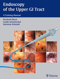
The essential guide to mastering endoscopic techniques of the upper GI tract
While technological advances have made endoscopy one of the most common procedures for examining the upper GI tract, learning how to maneuver the instruments and interpret the images can be frustrating for those without experience. Designed specifically for those in training, Endoscopy of the Upper GI Tract -- with its more than 770 illustrations and user-friendly format -- is the most comprehensive instructional guide available.
Beginning with a detailed introduction to all aspects of the endoscopic examination, this lavishly illustrated guide features:
- Clear descriptions and images of all of the instruments and how and when they are used
- Artfully combined photographs and 3D illustrations showing the exact location of the endoscope in relation to the anatomy of the immediate region
- Step-by-step instructions for handling the endoscope, such as insertion, air insufflation, irrigation, and more
- Useful checklists and tables that lay out the procedures from beginning to end, including preparations, necessary medication and anesthesia, required staff and supplemental equipment, potential risks and complications, etc.
The book also encompasses a complete full-color atlas that illustrates the entire spectrum of both normal and pathological findings. In addition to detailed explanations of each finding, the authors provide:
- The endoscopic criteria and the most important differential diagnoses for each disorder
- Series of images showing common variants, as well as comparison photographs of differential diagnoses
- Useful guidelines for proper documentation
A guide to interventional and extended examination techniques rounds out the text. All procedures, from treating upper gastrointestinal bleeding and collecting specimens to placing a duodenal tube and removing foreign bodies, are treated in full detail.
Wi
Das E-Book können Sie in Legimi-Apps oder einer beliebigen App lesen, die das folgende Format unterstützen:
Seitenzahl: 277
Veröffentlichungsjahr: 2004
Ähnliche


Library of Congress Cataloging-in-Publication Data is available from the publisher
This book is an authorized translation of the German edition published and copyrighted 2003 by Georg Thieme Verlag, Stuttgart, Germany. Title of the German edition: Der Gastroskopie-Trainer: Schritt-für-Schritt-Anleitungen für die Ösophago-, Gastro- und Duodenoskopie
Translator: Terry C Telger, Fort Worth, TX, USA
© 2004 Georg Thieme Verlag,
Rüdigerstrasse 14, 70469 Stuttgart, Germany
http://www.thieme.de
Thieme New York, 333 Seventh Avenue,
New York, NY 10001 USA
http://www.thieme.com
Typesetting by primustype Hurler, Notzingen Printed in Germany by Grammlich, Pliezhausen
ISBN 3-13-136731-8 (GTV)
ISBN 1-58890-239-0 (TNY)
Important note: Medicine is an ever-changing science undergoing continual development. Research and clinical experience are continually expanding our knowledge, in particular our knowledge of proper treatment and drug therapy. Insofar as this book mentions any dosage or application, readers may rest assured that the authors, editors, and publishers have made every effort to ensure that such references are in accordance with the state of knowledge at the time of production of the book.
Nevertheless, this does not involve, imply, or express any guarantee or responsibility on the part of the publishers in respect to any dosage instructions and forms of applications stated in the book. Every user is requested to examine carefully the manufacturers' leaflets accompanying each drug and to check, if necessary in consultation with a physician or specialist, whether the dosage schedules mentioned therein or the contraindications stated by the manufacturers differ from the statements made in the present book. Such examination is particularly important with drugs that are either rarely used or have been newly released on the market. Every dosage schedule or every form of application used is entirely at the user's own risk and responsibility. The authors and publishers request every user to report to the publishers any discrepancies or inaccuracies noticed.
Some of the product names, patents, and registered designs referred to in this book are in fact registered trademarks or proprietary names even though specific reference to this fact is not always made in the text. Therefore, the appearance of a name without designation as proprietary is not to be construed as a representation by the publisher that it is in the public domain.
This book, including all parts thereof, is legally protected by copyright. Any use, exploitation, or commercialization outside the narrow limits set by copyright legislation, without the publisher's consent, is illegal and liable to prosecution. This applies in particular to photostat reproduction, copying, mimeographing, preparation of microfilms, and electronic data processing and storage.
Preface
Attempts to look into human body orifices and body cavities date back to antiquity. Most of these efforts met with little success because of poor illumination. A breakthrough came in 1806 when Philip Bozzini introduced his Lichtleiter (“light conductor”), which supplied at least a theoretical solution to the problem. Bozzini was the first to envision the future application of endoscopes in urology, gynecology, and gastroenterology and the eventual development of laparoscopy.
Adolf Kussmaul introduced the rigid gastroscope in the 1890s. The gastroscopes used during the first half of the 20th century were semirigid devices in which lens systems transmitted the image to an eyepiece. A major advance came in the mid-20th century, when Basil Hirschowitz developed a flexible fiberoptic endoscope. But even this technology appears to have been superseded by the development of video endoscopy and, more recently, by wireless capsule endoscopy.
As endoscopy has evolved, the instruments have become more flexible and their outer diameters smaller, making the examination much easier for both the endoscopist and the patient. Today, upper gastrointestinal endoscopy is the most rewarding procedure for investigating complaints of the upper gastrointestinal tract. Visual inspection, specimen collection, and any necessary interventions can be carried out in the same sitting. Upper gastrointestinal endoscopy is safe and easy to perform for experienced endoscopists.
The quality of the examination depends upon the interplay between the endoscopic technique and the interpretation of the images. Anyone who is learning endoscopy is bound to encounter technical difficulties at first. For this reason, we have provided ample didactic information to supplement the atlas portions of this book.
Endoscopic interventional procedures have been practiced for more than 30 years. The range of endoscopic treatment options is constantly expanding, and examiners are often expected to perform these interventions in the early phase of their endoscopic training. Established therapeutic procedures are described in some detail, therefore.
We hope to provide our readers with an easy-to-use, comprehensive introduction to the method and its capabilities, and we wish them much success and satisfaction in the practice of gastrointestinal endoscopy.
Braunschweig and Berlin, spring 2004
Berthold Block
Guido Schachschal
Hartmut Schmidt
Acknowledgments
We wish to recognize all those who contributed to the success of this book. We thank the following colleagues for providing endoscopic images:
Dr. med. Dirk Bandorski, Fulda Medical Center, Medical Clinic II, Pacelliallee 4, 36043 Fulda.
Dr. med. Christian Bömecke, Agnes Karl Hospital, Hildesheimer Strasse 158, 30880 Hanover-Laatzen.
Dr. med. Thomas Koch, Medical Clinic I, St. Walburga Hospital, Schederweg 12, 59872 Meschede. (http://www.info-endoskopie.de)
Dr. med. Werner Schmidtbaur, Augsburg Medical Center, Medical Clinic III, Stenglinstrasse 2, 68156 Augsburg.
We are grateful to Mr. Horst Wesche, of the German Photographic Society in Hanover, for kindly providing the images pertaining to duodenal tube placement.
We thank Mrs. Stephanie Gay and Mr. Bert Sender of Bremen for turning our rough drawings into superb illustrations. Some of
the graphics were based on drawings by Mr. Michael Gradias of Wolfenbüttel (originally done for the Teaching Atlas of Gastroscopy. Stuttgart: Thieme; 1997).
We thank the excellent staff at our endoscopy unit at the Charité Hospital for their help and patience in obtaining the endoscopic images: Mrs. Ingrid Olerich, Mrs. Silvia Meinert, Mrs. Dagmar Nitschke, Mrs. Martina Linser, Mrs. Marion Doss, Mrs. Marion Strelow, Mrs. Grit Gartmann, Mrs. Annette Klameth, and Mr. Frank Maltzahn.
It is one thing to write text and obtain endoscopic images, but it is quite another to turn them into a book. We are grateful to the staff at Thieme Medical Publishers for presenting our text and endoscopic images in such an exquisite form. We thank Dr. Antje Schönplug and Mrs. Marion Holzer for their tireless efforts. We also thank Dr. Markus Becker, who contributed so much to the success of this book during all phases of its planning and production.
Berthold Block
Guido Schachschal
Hartmut Schmidt
Abbreviations
ALA aminolevulinic acidAP anteroposteriorARDS adult respiratory distress syndromeCMV cytomegalovirusCT computed tomographyECG echocardiogramEGD esophagogastroduodenoscopyENT ear, nose, and throatGAVE gastric antral venous ectasiaGI gastrointestinal (only in “upper GI endoscopy”)HSV herpes simplex virusLES lower esophageal sphincterMALT mucosa-associated lymphoid tissueNPO nothing by mouthNSAIDs nonsteroidal anti-inflammatory drugsPCR polymerase chain reactionPEG percutaneous endoscopic gastrostomyPEJ percutaneous endoscopic jejunostomyPPI proton pump inhibitorsTIPS transjugular intrahepatic portosystemic shuntingTTC through-the-channelTTS through-the-scopeContents
1 General
Indications and ContraindicationsRisks and Complications: Cardiac and PulmonaryLocal AnesthesiaSedation and AnalgesiaCardiac ComplicationsRespiratory ComplicationsRisks and Complications: GastrointestinalPerforation and BleedingInfectionEndoscopy Suite: Facilities and StaffProcedure RoomStaffEndoscopy UnitEndoscopy Suite: EndoscopeEndoscopy Suite: AccessoriesEndoscopic AccessoriesEmergency EquipmentDocumentation of FindingsPreparations for Endoscopy: Informed ConsentDisclosure and Informed ConsentPreparations for Endoscopy: Medications (1)Premedication and Medications Used During the ProcedurePreparations for Endoscopy: Medications (2)Checklists Before, During, and After the ExaminationDiagnosis and Treatment of Complications (1)Complications During the ExaminationDiagnosis and Treatment of Complications (2)Diagnosis and Treatment of Complications (3)Complications Immediately After the Examination or After a Complaint-Free IntervalDiagnosis and Treatment of Complications (4)Diagnosis and Treatment of Complications (5)Endoscopic Technique: Steps in LearningLearning the Examination TechniqueHandling the EndoscopeEndoscopic Technique: Maneuvering the ScopeEndoscopic Technique: Functions2 Examination Technique and Normal Findings
Inserting the EndoscopeBasic RulesBlind InsertionWith Visual ControlPassage through the Upper Esophageal SphincterDirect-Vision InsertionProblemsDirect-Vision Insertion: Four PhasesUpper Esophageal SphincterAnatomyPhysiologyCervical EsophagusAnatomyMiddle Esophageal ConstrictionAnatomyInspecting the Middle Esophageal ConstrictionRetrocardiac EsophagusAnatomyDistal Esophagus and Lower Esophageal ConstrictionAnatomyGastroesophageal JunctionAnatomyPhysiologyClosure MechanismsLower Esophageal Sphincter and Diaphragmatic HiatusAnatomyZ-LineAnatomySwallowing and Esophageal MotilityPhysiologyTerminology of Esophageal MotilityExamining the Stomach During InsertionTechnical AspectsAdvancing the EndoscopeViews: Fundus-Body Junction and Gastric BodyViews: Body-Antrum Junction and AntrumViews: PylorusGastric BodyDetailsBody-Antrum Junction and AntrumDetailsAntral RegionAntral PeristalsisRetroflexion ManeuverComponents of RetroflexionRetroflexion in the StomachManeuvering the EndoscopeInspection of the FundusManeuvering the EndoscopeInspection of the CardiaManeuvering the EndoscopeRelations of the Stomach: Abdominal WallAnatomyIdentifying the Abdominal WallRelations of the Stomach: PancreasImpression from the PancreasRelations of the Stomach: LiverImpression from the Inferior Hepatic BorderRelations of the Stomach: Heart and SpleenCardiac Notch, Impressions from the Heart and SpleenPassage into the Duodenal BulbPassing the EndoscopePassage into the Descending DuodenumManeuvering the EndoscopeViews: Bulbar and Proximal DuodenumViews: Descending Duodenum and Superior Duodenal AngleDuodenal BulbDetailsDescending DuodenumDetails3.1 Pathological Findings: Esophagus
Overview of Pathological Findings in the EsophagusCardial IncompetenceDefinition and Clinical AspectsDiagnosisHiatal Hernia: Axial Sliding HerniaDefinitions and Clinical AspectsDiagnosisHiatal Hernia: Paraesophageal HerniaDefinitionDiagnosisHiatal Hernia: Upside-Down StomachDefinitionDiagnosisGastroesophageal ProlapseDefinition and Clinical AspectsDiagnosisMallory-Weiss Lesion and Boerhaave SyndromeDefinitions and Clinical AspectsDiagnosisGastroesophageal Reflux and Reflux Esophagitis: Clinical AspectsGastroesophageal RefluxReflux EsophagitisReflux Esophagitis: Diagnosis and TreatmentDiagnosisTreatmentReflux Esophagitis: GradingGrade I-IV Reflux EsophagitisComplications of Reflux Esophagitis: Barrett EsophagusDefinition and Clinical AspectsDiagnosisComplications of Reflux Esophagitis: Management of Barrett EsophagusDiagnosisTreatment and Follow-UpComplications of Reflux Esophagitis: Peptic StricturePathophysiologyDiagnosisCandida EsophagitisPathophysiologyClinical AspectsDiagnosisViral Esophagitis: Herpes simplex and CytomegalovirusPathophysiologyClinical AspectsDiagnosisTreatmentEsophagitis due to Medications, Alcohol, or Foreign BodiesPathophysiologyDiagnosisAlkaline Reflux Esophagitis and Radiogenic EsophagitisAlkaline Reflux EsophagitisDiagnosisRadiogenic EsophagitisDiagnosisEsophagitis due to Corrosive Ingestion or Crohn DiseaseCorrosive IngestionDiagnosisCrohn Disease of the EsophagusSynopsis of Inflammatory Lesions of the EsophagusClinical ComplaintsCauses of ComplaintsDifferential DiagnosisDiverticula: Definitions and DiagnosisDefinitionsDiagnosisDiverticula: DetailsCervical DiverticulumThoracic DiverticulumEpiphrenic DiverticulumAchalasia: Clinical Aspects and DiagnosisClinical AspectsDiagnosisAchalasia: TreatmentTreatment OptionsFollow-UpsDiffuse Esophageal SpasmClinical AspectsDiagnosisTreatmentNutcracker Esophagus and Motility Disorders in SclerodermaNutcracker EsophagusDiagnosisEsophageal Motility Disorders in Systemic Diseases: SclerodermaDiagnosisNormal VariantsEsophageal VaricesAnatomyClinical AspectsDiagnosisEsophageal Varices: GradingEsophageal Varices: Signs of High Bleeding RiskAssessing the Risk of BleedingEsophageal Varices: TreatmentTherapyEsophageal Tumors: Overview of Benign Esophageal TumorsComplaintsDifferentiation of Benign Esophageal TumorsManagementBenign Esophageal Tumors: DiagnosisMalignant Esophageal Tumors: DiagnosisSquamous Cell CarcinomaAdenocarcinomaMalignant Esophageal Tumors: Treatment and Follow-UpTreatmentFollow-UpsPostoperative ConditionsType of Operation3.2 Pathological Findings: Stomach
Overview of Pathological Findings in the Stomach ...Gastritis: Clinical AspectsAcute GastritisAcute Gastritis: DiagnosisAcute Gastritis: Differential Diagnosis and TreatmentTreatmentChronic Gastritis: Clinical Aspects and ClassificationClinical FeaturesClassificationsChronic Gastritis: Diagnosis, Giant Fold Gastritis, and Ménétrier DiseaseDiagnosis of Chronic GastritisGiant Fold GastritisMénétrier DiseaseGastric Ulcer: Clinical Aspects and DiagnosisDefinition and PathophysiologyClinical FeaturesLocationDiagnosisGastric Ulcer: ManagementTreatment and Follow-UpGastric Ulcer: Helicobacter pyloriTests for Detection of Helicobacter pyloriMass, Tumor, Malignancy: OverviewClassificationRole of EndoscopySynopsisMass, Tumor, Malignancy: DiagnosisExtrinsic IndentationMass, Tumor, Malignancy: Intramural TumorsClassificationDiagnosisManagement StrategyPolypoid Lesions: Benign TumorsDefinitionClassification of Benign Gastric TumorsFrequencyDiagnosisPolypoid Lesions: Differential Diagnostic CriteriaPolypoid Lesions: Elster Glandular Cysts and Hyperplastic PolypsElster Glandular CystsHyperplastic PolypsPolypoid Lesions: Focal Hyperplasia and Chronic ErosionsFocal HyperplasiaChronic ErosionsPolypoid Lesions: Adenoma and Rare FindingsAdenomaHeterotopic Brunner GlandsCarcinoidHeterotopic Pancreatic TissuePeutz-Jeghers SyndromePolypoid Lesions: ManagementTreatment and Follow-UpMalignant Diseases of the Stomach: Gastric Carcinoma, Early CarcinomaGastric CarcinomaEarly CarcinomaMalignant Diseases of the Stomach: Advanced Gastric CarcinomaClassificationMalignant Diseases of the Stomach: Diagnosis of Gastric CarcinomaDiagnosisTreatmentMalignant Diseases of the Stomach: Gastric LymphomaClinical AspectsDiagnosisMalignant Diseases of the Stomach: Gastric Lymphoma, TreatmentStages and Treatment OptionsPortal Hypertension and Hypertensive Gastropathy: Clinical AspectsCauses and FindingsTreatmentPortal Hypertension and Hypertensive Gastropathy: DiagnosisThe Operated StomachSpecial ConsiderationsSystematic ExaminationThe Operated Stomach: Endoscopically Identifiable Lesions and DiseasesAlkaline Reflux GastropathyReflux EsophagitisStenosesUlcersSuture GranulomaBezoarsGastric Remnant CarcinomaRecurrent CarcinomaTotal GastrectomyPartial Gastrectomy: Types and FindingsTypesEndoscopic AppearancePartial Gastrectomy: ExaminationVagotomy and FundoplicationVagotomy and PyloroplastyEndoscopic AppearanceFundoplicationEndoscopic AppearanceAngiodysplasiasDefinitionDiagnosisDiverticula, Abnormal Gastric ContentsDiverticulaAbnormal Gastric ContentsMiscellaneous3.3 Pathological Findings: Duodenum
Overview of Pathological Findings in the DuodenumDuodenal Ulcer: Clinical FeaturesDefinition and CausesClinical AspectsLocationDuodenum Ulcer: Diagnosis and TreatmentDiagnosisTreatment and Follow-UpDuodenal Ulcer: ComplicationsBleeding, Penetration, and PerforationBulbitisCauseDiagnosisPolypoid Lesions in the DuodenumClassificationPolypoid Lesions in the Duodenum: DiagnosisSprue, Crohn Disease, and Whipple DiseaseSprueDiagnosisCrohn DiseaseWhipple DiseaseDuodenal DiverticulaClinical AspectsLocationDiagnosisDuodenal Changes Associated with Diseases in Adjacent OrgansAdjacent Organs4 Interventional Procedures and Extended Endoscopic Examination Methods
Overview of Interventional EndoscopyUpper Gastrointestinal Bleeding: Incidence and SignsIncidenceCausesSymptomsUpper Gastrointestinal Bleeding: Primary TreatmentHemodynamic StabilizationMaintaining Adequate RespirationIdentify the Source of Bleeding and Stop the BleedingBleeding Esophageal Varices and Fundic Varices: Medications and TubesTreatment MethodsPharmacological Therapy of Bleeding Esophageal VaricesBalloon TamponadeBleeding Esophageal Varices: SclerotherapyEndoscopic TreatmentsSclerotherapy with Polidocanol (Ethoxysclerol)Bleeding Esophageal Varices: BandingSclerotherapy of Fundic Varices, TIPS, and Operative TreatmentTransjugular Intrahepatic Portosystemic Shunt (TIPS)Operative TreatmentBleeding Ulcers: Nonoperative TherapiesIncidence and SymptomsNonoperative Treatment MethodsIndications for Endoscopic TreatmentBleeding Ulcers: Forrest ClassificationBleeding Ulcers: Pharmacological Therapy and Injection TechniquesPharmacological Therapy of Bleeding UlcersEndoscopic TechniquesInjection TherapyBleeding Ulcers: Hemoclip Application and Thermal MethodsHemoclip ApplicationThermal MethodsBleeding Ulcers: Management after Primary Hemostasis and in Special CasesManagement after Primary HemostasisHemostasis of a Mallory-Weiss LesionHemostasis of a Bleeding Dieulafoy UlcerHemostasis in Hemorrhagic GastritisSpecimen CollectionBiopsyBrush CytologyFluid SamplingEndoscopic Treatment of Precancerous Lesions and Early CarcinomaOpen ResectionEndoscopic TreatmentsPolypectomyEndoscopic MucosectomyPhotodynamic TherapyForeign Body RemovalIncidence and LocationSymptomsRules for ManagementForeign Body Removal: Types of Objects SwallowedCoinsMarblesBatteriesPieces of MeatPartial DenturesNeedles, ToothpicksForeign Bodies with a HolePercutaneous Endoscopic Gastronomy (PEG) BumperCondoms Containing NarcoticsPEG Placement: Principle, Indications, and ContraindicationsPrinciplePEG TubesAlternative Methods of Tube PlacementIndicationsContraindicationsInformed ConsentComplicationsPreparationsAftercarePEG Placement: Technique (1)PEG Placement: Technique (2)PEG Placement: Technique (3)PEG Placement: Technique (4)Duodenal Tube: Insertion through a PEGPEJ, Special Cases, and Complications during Tube PlacementRemoval of PEG Tube, Placement of Duodenal TubePEG Tube RemovalPlacement of a Duodenal TubeDuodenal Tube Placement: Technique (1)Duodenal Tube Placement: Technique (2)Duodenal Tube Placement: Technique (3)Upper Gastrointestinal Stenoses, Malignant StricturesCauses and Sites of OccurrenceTreatment OptionsMalignant StricturesEsophageal CarcinomaBronchial CarcinomaBenign StenosesPeptic Esophageal StrictureGastric Outlet StrictureCorrosive IngestionRadiationAchalasiaFundoplicationAnastomotic StenosisBanding and Sclerotherapy of VaricesUpper Gastrointestinal Stenoses: Dilation MethodsBougie DilationBalloon DilationUpper Gastrointestinal Stenoses: Incision, Self-Expanding StentsIncision of StricturesSelf-Expanding StentsUpper Gastrointestinal Stenoses: Intubation, Laser TreatmentIntubationLaser TreatmentUpper Gastrointestinal Stenoses: Coagulation and Botulinum ToxinArgon Plasma CoagulationInjection of Botulinum ToxinChromoendoscopy: Lugol SolutionDefinition and StainsLugol SolutionChromoendoscopy: Methylene BlueMethylene Blue and Barrett EpitheliumChromoendoscopy: Indigo Carmine, Uses of Vital StainsIndigo CarminePossible Uses of Vital Stains in the Upper Gastrointestinal TractFluorescent Endoscopy and Magnification EndoscopyPrinciple of Fluorescent EndoscopyMagnification EndoscopyFluorescent Endoscopy with 5-ALAEnteroscopySonde EnteroscopyPush EnteroscopyIntraoperative EnteroscopyCapsule EndoscopyAppendix Subject Index
Systematic Endoscopic ExaminationLocation of AbnormalitiesCharacterization of Abnormal FindingsSubject Index
1 General
Indications and Contraindications
Risks and Complications: Cardiac and Pulmonary
Risks and Complications: Gastrointestinal
Endoscopy Suite: Facilities and Staff
Endoscopy Suite: Endoscope
Endoscopy Suite: Accessories
Preparations for Endoscopy: Informed Consent
Preparations for Endoscopy: Medications (1)
Preparations for Endoscopy: Medications (2)
Checklists Before, During, and After the Examination
Diagnosis and Treatment of Complications (1)
Diagnosis and Treatment of Complications (2)
Diagnosis and Treatment of Complications (3)
Diagnosis and Treatment of Complications (4)
Diagnosis and Treatment of Complications (5)
Endoscopic Technique: Steps in Learning
Endoscopic Technique: Maneuvering the Scope
Endoscopic Technique: Functions
Indications and Contraindications
Upper gastrointestinal endoscopy, known also as upper GI endoscopy or esophagogastroduodenoscopy (EGD), is the method of choice for examining the esophagus, stomach, and duodenum. In one sitting, it permits the gross visual inspection of the upper gastrointestinal tract, the collection of tissue and fluid samples, as well as elective and emergency therapeutic interventions. It can be performed quickly and safely with good patient tolerance and without extensive patient preparations. The requirements in terms of equipment and operator proficiency are relatively modest.
Indications
Upper GI endoscopy has a broad range of indications. It is used to confirm or exclude a particular diagnosis in patients with upper gastrointestinal complaints, to monitor the progression of a known disease, and for staging in patients with a systemic disease (Fig. 1.1).
Contraindications
An absolute contraindication to elective upper GI endoscopy is lack of informed consent from a mentally competent patient. Relative contraindications are organ perforations and states of cardiac or respiratory decompensation (Fig. 1.2).
Fig. 1.1 Indications
Fig. 1.2 Relative contraindications
Risks and Complications: Cardiac and Pulmonary
The rate of serious complications in upper GI endoscopy is small and is measured in tenths of a percent (Table 1.1). Reports based on larger reviews show that the mortality rate is less than 0.01%.
It should be emphasized that most complications do not involve the gastrointestinal tract itself but consist of respiratory or cardiovascular incidents, especially in sick or sedated patients (Table 1.2).
Complications can result from local anesthesia, sedation, or the endoscopy itself. They consist mainly of respiratory and cardiovascular events, mechanical injuries, hemorrhages, and infections.
Table 1.1 Complication rates in upper GI endoscopyComplicationComplication ratePercentage of all complicationsCardiac1:200060%Pulmonary1:400030%Perforation, bleeding1:150009%Infection1:500001%Table 1.2 Risk factors and high-risk patients Advanced age NYHA class III-IV heart failure Grade III-IV aortic stenosis Severe pulmonary disease Bleeding tendency (Quick prothrombin < 50 %, thrombopenia < 50 000/μL) Anemia (Hb <8g/dL) Emergency procedures
Local Anesthesia
Anesthetic throat sprays have the potential to incite an allergic reaction, produce cardiac side effects, and promote aspiration. The overall risk of complications from pharyngeal anesthesia is approximately 1:10 000. The risk of fatal complications is considerably lower.
Sedation and Analgesia
Benzodiazepines. The use of benzodiazepines is often associated with a decrease in arterial oxygen saturation, but this is rarely significant. The risk is increased in older patients, patients with chronic respiratory failure, coronary heart disease, or hepatic insufficiency, and in emergency endoscopy.
The principal risks are a fall in blood pressure and hypoxemia-induced cardiac arrhythmia. Myocardial infarctions during endoscopy are rare. Respiratory complications can range from hypoventilation to apnea. The most common problem is aspiration. Sedation is believed to be the principal risk factor for aspiration pneumonia.
Narcotics. The use of narcotic analgesics, such as Pethidine, can lead to hypotension and bradycardia.
Cardiac Complications
Approximately 50 % of the complications that occur in upper GI endoscopy are cardiac in nature. They consist of heart rate changes, arrhythmias, and repolarization abnormalities. The mortality rate of cardiac complications ranges from 1:20000 to 1:50 000.
Arrhythmias. The most common arrhythmias are tachycardia and extrasystoles, which usually have no clinical significance and are spontaneously reversible. Bradycardia is observed in fewer than 5 % of patients. Significant tachyarrhythmias are also rare.
Repolarization abnormalities. These occur predominantly in patients with coronary heart disease. They reflect a myocardial ischemia, usually clinically silent, that is caused by arterial hypoxia due to the increased cardiac work load.
Respiratory Complications
Respiratory complications consist of hypoventilation, apnea, and aspiration, usually in connection with premedication. Their overall incidence is low, however. The mortality rate is less than 1:50 000.
Risks and Complications: Gastrointestinal
Perforation and Bleeding
Although perforation and bleeding from gastroscopy are the complications that patients fear the most, they account for less than 10% of all complications in diagnostic endoscopy.
The most common sites of perforation, in descending order of frequency, are the esophagus, hypopharynx, duodenum, and stomach. Predisposing factors are diverticula, severe cervical spondylosis, and endoscopic interventions such as dilation, prosthesis insertion, and laser therapy (Fig. 1.3). Severe postbiopsy bleeding during or after endoscopy is rare.
Infection
The risk of clinically overt infection after upper GI endoscopy is extremely small, but does exist. Bacteremia is a common occurrence, however. Three factors are relevant in the pathogenesis of infection: the transmission of infectious organisms, the nature of the procedure, and patient-associated risks (Table 1.3).
Disease Transmission
The direct transmission of microorganisms from patient to patient by contaminated endoscopes has been described for Salmonellae, mycobacteria, Helicobacter pylori, hepatitis B virus, and other pathogens. The endoscopic transmission of HIV infection has not yet been definitely confirmed.
Bacteremia is not uncommon after endoscopy (up to 5 % of cases) but usually has no clinical significance. The endoscope itself can be a reservoir for pathogenic microorganisms (including pseudomonas). Potential sources of infection are contaminated water bottles and the endoscope channels that are more difficult to access and clean. Meticulous cleaning and disinfection after each endoscopy and before the first endoscopy of the day are essential elements of risk management.
Nature of the Procedure
It is clear that procedures that inflict mucosal injuries are associated with a higher infection risk than a simple, uncomplicated endoscopy. Antibiotic prophylaxis should be used liberally in cases deemed to be at risk.
Patient-Associated Risks
These risks consist mainly of cardiac anomalies, prosthetic valves, and immunosuppression. The regimen shown in Table 1.4 is recommended for general antibiotic prophylaxis but should be tailored to suit individual clinical requirements.
Fig. 1.3Perforation and bleeding. Predisposing factors
Endoscopy Suite: Facilities and Staff
The size, equipment, and organization of the endoscopy suite are determined by the frequency of endoscopic procedures and the requirements that they must satisfy.
Procedure Room
Room. The procedure room should be large enough to accommodate all necessary instruments and equipment, the recumbent patient, and at least two other people. The room should have bright lighting that can be dimmed when necessary. Access to fresh air is desirable. Cleaning requirements should be considered during planning and setup of the room. A toilet and recovery area should be easily accessible.
Equipment. The minimum equipment and instruments needed for an endoscopy suite are the endoscope and supply unit, a cleaning area, examination table, sinks, emergency equipment, storage space for drugs, disposables, and accessories, and places for the patient and examiner to sit down.
Staff
Assistants. Although an experienced endoscopist can work successfully with inexperienced assistants, specially trained assistants are essential for more complex examinations and for procedures in high-risk patients. An experienced, efficient endoscopy nurse is an invaluable asset to the beginner.
Functions. The functions of the endoscopy nursing staff include setting up the necessary equipment, preparing the patient, assisting the endoscopist in inserting and advancing the instrument, observing the patient, comforting and reassuring the patient during the procedure, assisting with specimen collection, monitoring the patient's recovery, and cleaning and processing the equipment. The nursing staff should know the basic rules of emergency care in the event that complications arise. Endoscopy team members with a negative hepatitis A or B immune status should be immunized without delay.
Fig. 1.4Endoscopy unit
Endoscopy Unit
The endoscopy unit in the strict sense consists of the supply unit, the endoscope, and the cleaning area (Fig. 1.4).
Supply Unit
The supply unit consists of a light source, a compressed air pump for delivering air and water, a suction pump, and a video processor (for video endoscopy). These units converge at the supply plug of the endoscope.
Endoscopy Suite: Endoscope
Components of the Endoscope
The endoscope consists of the supply plug, umbilical cord, control head, insertion tube (shaft), and bending section. A fiberoptic endoscope has an eyepiece, while a video endoscope has remote control buttons for the video control unit (Fig. 1.5).
Supply plug and umbilical cord. The supply plug at the end of the umbilical cord has distal connectors for the light guide and air supply, side connectors for the water bottle and suction, and an air vent, which is not functional during endoscopy. The umbilical cord connects the supply plug to the control head.
Control head and insertion tube. Between the umbilical cord and insertion tube is the control head, which has controls for air insufflation, irrigation, suction, and for the bending section at the distal end of the scope. At the intersection of the insertion tube and control head, there is a biopsy port for passing instruments down the endoscope shaft. The insertion tube has a distal bending section, whose tip carries the illuminating end of the light guide, air and water jets, the distal opening of the biopsy channel, and the lens or video chip.
Endoscope handling and operation are described elsewhere (see p. 18).
Fig. 1.5 Endoscope
Fiberoptic vs. Video Endoscopy
In a fiberoptic endoscope, light is conducted from the distal lens to the eyepiece by bundles of optical glass fibers. In a video endoscope, the image is captured with a video chip at the distal end of the endoscope, transmitted electronically, and displayed on a monitor.
Advantages and disadvantages. Video endoscopy offers several advantages: high resolution; the convenience of a monitor display, which permits others in the room to view the image; easier handling of the endoscope during the procedure; and easier documentation of images, which can be digitally processed and stored. The main disadvantage of video endoscopy is its high cost.
A video camera can also be coupled to the eyepiece of a conventional fiberoptic endoscope, providing a monitor display. But the image quality is markedly reduced compared with direct video endoscopy.
Endoscopy Suite: Accessories
Endoscopic Accessories
The great advantage of endoscopy lies in the option of using both diagnostic and therapeutic instruments in one session. The necessary endoscopic accessories will depend on the requirements of the endoscopy department. Standard accessories consist of irrigation and suction tubes, cytology brushes, biopsy forceps, foreign-body retrieval forceps, and injection needles. Optional accessories include polypectomy snares, extraction baskets, dilators, and dilation sets.
Emergency Equipment
Upper GI endoscopy should be performed only if proper emergency supplies are within reach. The items listed in Table 1.5 should be available.
Necessary emergency medications are listed in Table 1.6.
Documentation of Findings
The cornerstone of documentation is the written endoscopy report. Images documenting abnormal findings and even normal findings in selected cases are a desirable adjunct to the written report.
Image documentation. Selected images can be printed out on a video printer. Documentation on videotape can provide a complete record of the examination procedure and findings, but videotapes can be costly to archive. A standard written report that gives a brief description of all inspected areas, including normal findings, is unsurpassed for its reproducibility and its value as a baseline for future examinations.
Guidelines for endoscopic reporting are provided on page 182 ff.
Table 1.5 Emergency equipment Suction apparatus Oxygen Intubation set Ventilation bag Defibrillator Pulse oximeter ECG monitor Sphygmomanometer Indwelling venous cannula Sengstaken tube SclerosantTable 1.6 Emergency medications (selection)AtropineAtropine, 0.5-mg ampulesEpinephrineSuprarenin, 1-mL ampules 1:1000FlumazenilAnexate, 0.5-mg ampulesLidocaineXylocaine, 0.5%5mLNaloxoneNarcanti, 0.4-mg ampulesTheophyllineEuphyllin, 240-mg ampulesPrednisoloneSolu-Decortin, 250-mg ampulesNitro sprayNitrolingual SprayNifedipineAdalat capsulesClemastineTavegil, 2-mg ampulesInfusion solutions
Preparations for Endoscopy: Informed Consent
Disclosure and Informed Consent
Disclosure should be taken very seriously. It is generally known that the great majority of lawsuits brought by patients against physicians stem from inadequate disclosure rather than treatment errors. Despite its relatively high tolerance and low complication risks, upper GI endoscopy is still an invasive examination. The patient should sign a consent form confirming that the physician has fully explained the nature and risks of the procedure, and the consent form should accurately reflect the information that the patient has received.
General Policies
Disclosure is provided by a physician.
Disclosure should be given at least 24 hours prior to the examination.
Disclosure is given verbally and in writing (aided by a standard information sheet).
The consent form should reflect the information that has been disclosed.
The patient willingly consents to the procedure in writing.
Exceptions
Disclosure may be waived for patients who are not verbally responsive.
Disclosure may be waived in emergencies and for incompetent patients.
Content of Disclosure
Adequate disclosure should include the reasons for performing the examination and the details of the procedure itself, patient-associated risks and special circumstances, the risks of the examination and of medications used during the procedure, and patient instructions before and after the examination.
General information about the procedure
Reason for the examination
Nature and conduct of the examination
Alternatives


