147,99 €
Mehr erfahren.
- Herausgeber: John Wiley & Sons
- Kategorie: Wissenschaft und neue Technologien
- Sprache: Englisch
Fundamentals of Light Microscopy and Electronic Imaging, Second Edition provides a coherent introduction to the principles and applications of the integrated optical microscope system, covering both theoretical and practical considerations. It expands and updates discussions of multi-spectral imaging, intensified digital cameras, signal colocalization, and uses of objectives, and offers guidance in the selection of microscopes and electronic cameras, as well as appropriate auxiliary optical systems and fluorescent tags.
The book is divided into three sections covering optical principles in diffraction and image formation, basic modes of light microscopy, and components of modern electronic imaging systems and image processing operations. Each chapter introduces relevant theory, followed by descriptions of instrument alignment and image interpretation. This revision includes new chapters on live cell imaging, measurement of protein dynamics, deconvolution microscopy, and interference microscopy.
PowerPoint slides of the figures as well as other supplementary materials for instructors are available at a companion website:
Das E-Book können Sie in Legimi-Apps oder einer beliebigen App lesen, die das folgende Format unterstützen:
Veröffentlichungsjahr: 2012
Ähnliche


Table of Contents
COVER
TITLE PAGE
COPYRIGHT PAGE
PREFACE
ACKNOWLEDGMENTS
CHAPTER 1 FUNDAMENTALS OF LIGHT MICROSCOPY
OVERVIEW
OPTICAL COMPONENTS OF THE LIGHT MICROSCOPE
APERTURE AND IMAGE PLANES IN A FOCUSED, ADJUSTED MICROSCOPE
KOEHLER ILLUMINATION
ADJUSTING THE MICROSCOPE FOR KOEHLER ILLUMINATION
FIXED TUBE LENGTH VERSUS INFINITY OPTICAL SYSTEMS
PRECAUTIONS FOR HANDLING OPTICAL EQUIPMENT
CARE AND MAINTENANCE OF THE MICROSCOPE
CHAPTER 2 LIGHT AND COLOR
OVERVIEW
LIGHT AS A PROBE OF MATTER
THE DUAL PARTICLE- AND WAVE-LIKE NATURE OF LIGHT
THE QUALITY OF LIGHT
PROPERTIES OF LIGHT PERCEIVED BY THE EYE
PHYSICAL BASIS FOR VISUAL PERCEPTION AND COLOR
ADDITION AND SUBTRACTION COLORS
CHAPTER 3 ILLUMINATORS, FILTERS, AND THE ISOLATION OF SPECIFIC WAVELENGTHS
OVERVIEW
ILLUMINATORS AND THEIR SPECTRA
ILLUMINATOR ALIGNMENT AND BULB REPLACEMENT
FILTERS FOR ADJUSTING THE INTENSITY AND WAVELENGTH OF ILLUMINATION
EFFECTS OF LIGHT ON LIVING CELLS
CHAPTER 4 LENSES AND GEOMETRICAL OPTICS
OVERVIEW
REFLECTION AND REFRACTION OF LIGHT
IMAGE FORMATION BY A SIMPLE LENS
RULES OF RAY TRACING FOR A SIMPLE LENS
OBJECT–IMAGE MATH
THE PRINCIPAL ABERRATIONS OF LENSES
DESIGNS AND SPECIFICATIONS OF OBJECTIVES
CONDENSERS
OCULARS
MICROSCOPE SLIDES AND COVERSLIPS
THE CARE AND CLEANING OF OPTICS
CHAPTER 5 DIFFRACTION AND INTERFERENCE IN IMAGE FORMATION
OVERVIEW
DIFFRACTION AND INTERFERENCE
THE DIFFRACTION IMAGE OF A POINT SOURCE OF LIGHT
THE CONSTANCY OF OPTICAL PATH LENGTH BETWEEN OBJECT AND IMAGE
EFFECT OF APERTURE ANGLE ON DIFFRACTION SPOT SIZE
DIFFRACTION BY A GRATING AND CALCULATION OF ITS LINE SPACING, D
ABBÉ’S THEORY FOR IMAGE FORMATION IN THE MICROSCOPE
A DIFFRACTION PATTERN IS FORMED IN THE REAR APERTURE OF THE OBJECTIVE
PRESERVATION OF COHERENCE: ESSENTIAL REQUIREMENT FOR IMAGE FORMATION
CHAPTER 6 DIFFRACTION AND SPATIAL RESOLUTION
OVERVIEW
NUMERICAL APERTURE
SPATIAL RESOLUTION
DEPTH OF FIELD AND DEPTH OF FOCUS
OPTIMIZING THE MICROSCOPE IMAGE: A COMPROMISE BETWEEN SPATIAL RESOLUTION AND CONTRAST
CHAPTER 7 PHASE CONTRAST MICROSCOPY AND DARKFIELD MICROSCOPY
OVERVIEW
PHASE CONTRAST MICROSCOPY
THE BEHAVIOR OF WAVES FROM PHASE OBJECTS IN BRIGHTFIELD MICROSCOPY
DARKFIELD MICROSCOPY
CHAPTER 8 PROPERTIES OF POLARIZED LIGHT
OVERVIEW
THE GENERATION OF POLARIZED LIGHT
POLARIZATION BY REFLECTION AND SCATTERING
VECTORIAL ANALYSIS OF POLARIZED LIGHT USING A DICHROIC FILTER
DOUBLE REFRACTION IN CRYSTALS
KINDS OF BIREFRINGENCE
PROPAGATION OF O AND E WAVEFRONTS IN A BIREFRINGENT CRYSTAL
BIREFRINGENCE IN BIOLOGICAL SPECIMENS
GENERATION OF ELLIPTICALLY POLARIZED LIGHT BY BIREFRINGENT SPECIMENS
CHAPTER 9 POLARIZATION MICROSCOPY
OVERVIEW
OPTICS OF THE POLARIZING MICROSCOPE
ADJUSTING THE POLARIZING MICROSCOPE
APPEARANCE OF BIREFRINGENT OBJECTS IN POLARIZED LIGHT
PRINCIPLES OF ACTION OF RETARDATION PLATES AND THREE POPULAR COMPENSATORS
CHAPTER 10 DIFFERENTIAL INTERFERENCE CONTRAST MICROSCOPY AND MODULATION CONTRAST MICROSCOPY
OVERVIEW
THE DIC OPTICAL SYSTEM
MODULATION CONTRAST MICROSCOPY
CHAPTER 11 FLUORESCENCE MICROSCOPY
OVERVIEW
APPLICATIONS OF FLUORESCENCE MICROSCOPY
PHYSICAL BASIS OF FLUORESCENCE
PROPERTIES OF FLUORESCENT DYES
AUTOFLUORESCENCE OF ENDOGENOUS MOLECULES
FLUORESCENT DYES AND PROTEINS IN FLUORESCENCE MICROSCOPY
ARRANGEMENT OF FILTERS AND THE EPI-ILLUMINATOR IN THE FLUORESCENCE MICROSCOPE
OBJECTIVES AND SPATIAL RESOLUTION IN FLUORESCENCE MICROSCOPY
CAUSES OF HIGH FLUORESCENCE BACKGROUND
THE PROBLEM OF BLEEDTHROUGH WITH MULTIPLY STAINED SPECIMENS
QUENCHING, BLINKING, AND PHOTOBLEACHING
EXAMINING FLUORESCENT MOLECULES IN LIVING CELLS
CHAPTER 12 FLUORESCENCE IMAGING OF DYNAMIC MOLECULAR PROCESSES
OVERVIEW
MODES OF DYNAMIC FLUORESCENCE IMAGING
FÖRSTER RESONANCE ENERGY TRANSFER
APPLICATIONS
FLUORESCENCE RECOVERY AFTER PHOTOBLEACHING
TIRF MICROSCOPY: EXCITATION BY AN EVANESCENT WAVE
ADVANCED AND EMERGING DYNAMIC FLUORESENCE TECHNIQUES
CHAPTER 13 CONFOCAL LASER SCANNING MICROSCOPY
OVERVIEW
THE OPTICAL PRINCIPLE OF CONFOCAL IMAGING
ADVANTAGES OF CLSM OVER WIDEFIELD FLUORESCENCE SYSTEMS
CRITERIA DEFINING IMAGE QUALITY AND THE PERFORMANCE OF AN ELECTRONIC IMAGING SYSTEM
CONFOCAL ADJUSTMENTS AND THEIR EFFECTS ON IMAGING
PHOTOBLEACHING
GENERAL PROCEDURE FOR ACQUIRING A CONFOCAL IMAGE
PERFORMANCE CHECK OF A CONFOCAL SYSTEM
FAST (REAL-TIME) IMAGING IN CONFOCAL MICROSCOPY
SPECTRAL ANALYSIS: A VALUABLE ENHANCEMENT FOR CONFOCAL IMAGING
OPTICAL SECTIONING BY STRUCTURED ILLUMINATION
DECONVOLUTION MICROSCOPY
CHAPTER 14 TWO-PHOTON EXCITATION FLUORESCENCE MICROSCOPY
OVERVIEW
THE PROBLEM OF PHOTON SCATTERING IN DEEP TISSUE IMAGING
TWO-PHOTON EXCITATION IS A NONLINEAR PROCESS
LOCALIZATION OF EXCITATION
WHY TWO-PHOTON IMAGING WORKS
RESOLUTION
EQUIPMENT
THREE-PHOTON EXCITATION
SECOND HARMONIC GENERATION MICROSCOPY
CHAPTER 15 SUPERRESOLUTION IMAGING
OVERVIEW
THE RESOLFT CONCEPT
SINGLE-MOLECULE LOCALIZATION MICROSCOPY
STRUCTURED ILLUMINATION MICROSCOPY
STIMULATED EMISSION DEPLETION (STED) MICROSCOPY: SUPERRESOLUTION BY PSF ENGINEERING
CHAPTER 16 IMAGING LIVING CELLS WITH THE MICROSCOPE
OVERVIEW
LABELING STRATEGIES FOR LIVE-CELL IMAGING
CONTROL OF ILLUMINATION
CONTROL OF ENVIRONMENTAL CONDITIONS
OPTICS, DETECTORS, AND HARDWARE
EVALUATING LIVE-CELL IMAGING RESULTS
CHAPTER 17 FUNDAMENTALS OF DIGITAL IMAGING
OVERVIEW
THE CHARGE-COUPLED DEVICE (CCD IMAGER)
CCD DESIGNS
BACK-THINNED SENSORS
EMCCD CAMERAS: HIGH PERFORMANCE DESIGN FOR GREATEST SENSITIVITY
SCIENTIFIC CMOS: THE NEXT GENERATION OF SCIENTIFIC IMAGERS
CAMERA VARIABLES AFFECTING CCD READOUT AND IMAGE QUALITY
SIX TERMS DEFINE IMAGING PERFORMANCE
ALIASING
COLOR CAMERAS
CHAPTER 18 DIGITAL IMAGE PROCESSING
OVERVIEW
PRELIMINARIES: IMAGE DISPLAY AND DATA TYPES
HISTOGRAM ADJUSTMENT
ADJUSTING GAMMA (γ) TO CREATE EXPONENTIAL LUTS
FLAT-FIELD CORRECTION
IMAGE PROCESSING WITH FILTERS
SIGNAL-TO-NOISE RATIO
THE USE OF COLOR
IMAGES AS RESEARCH DATA AND REQUIREMENTS FOR SCIENTIFIC PUBLICATION
APPENDIX A: ANSWER KEY TO EXERCISES
CHAPTER 1
CHAPTER 2
CHAPTER 4
CHAPTER 6
CHAPTER 7
CHAPTER 9
CHAPTER 13
CHAPTER 16
APPENDIX B: MATERIALS FOR DEMONSTRATIONS AND EXERCISES
CHAPTER 1: CALIBRATION OF MAGNIFICATION
CHAPTER 2: COMPLEMENTARY COLORS
CHAPTER 3: SPECTRA OF COMMON LIGHT SOURCES
CHAPTER 4: OPTICAL BENCH MICROSCOPE, LENS ABERRATIONS
CHAPTER 5: DIFFRACTION IMAGES IN REAR FOCAL PLANE OF OBJECTIVE
CHAPTER 6: RESOLUTION OF STRIAE IN DIATOMS
CHAPTER 7: PHASE CONTRAST MICROSCOPY
CHAPTER 8: POLARIZED LIGHT
CHAPTER 9: POLARIZATION MICROSCOPY
CHAPTER 10: DIC MICROSCOPY
CHAPTER 11: FLUORESCENCE MICROSCOPY
CHAPTER 16: LIVE CELL IMAGING
APPENDIX C: SOURCES OF MATERIALS FOR DEMONSTRATIONS AND EXERCISES
GLOSSARY
MICROSCOPY WEB RESOURCES
MICROSCOPY EDUCATION
MICROSCOPE MANUFACTURERS
SCIENTIFIC DIGITAL CAMERA MANUFACTURERS
FLUORESCENCE FILTERS
FLUORESCENT DYES AND PROTEINS
SPECTRAL VIEWERS FOR FLUORESCENT DYES AND FILTERS
SOFTWARE FOR ACQUISITION, PROCESSING, AND ANALYSIS
MICROSCOPY SOCIETIES
MICROSCOPY COURSES AND WORKSHOPS
LIGHT SOURCES, SHUTTERS, AND FILTER WHEELS
LIVE-CELL IMAGING
MICROSCOPY JOURNALS
RECOMMENDED READING
REFERENCES
INDEX
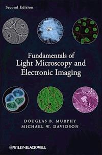
Cover Image: Courtesy of Michael W. Davidson
Copyright © 2013 by Wiley-Blackwell. All rights reserved
Published by John Wiley & Sons, Inc., Hoboken, New Jersey
Published simultaneously in Canada
No part of this publication may be reproduced, stored in a retrieval system, or transmitted in any form or by any means, electronic, mechanical, photocopying, recording, scanning, or otherwise, except as permitted under Section 107 or 108 of the 1976 United States Copyright Act, without either the prior written permission of the Publisher, or authorization through payment of the appropriate per-copy fee to the Copyright Clearance Center, Inc., 222 Rosewood Drive, Danvers, MA 01923, (978) 750-8400, fax (978) 750-4470, or on the web at www.copyright.com. Requests to the Publisher for permission should be addressed to the Permissions Department, John Wiley & Sons, Inc., 111 River Street, Hoboken, NJ 07030, (201) 748-6011, fax (201) 748-6008, or online at http://www.wiley.com/go/permissions.
Limit of Liability/Disclaimer of Warranty: While the publisher and author have used their best efforts in preparing this book, they make no representations or warranties with respect to the accuracy or completeness of the contents of this book and specifically disclaim any implied warranties of merchantability or fitness for a particular purpose. No warranty may be created or extended by sales representatives or written sales materials. The advice and strategies contained herein may not be suitable for your situation. You should consult with a professional where appropriate. Neither the publisher nor author shall be liable for any loss of profit or any other commercial damages, including but not limited to special, incidental, consequential, or other damages.
For general information on our other products and services or for technical support, please contact our Customer Care Department within the United States at (800) 762-2974, outside the United States at (317) 572-3993 or fax (317) 572-4002.
Wiley also publishes its books in a variety of electronic formats. Some content that appears in print may not be available in electronic formats. For more information about Wiley products, visit our web site at www.wiley.com.
Library of Congress Cataloging-in-Publication Data:
Murphy, Douglas B.
Fundamentals of light microscopy and electronic imaging / Douglas B. Murphy, Michael W. Davidson. – 2nd ed.
p. ; cm.
Includes bibliographical references and index.
ISBN 978-0-471-69214-0 (cloth)
1. Microscopy. I. Davidson, Michael W. (Michael Wesley), 1950- II. Title.
[DNLM: 1. Microscopy. 2. Image Processing, Computer-Assisted. QH 211]
QH205.2.M87 2013
502.8'2–dc23
2012009798
PREFACE
In the 10 years since this book first appeared, much has happened to catapult light microscopy into the forefront of biomedical research methodologies. The advances include: fundamentally new optical methods and imaging technologies for “superresolution” imaging; an explosion of new fluorescent dyes and fluorescent protein probes; new designs for objectives and thin-film interference filters; new designs of illuminators, including LED illuminators used in fluorescence imaging; new generations of silicon-based detectors, such as EMCCD cameras and scientific CMOS cameras, and many other developments. We have modified many of the chapters to include these topics, and we have added new chapters to cover essential new areas in fluorescence microscopy: fluorescence dynamics with FRET, FRAP, and TIRF; two-photon microscopy and second harmonic generation imaging; superresolution imaging including methods for single-molecule localization imaging, structured illumination imaging, and stimulated emission depletion microscopy; and a new chapter on live-cell imaging methods. But we removed a chapter on video microscopy, in keeping with the technology of the new digital age. We have also kept: demonstrations and laboratory exercises to help master new principles; a glossary of terms, appendices that supplement the exercises; and a new Webliography of basic resources on the internet.
We hope the new will help promote interest in microscopy and provide investigators with the information necessary to get the best performance from their imaging equipment.
Douglas B. MurphyManager of Light Microscope Imaging,HHMI Janelia Farm Research Campus, Ashburn, VAAdjunct Professor of Cell BiologyJohns Hopkins Medical School, Baltimore, MDMichael W. DavidsonOptical Microscopy Facility,National High Magnetic Field LaboratoryFlorida State University, Tallahassee, FL
ACKNOWLEDGMENTS
We wish to thank our many friends and colleagues who made this work possible, foremost our spouses, Christine Murphy and Pamela Davidson, for their great patience and encouragement throughout the project. Without their cooperation and understanding, the book could not have been written. We thank Christine Murphy, Tadja Dragoo, and Erin Wilson for help with the writing and for proofreading the text.
Special thanks are due to many individuals who made this work possible.
I (DBM) would like to thank the many students who have taken my microscope courses over the years at Johns Hopkins and Janelia Farm, who inspired me to write the book and gave valuable advice. In particular, I would like to thank my colleagues at Johns Hopkins and others who helped me with the first edition: Drs. Bill Earnshaw (University of Edinburgh), Gordon Ellis (University of Pennsylvania), Joe Gall (Carnegie Institution, Department of Embryology), Shinya Inoue (Marine Biological Laboratory), Ernst Keller (Carl Zeiss, Inc.), John Russ (North Carolina State University), Kip Sluder (University of Massachusetts Medical School), and Ken Spring (National Institutes of Health). For help with the second edition, I am deeply indebted to Eric Betzig, Mats Gustafsson, Harald Hess, Na Jie, and Karel Svoboda at Janelia Farm, as well as many Janelia Farm fellows and colleagues, including: Alma Arnold, Tim Brown, Reto Fiolka, Margaret Jefferies, Gleb Shtengel, Johannes Seelig, and Lin Shao. Rebecca Williams (Cornell University) and Jerome Mertz (Boston University) also helped in important ways. I especially want to give special thanks to Reed George and Gerry Rubin at Janelia Farm whose cooperation made it possible for me to work on this project, and Peter Devreotes at Johns Hopkins Medical School for encouragement and support.
I (MWD) would like to thank (in addition to most of the folks listed above) the many graphics artists, laboratory technicians, graduate students, and programmers who have worked so diligently on the Molecular Expressions, Nikon MicroscopyU, Zeiss Campus, and Olympus Resource Center websites over the past decade and assisted in the creation of figures and images in the second edition. These include: Adam Rainey, Tony Gines, Chris Burdette, Aaron Baillie, Nathan Kennedy, Kevin John, Rich Ludlow, John Childs, Chris Steenerson, Lane Henderson, Pablo Montoya, Steve Price, David Howard, John Bouma, Sean Fink, Shane Hewett, Stephanie Corn, Matt Parry-Hill, John Long, Matt De Marco, Lionel Parsons, Nate Bibler, Bo Flynn, Nathan Claxton, Korey Wilson, Ericka Ramko, Michelle Baird, Paula Cranfill, John Allen, Sarah Gilbert, Patrick Roche, John Griffin, Tom Fellers, Shannon Neaves, Riley Evans, Brittany Sell, and David Homan. I would also like to thank my colleagues at Florida State University and the National High Magnetic Field Laboratory who have provided help in all phases of the development of our Optical Microscopy facility: Jack Crow, Brian Fairhurst, Greg Boebinger, Clyde Rea, Alan Marshall, Dave Gilbert, Ross Ellington, Tim Cross, Lei Zhu, Tom Roberts, Kim Riddle, Steve Lenhert, Randy Rill, Dave van Winkle, Kirby Kemper, Bob Johnson, Ray Bye, Betty Southard, and John Fraser. Colleagues from other universities have also contributed by teaching us new tricks in microscopy and fluorescent probe development: Robert Campbell (University of Alberta), Dave Piston (Vanderbilt University), Jason Swedlow (University of Dundee), Jennifer Lippincott-Schwartz (NIH), George Patterson (NIH), Clare Waterman (NIH), Kurt Thorn (University of California, San Francisco), Rich Day (Indiana University), Dmitriy Chudakov (Russian Academy of Science), Vlad Verkhusha (Albert Einstein), and Tom Deerinck (University of California, San Diego).
We give special acknowledgment and thanks to our colleagues at Carl Zeiss, Leica Microsystems, Nikon USA, Olympus America, Molecular Probes (Life Technologies), Hamamatsu Photonics, and other companies for many years of support and for helpful information and details for many of the figures. We give special thanks and acknowledgment to: (Nikon) Stan Schwartz, Steve Ross, Nathan Claxton, Gary Laevsky, Joel Silfies, Eric Flem, Ed Lieser, Lee Shuett, Don Armstrong, Ric Villani, John Zentmeyer, Richard Gruskin, Allison Forlenza, Joe LoBiondo, Deborah Robbins, Tracey Webb, Jeff Larson, and Mike Davis. (Olympus) George Steares, Bill Fester (now at 3I), Nick George (now at Semrock), Ian Kirk, Ed Lachica (now at Lumen Dynamics), Monica Kirk (now at 3I), Kim Wicklund, Tim Randall, Chris Higgins, Stuart Shand, Kenji Matsuba, Richard Baucom, Brad Burklow, Laura Ferguson, Brendan Brinkman, Sam Tesfai, Thomas Geer, and Paul Jantzen. (Zeiss) Alex Söll, Jochen Tham, Maya Everett, Rudi Rottenfusser, Scott Olenych, Matthias Langhorst, Kenny Patterson, Elise Shumsky, Brian Crooks, and Klaus Weisshart. (Leica) Sebastian Tille, Bernd Sägmüller, Sean Garvey, Doug Reed, Anthony Santerelli, and Geoff Daniels.(Hamamatsu) Butch Moomaw, Ken Kaufmann, and Mark Hobson. (Semrock) Turan Erdogan, Nick George, and Prashant Prabhat. (Photometrics) Chris Murphy, Hilary Hicks, and David Barnes. (Chroma) Mike Stanley and Chris Bauman. (Omega) Dan Osborn. (Molecular Probes) Mike Ignatius, Mike O’Grady, Nick Dolman, Cathy Erickson, Jason Kilgore, Magnus Persmark, and Iain Johnson. (Hunt Optics and Imaging) Andrew Hunt and John Marchlenski. (BioVision) Ken Anderson, Fernando Delaville (now at Leica).
Finally, we thank our editors at John Wiley & Sons for their great patience in receiving the manuscript and managing the production of the book.
D.B.M.M.W.D.
Brightfield microscopy of stained mesophyll cells in a leaf section.
CHAPTER 1
FUNDAMENTALS OF LIGHT MICROSCOPY
OVERVIEW
In this chapter, we examine the optical design of the light microscope and review procedures for adjusting the microscope and its illumination to obtain the best optical performance. The light microscope contains two distinct sets of interlaced focal planes, eight planes in all, between the illuminator and the eye. All of these planes play an important role in image formation. As we will see, some planes are not fixed, but vary in their location depending on the focus position of the objective and condenser lenses. Therefore, an important first step is to adjust the microscope and its illuminator for Koehler illumination, a method of illumination introduced by August Koehler in 1893 that gives bright, uniform illumination of the specimen and simultaneously positions the sets of image and diffraction planes at their proper locations. We will refer to these locations frequently throughout the book. Indeed, microscope manufacturers build microscopes so that filters, prisms, and diaphragms are located at precise physical locations in the microscope body, assuming that certain focal planes will be precisely located after the user has adjusted the microscope for Koehler illumination. Finally, we will practice adjusting the microscope for examining a stained histological specimen, review the procedure for determining magnification, and measure the diameters of cells and nuclei in a tissue sample.
OPTICAL COMPONENTS OF THE LIGHT MICROSCOPE
A compound light microscope is an optical instrument that uses visible light to produce a magnified image of an object (or specimen) that is projected onto the retina of the eye or onto the photosensitive surface of an imaging device. The word compound refers to the fact that two lenses, the objective and the eyepiece (or ocular), work together to produce the final magnification M of the image such that:
Two microscope components are of critical importance in forming the image: (1) the objective, which collects light diffracted by the specimen and forms a magnified real image at what is called the real intermediate image plane near the eyepieces or oculars, and (2) the condenser, which focuses light from the illuminator onto a small area of the specimen. (We define real vs. virtual images and examine the geometrical optics of lenses and magnification in Chapter 4; a real image can be viewed on a screen or exposed on a sheet of film, whereas a virtual image cannot.) The arrangement of these and other components in an upright stand research level microscope is shown in Figure 1.1, and for an inverted research microscope in Figure 1.2. Two lamps provide illumination for brightfield and interference (illumination from below: diascopic) and fluorescence (illumination from above: episcopic) modes of examination. Both the objective and condenser contain multiple lens elements that perform close to their theoretical limits and are therefore expensive. As these optics are handled frequently, they require careful attention. Other components less critical to image formation are no less deserving of care, including the tube lens and eyepieces, the lamp collector and lamp socket and its cord, filters, polarizers, retarders, and the microscope stage and stand with coarse and fine focus.
Figure 1.1The research light microscope with upright stand. Two lamps provide transmitted and reflected light illumination. Note the locations of the knobs for the specimen and condenser lens focus adjustments. Also note the positions of two variable iris diaphragms: the field diaphragm near the illuminator, and the condenser diaphragm at the front aperture of the condenser. Each has an optimum setting in a properly adjusted microscope. Above: Nikon Eclipse 80i upright microscope; below: Olympus BX71 upright microscope.
Figure 1.2The research light microscope with inverted stand. As in upright designs, two lamps provide transmitted and reflected light illumination. Note the locations of the knobs for the specimen and condenser lens focus adjustments, which are often in different locations on inverted microscopes. Also note the positions of two variable iris diaphragms: the field diaphragm near the illuminator, and the condenser diaphragm at the front aperture of the condenser. Each has an optimum setting in a properly adjusted microscope. Above: Leica Microsystems DMI6000 B inverted microscope; below: Zeiss Axio Observer inverted microscope.
At this point, take time to examine Figure 1.3, which shows how an image becomes magnified and is perceived by the eye. The figure also points out the locations of important focal planes in relation to the objective, the ocular, and the eye. The specimen on the microscope stage is examined by the objective, which produces a magnified real image of the object in the image plane of the ocular. When looking in the microscope, the ocular acting together with the eye’s cornea and lens projects a still more magnified real image onto the retina, where it is perceived and interpreted by the brain as a magnified virtual image about 25 cm (10 in) in front of the eye. For photography, the intermediate image is recorded directly or projected as a real image onto a camera.
Figure 1.3Perception of a magnified virtual image of a specimen in the microscope. The objective forms a magnified image of the object (called the real intermediate image) in the eyepiece; the intermediate image is examined by the eyepiece and eye, which together form a real image on the retina. Because of the perspective, the retina and brain interpret the scene as a magnified virtual image about 25 cm in front of the eye.
Microscopes come in both inverted and upright designs (Figs. 1.1 and 1.2). In both designs the location of the real intermediate image plane at the eyepiece is fixed, and the focus dial of the microscope is used to position the image at precisely this location. In most conventional upright microscopes, the objectives are attached to a nosepiece turret on the microscope body, and the focus control moves the specimen stage up and down to bring the image to its proper location in the eyepiece. In inverted designs, the stage itself is fixed, being bolted to the microscope body, and the focus dials move the objective turret up and down to position the image in the eyepieces. Inverted microscopes are rapidly gaining in popularity because one can examine living cells in culture dishes filled with medium using standard objectives and avoid the use of sealed flow chambers, which can be awkward. One also has better access to the stage, which can serve as a rigid working platform for microinjection and physiological recording equipment. Inverted designs also have their center of mass closer to the lab bench and are therefore less sensitive to vibration. However, there is some risk of physical damage, as objectives may rub against the bottom surface of the stage during rotation of the objective turret. Oil immersion objectives are also at risk, because gravity can cause oil to drain down and enter the crevice between the nose and barrel, potentially contaminating internal lens surfaces, ruining the optical performance and resulting in costly lens repair. This can be prevented by wrapping a pipe cleaner or hair band around the upper part of the lens to catch excess drips of oil. Therefore, despite many advantages, inverted research microscopes require a little more attention than do standard upright designs.
APERTURE AND IMAGE PLANES IN A FOCUSED, ADJUSTED MICROSCOPE
Principles of geometrical optics show that a microscope has two sets of interlaced conjugate focal planes, a set of four object or field planes, and a set of 4 aperture or diffraction planes, that have fixed, defined locations with respect to the object, optical elements, the light source, and the eye or camera. Each plane within a set is conjugate with the other planes, with the consequence that all of the planes of a given set can be seen simultaneously when looking in the microscope. The field planes are observed in normal viewing mode using the eyepieces. This mode of viewing is called the normal, or object, or orthoscopic mode, and the real image of an object is called an orthoscopic image. Viewing the aperture or diffraction planes requires using an eyepiece telescope or Bertrand lens, which is focused on the rear aperture of the objective (see Note). This mode of viewing is called the aperture, or diffraction, or conoscopic mode, and the image of the diffraction plane viewed at this location is called the conoscopic image. In this text, we refer to the two viewing modes as the normal and aperture viewing modes and do not use the terms orthoscopic and conoscopic, although these terms are common in other texts.
Figure 1.4Objective and eyepiece diagrams. (a) Cross section of an objective showing the location of the back or rear aperture. (b) Cross sectional view of a focusable eyepiece, showing the location of the real intermediate image, in this case, containing an eyepiece reticule. Notice the many lens elements that make up these basic optics.
The identities of the sets of conjugate focal planes are listed in Table 1.1, and their locations in the microscope under conditions of Koehler illumination are shown in Figure 1.5. The terms front aperture and rear aperture refer to the openings at the front and rear focal planes of a lens from the perspective of a light ray traveling from the lamp to the retina. Knowledge of the location of these planes is essential for adjusting the microscope and for understanding the principles involved in image formation. Indeed, the entire design of a microscope is based around these planes and the user’s need to have access to them.
TABLE 1.1 Conjugate Planes in Optical Microscopy
Field Planes
Aperture Planes
(Normal view through the eyepieces)
(Aperture view through the eyepiece telescope)
Lamp (field) diaphragm
Lamp filament
Object (specimen) or field plane (diaphragm)
Front aperture of condenser
Real intermediate image plane (eyepiece field stop)
Rear aperture of objective
Retina or camera sensor face
Exit pupil of eyepiece (coincident with pupil of eye)
Figure 1.5Conjugate and aperture planes in Koehler illumination. Arrows mark the conjugate focal planes. Note the locations of four conjugate field planes (red arrows; left) and four conjugate aperture planes (blue arrows; right) indicated by the crossover points of rays in the diagrams. The left-hand diagram shows that the specimen or object plane is conjugate with the real intermediate image plane in the eyepiece, the retina of the eye, and the field stop diaphragm between the lamp and the condenser. The right-hand drawing shows that the lamp filament is conjugate with aperture planes at the front focal plane of the condenser, the rear focal plane of the objective, and the pupil of the eye. The two sets of conjugate planes interdigitate with one another.
The exit pupil of the eyepiece, one of the microscope’s aperture planes, is the disk of light that appears to hang in space a few millimeters above the back lens of the eyepiece; it is simply the image of the illuminated rear aperture of the objective. Normally, we are unaware that we are viewing four conjugate field planes when looking through the eyepieces of a microscope. As an example of the simultaneous visibility of conjugate focal planes, consider that the image of a piece of dirt on the focused specimen could lie in any one of the four field planes of the microscope: floaters near the retina, dirt on an eyepiece reticule, dirt on the specimen itself, and dirt on the glass plate covering the field diaphragm. With knowledge of the locations of the conjugate field planes, one can quickly determine the location of the dirt by rotating the eyepiece, moving the microscope slide, or wiping the cover plate of the field diaphragm. Before proceeding, you should take the time to identify the locations of the field and aperture planes on your microscope in the laboratory.
KOEHLER ILLUMINATION
Illumination is a critical determinant of optical performance in light microscopy. Apart from the intensity and wavelength range of the light source, it is important that a large cone of light emitted from each source point be collected by the lamp collector and that the source be imaged onto the front aperture of the condenser. From there, each point of the source image is projected through the specimen and to infinity as a parallel collimated pencil of light. The size of the illuminated field at the specimen is adjusted so that it matches the specimen field diameter of the objective being employed. Because each source point contributes equally to illumination in the specimen plane, variations in intensity in the image are attributed to the object and not to irregular illumination from the light source. The method of illumination introduced by August Koehler in the late nineteenth century fulfills these requirements and is the standard method of illumination used in light microscopy (Fig. 1.6). Under the conditions set forth by Koehler, a collector lens on the lamp housing is adjusted so that it focuses an image of the lamp filament at the front focal plane of the condenser while completely filling the aperture with light. Under this condition, illumination of the specimen plane is bright and even. Achieving this condition also requires focusing the condenser using the condenser focus knob, an adjustment that brings two sets of conjugate focal planes into precise physical locations in the microscope, a requirement for a wide range of image contrasting techniques that are discussed later in Chapters 7–11. The main advantages of Koehler illumination in image formation are:
Bright and even illumination in the specimen plane and in the conjugate image plane.
Even when illumination is provided by an irregular light source, such as a lamp filament, illumination of the object-specimen is remarkably uniform across an extended area. Under these conditions of illumination, a given point in the specimen is illuminated by every point in the light source, and conversely, a given point in the light source illuminates every point in the specimen.
Positioning of two different sets of conjugate focal planes at specific locations along the optical axis of the microscope
, a strict requirement for maximal spatial resolution and optimal image formation for a variety of optical modes. As we will see, stage focus and condenser focus and centration position the focal planes correctly, while correct settings of the field diaphragm and the condenser aperture diaphragm give control over resolution and contrast. Once properly adjusted, it is easier to locate and correct faults, such as dirt and bubbles that can degrade optical performance.
Figure 1.6August Koehler introduced a new method of illumination that greatly improved image quality and revolutionized light microscope design. Koehler introduced the system in 1893 while he was a university student and instructor at the Zoological Institute in Giessen, Germany, where he performed photomicrography for taxonomic studies on limpets. Using the traditional methods of critical illumination, the glowing mantle of a gas lamp was focused directly on the specimen with the condenser, but the images were unevenly illuminated and dim, making them unsuitable for photography using slow-speed emulsions. Koehler’s solution was to reinvent the illumination scheme. He introduced a collector lens for the lamp and used it to focus the image of the lamp on the front aperture of the condenser. A luminous field stop (the field diaphragm) was then focused on the specimen with the condenser focus control. The method provided bright, even illumination, and fixed the positions of the focal planes of the microscope optics. In later years, phase contrast microscopy, fluorescence microscopy with epi-illumination, differential interference contrast microscopy, and confocal optical systems would all utilize and be critically dependent on the action of the collector lens, the field diaphragm, and the presence of fixed conjugate focal planes that are inherent to Koehler’s method of illumination. The interested reader should refer to the special centenary publication on Koehler by the Royal Microscopical Society (see Koehler, 1893).
ADJUSTING THE MICROSCOPE FOR KOEHLER ILLUMINATION
Review Figure 1.5 again to familiarize yourself with the locations of the two sets of focal planes, one set of four field planes, and one set of four aperture planes. You will need an eyepiece telescope or Bertrand lens to examine the aperture planes and to make certain adjustments. In the absence of a telescope lens, one may simply remove an eyepiece and look straight down the optical axis at the objective aperture; however, without a telescope, the aperture diameter is small and difficult to see clearly. The adjustment procedure is given in detail below. You will need to check your alignment every time you change a lens to examine the specimen at a different magnification.
Preliminaries
.
Place a specimen slide, such as a stained histological specimen, on the stage of the microscope. Adjust the condenser height with the condenser-focusing knob so that the front lens element of the condenser comes within ∼1–2 mm of the specimen slide. Do the same for the objective. Be sure all diaphragms are open so that there is enough light (includes illuminator’s field diaphragm, the condenser’s front aperture diaphragm, and in some cases, a diaphragm in the objective itself). Adjust the lamp power supply so that the illumination is bright but comfortable when viewing the specimen through the eyepieces.
Check that the lamp fills the front aperture of the condenser
.
Inspect the front aperture of the condenser by eye and ascertain that the illumination fills most of the aperture. It helps to hold a piece of lens tissue against the aperture to check the area of illumination (
Fig. 1.7
). Using an eyepiece telescope or Bertrand lens, examine the rear aperture of the objective and its conjugate planes, the front aperture of the condenser, and the lamp filament. Be sure the lamp filament is centered, using the adjustment screws on the lamp housing if necessary, and confirm that the lamp filament is focused in the plane of the condenser diaphragm. This correction is made by adjusting the focus dial of the collector lens on the lamp housing. Once these adjustments are made, it is usually not necessary to repeat the inspection every time the microscope is used. Instructions for centering the lamp filament or arc are given in Chapter 3. Lamp alignment should be rechecked after the other steps have been completed.
Focus the specimen
.
Bring a low power objective to within 1 mm of the specimen, and looking in the microscope, carefully focus the specimen using the microscope’s coarse and fine focus dials. It is helpful to position the specimen with the stage controls so that a region of high contrast is centered on the optical axis before attempting to focus. It is also useful to use a low magnification “dry” objective (10–25×, used without immersion oil) first, since the
working distance
, the distance between the coverslip and the objective, is 2–5 mm for a low power lens. This reduces the risk of plunging the objective into the specimen slide and causing damage. Since the lenses on most microscopes are
parfocal
(see Chapter 4), higher magnification objectives will already be in focus or close to focus when rotated into position.
Focus and center the condenser
.
With the specimen in focus, close down (stop down) the
field diaphragm
and then, while examining the specimen through the eyepieces, focus the angular outline of the diaphragm’s periphery using the condenser’s focusing knob (
Fig. 1.8
). If there is no light, turn up the power supply and bring the condenser closer to the microscope slide. If light is seen but seems to be far off axis, switch to a low power lens and move the condenser positioning knobs slowly to bring the center of the illumination into the center of the field of view. Focus the image of the field diaphragm and center it using the condenser’s two centration adjustment screws (
Fig. 1.9
). The field diaphragm is then opened just enough to accommodate the object or the field of a given detector. This helps reduce scattered or stray light and improves image contrast. The condenser is now properly adjusted. We are nearly there! The conjugate focal planes that define Koehler illumination are now at their proper locations in the microscope.
Adjust the condenser diaphragm while viewing the objective rear aperture with an eyepiece telescope or Bertrand lens
.
Finally, the condenser diaphragm is adjusted to obtain the best resolution and contrast, but is not closed so far as to degrade the resolution. In viewing the condenser front aperture using a telescope, the small bright disc of light seen in the telescope represents the objective’s rear aperture plus the superimposed image of the condenser’s front aperture diaphragm. As you close down the condenser diaphragm, you will see its edges enter the aperture opening and limit the objective aperture’s diameter. Focus the telescope so the edges of the diaphragm are seen clearly. Stop when ∼3/4 of the maximum diameter of the aperture remains illuminated and use this setting as a starting position for subsequent examination of the specimen (
Fig. 1.10
). As pointed out in Chapter 6, the setting of this aperture is crucial, because it determines the resolution of the microscope, affects the contrast of the image, and establishes the depth of field. It is usually impossible to optimize for resolution and contrast at the same time, so the 3/4 open position indicated here is a good starting position. The final setting depends on the inherent contrast of the specimen.
Adjust the lamp brightness
.
Image brightness is controlled by regulating the lamp voltage, or if the voltage is nonadjustable, by placing neutral density filters in the light path near the illuminator in specially designed filter holders.
The aperture diaphragms should never be closed down as a way to reduce light intensity
, because this action reduces the resolving power and may blur fine details in the image. We will return to this point in Chapter 6.
Figure 1.7Examining the area of illumination at the condenser front aperture.
Figure 1.8Adjusting the field diaphragm opening size and focusing the condenser.
Figure 1.9Adjusting the condenser centering knobs during alignment of the microscope for Koehler illumination.
Figure 1.10Adjusting the condenser diaphragm opening size to maximize contrast.
The procedure for adjusting the microscope for Koehler illumination seems invariably to stymie most newcomers. With so many planes and devices to think about, this is perhaps to be expected. To get you on your way, try to remember this simple two step guide: Focus on a specimen and then focus and center the condenser. Post this reminder near your microscope. If you do nothing else beyond this, you will have properly adjusted the image and aperture planes of the microscope, and the rest will come quickly enough after practicing the procedure a few times. Although the adjustments sound complex, they are simple to perform, and their significance for optical performance cannot be overstated. The advantages of Koehler illumination for a number of optical contrasting techniques will be revealed in the next several chapters.
FIXED TUBE LENGTH VERSUS INFINITY OPTICAL SYSTEMS
Until the late 1980s, most microscopes had a fixed tube length with a specified distance between the nosepiece opening, where the objective is attached, and the eyepiece seat in the observation tubes. This distance is known as the mechanical tube length of the microscope. The design assumes that when the specimen is placed in focus, it is a few micrometers further away than the front focal plane of the objective (Fig. 1.11a). Finite tube lengths were standardized at 160 mm during the nineteenth century by the Royal Microscopical Society (RMS), and were in use for over 100 years. Objectives designed to be used with a microscope having the industry standard tube length of 160 mm are inscribed with “160” on the barrel.
Figure 1.11Finite and infinity corrected microscope optical configuration. (a) Finite microscope optical train showing focused light rays from the objective at the intermediate image plane. (b) Infinity-corrected microscope with a parallel light beam between the objective and tube lens. This is the region of the optical train that is designed for auxiliary components, such as DIC prisms, polarizers, and filters.
Adding optical accessories into the light path (between the microscope frame and observation tube head) of a fixed tube length microscope increases the effective tube length to a value greater than 160 mm. Therefore, inserting auxiliary components, such as a reflected light or fluorescence illuminator, polarizers, filters, and DIC prisms, can introduce spherical aberration and “ghost images” into an otherwise perfectly corrected optical system. During the period when most microscopes had fixed tube lengths, manufacturers were forced to place additional optical elements into these accessories to reestablish the effective 160-mm tube length of the microscope. The optical cost was an increase in magnification and reduced light intensity in resulting images. To circumvent these artifacts, the German microscope manufacturer Reichert pioneered the concept of infinity optics. The company started experimenting with infinity-corrected optical systems as early as the 1930s, but this concept did not become standard equipment for most manufacturers until 50 years later.
Infinity optical systems have a different objective design that produces a flux of parallel light wavefronts imaged at infinity, which are then brought into focus at the intermediate image plane by a special optic termed a telan or tube lens. The region between the objective rear aperture and the tube lens is called infinity space, where auxiliary components can be introduced into the light path without producing focus artifacts or optical aberrations (Fig. 1.11b). Correction for optical aberration in infinity microscopes is accomplished by modifying either the tube lens or the objective, or both. Infinity microscopes can maintain parfocality between objectives even when auxiliary components are introduced, and these components are designed to produce exactly 1× magnification to enable comparison of specimens using a combination of several optical techniques, such as phase contrast and DIC with fluorescence. This is possible because optical accessories (such as DIC prisms) placed in the infinity space do not shift the location or focal point of the image. A note of caution: objectives designed for older 160-mm fixed tube length microscopes are not interchangeable with newer infinity-corrected microscopes.
PRECAUTIONS FOR HANDLING OPTICAL EQUIPMENT
Never strain, twist, or drop objectives or other optical components
. Optics for polarization microscopy are especially susceptible to failure due to mishandling.
Never force the focus controls
of the objective or condenser, and always watch lens surfaces as they approach the specimen. This is especially important for high power, oil-immersion lenses.
Never touch optical surfaces
. In some cases, just touching an optical surface can remove unprotected coatings and ruin filters costing hundreds of dollars. Carefully follow the procedures for cleaning lenses and optical devices.
CARE AND MAINTENANCE OF THE MICROSCOPE
Microscopes are sophisticated instruments that require periodic maintenance and cleaning to guarantee satisfactory performance. When neglected by continuous exposure to dust, lint, pollen, dirt, and failure to remove immersion oil after use, the optical performance can deteriorate to the point that images are negatively affected. Likewise, regular maintenance of the microscope’s mechanical and electrical components is equally important to prevent a gradual degradation in the operation of the focusing rack, stage translation mechanism, adjustable diaphragms, filter sliders, and auxiliary electronics. Dust covers are usually provided with microscopes when they are purchased and should be installed whenever the instrument is not in use to prevent contamination from airborne particles drifting through the laboratory. However, even when the microscope is routinely covered during periods of inactivity, those instruments that are used on a daily basis are still likely to experience a slow buildup of contaminants over time.
Particulates, such as dust, lint, fibers, and general debris (collectively referred to as dirt), can seriously affect the quality of an image if they land on a glass surface in a plane near the specimen or the camera sensor. Critical areas to examine for dirt contamination are the objective front lens element, the surface of the camera sensor (and its protective glass cover), both surfaces of the cover slip, the surface of the microscope slide, camera adapter optical surfaces, the upper lens of the condenser, the eyepiece lenses, both surfaces of the reticule, and other glass surfaces in the light path, including lamps, filters, beamsplitters, collector lenses, and heat filters. Cleaning of the microscope optical components is discussed in Chapter 4. Problems with the focusing rack or mechanical stage controls should be left for qualified microscope technicians. External painted surfaces on most microscopes are extremely durable. However, they can be cleaned when needed using a lightly moistened microfiber cloth. Remove loose dust and dirt using a soft hairbrush or ear syringe (available in drugstores). Avoid using compressed air as the propellant can leave unwanted deposits on painted and glass surfaces.
Figure 1.12The eyepiece reticule and stage micrometer used for determining magnification. The typical eyepiece reticule is divided into 1/100 cm (100 µm unit) divisions, and the stage micrometer into 1/100 mm (10 µm unit) divisions. The appearance of the two overlapping scales is shown in the middle of the figure.
Interpretive drawing of cone cells in the human fovea.
CHAPTER 2
LIGHT AND COLOR
OVERVIEW
In this chapter, we review the nature and action of light as a probe to examine objects in the light microscope. Knowledge of the wave nature of light is essential for understanding the physical basis of color, polarization, diffraction, image formation, and many other topics covered in this book. The eye–brain visual system is responsible for the detection of light, including the perception of color and differences in light intensity that we recognize as contrast. The eye is also a remarkably designed detector in an optical sense—the spacing of photoreceptor cells in the retina perfectly matches the requirement for resolving the finest image details formed by its lens (). Knowledge of the properties of light is important in selecting filters and objectives, interpreting colors, performing low-light imaging, and many other tasks.
Lesen Sie weiter in der vollständigen Ausgabe!
Lesen Sie weiter in der vollständigen Ausgabe!
Lesen Sie weiter in der vollständigen Ausgabe!
Lesen Sie weiter in der vollständigen Ausgabe!
Lesen Sie weiter in der vollständigen Ausgabe!
Lesen Sie weiter in der vollständigen Ausgabe!
Lesen Sie weiter in der vollständigen Ausgabe!
Lesen Sie weiter in der vollständigen Ausgabe!
Lesen Sie weiter in der vollständigen Ausgabe!
Lesen Sie weiter in der vollständigen Ausgabe!
Lesen Sie weiter in der vollständigen Ausgabe!
Lesen Sie weiter in der vollständigen Ausgabe!
Lesen Sie weiter in der vollständigen Ausgabe!
Lesen Sie weiter in der vollständigen Ausgabe!
Lesen Sie weiter in der vollständigen Ausgabe!
Lesen Sie weiter in der vollständigen Ausgabe!
Lesen Sie weiter in der vollständigen Ausgabe!
Lesen Sie weiter in der vollständigen Ausgabe!
Lesen Sie weiter in der vollständigen Ausgabe!
Lesen Sie weiter in der vollständigen Ausgabe!
Lesen Sie weiter in der vollständigen Ausgabe!
Lesen Sie weiter in der vollständigen Ausgabe!
Lesen Sie weiter in der vollständigen Ausgabe!
Lesen Sie weiter in der vollständigen Ausgabe!


