63,99 €
Mehr erfahren.
- Herausgeber: John Wiley & Sons
- Kategorie: Wissenschaft und neue Technologien
- Sprache: Englisch
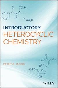
A unique approach to a core topic in organic chemistry presented by an experienced teacher to students and professionals
Heterocyclic rings are present in the majority of known natural products, contributing to enormous structural diversity. In addition, they often possess significant biological activity. Medicinal chemists have embraced this last property in designing most of the small molecule drugs in use today. This book offers readers a fundamental understanding of the basics of heterocyclic chemistry and their occurrence in natural products such as amino acids, DNA, vitamins, and antibiotics. Based on class lectures that the author has developed over more than 40 years of teaching, it focuses on the chemistry of such heterocyclic substances and how they differ from carbocyclic systems.
Introductory Heterocyclic Chemistry offers in-depth chapters covering naturally occurring heterocycles; properties of aromatic heterocycles; π-deficient heterocycles; π-excessive heterocycles; and ring transformations of heterocycles. It then offers an overview of 1,3-dipolar cycloadditions before finishing up with a back-to-basics section on nitriles and amidines.
- Presents a conversational approach to a fundamental topic in organic chemistry teaching
- Offers a unique look at this core organic chemistry topic via important naturally occurring and/or biologically active heterocycles
- Based on the author's many years of class lectures for teaching at the undergraduate and graduate level as well as pharmaceutical-industry courses
- Clear, concise, and accessible for advanced students of chemistry to gain a fundamental understanding of the basics of heterocyclic chemistry
Introductory Heterocyclic Chemistry is an excellent text for undergraduate and graduate students as well as chemists in industrial environments in chemistry, pharmacy, medicinal chemistry, and biology.
Sie lesen das E-Book in den Legimi-Apps auf:
Seitenzahl: 364
Veröffentlichungsjahr: 2018
Ähnliche


Table of Contents
Cover
Dedication
Preface
Acknowledgments
Chapter 1: Some Biologically Important Heterocycles of Nature
1.1 Vitamins
1.2 Antibiotics and Tetrapyrroles
References
Chapter 2: Orbitals and Aromaticity; Chemical Reactivity
References
Chapter 3: A Prelude to Synthesis
References
Chapter 4: π‐Deficient Heterocycles: Some Physical Properties
References
Chapter 5: π‐Deficient Heterocycles: De Novo Syntheses
5.1 De Novo Syntheses, Pyrimidines
5.2 Fused‐Ring Systems, Quinolines
References
Chapter 6: π‐Deficient Heterocycles: Introduction of New Substituents: Nucleophilic Substitution
References
Chapter 7: π‐Deficient Heterocycles: Introduction of New Substituents: Heterocyclic N‐Oxides
7.1 Further Reactions of N‐Oxides
References
Chapter 8: π‐Deficient Heterocycles: Introduction of New Substituents: Quinolines and Isoquinolines
References
Chapter 9: π‐Deficient Heterocycles: Manipulation of Existing Substituents
9.1 Summary
References
Chapter 10: π‐Excessive Heterocycles: General Properties
References
Chapter 11: π‐Excessive Heterocycles: De Novo Syntheses
11.1 Synthesis of 1,3‐Azoles
11.2 Synthesis of 1,2‐Azoles
11.3 Fischer Indole Synthesis
References
Chapter 12: π‐Excessive Heterocycles: Introduction of New Substituents
References
Chapter 13: Ring Transformations of π‐Excessive Heterocycles: Diels‐Alder Reactions
References
Chapter 14: Heterocycles as Synthons
References
Chapter 15: 1,3‐Dipolar Cycloadditions—An Overview
References
Chapter 16: Back to Basics
References
Chapter 17: A Brief Synopsis
Index
End User License Agreement
List of Illustrations
Chapter 1
Figure 1.1 Some naturally occurring heterocycles.
Figure 1.2 Naturally occurring heterocycles.
Figure 1.3 Heterocyclic vitamins.
Schema 1.1
Figure 1.4 Heterocyclic vitamins.
Figure 1.5 Antibiotics and and tetrapyrroles.
Chapter 2
Figure 2.1 Properties of aromatic heterocylics, orbitals.
Figure 2.2 Trends in resonance energy.
Figure 2.3 π‐Deficient heterocycles.
Figure 2.4 π‐Excessive heterocycles.
Chapter 3
Scheme 3.1 Why heterocycles?
Scheme 3.2 A laboratory analogy.
Figure 3.1 Common bond‐forming reactions.
Figure 3.2 Some remarkable transformations.
Chapter 4
Figure 4.1 π‐Deficient heterocycles: Solubility and tautomerization.
Figure 4.2 π‐Deficient heterocycles: basicity.
Chapter 5
Figure 5.1 Synthesis of π‐deficient heterocycles.
Scheme 5.1 Synthesis of π‐deficient heterocycles: pyridines.
Scheme 5.2 Synthesis of π‐deficient heterocycles: pyridines.
Figure 5.2 Synthesis of π‐deficient heterocycles: pyridines.
Figure 5.3 Synthesis of π‐deficient heterocycles: pyrimidines.
Scheme 5.3 Synthesis of π‐deficient heterocycles: quinolines.
Scheme 5.4 Synthesis of π‐deficient heterocycles: isoquinolines.
Scheme 5.5 Synthesis of π‐deficient heterocycles: isoquinolines.
Chapter 6
Figure 6.1 Introducing new substituents.
Figure 6.2 Introducing new substituents.
Scheme 6.1
Scheme 6.2 Nucleophilic displacement: potential complications.
Figure 6.3 Orbital requirements for fragmentation.
Scheme 6.3
Figure 6.4 A complication of a different sort.
Scheme 6.4 Nucleophilic displacement: a second complication.
Figure 6.5 Elimination‐addition: a competing mechanism.
Figure 6.6 Unequivocal generation of 3,4‐pyridyne.
Chapter 7
Figure 7.1 Introducing new substituents.
Figure 7.2 Pyridine N‐oxides.
Figure 7.3 Pyridine N‐oxides.
Figure 7.4 Pyridine N‐oxides, reduction.
Figure 7.5 A novel activating agent.
Figure 7.6 For weaker nucleophiles, activation is crucial.
Figure 7.7 Planning a synthesis of ricinine.
Figure 7.8 Synthesis of ricinine: tactical considerations.
Figure 7.9 Strategic considerations in the synthesis of ricinine.
Figure 7.10 Ricinine synthesis: a potential end game.
Scheme 7.1 Synthesis of ricinine: putting it all together.
Figure 7.11 Action of Ac
2
O on N‐oxides.
Figure 7.12 Action of Ac
2
O on N‐oxides.
Figure 7.13 Related transformations.
Figure 7.14 Synthesis of 2‐chloromethylpyridine.
Figure 7.15 Further reactions with sulfonyl chlorides.
Figure 7.16 Multiple Boekelheide rearrangements.
Figure 7.17 Rules for Ac
2
O arrangements.
Scheme 7.2 Application of the Beokelheide rearrangement to the synthesis of muscopyridine.
Figure 7.18 Cycloaddition routes to N‐oxides.
Figure 7.19 Cycloaddition routes to N‐oxides.
Figure 7.20 Cycloaddition routes to N‐oxides.
Chapter 8
Figure 8.1 Claisen’s synthesis of benzoyl cyanide.
Figure 8.2 Formation of a Reissert compound from quinoline.
Figure 8.3 Pyridine does not form a Reissert compound.
Figure 8.4 Chemistry of Reissert derivatives.
Scheme 8.1
Scheme 8.2 Aklylation of Reissert derivatives.
Scheme 8.3 Aklylation of Reissert derivatives.
Scheme 8.4 Aporphine ‐ the final steps.
Figure 8.5 Chemistry of Reissert derivatives.
Scheme 8.5 Chemistry of Reissert derivatives.
Figure 8.6 Chemistry of Reissert derivatives.
Scheme 8.6 Chemistry of Reissert derivatives.
Figure 8.7 A very selective reduction.
Figure 8.8 Chemistry of Reissert derivatives.
Figure 8.9 Some selective reductions employing Reissert derivatives.
Figure 8.10 Chemistry of Reissert derivatives.
Chapter 9
Figure 9.1 Manipulation of existing substituents: acidic alkyl groups.
Figure 9.2 Manipulation of existing substituents: active methylene chemistry.
Figure 9.3 Active methylene chemistry.
Figure 9.4 Mechanism of the Mannich reaction.
Figure 9.5 Synthesis of L‐699,392.
Figure 9.6 Manipulation of existing substituents: vinylpyridines.
Figure 9.7 1,4‐Dihydropyridines as latent 1,5‐dicarbonyl compounds.
Figure 9.8 Synthetic utility of vinyl pyridines.
Scheme 9.1 Vinyl pyridines as bis annulation reagents.
Scheme 9.2
Scheme 9.3 Vinyl pyridines as bis annulation reagents.
Figure 9.9 Manipulation of existing substituents.
Figure 9.10 Manipulation of existing substituents.
Figure 9.11 Variants of the Hammick reaction.
Figure 9.12 Manipulation of existing substituents.
Figure 9.13 Manipulation of existing substituents.
Scheme 9.4 Putting it all together: synthesis of quinine.
Chapter 10
Figure 10.1 π‐Excessive heterocycles: general properties
Figure 10.2 π‐Excessive heterocycles: general properties
Figure 10.3 π‐Excessive heterocycles: general properties
Figure 10.4 π‐Excessive heterocycles: general properties.
Figure 10.5 C‐2 versus C‐3 electrophilic aromatic substitution.
Chapter 11
Figure 11.1 Synthesis of π‐excessive heterocycles.
Figure 11.2 De Novo synthesis.
Figure 11.3 De Novo synthesis.
Figure 11.4 Mechanism of pyrrole synthesis.
Scheme 11.1 De Novo synthesis.
Scheme 11.2
Scheme 11.3 De Novo synthesis.
Scheme 11.4 Knorr pyrrole synthesis.
Scheme 11.5 Knorr pyrrole synthesis.
Figure 11.5 De Novo synthesis.
Figure 11.6 Interrupted Feist‐Benary reaction.
Scheme 11.6 De Novo synthesis.
Scheme 11.7
Scheme 11.8 De Novo synthesis.
Scheme 11.9
Figure 11.7 De Novo synthesis.
Scheme 11.10
Figure 11.8 De Novo synthesis.
Figure 11.9 De Novo synthesis.
Figure 11.10 De Novo synthesis.
Figure 11.11 De Novo synthesis.
Figure 11.12 De Novo synthesis.
Figure 11.13 De Novo synthesis.
Figure 11.14 De Novo synthesis.
Figure 11.15 De Novo synthesis.
Chapter 12
Figure 12.1 Synthesis of π‐excessive heterocycles.
Figure 12.2 Generation of acetyl nitrate.
Figure 12.3 Introduction of new substituents.
Figure 12.4 Introduction of new substituents.
Scheme 12.1 Introduction of new substituents.
Scheme 12.2 Introduction of new substituents.
Figure 12.5 Introduction of new substituents.
Figure 12.6 Conditions for Mannich condensations.
Scheme 12.3
Scheme 12.4 Putting it all together.
Figure 12.7 Lithiation of anisole.
Figure 12.8 Lithiation of methyl vinyl ether.
Figure 21.9 Lithiation of π‐excessive heterocycles.
Scheme 12.5
Chapter 13
Figure 13.1 Ring transformation of π‐excessive heterocycles: Diels‐Alder reactions.
Figure 13.2 Energy diagram for Diels‐Alder reaction of furan.
Figure 13.3 Ring transformation of π‐excessive heterocycles: Diels‐Alder reactions.
Figure 13.4 Ring transformation of π‐excessive heterocycles: Diels‐Alder reactions.
Figure 13.5 Ring transformation of π‐excessive heterocycles: Diels‐Alder reactions.
Figure 13.6 Ring transformation of π‐excessive heterocycles: Diels‐Alder reactions.
Figure 13.7 Ring transformation of π‐excessive heterocycles: Diels‐Alder reactions.
Scheme 13.1 Ring transformation of π‐excessive heterocycles: Diels‐Alder reactions.
Scheme 13.2 Ring transformation of π‐excessive heterocycles: Diels‐Alder reactions.
Figure 13.8 Ring transformation of π‐excessive heterocycles: Diels‐Alder reactions.
Scheme 13.3 Ring transformation of π‐excessive heterocycles: Diels‐Alder reactions.
Figure 13.9 Ring transformation of π‐excessive heterocycles: Diels‐Alder reactions.
Scheme 13.4 A novel synthesis of pyridoxine.
Figure 13.10 Ring transformation of π‐excessive heterocycles: Diels‐Alder reactions.
Figure 13.11 Ring transformation of π‐excessive heterocycles: Diels‐Alder reactions.
Scheme 13.5
Scheme 13.6 Ring transformation of π‐excessive heterocycles: Diels‐Alder reactions.
Chapter 14
Figure 14.1 Heterocycles as synthons
Scheme 14.1 Furan as a synthon.
Scheme 14.2 Furan as a synthon.
Scheme 14.3 Furan as a synthon.
Scheme 14.4 Putting it all together.
Scheme 14.5 Furan as a synthon.
Scheme 14.6 Furan as a synthon.
Figure 14.2 Heterocycles as a synthon.
Figure 14.3 Thiophene as a synthon.
Scheme 14.7 Thiophene as a synthon.
Scheme 14.8 Thiophene as a synthon.
Scheme 14.9 Thiophene as a synthon.
Scheme 14.10 Woodward’s synthesis of colchicine.
Figure 14.4 Heterocycles as synthons.
Scheme 14.11 Isoxazole as a synthon.
Scheme 14.12 Isoxazole as a synthon.
Scheme 14.13 Isoxazole as a synthon.
Scheme 14.14 Isoxazole as a synthon.
Scheme 14.15 Isoxazole as a synthon.
Figure 14.5 Isoxazole as a synthon.
Scheme 14.16 Isoxazole as a synthon.
Figure 14.6 Heterocycles as synthons.
Scheme 14.17 Pyrazoles as a synthon.
Chapter 15
Figure 15.1 Synthesis and reactions of 1,3‐dipoles.
Figure 15.2 Synthesis and reactions of 1,3‐dipoles.
Figure 15.3 Synthesis and reactions of 1,3‐dipoles.
Figure 15.4 Synthesis and reactions of 1,3‐dipoles.
Figure 15.5 Synthesis and reactions of 1,3‐dipoles.
Scheme 15.1
Figure 15.6 Synthesis and reactions of 1,3‐dipoles.
Figure 15.7 Synthesis and reactions of 1,3‐dipoles.
Figure 15.8 Nitrile oxides.
Scheme 15.2 Nitrile oxides.
Figure 15.9 Synthesis and reactions of 1,3‐dipoles.
Figure 15.10 Synthesis and reactions of 1,3‐dipoles.
Scheme 15.3
Figure 15.11 Synthesis and reactions of 1,3‐dipoles.
Scheme 15.4 Synthesis and reactions of 1,3‐dipoles.
Figure 15.12 Synthesis and reactions of 1,3‐dipoles.
Figure 15.13 Synthesis and reactions of 1,3‐dipoles.
Figure 15.14 Synthesis and reactions of 1,3‐dipoles.
Figure 15.15 Synthesis and reactions of 1,3‐dipoles.
Figure 15.16 Synthesis and reactions of 1,3‐dipoles.
Scheme 15.5 Synthesis and reactions of 1,3‐dipoles.
Scheme 15.6 Synthesis and reactions of 1,3‐dipoles.
Chapter 16
Figure 16.1 Back to basics.
Figure 16.2 Nitriles and amidines.
Figure 16.3 Nitriles and amidines.
Figure 16.4 Nitriles and amidines.
Scheme 16.1 Complex transformations.
Scheme 16.2 Mechanisms are good practice.
Guide
Cover
Table of Contents
Begin Reading
Pages
C1
iv
1
2
3
4
5
6
7
8
9
10
11
12
13
14
15
17
18
19
20
21
23
24
25
27
28
29
30
31
32
33
34
35
36
37
38
39
44
40
41
42
43
45
46
47
48
49
58
50
51
52
53
54
55
56
57
59
60
61
62
63
64
65
66
67
68
69
70
71
72
73
74
75
79
80
76
77
78
81
82
83
84
85
86
87
89
90
93
101
102
91
92
94
95
96
97
98
99
100
103
104
105
106
107
108
109
110
111
112
113
114
115
118
120
121
116
117
119
122
123
124
125
126
127
128
129
130
131
132
133
134
135
136
137
138
139
140
141
142
143
144
145
146
147
148
149
150
151
152
153
154
155
157
161
168
173
158
159
160
162
163
164
165
166
167
169
170
171
172
174
175
176
177
178
179
188
201
180
181
182
183
184
185
186
187
189
190
191
192
193
194
195
196
197
198
199
200
202
203
204
205
206
207
208
209
210
211
212
213
214
215
216
217
218
219
220
221
222
223
224
225
226
227
228
229
230
231
232
233
234
235
236
237
239
240
241
242
243
244
245
246
247
248
249
251
252
253
254
255
256
257
Introductory Heterocyclic Chemistry
Peter A. Jacobi
Dartmouth CollegeNew Hampshire, United States
Copyright
This edition first published 2019
© 2019 John Wiley and Sons Ltd
All rights reserved. No part of this publication may be reproduced, stored in a retrieval system, or transmitted, in any form or by any means, electronic, mechanical, photocopying, recording or otherwise, except as permitted by law. Advice on how to obtain permission to reuse material from this title is available at http://www.wiley.com/go/permissions.
The right of Peter A. Jacobi to be identified as the author of this work has been asserted in accordance with law.
Registered Office(s)
John Wiley & Sons, Inc., 111 River Street, Hoboken, NJ 07030, USA
John Wiley & Sons Ltd, The Atrium, Southern Gate, Chichester, West Sussex, PO19 8SQ, UK
Editorial Office
The Atrium, Southern Gate, Chichester, West Sussex, PO19 8SQ, UK
For details of our global editorial offices, customer services, and more information about Wiley products visit us at www.wiley.com.
Wiley also publishes its books in a variety of electronic formats and by print‐on‐demand. Some content that appears in standard print versions of this book may not be available in other formats.
Limit of Liability/Disclaimer of Warranty
In view of ongoing research, equipment modifications, changes in governmental regulations, and the constant flow of information relating to the use of experimental reagents, equipment, and devices, the reader is urged to review and evaluate the information provided in the package insert or instructions for each chemical, piece of equipment, reagent, or device for, among other things, any changes in the instructions or indication of usage and for added warnings and precautions. While the publisher and authors have used their best efforts in preparing this work, they make no representations or warranties with respect to the accuracy or completeness of the contents of this work and specifically disclaim all warranties, including without limitation any implied warranties of merchantability or fitness for a particular purpose. No warranty may be created or extended by sales representatives, written sales materials or promotional statements for this work. The fact that an organization, website, or product is referred to in this work as a citation and/or potential source of further information does not mean that the publisher and authors endorse the information or services the organization, website, or product may provide or recommendations it may make. This work is sold with the understanding that the publisher is not engaged in rendering professional services. The advice and strategies contained herein may not be suitable for your situation. You should consult with a specialist where appropriate. Further, readers should be aware that websites listed in this work may have changed or disappeared between when this work was written and when it is read. Neither the publisher nor authors shall be liable for any loss of profit or any other commercial damages, including but not limited to special, incidental, consequential, or other damages.
Library of Congress Cataloging‐in‐Publication Data applied for
Paperback : 9781119417590
Cover design by Wiley
Cover Images: © iyadsm/iStockphotoCourtesy of Peter A. Jacobi
Set in 10/12pt WarnockPro by SPi Global, Chennai, India
10 9 8 7 6 5 4 3 2 1
Dedication
In tribute to Ted, whose fascination with heterocyclic chemistry was boundless—a treasured friend, and an inspiring mentor to generations of heterocyclic chemists
Preface
Why heterocycles? Why has Nature chosen these ring systems as a foundation for so many of her life processes? And why have organic chemists focused so much effort on understanding and synthesizing these materials? One reason is that heterocyclic rings (i.e. those containing atoms other than carbon) are present in the majority of known natural products, contributing to enormous structural diversity. In addition, they often possess significant biological activity. Medicinal chemists have embraced this last property in designing most of the small molecule drugs in use today. Indeed, a 2014 study found that nearly 60% of all FDA‐approved drugs in this category incorporate a nitrogen heterocycle (totaling some 640!).* Oxygen and sulfur heterocycles are also well represented.
The chemistry of these substances is quite different from that encountered in carbocyclic systems, and an appreciation of these differences is important for understanding biochemistry and molecular biology at a fundamental level. Also, it is not unusual for heterocyclic species to undergo rearrangements and transformations having no parallel in carbocyclic chemistry, which provides a good training ground for sharpening mechanistic skills.
This leads to the question of at what level should the study of heterocyclic chemistry begin. It is fair to say that most courses in this area are geared toward graduate education, at least in the U.S. But this need not be the case, and the author has many years of experience teaching this material at the junior and senior undergraduate level. This text builds upon that experience, and it should be appropriate for introducing this topic to aspiring heterocyclic chemists at various stages of their careers. It should also find use as a resource for chemists in related fields who simply wish to “test the waters” of heterocyclic chemistry.
* Vitaku, E.; Smith, D. T.; Njardarson, J.T. J. Med. Chem. 2014, 57, 10257-10274.
Acknowledgments
The author is indebted to my good friend and colleague, Professor Gordon W. Gribble, for reading and commenting on the entire text. A special thanks is also due to Ms. Lora C. Leligdon, of Kresge Library, Dartmouth College, for her invaluable aid in tracking down innumerable references. And finally, I am grateful to my dear wife Lee Ann, who, although not a chemist persevered through the entire project and served as proofreader extraordinaire.
1Some Biologically Important Heterocycles of Nature
Heterocyclic rings come in many sizes and shapes, and they may be either aromatic or non‐aromatic, fused or non‐fused. This chapter provides a brief survey of some of the most biologically important heterocycles found in nature.
Let us begin our discussion with the three common amino acids tryptophan, proline, and histidine (Figure 1.1). Tryptophan contains an indole skeleton (blue), which is aromatic by virtue of having 10 π‐electrons in a cyclic conjugated array, two of which are donated by the ring nitrogen. Proline, on the other hand, is clearly non‐aromatic, since the pyrrolidine ring has only the free electron pair on nitrogen. But what about histidine, the distinguishing feature of which is an imidazole core with a total of eight electrons? Does this ring system satisfy Hückel's rule? The answer is yes, since one of the electron pairs resides in an orthogonal sp2‐orbital, leaving 6 π‐electrons to constitute the aromatic sextet. Lastly, while not an amino acid itself, we include in this introduction serotonin, a product of catabolism of tryptophan [1a]. The chief function of this indole alkaloid is as a neurotransmitter in the brain, and as such, it plays a key role in regulating mood. Drugs that alter the concentration of serotonin in the brain have found use in treating depression and anxiety disorders.
Figure 1.1 Some naturally occurring heterocycles.
As to their own biological role, amino acids are ubiquitous as the molecular building blocks of peptides, proteins, and enzymes. In addition to the three heterocyclic amino acids just described, 17 others make up the class of 20 naturally occurring amino acids, 9 of which are considered “essential” (i.e., they cannot be biosynthesized by humans, and must be provided by diet). In this context, one might wonder how nature functions at such a complex level with such a limited “tool chest.” Part of the answer is given by the simple equation N=20n, where N equals the number of possible peptides/proteins, 20 equals the number of monomer building blocks, and n equals the number of amino acids in the chain. The results can be staggering! For example, the number of structurally unique dipeptides would total 400; for n=5 this number jumps to 3,200,000; and for n=100, which is still quite a small protein, the possible combinations would be, well, astronomical (many orders of magnitude greater than the estimated number of atoms in the universe) [1b].
Proteins are one example of a class of compounds known as informational macromolecules, which control crucial life processes. We saw above how great complexity can be generated from a relatively small group of amino acid building blocks, even considering just the primary structure of the derived proteins. However, proteins are not unique in this capability, and it has been estimated that over 90% of all of the organic material found in living organisms, including many thousands of macromolecules, can be generated from about three dozen monomeric species [1a]. Of these, 20 constitute the naturally occurring amino acids. Another five are the DNA and RNA bases adenine (abbreviated A), thymine (T), guanine (G), and cytosine (C), all found in DNA, and uracil (U), which replaces thymine in RNA (Figure 1.2). A and G are examples of purine heterocycles, while T, C, and U are pyrimidines.
Figure 1.2 Naturally occurring heterocycles.
DNA is formally derived from A, T, G, and C by initial condensation of the NH groups shown in red with 2‐deoxyribose (an example of a furanose heterocycle, and another of nature's basic building blocks). The resultant deoxyribonucleosides then undergo phosphorylation at the primary hydroxyl group to afford the corresponding deoxyribonucleotides, which on polymerization lead to single stranded DNA. But this is not the end of the story. Most readers will be aware of the double helical nature of DNA, wherein base pairing through hydrogen bonding assures that identical concentrations of A and T will always be present. The same holds true for G and C. This observation was one of the keys to unraveling the structure of DNA, and it provided a molecular basis for the process of replication. RNA, produced from DNA by transcription, is a single stranded polynucleotide with uracil substituting for thymine, and ribose replacing deoxyribose. In the third step for processing genetic information, the message encoded in RNA is translated by ribosomes to that required for synthesizing specific proteins.
1.1 Vitamins
Who among us has not at some point been concerned with “getting enough vitamins?” Although generally required in only trace quantities, these micronutrients are one of five essential components of a healthy diet (the others being carbohydrates, fats, proteins, and certain mineral elements). Vitamins perform a myriad of biological functions, and they are generally classified as being either fat soluble or water soluble. All of the water‐soluble vitamins are heterocycles, a sampling of which are described below (Figure 1.3) [1].
Figure 1.3 Heterocyclic vitamins.
Pyridoxine is one of a group of three closely related pyridine heterocycles that constitute the vitamin B6 group of vitamins (Figure 1.3). These species are best known for their role in transamination reactions, wherein an amino group from one α‐amino acid is reversibly transferred to the α‐carbon of an α‐keto acid. This process is coupled with the interconversion of pyridoxal and pyridoxamine, according to the overall equation:
While the details for this enzyme catalyzed transformation are complex, we shall see later that each step finds precedent in common bond forming reactions employed in heterocycle synthesis.
Ascorbic acid (vitamin C) is best known as a preventive and curative agent for the debilitating disease scurvy, which is characterized by lethargy, brown spots on the skin and gums, open sores, and if left untreated, death from bleeding. Human beings are among the few vertebrates who cannot biosynthesize this substance from glucose. Up until the early 1800s scurvy was common among sailors and other adventurers lacking access to fresh citrus fruit, an excellent source of vitamin C. The British Navy is credited with making the observation that a daily ration of citrus juice, mixed of course with grog, prevented scurvy (hence the term “limey,” originally intended as derogatory slang for sailors in the Royal Navy). In this capacity, vitamin C serves as a co‐factor in the enzymatic hydroxylation of proline to afford 4‐hydroxyproline, a major component of connective tissue:
It is, of course, also well known for its antioxidant properties by virtue of its enediol functionality (shown in red in Figure 1.3).
As ascorbic acid is to scurvy, so vitamin B12 is to pernicious anemia, an autoimmune disorder in which the body fails to produce sufficient healthy red blood cells. A major breakthrough in treating this disease was made in 1926, when it was found that a diet rich in partially cooked liver effected a cure. It remained until the late 1940s for the anti‐pernicious anemia factor to be isolated in the crystalline state, in the form of cyanocobalamin (R=CN in Figure 1.3; in the physiologically active forms, R=5'‐deoxyadenosyl or CH3). Another 10 years were required to determine its very complex structure, containing a cobalt atom complexed in a corrin nucleus. In humans, vitamin B12 serves as a coenzyme in two important enzymatic processes. In the methylcobalamin form it functions as a methyltransferase, in which a methyl group transfers between two molecules. Alternatively, in the adenosylcobalamin form it functions as an isomerase, in which a hydrogen atom shifts from one carbon atom to an adjacent one, with concomitant exchange of a second group. See, for example, the conversion of glutamic acid to β‐methylaspartic acid below, where the group shown in red migrates from C3 to C4, exchanging with the hydrogen shown in blue [1a]:
Thiamin (vitamin B1) contains both a thiazole and a pyrimidine ring and it has played a rich role in the history of vitamin discovery. In fact, the name “vitamine” derives from early studies on this material, when it was recognized as a vital, amine containing dietary component in preventing beriberi (the “e” was later dropped when it was discovered that not all vitamins are amines). In animal tissues it is present mainly as a pyrophosphate derivative (TPP), in which form it serves as a coenzyme in several cellular processes. One of these involves decarboxylations of α‐keto acids, such as pyruvate, where TPP functions as a carrier of an intermediate aldehyde:
All such conversions exploit the unusually high acidity of the C‐2 hydrogen shown in red (pKa≈18 in the free state [2a]; thought to be considerably lower in the active site [2b]), and the nucleophilicity of the derived ylide (sometimes referred to as a heterocyclic carbene). Thus, the first step in pyruvate decarboxylation involves nucleophilic addition of TPP ylide across the ketone carbonyl group of pyruvate (Scheme 1.1). This is followed by decarboxylation to generate an enol, following a mechanism analogous to that observed with β‐ketoacids. Finally, protonation and rapid cleavage of the resultant hydroxyethyl group generates acetaldehyde and returns TPP.
Schema 1.1
In Scheme 1.1, thiamin pyrophosphate serves as a coenzyme in the decarboxylation of pyruvate to afford acetaldehyde. Biotin (vitamin H), on the other hand, is a coenzyme for carboxylase enzymes, involved in the synthesis of numerous biologically important molecules. As one example, the N‐H group highlighted in red (Figure 1.3) serves as a carrier of a carboxy group (−CO2−) in the carboxylation of pyruvate to oxaloacetate, the biological precursor to glucose. The driving force for this reaction is provided by adenosine triphosphate (ATP), which in the process is converted to ADP:
Historically, vitamin H has sometimes been referred to as the “beauty vitamin,” for its purported benefits to hair and skin, or, in Deutsch, “Haar und Haut.” Hence the letter “H.”
Finally, among the heterocyclic vitamins, folic acid (pteroylglutamic acid) deserves special mention (Figure 1.4). Indeed, it has been stated that “there are few examples in biochemistry where a single compound has provided the key to as many nutritional phenomena as pteroylglutamic acid [3].”
Figure 1.4 Heterocyclic vitamins.
Folic acid itself is found widely distributed in food sources, especially in leafy green vegetables such as spinach. However, as with most vitamins, this species must be converted to an activated form prior to functioning as a coenzyme. The initial step in this process involves enzymatic reduction at the C7‐N8 double bond to give 7,8‐dihydrofolic acid (FH2). Further reduction, catalyzed by dihydrofolate reductase, then affords the active coenzyme 5,6,7,8‐tetrahydrofolic acid (FH4), which serves as the carrier of one carbon fragments at the methyl, formaldehyde, and formate oxidation levels. Thus, in the pathway shown, serine hydroxymethyltransferase converts FH4 to N5,N10‐methylenetetrahydrofolate (5,10‐CH2‐THF), in which the methylene group to be transferred is highlighted in red. And to where is this group transferred? In a step catalyzed by thymidylate synthase, 5,10‐CH2‐THF and deoxyuridylate come together to produce deoxythymidylate, by a process involving reductive methylation (the product of oxidation in this step is FH2) [1].
The deciphering of this pathway had enormous implications. Since deoxythymidylate is one of the fundamental building blocks of DNA, which in turn is required of all living cells, it follows that disruption of any of the activating steps described above would lead to cell death. In fact, this strategy has been put to good practice in cancer chemotherapy for over 65 years, focusing both on inhibition of dihydrofolate reductase, as well as thymidylate synthase. Of the near relatives of folic acid, the drug methotrexate binds about 1,000 times stronger to dihydrofolate reductase than does the natural substrate [1b], a result that has been attributed to N‐1 protonation in the enzyme pocket, facilitated by the C‐4 amino group (Figure 1.4) [4]. Note also that N10 is blocked from activation by a methyl group. In any event, either alone or in combination with other drugs, methotrexate is still employed as a front‐line chemotherapy agent in combating a variety of cancers. It is far from a perfect drug, however. The holy grail of chemotherapy would be to find such an agent that was lethal only to cancer cells, which has proven to be an elusive goal. Drugs such as methotrexate owe their effectiveness to the fact that cancer cells replicate at a much greater rate than normal cells, with the consequence that the drug's impact is higher.
1.2 Antibiotics and Tetrapyrroles
No survey of biologically important heterocycles found in nature would be complete without some mention of her antibiotics and tetrapyrroles (Figure 1.5). Penicillin G occupies a special niche in the history of antibiotics, dating back to a chance observation by the Scottish bacteriologist Alexander Fleming in 1928. As the story goes, Fleming left a Petri dish containing the bacterium Staphylococcus aureus uncovered while on vacation. When he returned, he was astounded to find that the bacterium had died in some areas of the dish. The causative agent for this demise was eventually traced to a fungus of the genus Penicillium, which had apparently drifted in through an open window from a laboratory on the floor below. Fleming postulated that the effect was caused by an antibacterial component present in the fungus, which he named penicillin, but he was unable to further characterize this substance. It remained for Florey and Chain to succeed in purifying penicillin G in 1942, followed by its structure determination in 1945 by Dorothy Hodgkin. For their work, Florey, Chain, and Fleming shared the 1945 Nobel Prize in Medicine (Hodgkin would also eventually win a Nobel, “for her determinations by X‐ray techniques of the structures of important biochemical substances,” including vitamin B12). For his part, Fleming once alluded to his discovery with the statement “One sometimes finds what one is not looking for.” Surely this is one of the great understatements of scientific achievement on record. Indeed, were it not for Fleming's observation, the modern age of antibiotics would most likely have been considerably delayed.
Figure 1.5 Antibiotics and and tetrapyrroles.
Penicillin G was thus the first of the β‐lactam antibiotics to be isolated and purified, and compared to other drugs of the era, it had remarkable activity against a wide range of bacteria. This was coupled with low human toxicity. It exerts its activity by specifically inhibiting the transpeptidase that catalyzes the last step in cell wall biosynthesis, which is the cross linking of peptidoglycan. In this capacity the highly reactive β‐lactam ring functions as an irreversible acylating agent. Unfortunately, though, through decades of misuse, many bacteria have developed resistance to this class of antibiotics, chiefly through the production of β‐lactamases. To overcome this resistance, β‐lactam antibiotics are often co‐administered with β‐lactamase inhibitors.
Erythromycin is an example of a class of antibiotics characterized by incorporating a macrolide ring, and their mode of action is entirely different. In susceptible bacteria, macrolide antibiotics inhibit protein biosynthesis by a mechanism involving disruption of the enzyme peptidyltransferase, thereby interfering with ribosomal translation. Erythromycin and a variety of analogs are particularly useful for treating infections in patients having an allergy for penicillin. Finally, Linezolid is an example of a potent class of antibiotics containing an oxazolidinone ring, which also function by inhibiting the bacterial synthesis of proteins. While more toxic than either penicillin or erythromycin, oxazolidinone antibiotics are oftentimes employed as a “drug of last resort” in hospital settings, due to their effectiveness against gram‐positive bacteria that have developed resistance to first line drugs.
This brings us to the topic of tetrapyrroles, of which heme and chlorophyll a are the most abundant in nature (Figure 1.5). Heme, of course, is the iron porphyrin component found in hemoglobin, the oxygen binding protein of red blood cells. As such it is responsible for oxygen transport throughout the body, and when fully oxygenated is bright red in color. It is also found in myoglobin, the oxygen carrying and storage protein of muscle. Chlorophyll a is one of several closely related bright green pigments found in all higher plants, where it plays a key role as the photoreceptor in photosynthesis. This is the process whereby visible light provides the driving force for the conversion of water and carbon dioxide into glucose and oxygen. The balanced equation for this process is:
and it is endothermic by 686 kcal/mole. It has been estimated that more than 1017 kcal of free energy is stored annually in this manner by the plant world using solar energy [1a]. This far exceeds the per annum amount of energy produced worldwide by the burning of all fossil fuels.
Finally, we close this chapter with a brief discussion of phytochrome (Figure 1.5), which is an example of a billiprotein (i.e., containing both a linear tetrapyrrole chromophore and a covalently bound protein) [5a]. This deep blue pigment is found in only trace amounts in higher plants, but if chlorophyll is the engine of plant life, then phytochrome (Pr) is the driver. In green plants Pr functions as the “on‐off” switch for photomorphogenesis, the process by which light transmits growth regulatory information to a cell's genetic apparatus. Information of this type is crucial to the timing of seasonal phenomena, such as seed germination, flowering and fruiting, and chlorophyll production. Mechanistically, Pr functions by undergoing a photoreversible, photochromic interconversion with an activated species known as Pfr. An abundance of evidence points to this change involving photoisomerization about the C15–C16 double bond, with retention of a “semi‐extended” conformation [5b]. According to this model, Z,E‐isomerization induces a change in the tertiary structure of the surrounding protein, providing a molecular basis for transduction of the light signal to the cell's genetic regulatory apparatus. It should be noted that a similar mechanism is involved in the chemistry of vision, wherein 11‐cis retinal, the prosthetic group of rhodopsin, undergoes light‐induced isomerization to the all trans form, triggering a nerve impulse.
References
1 (a) Lehninger, A. L.,
Principles of Biochemistry
, Worth Publishers, Inc., New York,
1982
. (b) Voet, D.; Voet, J. G.,
Biochemistry
, 4
th
edition, John Wiley & Sons, Inc., New York,
2011
.
2 (a) Washabaugh, M. W.; Jencks, W. P.
Biochemistry
1988
,
27
, 5044–5053. (b) Jordon, F.; Li, H.; Brown, A.
Biochemistry
1999
,
38
, 6369–6373.
3 Stokstad, E. L. R.; Harris, R. S.; Bethel, F. H.,
The Vitamins
, Vol. 3, Sebrell, W. H.; Harris, R. S., Eds., Academic Press, New York,
1954
.
4 Blakley, R. L.; Cocco, L.
Biochemistry
1985
,
24
, 4704–4709.
5 (a)
Phytochrome and Photoregulation in Plants
, Furuya, M., Ed., Academic Press, New York,
1987
. (b) Rockwell, N. C.; Lagarias, J. C.
ChemPhysChem
2010
,
11
, 1172–1180. For synthetic aspects, see (c) Jacobi, P. A.; Odeh, I. M. A.; Buddhu, S. C.; Cai, G.; Rajeswari, S.; Fry, D.; Zheng, W.; DeSimone, R. W.; Guo, J.; Coutts, L. D.; Hauck, S. I.; Leung, S. H.; Ghosh, I.; Pippin, D.
Synlett
2005
,
19
, 2861–2885.


