Neonatology at a Glance E-Book
38,99 €
Mehr erfahren.
- Herausgeber: John Wiley & Sons
- Kategorie: Fachliteratur
- Serie: At a Glance
- Sprache: Englisch
Written by a team of leading international experts, Neonatology at a Glance provides a concise and easy-to-read overview of neonatal medicine. Each topic is clearly explained over a two-page spread, aided by numerous diagrams and illustrations. It has been extensively updated to include recent advances in perinatal medicine, genetics, respiratory support, therapeutic hypothermia, antimicrobial stewardship, and family integrated care. The book covers the wide range of problems encountered in looking after newborn babies, from normal newborn infants to the complexities of neonatal intensive care.
Neonatology at a Glance:
- Provides up-to-date coverage of the important conditions you will encounter, including neonatal resuscitation and care of preterm infants
- Covers challenging topics including pain, ethical issues, patient safety, evidence-based medicine, and palliative and end of life care
- Includes details of a wide range of practical procedures, including less invasive surfactant administration, cranial ultrasound, brain monitoring and neuroimaging, and neonatal transport
Neonatology at a Glance is the perfect guide for all health professionals looking after newborn infants, including pediatric trainees, medical students, neonatal nurse practitioners and neonatal nurses, therapists, and midwives. For neonatologists, pediatricians, and neonatal lecturers, it is a valuable resource to assist with teaching.
Sie lesen das E-Book in den Legimi-Apps auf:
Seitenzahl: 620
Veröffentlichungsjahr: 2020
Ähnliche


Table of Contents
Preface
Contributors
Acknowledgments
1 Milestones in neonatology
Thermal regulation
Nutrition
Rhesus hemolytic disease
Antibiotics
Respiratory distress syndrome (RDS)
Development of neonatal intensive care
Challenges for the future
2 Epidemiology
Births
Maternal mortality
Perinatal mortality
Neonatal mortality
Epidemiologic data collection
Infant mortality
3 Perinatal medicine overview
Neonatal involvement in perinatal care
Neonatal networks
4 Prepregnancy care, prenatal screening, and fetal medicine
Fetal medicine
5 Maternal medical conditions
Diabetes mellitus
Type 2 and gestational diabetes
Maternal red blood cell alloimmunization
Perinatal alloimmune thrombocytopenia
6 Intrauterine growth restriction
Definition
Etiology
Pathophysiology
Management
7 Multiple births
Fetal complications
Neonatal complications
8 Preterm delivery
Causes
Risk factors
Prevention
Management
Timing of delivery
9 Maternal drugs affecting the fetus and newborn infant
Neonatal abstinence (withdrawal) syndrome (NAS)
Clinical assessment
Management
Cocaine
Teratogenic medicines
10 Congenital infection
Diagnosis
Clinical features
Congenital cytomegalovirus (CMV) infection
Congenital toxoplasmosis
Congenital rubella
Congenital syphilis
Varicella: chickenpox, varicella zoster virus (VZV) infection
Congenital parvovirus B19
Congenital zika virus
11 Genetics
Congenital anomalies
Specific genetic disorders
Genetic testing
12 Adaption to extra‐uterine life
Physiologic changes in fetal–neonatal transition
Abnormal transition from fetal to extrauterine life
The Apgar score
Asphyxia
13 Neonatal resuscitation and post‐resuscitation care
Preparation
Cord clamping
Temperature control
Initial assessment at birth
A – Airway
B – Breathing
Endotracheal intubation
C – Circulation
Drugs
Withholding and discontinuing resuscitation
Post‐resuscitation care in the delivery room
14 Birth injuries
Injuries to the head
Injuries to the face
Injury to the neck, shoulders and limbs
15 Routine care of the newborn infant
Screening
Health promotion
Discharge
16 Routine examination of the newborn infant
Preparation
The infant
Routine examination of newborn infants
17 Neurology examination
States of alertness
Tone and posture
Reflexes
Spontaneous movements
Abnormal signs
Behavior
18 Feeding
Nutritional characteristics of human milk compared with unmodified cow's milk
Formula
Feeding advice resources
19 Parental attachment
Communicating with parents
Antenatal identification of fetal abnormality or potential abnormality
Admission of the infant to the neonatal unit
Infants with serious congenital malformations
20 Minor abnormalities in the first few days
21 Overview of common problems of term infants
Anticipation based on history
Overview of common medical problems
22 Admission to the neonatal unit
Welcoming parents and families
Open access
Explanation and facilitating communication
Assisting attachment
Providing a family‐friendly environment
23 Stabilizing the sick newborn infant
Airway – see chapters 13, 41 and 77
Breathing – see chapters 13 and 24
Circulation
Shock
Disability and Dextrose (Neurologic system) – see Chapter 16 and 51
Environment and systematic examination
Family
24 Respiratory support
Forms of respiratory support
Supplemental oxygen therapy
Continuous positive airway pressure (CPAP)
Conventional mechanical ventilation (via a tracheal tube)
Synchronized ventilation modes
High‐frequency oscillatory ventilation (HFOV)
High frequency jet ventilation (HFJV)
Inhaled nitric oxide (iNO)
Respiratory failure
Extracorporeal membrane oxygenation (ECMO)
25 Developmental care
Observing newborn behavior
The nursery environment
Adapting care
26 Family integrated care
27 Preterm infants and their complications
Short term complications
28 Lung development and surfactant
Structural development
Surfactant
Clinical implications of surfactant deficiency
Antenatal corticosteroids
Surfactant therapy
29 Respiratory distress syndrome
Risk factors
Pathology
Pathogenesis
Clinical features
Natural course
Management
Complications
30 Temperature control
Hypothermia
Evaporative heat loss in preterm infants
Keeping neonates warm
31 Growth and nutrition
Growth
Nutrition
Enteral feeding
Parenteral nutrition (PN)
Supplements
Osteopenia of prematurity
32 Intraventricular hemorrhage and periventricular leukomalacia
Diagnosis
Clinical features
Laboratory findings
Management
Prognosis
Prevention
33 Patent ductus arteriosus (PDA)
Ductal closure
Risk factors
Clinical features
Investigations
Management
34 Apnea, infection, anemia, and jaundice
Apnea, bradycardia and desaturations
Infection
Anemia
Jaundice
35 Retinopathy of prematurity
Pathogenesis
Screening
Treatment of ROP
36 Necrotizing enterocolitis
Risk factors
Clinical features
Laboratory findings
Radiologic abnormalities
Management
37 Bronchopulmonary dysplasia
Definition
Predisposing factors
Clinical features
Investigations
Management
Strategies for prevention
38 Discharge of preterm infants from hospital
Discharge planning
39 Outcome of preterm infants
Survival
Outcome
Growth
Medical complications
Disability and impairment
Neurosensory impairment
Cognitive impairment
Behavioral outcomes
School performance
Adult outcomes
40 Respiratory distress in term infants
Common causes
Less common causes
Rare causes
41 Upper airway disorders
Cleft lip and palate
Choanal atresia
Pierre Robin sequence
Laryngomalacia
Subglottic Stenosis
42 Jaundice
Significance of severe hyperbilrubinemia
Causes of early‐onset jaundice (<24 hours) (Table 42.1)
Causes of jaundice 24 hours to 2 weeks
Clinical examination and assessment
Investigations
Management
Prolonged jaundice (>14 days)
43 Neonatal infection
Bacterial sepsis
Risk factors
Clinical presentation
Investigations
Interpretation
44 Antimicrobial stewardship
Global neonatal antibiotic use and resistance
45 Specific bacterial infections
Group B streptococcal (GBS) infection
Listeria monocytogenes
Gram‐negative infection (e.g. E. Coli, Pseudomonas, Klebsiella)
Conjunctivitis
Skin
46 Viral infections
Herpes simplex virus (HSV)
Hepatitis B (HBV)
Hepatitis C
HIV
47 Hypoglycemia and hyperglycemia
Hypoglycemia
Hyperglycemia
48 Gastrointestinal disorders
Vomiting
Gastroesophageal reflux
Esophageal atresia and tracheoesophageal fistula
Abdominal masses
Abdominal wall defects
Imperforate anus
49 Gastrointestinal obstruction
Causes (See Figure 49.1)
Diagnostic clues
Clinical presentation
Diagnosis
Management
Some specific conditions
50 Cardiac disorders
Risk factors
Presentation
Heart murmur
Heart failure
Selected causes of cyanotic congenital heart disease
Oxygen saturation screening for critical congenital heart defects
Investigations
Management of congenital heart disease
51 Hypoxic–ischemic encephalopathy
Pathogenic mechanisms of HIE
Compensatory mechanisms
Primary and delayed injury
Clinical staging
Clinical features, investigations, and management
Therapeutic hypothermia
Cerebral function tests and neuroimaging
Outcome
52 Seizures and perinatal strokes
53 Neural tube defects and hydrocephalus
Neural tube defects
Anencephaly
Encephalocele
Spina bifida
Hydrocephalus
54 The hypotonic infant
Clues from the history
Causes, clinical features, and investigations
Some specific conditions
55 Renal and urinary tract anomalies diagnosed prenatally
Embryology
Structural abnormalities of the kidneys
56 Renal and urinary tract disorders
Electrolyte problems
Urinary tract infection (UTI)
Acute kidney injury, AKI
57 Genital disorders
Inguinal hernia
Hydrocele
Undescended testis
Torsion of the testis
Hypospadias
Circumcision
58 Disorders of sex development
Sex development
Congenital adrenal hyperplasia
59 Anemia and polycythemia
Anemia
Polycythemia
60 White cell disorders
White blood cells in the newborn
Neutrophilia
Neutropenia
Blood abnormalities in infants with Down syndrome (trisomy 21)
61 Coagulation and thrombotic disorders
Thrombocytopenia
Abnormal coagulation
Thrombotic disorders (thrombophilia)
62 Dermatological disorders
Goals of neonatal skin care
Diaper (nappy) dermatitis
Infection
Vascular skin lesions
63 Bone and joint disorders
Congenital abnormalities of the hip and feets
Infection
Skeletal dysplasias
64 Inborn errors of metabolism
Age of presentation
When to suspect an inborn error of metabolism
Management
65 Hearing and vision
Hearing
Vision
66 Pain
Development of pain pathways in the fetus and preterm infant
Factors that modify pain responses
Assessment of pain
Pain assessment scales
Minimizing pain
67 Pharmacology
Drug dosing
Drug monitoring
Drugs in breast milk
Drug licensing and neonatology
68 Quality improvement
Identifying areas of improvement
Improvement science
69 Patient safety
Briefing
Hand‐off (hand‐overs)
Simulation
Modes of simulation
Critical incidents
Hospital associated infections
Extravasation of intravenous infusions
Excessive fluid volume infused
Unplanned extubation
Giving wrong breast milk to wrong patient
Complications of umbilical arterial catheters (UAC)
Thrombosis/emboli/vasoconstriction
Blood loss from arterial catheters
Ischemic damage from peripheral artery catheters
Portal vein thrombosis from umbilical venous catheters
Extravasation of parenteral nutrition (PN) from central venous lines
Burns and scalds
Scarring of skin
Nasal damage from tracheal tube
Nasal damage from nasal CPAP
Tracheal stenosis
Aspiration pneumonia from misplaced gavage (nasogastric) feeding tubes
70 Evidence‐based practice
What is evidence‐based practice (EBP)?
Steps in evidence‐based practice
Examples of evidence‐based practice in neonatology
71 Ethics
72 Research and consent
Research
Practical difficulties in conducting research in infants
Consent
Consent issues in clinical practice
73 Palliative and end‐of‐life care
Care plans
Place of care
Support for the parents, siblings, and family
Care after death
Caring for staff
Organ donation
Autopsy
74 Follow‐up of high‐risk infants
Goals
Criteria
Organization and timing
Who should conduct neonatal follow‐up?
Components
Outcome measures
75 Global neonatology
Geography of newborn deaths
Causes of newborn deaths
Timing of newborn deaths
Reducing neonatal mortality in low‐income countries
76 Transport of the sick newborn infant
Infrastructure
Why transfer?
Equipment
Documentation
77 Intubation
Endotracheal intubation
INSURE
Less Invasive Surfactant Administration (LISA)
Video laryngoscopy
78 Chest tubes
Needle thoracotomy (chest needling)
Chest tubes (chest drain)
Pleural tap
79 Common practical procedures
80 Umbilical catheters and intraosseous cannulation
Umbilical catheters
Intraosseous cannulation
81 Central venous catheters and exchange transfusions
Central venous catheters (CVC)
Exchange transfusion
82 Cranial ultrasound
Limitations of ultrasound
Lesions that can be identified
Practical issues
Lesions detectable on cranial ultrasound (Figures 82.4–82.11)
Germinal matrix hemorrhage, GMH (Grade I) (Figure 82.3)
Intraventricular hemorrhage, GMH‐IVH (Grade II – no ventricular dilatation) (Figure 82.4)
Intraventricular hemorrhage, GMH‐IVH with dilatation (Grade III – ventricular dilatation) (Figure 82.5)
Hemorrhagic parenchymal infarct (Grade IV) (Figure 82.6)
Porencephalic cyst (Figure 82.7)
Post‐hemorrhagic ventricular dilatation (PHVD) (Figure 82.8)
Ventricular index (Figure 82.9)
Echodensities (Figure 82.10)
Cystic periventricular leukomalacia (PVL) (Figure 82.11)
View from additional window (Figure 82.12)
Color Doppler flow velocity measurements
Additional windows
83 Brain monitoring
Electroencephalography (EEG) and amplitude‐integrated electroencephalography (aEEG)
Use of aEEG and EEG in neonates
Electroencephalography (EEG)
Amplitude integrated EEG (aEEG)
Near infrared spectroscopy (NIRS)
Reference values for NIRS
84 Perinatal neuroimaging with MRI
Functional MRI
The connectome and network theory
Practical and safety considerations of MRI
Prognostic information
85 Echocardiography for the neonatologist
Standard views
Assessment of the PDA
Assessment of left ventricular function in critically ill neonates
Assessment of pulmonary hypertension (PH)
Assessment of neonatal hypotension or shock
Identify the position of umbilical and central venous lines
Appendix Gestational age assessment, BP, Newborn Early Warning Trigger and Track (NEWTT) chart, Jaundice, Hypoglycemia, Growth charts
Gestational age assessment: Ballard exam
Calculating an estimated gestational age
Blood pressure charts
Jaundice indications for phototherapy and exchange transfusion
Newborn early warning trigger and track (NEWTT)
Hypoglycemia prevention and treatment and persistent or symptomatic hypoglycemia screening tests
Growth charts
Further reading
Chapter 9
Chapter 10
Chapter 17
Chapter 18
Chapter 26
Chapter 32
Chapter 35
Chapter 44
Chapter 45
Chapter 46
Chapter 47
Chapter 54
Chapter 60
Chapter 62
Chapter 65
Chapter 69
Chapter 75
Chapter 79
Index
End User License Agreement
List of Tables
Chapter 5
Table 5.1 Effect of maternal thyroid disease on the newborn infant.
Chapter 9
Table 9.1 Potential consequences for the fetus or infant of perinatal d...
Table 9.2 Clinical features of opiate withdrawal.
Table 9.3 Some recognizable patterns of malformation or neonatal proble...
Chapter 10
Table 10.1 Range of methods to diagnose congenital infections.
Table 10.2 Transmission rate and treatment of toxoplasmosis.
Chapter 11
Table 11.1 Causes of congenital anomalies.
Table 11.2 Investigations to consider.
Table 11.3 Risk of trisomy 21 in liveborn infants by maternal age.
Table 11.4 Types of genetic testing used in neonatal infants.
Table 11.5 Range of results generated in genetic testing.
Chapter 12
Table 12.1 Conditions associated with abnormal neonatal adaptation to e...
Table 12.2 Apgar score.
Chapter 13
Table 13.1 Resuscitation drugs.
Chapter 16
Table 16.1 Significant congenital abnormalities which may be identified...
Chapter 19
Table 19.1 How parents wish to be told about a serious problem or life‐...
Chapter 21
Table 21.1 Neonatal problems associated with maternal conditions.
Table 21.2 Neonatal problems associated with fetal conditions.
Table 21.3 Neonatal problems associated with abnormal labor and deliver...
Chapter 23
Table 23.1 Some causes of isolated changes in heart rate.
Chapter 24
Table 24.1 Conditions that may require ECMO.
Chapter 25
Table 25.1 Behavioral observation.
Chapter 26
Table 26.1 Examples of topics covered in parent education sessions.
Table 26.2 Advantages and disadvantages of parents' presence at ward roun...
Chapter 29
Table 29.1 Causes of respiratory distress in preterm infants.
Chapter 31
Table 31.1 Different growth charts.
Table 31.2 Nutritional requirements of preterm babies (ESPGHAN 2010).
Table 31.3 Comparison of various milks (ESPGHAN 2010).
Table 31.4 Typical total fluid intake according to postnatal age.
Chapter 32
Table 32.1 Incidence of severe intraventricular hemorrhage (IVH) and cy...
Table 32.2 A classification of lesions identified on intracranial ultra...
Chapter 34
Table 34.1 Transfusion thresholds in very preterm infants (<32 weeks)
Chapter 35
Table 35.1 International classification of retinopathy of prematurity.
Table 35.2 Recommended timing of first ROP examination based on GA (ges...
Chapter 36
Table 36.1 Clinical signs of peritonitis/perforation.
Chapter 39
Table 39.1 Factors which may improve survival and outcome of low birthw...
Table 39.2 Definitions of disability for use at 24 months of corrected ...
Chapter 42
Table 42.1 Causes of jaundice by age of onset.
Table 42.2 Bilirubin treatment thresholds by age for term infants from ...
Chapter 44
Table 44.1 Reasons antimicrobials are prescribed to neonates.
Table 44.2 CDC Recommendations for seven core elements to an antimicrob...
Chapter 47
Table 47.1 Clinical features associated with persistent hypoglycemia.
Chapter 48
Table 48.1 Vomiting – investigations to consider and their purpose.
Chapter 50
Table 50.1 Classification of congenital heart disease, with examples an...
Chapter 51
Table 51.1 Modified Sarnat staging of hypoxic–ischemic encephalopathy.
Table 51.2 Cerebral function tests and neuroimaging and their indicatio...
Table 51.3 Postnatal markers of poor prognosis.
Chapter 52
Table 52.2 Types of perinatal strokes.
Chapter 56
Table 56.1 Causes of acute kidney injury (acute renal failure) in neona...
Chapter 57
Table 57.1 Features of normal male genitalia at term.
Chapter 59
Table 59.1 Clinical features of anemia.
Chapter 60
Table 60.1 Changes in white blood cells and their clinical significance...
Table 60.2 Clinical and laboratory features of Transient Myeloprolifera...
Chapter 61
Table 61.1 Classification of fetal and neonatal thrombocytopenia (most ...
Table 61.2 Bleeding disorders.
Table 61.3 Interpretation of abnormal clotting studies.
Chapter 62
Table 62.1 Developmental differences between the skin of infants and ad...
Table 62.2 Toxicity reported from topical antiseptic use in preterm inf...
Table 62.3 Some skin lesions associated with genetic syndromes.
Chapter 64
Table 64.1 Selected inborn errors of metabolism that may present in the n...
Table 64.2 First‐line investigations when inborn error of metabolism is s...
Table 64.3 Second‐line investigations, guided by clinical picture and dis...
Table 64.4 Approach to management.
Table 64.5 Examples of vitamins and supplements used to treat IEM.
Chapter 65
Table 65.1 Risk factors for hearing loss.
Table 65.2 Rationale for universal hearing screening.
Chapter 66
Table 66.1 Some validated pain assessment scales in newborn and preterm...
Chapter 67
Table 67.1 Examples of drugs used in breast‐feeding mothers that may af...
Chapter 68
Table 68.1 National neonatal audit items for all neonatal units in Engl...
Table 68.2 Summary of some of the potentially better practices undertak...
Chapter 69
Table 69.1 Reporting of critical incidents.
Table 69.2 Incentives and disincentives to reporting safety incidents.
Table 69.3 Hierarchy of risk reduction strategies.
Chapter 71
Table 71.1 Ethical questions in neonatal care.
Table 71.2 Situations in which it may be ethical to withdraw or withhol...
Table 71.3 Situations where treatment of disabled infants can be withheld in the...
Chapter 74
Table 74.1 Widely used developmental assessments.
Chapter 77
Table 77.1 Guide to endotracheal tube size.
Chapter 80
Table 80.1 Formula to calculate length of umbilical lines.
Appendix
Table 86.1 Physical maturity scores
Table 86.2 Gestational age estimated from summed neuromuscular and phys...
Table 86.3 American Academy of Pediatrics indications for phototherapy ...
Table 86.4 Investigations for persistent or symptomatic hypoglycemia.
List of Illustrations
Chapter 1
Figure 1.1 The Tarnier incubator. The water was heated by the oil flame. Heate...
Figure 1.2 Incubators with premature babies at the Pan‐American Exposition, Bu...
Figure 1.3 Change with time of main organisms causing neonatal infection.
Chapter 2
Figure 2.1 Extended perinatal mortality (stillbirths + neonatal mortality) in ...
Figure 2.2 (a) Decline in infant and neonatal and postneonatal mortality in th...
Figure 2.3 Percentage of live births born preterm, low birthweight (<2.5 kg) a...
Figure 2.4 Percentage of neonatal, postneonatal, and infant deaths caused by i...
Chapter 3
Figure 3.1 Organization of tertiary perinatal care.
Figure 3.2 Significant fetal abnormalities detected on prenatal ultrasound scr...
Figure 3.3 An infant on extracorporeal membrane oxygenation (ECMO), which is p...
Figure 3.4 Levels of neonatal care.
Chapter 4
Figure 4.1 Ultrasound showing sacral myelocele.
Figure 4.2font-weight:bold;" Figure 4.2 Ultrasound showing talipes equinovarus...
Figure 4.3 Bilateral talipes equinovarus. 3D image reconstruction of fetal MRI...
Figure 4.4 Techniques in fetal medicine and their indications.
Figure 4.5 Fetus with pigtail catheter to drain a pleural effusion.
Chapter 5
Figure 5.1 Macrosomic infant with birthweight 4.8 kg at 38 weeks' gestation. T...
Figure 5.2 Etiology and clinical features of Rhesus hemolytic disease.
Chapter 6
Figure 6.1 Chart showing increase in birthweight with gestational age. Most sm...
Figure 6.2 Reduction of birthweight with maternal smoking.
Figure 6.3 Consequences of progressive uteroplacental failure with increasing ...
Figure 6.4 Umbilical artery Doppler waveforms and diagrammatic representation....
Figure 6.5 Doppler flow velocity waveform of the ductus venosus showing (a) no...
Chapter 7
Figure 7.1 Change in the number of multiple births in the UK since 1980. There...
Figure 7.2 Relationship between chorionicity and zygosity in twins. Placentati...
Figure 7.3 Twin–twin transfusion syndrome (TTTS) in monochorionic twin pregnan...
Figure 7.4 Quintuplets. Multiple births look endearing, but families may need ...
Chapter 8
Figure 8.1 Estimates of global burden of mortality and morbidity for the 15 mi...
Figure 8.2 Conditions associated with prematurity. (IUGR, intrauterine growth ...
Chapter 9
Figure 9.1 Features of fetal alcohol syndrome.
Figure 9.2 Severe limb shortening (phocomelia, “like a seal”) from maternal th...
Chapter 10
Figure 10.1 Congenital and neonatal infections. (CMV – cytomegalovirus; VZV – ...
Figure 10.2 The symptomatic infant.
Figure 10.3font-weight:bold;" Figure 10.3 Blueberry muffin rash in rubella and...
Figure 10.4 Postnatal CT scan of the brain showing intracranial calcification ...
Figure 10.5 Retinitis from toxoplasmosis. This may present many years later.
Figure 10.6 Characteristic rash and desquamation on the feet in congenital syp...
Figure 10.7 Characteristic rash and desquamation on hands in congenital syphil...
Figure 10.8 X‐rays in congenital syphilis showing bilateral metaphyseal lucenc...
Chapter 11
Figure 11.1 Clinical approach to congenital anomalies.
Figure 11.2 Trisomy 21 due to non‐disjunction.
Figure 11.3 Trisomy 21 (Down syndrome). (a) Facial features – upward slant of ...
Figure 11.4 Characteristic abnormalities of trisomy 18 (Edwards syndrome). (a)...
Figure 11.5 Characteristic abnormalities of trisomy 13 (Patau syndrome). (a) S...
Figure 11.6 Example of microarray showing 22q11 deletion (Di George syndrome)....
Chapter 12
Figure 12.1 Changes in the circulation at birth. (a) Fetal circulation. (b) Ne...
Figure 12.2 Schematic representation of physiologic responses to intrapartum a...
Chapter 13
Figure 13.1 Head position is the key to airway management. (a) Head in the cor...
Figure 13.2 Correct size and position of face mask. It should cover the mouth,...
Figure 13.3 Mask ventilation via a T‐piece connected to air/oxygen blender fro...
Figure 13.4 Two‐person airway control – consider if mask inflation ineffective...
Figure 13.5 (a) Apply pressure to lower third of sternum, just below an imagin...
Figure 13.6 UK algorithm for newborn life support.
Figure 13.7 US neonatal resuscitation algorithm. ETT Endotracheal tube, HR Hea...
Figure 13.8 Helping Babies Breathe action plan following delivery. Source: Wit...
Figure 13.9 Post‐resuscitation care in the delivery room.
Chapter 14
Figure 14.1 Anatomic location of injuries to the head.
Figure 14.2 Chignon.
Figure 14.3 Cephalhematoma.
Figure 14.4 Subgaleal/subaponeurotic hemorrhage.
Figure 14.5 Skull fractures.
Figure 14.6 Scalp lacerations.
Figure 14.7 Facial palsy.
Figure 14.8 Asymmetric crying facies.
Figure 14.9 Fractured clavicle.
Figure 14.10 Erb palsy.
Figure 14.11 Klumpke palsy.
Figure 14.12 Humeral or femoral fractures.
Chapter 15
Figure 15.1 Advice for parents to reduce the risk of SIDS. (a) Back to sleep. ...
Chapter 16
Figure 16.1 (a) Barlow test for dislocatable hip. The hip is held flexed and t...
Figure 16.2 Checking for red reflex. If partially or completely obscured (cata...
Figure 16.3 Routine examination of newborn infants. In the UK the Newborn Infa...
Chapter 17
Figure 17.1 Passive tone in limbs and trunk.
Figure 17.2 Active tone in limbs and trunk.
Figure 17.3 Primary reflexes.
Chapter 18
Figure 18.1 Positioning for breast‐feeding.
Figure 18.2 Preterm twins successfully learning to feed at the breast.
Chapter 19
Figure 19.1 Steps in normal attachment.
Chapter 20
Figure 20.1 Some minor abnormalities which may be noted in the first few days ...
Figure 20.2 Breast enlargement.
Figure 20.3 Positional talipes. (a) Position of the feet. (b) The foot can be ...
Figure 20.4 Stork mark (nevus simplex, salmon patch).
Figure 20.5 (a) Erythema toxicum showing patchy pustules on erythematous base....
Figure 20.6 Mongolian spot.
Figure 20.7 Natal teeth. Front lower incisors present at birth. Remove if loos...
Figure 20.8 Extra digits. Usually connected by a skin tag but may contain bone...
Figure 20.9 Ear tags. Consult plastic surgeon. Check that the ear and hearing ...
Chapter 21
Figure 21.1 Common medical problems of term infants in the first few days of l...
Chapter 22
Figure 22.1 Encourage parents to come to the unit at any time.
Figure 22.2 Grandparents on the neonatal unit visiting the latest additions to...
Figure 22.3 Supervised sibling visits should be encouraged.
Figure 22.4 Ventilated baby gripping her mother's hand.
Figure 22.5 Mother gavage (tube) feeding her baby. Parents also need to feel c...
Figure 22.6 Mother gavage (tube) feeding her baby. One of many activities that...
Figure 22.7 (a and b) Skin‐to‐skin contact with parent. In many low and middle...
Figure 22.8 Parents may like to add personal touches to their baby's bed, with...
Chapter 23
Figure 23.1 Stabilization in the neonatal unit. (PPHN, Pulmonary hypertension ...
Figure 23.2 Causes of shock.
Figure 23.3 Example of a guideline for management of low blood pressure or sho...
Chapter 24
Figure 24.1 Changes in use of respiratory interventions in VLBW (very low birt...
Figure 24.2 (a) Oxygen delivered via nasal cannula. (b) Humidified high flow n...
Figure 24.3 Saturation measurements above 95% in preterm infants receiving sup...
Figure 24.4 Nasal CPAP (continuous positive airway pressure) with flow driver ...
Figure 24.5 Diagram of bubble CPAP. Humidified blended gases are delivered via...
Figure 24.6 Intermittent positive‐pressure ventilation (IPPV). Diagram...
Figure 24.7 Pressure − volume loop.
Figure 24.8 Pressure, flow and time waveform with volume guided ventilation.
Figure 24.9 High – frequency oscillatory ventilation (HFOV). Diagram showing c...
Figure 24.10 Components in delivery of inhaled nitric oxide. There is a scaven...
Figure 24.11 ECMO (extracorporeal membrane oxygenation) circuit. The infant's ...
Chapter 25
Figure 25.1 (a) This baby's controlled posture and focused expression show suc...
Figure 25.2 Promotion of parental attachment by involvement with their baby's ...
Figure 25.3 Promotion of parental attachment through touch.
Figure 25.4 Incubator covered to shade the baby. A flap always folded back so ...
Figure 25.5 Soft bedding with supportive nesting can contain disorganized move...
Figure 25.6 Help sensitive infants to find bathing pleasurable by loosely wrap...
Figure 25.7 Many activities can be done with the baby lying on one side to giv...
Chapter 26
Figure 26.1 Relationship of family integrated care and FCC family centered car...
Figure 26.2 Early skin‐to‐skin care promotes adoption of 24‐hour kangaroo moth...
Figure 26.3 The four pillars of family integrated care.
Chapter 27
Figure 27.1 Maturational changes in appearance, posture and development with a...
Table 27.1 Changes of appearance of infants with gestation showing an infa...
Figure 27.2 Preterm infant at 23 weeks' gestation, showing thin, gelatinous sk...
Figure 27.3 Preterm infant at 30 weeks' gestation, showing medium‐thickness sk...
Figure 27.4 Term infant showing flexed posture and thick skin, and well‐formed...
Figure 27.5 Main forms of respiratory support and short‐term complications of ...
Chapter 28
Figure 28.1 Phases of lung development.
Figure 28.2 (a) It is hard to blow up a balloon that is collapsed (small radiu...
Figure 28.3font-weight:bold;" Figure 28.3 In the absence of surfactant, the pr...
Figure 28.4 Composition of surfactant.
Figure 28.5 Difference in lung volume for a given airway pressure between norm...
Figure 28.6 Effect of surfactant deficiency and lung immaturity in preterm inf...
Chapter 29
Figure 29.1 Histology showing (a) characteristic features of RDS. The hyaline ...
Figure 29.2 Chest retraction in a preterm infant with respiratory distress.
Figure 29.3 Characteristic chest X‐ray (after four hours of age) in RDS showin...
Figure 29.4 Transillumination of the chest with a fiberoptic light source show...
Chapter 30
Figure 30.1 (a–d) How newborn infants lose heat.
Figure 30.2 (a) Transepidermal water loss increases with decreasing gestation....
Figure 30.3 The neutral thermal environment is the temperature range where hea...
Chapter 31
Figure 31.1 Mean growth curves of weight of infants 22–31 weeks by postmenstru...
Figure 31.2 Preterm infant learning to suck at the breast whilst still on cont...
Figure 31.3 Preterm infant learning to breast‐feed whilst still receiving gava...
Figure 31.4 Osteopenia of prematurity. (a) Reduced bone mineralization with so...
Chapter 32
Figure 32.1 Pathogenesis of cerebral hemorrhage and cystic periventricular leu...
Figure 32.2 Autopsy specimen showing (a) large parenchymal and intraventricula...
Figure 32.3 Natural history and complications of cerebral hemorrhage and periv...
Chapter 33
Figure 33.1 Anatomy of the ductus arteriosus (after birth), with left to right...
Figure 33.2 Chest X‐ray showing increased pulmonary vasculature markings and c...
Figure 33.3 (a) Ultrasound of four chamber view showing dilated left ventricle...
Chapter 34
Figure 34.1 Apnea is absence of breathing for more than 10–15 seconds and may ...
Figure 34.2 Organisms causing late‐onset sepsis in very low birthweight infant...
Chapter 35
Figure 35.1 Diagrammatic proforma for recording ROP screening examination.
Figure 35.2 Wide field retinal imaging illustrating progression of ROP with re...
Chapter 36
Figure 36.1 Risk factors in the pathogenesis of necrotizing enterocolitis.
Figure 36.2 Abdominal distension and shiny discolored, abdominal skin in sever...
Figure 36.3 Abdominal X‐ray showing intramural air (arrow). There are also dis...
Figure 36.4 Air in portal venous system (arrow). This is often a transient sig...
Figure 36.5 Bowel perforation showing air under the diaphragm on lateral X‐ray...
Chapter 37
Figure 37.1 Pathogenesis of bronchopulmonary dysplasia.
Figure 37.2 Chest X‐ray in bronchopulmonary dysplasia showing generalized, pat...
Figure 37.3 Infant with bronchopulmonary dysplasia receiving low‐flow nasal ox...
Chapter 38
Figure 38.1 Transition from intensive care to home.
Figure 38.2 (a) Infant receiving oxygen therapy and gavage (nasogastric) feedi...
Figure 38.3 Parents and their baby leaving the neonatal unit. Some of the item...
Chapter 39
Figure 39.1 Survival by gestational age of very low birthweight (VLBW) infants...
Figure 39.2 Survival by birthweight of very low birthweight (VLBW) infants. Ov...
Figure 39.3 Results of labor ward management for extremely preterm births. Whi...
Figure 39.4 Outcomes for extremely premature infants admitted to neonatal unit...
Figure 39.5 Birthweight‐specific prevalence of cerebral palsy in Europe 1990–2...
Figure 39.6 Prevalence of special educational needs (SEN) by gestational age a...
Figure 39.7 Increased incidence of impaired cognitive function, academic skill...
Figure 39.8 Stability of cognitive and IQ measures over childhood to 19 years ...
Chapter 40
Figure 40.1 Clinical features of respiratory distress.
Figure 40.2 Causes of respiratory distress in term infants.
Figure 40.3 Lung liquid in the mouth of a newborn term infant with transient t...
Figure 40.4 Chest X‐ray in transient tachypnea of the newborn showing fluid in...
Figure 40.5 Chest X‐ray of pneumonia showing widespread patchy consolidation. ...
Figure 40.6 Chest X‐ray in meconium aspiration. There is hyperinflation of the...
Figure 40.7 Transillumination of the chest showing the presence of pneumothora...
Figure 40.8 Bilateral pneumothoraces. Border of lungs are shown with arrows; l...
Figure 40.9 Pulmonary hypertension leads to right‐to‐left shunting.
Figure 40.10 Chest X‐ray showing diaphragmatic hernia. There is bowel in the l...
Chapter 41
Figure 41.1 Types of cleft lip and palate. (a) Unilateral cleft lip. (b) Unila...
Figure 41.2 Bilateral cleft lip and palate. The deformity looks very unsightly...
Figure 41.3 Showing parents photographs (a) before and (b) after cleft lip sur...
Figure 41.4 Choanal atresia on MRI scan. There is a bony bar across the poster...
Figure 41.5 Pierre Robin sequence showing micrognathia.
Chapter 42
Figure 42.1 Metabolism of bilirubin. Bilirubin is the product of the metabolis...
Figure 42.2 Opisthotonus from kernicterus. This is now rarely seen in high‐inc...
Figure 42.3 (a) Cross‐section of the brain at autopsy showing yellow staining,...
Figure 42.4 Bilirubin chart of bilirubin level and time from birth, showing th...
Figure 42.5 Nomogram for determination of risk of development of severe hyperb...
Chapter 43
Figure 43.1 Overview of neonatal infection. (For Congenital infection see Figu...
Chapter 44
Figure 44.1 Organisms causing neonatal infection in neonatal intensive care un...
Figure 44.2 European resistance map showing percentage (%) of invasive Escheri...
Figure 44.3 Proportion of prescribed antibiotics among neonates (30 days) by g...
Chapter 45
Figure 45.1 a) Group B streptococcal (GBS) antibiotic prophylaxis guidelines...
Figure 45.2 Some specific sites of bacterial infection.
Figure 45.3 Purulent conjunctivitis with swelling of eyelids at six days from
Figure 45.4 Bullous impetigo. There are superficial blisters; some have been d...
Chapter 46
Figure 46.1 Global overview of prevalence of maternal HbsAg (hepatitis B surfa...
Figure 46.2 Global new HIV infections (green) and averted by PMTCT (Prevention...
Chapter 47
Figure 47.1 An example of a guideline for the prevention and treatment of hypo...
Figure 47.2 Causes of persistent hypoglycemia.
Chapter 48
Figure 48.1 This infant presented with blood‐stained vomiting at 12 hours of a...
Figure 48.2 Abdominal X‐ray showing distended loops of bowel in an infant with...
Figure 48.3 Abdominal distension from Hirschsprung disease.
Figure 48.4 Different types of esophageal atresia and tracheoesophageal fistul...
Figure 48.5 Frothing of oral secretions after birth from esophageal atresia.
Figure 48.6 Abdominal masses and their causes.
Figure 48.7 Omphalocele.
Figure 48.8 Gastroschisis.
Figure 48.9 Gastroschisis on prenatal ultrasound scan.
Figure 48.10 Gastroschisis in silastic silo. There is also a central venous ca...
Chapter 49
Figure 49.1 Causes of intestinal obstruction.
Figure 49.2 Abdominal X‐ray showing double bubble in duodenal atresia.
Figure 49.3 Dilated loops of small and large bowel secondary to distal obstruc...
Chapter 50
Figure 50.1 Fetal ultrasound showing atrioventricular septal defect (AVSD).
Figure 50.2 Clinical features of heart failure.
Figure 50.3 Hypoplastic left heart syndrome; the left ventricle, aortic valve ...
Figure 50.4 ECG showing supraventricular tachycardia.
Figure 50.5 Transposition of the great arteries. a) The aorta arises from the ...
Figure 50.6 Tetralogy of Fallot. There is pulmonary infundibular stenosis caus...
Figure 50.7 Severe coarctation of the aorta, an example of duct‐dependent syst...
Figure 50.8 Pulmonary atresia, an example of duct‐dependent pulmonary circulat...
Chapter 51
Figure 51.1 Antepartum and intrapartum factors preceding neonatal hypoxic–isch...
Figure 51.2 Schematic diagram showing potential for prevention of secondary ne...
Figure 51.3 Clinical manifestations, investigations, and management of hypoxic...
Figure 51.4 Therapeutic cooling for moderate or severe HIE.
Figure 51.5 Amplitude‐integrated EEG (aEEG) trace (upper panel) and raw EEG (l...
Figure 51.6 Doppler ultrasound to measure the blood flow velocity wave form in...
Figure 51.7 Acute changes typically seen in the first week after perinatal asp...
Figure 51.8 Cerebral atrophy on MRI (axial T1W) developing several weeks after...
Chapter 52
Table 52.1 Recognition, causes, investigation, and management of seizures....
Figure 52.1 EEG being performed.
Figure 52.2 MRI images showing left middle cerebral artery infarction (arrow)....
Chapter 53
Figure 53.1 Occipital encephalocele.
Figure 53.2 (a) Spina bifida occulta. Defect in the vertebral arch with intact...
Figure 53.3 Complications associated with severe myelomeningocele. These depen...
Figure 53.4 CT scan axial view showing ventricular dilatation in a term infant...
Chapter 54
Figure 54.1 Clinical features of hypotonia. (a) On “shoulder suspension” when ...
Figure 54.2 Causes, clinical features, and investigations.
Figure 54.3 Clinical features that may be present with a peripheral neuromuscu...
Figure 54.4 Prader–Willi syndrome. Characteristic facies and hypogonadism. The...
Chapter 55
Figure 55.1 Features of unilateral and bilateral hydronephrosis.
Figure 55.2 Ultrasound showing hydronephrosis. As a measure of its severity, t...
Figure 55.3 Example of a guideline of the initial management of renal and urin...
Figure 55.4 Potter syndrome/sequence.
Figure 55.5 (a) Autosomal dominant polycystic kidney disease (ADPKD). There ar...
Figure 55.6 (a) Multicystic dysplastic kidney (MCDK). The kidney is replaced b...
Chapter 56
Figure 56.1 Some key points about renal function in newborn infants.
Figure 56.2 VCUG (voiding cystourethrogram, micturating cystourethrogram) show...
Figure 56.3 Bilateral renal scarring, more severe on right, on DMSA radionucli...
Chapter 57
Figure 57.1 (a) Embryology of testicular descent. The testis migrates from the...
Figure 57.2 Inguinal hernias in a preterm infant.
Figure 57.3 Hydrocele on transillumination.
Figure 57.4 Classification of hypospadias.
Figure 57.5 Hypospadias. The arrow shows the urethral meatus.
Chapter 58
Figure 58.1 Sex development in the fetus.
Figure 58.2 Ambiguous genitalia at birth. Do not assign a gender before expert...
Figure 58.3 Abnormal adrenal steroid biosynthesis in the commonest form of con...
Figure 58.4 Virilized female from congenital adrenal hyperplasia. There is cli...
Chapter 59
Figure 59.1 Oxygen dissociation curve showing the higher oxygen affinity of ne...
Figure 59.2 Causes and investigation of anemia.
Figure 59.3 Fetomaternal hemorrhage. (a) Anemia (reduced red cells), nucleated...
Figure 59.4 Plethoric term infant. Nasogastric tube is because of poor feeding...
Chapter 60
Figure 60.1 Total neutrophil count, showing the rise with age and the normal r...
Figure 60.2 Blood smear showing four neutrophil “band” cells in a neonate with...
Figure 60.3 Blood film from a neonate with Down syndrome and TMD/TAM. The blas...
Chapter 61
Figure 61.1 Petechiae from thrombocytopenia in an infant.
Figure 61.2 The coagulation pathway. Cell‐based coagulation model with an init...
Figure 61.3 Infant with microthrombi in the skin from protein C deficiency.
Chapter 62
Figure 62.1 (a) Port wine stain with trigeminal distribution (Sturge‐Weber syn...
Figure 62.2 Strawberry nevus.
Figure 62.3 Giant congenital melanocytic nevus (GCMN). Rare but serious condi...
Figure 62.4 Epidermolysis bullosa. Rare group of disorders. Bullae, or blister...
Chapter 63
Figure 63.1 Pavlik harness for treatment of developmental dysplasia of the hip...
Figure 63.2 (a and b) Talipes equinovarus. The foot is inverted and supinated ...
Figure 63.3 Treatment of talipes equinovarus with serial plaster casts
Figure 63.4 Septic arthritis showing swollen left knee (arrow).
Figure 63.5 (a and b) X‐rays of osteogenesis imperfecta showing osteopenia, mu...
Chapter 64
Figure 64.1 Simplified diagnostic approach to investigation of significant hyp...
Chapter 65
Figure 65.1 Otoacoustic emissions (OAE).
Figure 65.2 Auditory brainstem response (ABR).
Figure 65.4 Cataracts in both eyes (more severe in the right eye) of an infant...
Figure 65.5 Bilateral congenital glaucoma.
Figure 65.3 Iris coloboma. Keyhole‐shaped pupil due to defect of the iris infe...
Chapter 66
Figure 66.1 Postulated hierarchy of pain from procedures.
Figure 66.2 Containing the infant helps reduce pain. This involves secure, sup...
Chapter 67
Figure 67.1 Key physiologic factors affecting neonatal pharmacology.
Figure 67.2 Drug monitoring – steady‐state, peak and trough levels.
Chapter 68
Figure 68.1 (a) The PDSA cycle showing the three key questions (Model for Impr...
Figure 68.2 Reduction in blood stream infections (BSI)/1000 bed days from 7.8 ...
Chapter 69
Figure 69.1 A low fidelity manikin used in a clinical setting to help train th...
Figure 69.2 Extravasation injury.
Figure 69.3 Scarring from extravasation injury.
Figure 69.4 X‐ray showing umbilical arterial and venous catheters. Catheter in...
Figure 69.5 Ischemic damage from radial artery catheter.
Figure 69.6 Scalding of skin from excessive heat from radiant warmer after dis...
Figure 69.7 Scarring from chest tubes.
Figure 69.8 Tracheal stenosis following prolonged mechanical ventilation. The ...
Chapter 70
Figure 70.1 Steps in evidence‐based practice for assessing if therapeutic hypo...
Figure 70.2 Meta‐analysis of prophylactic corticosteroids for preterm birth sh...
Figure 70.3 Changes in oxygen therapy with time.
Figure 70.4 Outcomes in preterm infants with lower or higher oxygen saturation...
Chapter 71
Figure 71.1 Gestational age thresholds for resuscitation in three Northern Eur...
Figure 71.2 The role of parents in decisions about medical treatment for a chi...
Figure 71.3 A stepwise approach to resolve disagreements.
Figure 71.4 High‐profile court cases have highlighted the ethical dilemmas aro...
Figure 71.5 Outcome for babies born alive between 22 & 26 weeks' gestation....
Chapter 72
Figure 72.1 Clinical equipoise, a requirement for research on newborns, stands...
Chapter 73
Figure 73.1 Memory boxes can be used to collect mementoes of the baby's life.
Figure 73.2 Cold mattresses can be used with traditional cots (cribs) to prolo...
Chapter 74
Figure 74.1 An example of a high‐risk follow‐up program.
Figure 74.2 Tiny preterm babies do grow up! Sally and William, from birth at 2...
Chapter 75
Figure 75.1 Global progress showing rate of decline by geographical region for...
Figure 75.2 Variation between countries in Neonatal Mortality Rates in 2016.
Figure 75.3 Causes of death for children under the age of five in 2016.
Figure 75.4 Women's group meeting in Nepal.
Figure 75.5 Kangaroo mother care for a preterm newborn.
Chapter 76
Figure 76.1 Specialized transport equipment secured in ambulance.
Figure 76.2 Lightweight transport incubator in air ambulance.
Figure 76.3font-weight:bold;" Figure 76.3 Helicopter transfer in Northern Cana...
Figure 76.4 Long distance repatriation of twins by fixed wing airplane.
Chapter 77
Figure 77.1 (a) Technique of laryngoscopy for endotracheal intubation. Place t...
Figure 77.2 Administration of surfactant (a) down a catheter passed into a tra...
Figure 77.3 An elective tube change using a video laryngoscope. (a) Endotrache...
Chapter 78
Figure 78.1 Chest X ray showing a large tension pneumothorax on the left side ...
Figure 78.2 Needle thoracotomy with a butterfly drain
Figure 78.3 Insertion of guidewire into needle (Pink) using the Seldinger tech...
Figure 78.4 Chest tube has been threaded over guidewire, which has been withdr...
Figure 78.5 X‐ray showing lung re‐expansion and a left chest tube (pigtail) to...
Figure 78.6 Chest X‐ray showing bilateral pleural effusions – there is a clear...
Figure 78.7 Simulation of thoracocentesis (aspiration of pleural fluid) using ...
Chapter 79
Table 79.1 Some common procedures.
Figure 79.1 Shaded areas show sites for capillary sampling.
Figure 79.2 Venous blood sampling from back of the hand.
Figure 79.3 Peripheral venous cannulation. When blood flows back, advance cann...
Figure 79.4 Peripheral arterial cannulation. Check for collateral circulation ...
Figure 79.5 Suprapubic aspiration under ultrasound guidance. Source: Adapted...
Figure 79.6 Lumbar puncture.(a) Back curved. Back upright and parallel to edge...
Chapter 80
Figure 80.1 Two arteries and one vein in umbilicus. The arteries are small, c...
Figure 80.2 Fixation of umbilical artery catheter. The catheter is looped and...
Figure 80.3 Position of catheters. For an arterial catheter (red), the high p...
Figure 80.4 X‐ray to confirm position of the umbilical artery (red) and umbil...
Figure 80.5 (a) Intraosseous infusion into tibia. (b) Use of an inter‐osseous...
Chapter 81
Figure 81.1 An example of placing a peripherally inserted central catheter (PI...
Figure 81.2 The cannula is removed and then split, leaving the long line in pl...
Figure 81.3 Securing the line using sterile strips followed by clear adhesive ...
Figure 81.4 X‐ray demonstrating the importance of confirming catheter position...
Figure 81.5 An example of exchange transfusion via umbilical vein with 10 ml a...
Chapter 82
Figure 82.1 Standard coronal views. (a) Frontal lobe. (b) Lateral ventricles (...
Figure 82.2 Standard sagittal views. (a) Midline (b) Parasagittal (c) Tangenti...
Figure 82.3 Left germinal matrix (subependymal) hemorrhage (Grade I). (a) Coro...
Figure 82.4 Bilateral intraventricular hemorrhage, GMH‐IVH (Grade II). (a) Cor...
Figure 82.5 Bilateral intraventricular hemorrhage, GMH‐IVH with ventricular di...
Figure 82.6 Right hemorrhagic parenchymal infarct (Grade IV). (a) Coronal view...
Figure 82.7 Porencephalic cyst at site of a unilateral hemorrhagic parenchymal...
Figure 82.8 Marked bilateral post‐hemorrhagic ventricular dilatation (PHVD) an...
Figure 82.9 Ventricular index, measured from the midline to the lateral border...
Figure 82.10 Bilateral echodensities on coronal view.
Figure 82.11 Widespread periventricular cysts on day 55 in (a) coronal and (b)...
Figure 82.12 Mastoid fontanelle view showing cerebellum.
Chapter 83
Figure 83.1 (a) A standard neonatal EEG montage (b) electrode positions accord...
Figure 83.2 Classification of aEEG. This is based on voltage (upper and lower ...
Figure 83.3 Seizures on EEG (lower panel) and four channel aEEG (upper panel)....
Figure 83.4 Seizures and response to anticonvulsants on aEEG recorded on a CFM...
Figure 83.5 Diagram demonstrating how the near infrared light beam passes thro...
Chapter 84
Figure 84.1 Three‐dimensional rendered T2‐weighted images in preterm infants a...
Figure 84.2 Tissue‐specific changes during brain maturation. Early in the thir...
Figure 84.3 Diffusion MRI can accurately delineate axon fiber bundles and prov...
Figure 84.4 A combination of diffusion and fMRI is used to assess the response...
Chapter 85
Figure 85.1 Positions of probe and views obtained.
Figure 85.2 The planes of the long‐axis, short‐axis and four‐chamber apical or...
Figure 85.3 (a) Long axis view. (b) Ultrasound showing long‐axis view. (RV, Ri...
Figure 85.4 (a) Short‐axis view. (b) Ultrasound of short‐axis view. RA, Right ...
Figure 85.5 (a) Four‐chamber view. The right ventricle can be identified from ...
Figure 85.6 Assessment of pulmonary hypertension.
Figure 85.7 Parasternal short axis view illustrating an umbilical vein cathete...
Appendix
Figure 86.1 Neuromuscular maturity.
Figure 86.2 Increase in (a) systolic and (b) mean blood pressure measured osci...
Figure 86.3 (a) Newborn Early Warning Trigger and Track (NEWTT) form. (b) Chec...
Figure 86.4 Adaptation of the guideline for the prevention and treatment of hy...
Figure 86.5 UK‐WHO growth chart for girls. (Courtesy of Royal College of Paedi...
Figure 86.6 UK‐WHO growth chart for boys.
Guide
Cover
Table of Contents
Begin Reading
Pages
iv
vii
viii
ix
x
xi
xii
1
2
3
4
5
6
7
8
9
10
11
12
13
14
15
16
17
18
19
20
21
22
23
24
25
26
27
29
30
31
32
33
34
35
36
37
38
39
40
41
42
43
44
45
46
47
48
49
50
51
52
53
54
55
56
57
58
59
60
61
62
63
64
65
66
67
68
69
70
71
72
73
74
75
76
77
78
79
80
81
82
83
84
85
86
87
88
89
90
91
92
93
94
95
96
97
98
99
100
101
102
103
104
105
106
107
108
109
110
111
112
113
114
115
116
117
118
119
120
121
122
123
124
125
126
127
128
129
130
132
133
134
135
136
137
138
139
140
141
142
143
144
145
146
147
148
149
150
151
152
153
154
155
156
157
158
159
160
161
162
163
164
165
166
167
168
169
170
171
172
173
174
175
176
177
178
179
180
181
182
183
184
185
186
187
188
189
190
191
192
193
194
195
196
197
198
199
200
201
202
203
204
205
206
207
208
209
210
211
212
213
214
215
216
217
218
219
220
221
222
223
224
225
226
227
228
230
231
232
234
235
236
237
238
239
240
241
242
243
Neonatology at a Glance
Edited by
TOM LISSAUER, MB, BCHIR, FRCPCHHonorary Consultant NeonatologistImperial College Healthcare TrustLondon, UK
AVROY A. FANAROFF, MD, FRCPE, FRCPCHEmeritus Eliza Henry Barnes Professor of NeonatologyRainbow Babies & Children’s HospitalEmeritus Professor of PediatricsCase Western Reserve University School of MedicineCleveland, Ohio, USA
LAWRENCE MIALL, MBBS, BSC, MMEDSC, FRCPCHConsultant NeonatologistLeeds Children’s HospitalLeeds, UKHonorary Senior Lecturer, University of LeedsLeeds, UK
JONATHAN FANAROFF, MD, JD, FAAPProfessor of Pediatrics Case Western Reserve University School of Medicine Director, Rainbow Center for Pediatric EthicsRainbow Babies & Children's HospitalDivision of NeonatologyRainbow Babies & Children’s HospitalCleveland, Ohio, USA
Fourth Edition
Copyright
This edition first published 2020 © 2020 by John Wiley & Sons
Edition History 1e, 2006, 2e, 2011, 3e, 2015.
All rights reserved. No part of this publication may be reproduced, stored in a retrieval system, or transmitted, in any form or by any means, electronic, mechanical, photocopying, recording or otherwise, except as permitted by law. Advice on how to obtain permission to reuse material from this title is available at http://www.wiley.com/go/permissions.
The rights of Tom Lissauer, Avroy A. Fanaroff, Lawrence Miall and Jonathan Fanaroff to be identified as the authors of editorial work has been asserted in accordance with law.
Registered Offices
John Wiley & Sons, Inc., 111 River Street, Hoboken, NJ 07030, USA
John Wiley & Sons Ltd, The Atrium, Southern Gate, Chichester, West Sussex, PO19 8SQ, UK
Editorial Office
9600 Garsington Road, Oxford, OX4 2DQ, UK
For details of our global editorial offices, customer services, and more information about Wiley products visit us at www.wiley.com.
Wiley also publishes its books in a variety of electronic formats and by print‐on‐demand. Some content that appears in standard print versions of this book may not be available in other formats.
Limit of Liability/Disclaimer of Warranty
The contents of this work are intended to further general scientific research, understanding, and discussion only and are not intended and should not be relied upon as recommending or promoting scientific method, diagnosis, or treatment by physicians for any particular patient. In view of ongoing research, equipment modifications, changes in governmental regulations, and the constant flow of information relating to the use of medicines, equipment, and devices, the reader is urged to review and evaluate the information provided in the package insert or instructions for each medicine, equipment, or device for, among other things, any changes in the instructions or indication of usage and for added warnings and precautions. While the publisher and authors have used their best efforts in preparing this work, they make no representations or warranties with respect to the accuracy or completeness of the contents of this work and specifically disclaim all warranties, including without limitation any implied warranties of merchantability or fitness for a particular purpose. No warranty may be created or extended by sales representatives, written sales materials or promotional statements for this work. The fact that an organization, website, or product is referred to in this work as a citation and/or potential source of further information does not mean that the publisher and authors endorse the information or services the organization, website, or product may provide or recommendations it may make. This work is sold with the understanding that the publisher is not engaged in rendering professional services. The advice and strategies contained herein may not be suitable for your situation. You should consult with a specialist where appropriate. Further, readers should be aware that websites listed in this work may have changed or disappeared between when this work was written and when it is read. Neither the publisher nor authors shall be liable for any loss of profit or any other commercial damages, including but not limited to special, incidental, consequential, or other damages.
Library of Congress Cataloging‐in‐Publication Data
Names: Lissauer, Tom, editor. | Fanaroff, Avroy A, editor. | Miall,
Lawrence, editor. | Fanaroff, Jonathan M., editor.
Title: Neonatology at a glance / edited by Tom Lissauer, Avroy A. Fanaroff,
Lawrence Miall, Jonathan Fanaroff.
Other titles: At a glance series (Oxford, England)
Description: Fourth edition. | Hoboken, NJ : Wiley‐Blackwell, 2020. |
Series: At a glance series | Includes index.
Identifiers: LCCN 2019029952 (print) | ISBN 9781119513193 (paperback) |
ISBN 9781119513209 (adobe pdf) | ISBN 9781119513223 (epub)
Subjects: MESH: Infant, Newborn | Infant Care | Infant, Newborn,
Diseases–therapy | Neonatology–methods | Handbook
Classification: LCC RJ254 (print) | LCC RJ254 (ebook) | NLM WS 39 | DDC
618.92/01–dc23
LC record available at https://lccn.loc.gov/2019029952
LC ebook record available at https://lccn.loc.gov/2019029952
Cover Design: Wiley
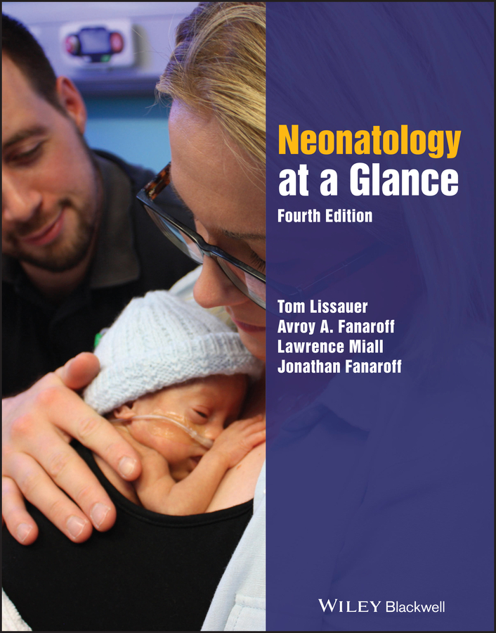
Cover Image: Courtesy of Lawrence Miall
Preface
This book provides a concise, illustrated overview of neonatal medicine. It is written for pediatric interns and residents, medical students, neonatal nurse practitioners, neonatal nurses, therapists, and midwives who care for newborn babies either on a neonatal unit or with their mothers in the normal newborn nursery (postnatal wards). For neonatologists, pediatricians, and nurse tutors it will be a useful aid to teaching.
We have divided all of neonatology into only 85 topics, with each covered in one or occasionally two to four double pages. This has been a challenging exercise; it would have been easier to write a longer book, but this format has forced us to identify the most important points and omit unnecessary details. The book has been designed to facilitate learning and to make it enjoyable. Modern education emphasizes visual impact and this is reflected in this book. The layout, photographs, and illustrations have been chosen to assist learning and make the book attractive, stimulating, and interesting. In addition, there are specific aids to learning, with boxes to highlight key points and questions and answers embedded in the text.
The book covers the wide range of common or important neonatal clinical conditions and their management. It also puts neonatology into context, with sections on its history, epidemiology, evidence‐based practice, research and consent, ethics, and global neonatology. It covers perinatal medicine, stabilizing the sick newborn, and admission to the neonatal unit, including developmental and family integrated care. The specific problems of preterm infants are covered in detail, including discharge from hospital and outcome. The medical problems encountered in neonatal care are considered, as well as challenging topics such as pain management, quality improvement, patient safety, and palliative and end of life care. Practical procedures are described, including neonatal resuscitation, neonatal transport, cranial ultrasound, brain monitoring, neuroimaging, and echocardiography for the neonatologist to inform the practicing clinician about them even if they do not perform all these procedures themselves. This new edition has allowed us to thoroughly update and revise the book. New topics have been added, such as recently developed forms of genetic testing, family integrated care, antimicrobial stewardship, and brain monitoring. A list of further reading is also included.
Whilst the book describes the salient features of intensive care, such as stabilizing the sick infant and respiratory support, it is not a manual of neonatal intensive care, of which there are many.
The book has been a collaborative project between editors and contributors from both the UK and North America. Where practices differ between the two sides of the Atlantic this has been acknowledged and described. This collaboration has been highly educational and hugely enjoyable for the editors and contributors as well as improving the book by forcing us to concentrate on the principles of practice instead of the details.
We would like to thank our many colleagues who have given their time to write, revise or review chapters and offer advice on improvements. Others have willingly contributed photographs and other images that enhance the book immensely. We are grateful to the many doctors, nurses and therapists whose positive comments about the book encouraged us to produce this fourth edition. We would also like to thank our families for allowing us to spend so much time over many years on this project.
Tom LissauerAvroy A. FanaroffLawrence MiallJonathan Fanaroff
Contributors
The Editors are indebted to the following for writing or contributing to chapters for this edition, many of whom also contributed to previous editions of the book:
Louise Allen
Consultant Pediatric Ophthalmologist
Ophthalmology Department
Cambridge University NHS Foundation Trust
Cambridge, UK
Retinopathy of prematurity, Hearing and vision
Mark Anderson
Consultant Pediatrician, Newcastle upon Tyne Hospitals
NHS Trust, Newcastle upon Tyne, UK
Pharmacology
Tomoki Arichi
Senior Lecturer in Perinatal Imaging King’s College London, UK
Perinatal neuroimaging with MRI
Denis Azzopardi
Professor of Neonatal Medicine, Centre for the
Developing Brain, King's College London, UK
Brain monitoring
Hannah Blencowe
Research Fellow, MARCH Centre, London School of
Hygiene and Tropical Medicine, London, UK
Global neonatology
Katherine Burke
Clinical Lecturer, Cardiff University
Cardiff, Wales, UK
Genetics
Neidin Bussmann
Senior Clinical Research Fellow
The Rotunda Hospital, Dublin Ireland
Clinical Tutor in Neonatology
Royal College of Surgeons in Ireland
Patent ductus arteriosus, Echocardiography
for the neonatologist
Subarna Chakravorty
Consultant Pediatric Hematologist
King’s College Hospital
London, UK
Anemia and polycythemia, White cell disorders
Coagulation
Angus Clarke
Professor and Honorary Consultant in Clinical
Genetics, Institute of Medical Genetics
University Hospital of Wales, Cardiff, UK
Genetics
Leigh Dyet
Consultant Neonatologist
Neonatal Department, University College London Hospital (UCLH), London, UK
Seizures and perinatal strokes
David Edwards
Professor of Pediatrics and Neonatology
King’s College London, UK
Perinatal neuroimaging with MRI
Afif EL‐Khuffash
Consultant Neonatologist, The Rotunda Hospital, Dublin Ireland
Honorary Clinical Associate Professor
Royal College of Surgeons, Ireland.
Patent ductus arteriosus, Echocardiography for the neonatologist
Sharon English
Consultant Neonatologist, Leeds Children's Hospital
Leeds, UK
Palliative and end‐of‐life care
Chris Foster
Consultant Neonatologist
Leeds Children's Hospital, Honorary Senior Lecturer
University of Leeds
Leeds, UK
Growth and nutrition
Jane E Harding
Professor of Neonatology
The University of Auckland
Auckland, New Zealand
Hypoglycemia and hyperglycemia
Cath Harrison
Consultant Neonatologist, Leeds Children’s Hospital
Neonatal Lead, Embrace Infant and Child Transport Team
Leeds, UK
Transport of the sick newborn infant
James Hatcher
Consultant in Microbiology and Infectious Diseases
Great Ormond Street Hospital for Children, London, UK
Antimicrobial stewardship
Jo Hegarty
Neonatologist, Newborn Services, Auckland City Hospital
Auckland, New Zealand
Hypoglycemia and hyperglycemia
Angela Huertas
Consultant Neonatologist
Neonatal Department, University College London Hospital (UCLH), London, UK
Neurologic examination
Kathryn Johnson
Consultant Neonatologist & Research Lead
Leeds Neonatal Service, Leeds, UK


