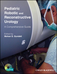
Pediatric Robotic and Reconstructive Urology E-Book
206,99 €
Mehr erfahren.
- Herausgeber: John Wiley & Sons
- Kategorie: Fachliteratur
- Sprache: Englisch
Robotic urological surgery is one of the most significant urological developments in recent years. It allows for greater precision than laparoscopic methods while retaining quicker recovery time and reduced morbidity over classical open surgical techniques. For children, where the room for error is already reduced because of smaller anatomy, it takes on even more importance for urologists. As a result, robotic surgery is rightly considered one of the most exciting contemporary developments in pediatric urology.
Pediatric Robotic and Reconstructive Urology: A Comprehensive Guide provides specialist and trainees with an innovative text and video guide to this dynamic area, in order to aid mastery of robotic approaches and improve the care of pediatric patients.
Full-color throughout and including over 130 color images, this comprehensive guide covers key areas including:
- Training, instrumentation and physiology of robotic urologic surgery
- Surgical planning and techniques involved
- Adult reconstructive principles applicable to pediatrics
- Management of complications, outcomes and future perspectives for pediatric urologic surgery
Also included are 30 high-quality surgical videos illustrating robotic surgery in action, accessed via a companion website, thus providing the perfect visual tool for the user.
With chapters authored by the leading names in the field, and expertly edited by Mohan Gundeti, this ground-breaking book is essential reading for all pediatric urologists, pediatric surgeons and general urologists, whether experienced or in training.
Of related interest
Smith's Textbook of Endourology, 3E
Smith, ISBN 9781444335545
Pediatric Urology: Surgical Complications and Management
Wilcox, ISBN 9781405162685
Sie lesen das E-Book in den Legimi-Apps auf:
Seitenzahl: 864
Veröffentlichungsjahr: 2012
Ähnliche


Contents
Cover
Online companion teaching video
Title Page
Copyright
Dedication
List of Contributors
Forewords
Preface
The Evolution and Future of Robotic Surgery in Pediatric Urology
Acknowledgments
I: History, Training, Instrumentation, and Physiology
1: The Evolution of Robotic Surgery and Its Clinical Applications
Introduction
Evolution of surgical access
Evolution of vision systems
Telepresence platforms
Clinical applications
Conclusion
2: Stepwise Approach to Training for Robotic Surgery and Credentialing
Introduction
Robotic surgery training for residents and fellows
Postgraduate robotic surgery procedural training
Credentialing
Conclusion
3: Role of Simulators in Robotic Surgery
Introduction
Challenges in the wider adoption of robot-assisted surgery
Robotic surgical simulator (RoSS)
Validation studies
Discussion
Disclosure
4: Operating Room Setup and Instrumentation
Introduction
Starting a pediatric da Vinci robotic program
The da Vinci operating room
Operating room setup
The da Vinci pediatric surgical patient
da Vinci instruments
Patient positioning
Port placement [4]
Powering on, homing, and draping
Docking
Passing needles in and out
Troubleshooting
Emergency procedures, complications, and conversion
Conclusion
5: Different Robotic Surgical Systems and Instruments: Advantages and Disadvantages
Introduction
New technology
Surgical systems
Instruments
Conclusion
6: Transition to Robotic from Laparoscopic Surgery: Lessons Learned
Introduction
Preoperative considerations
Intraoperative considerations
Postoperative considerations
Conclusion
7: Laparoscopy as a Foundation and Its Limitations and Pitfalls in Reconstructive Pediatric Urology
Limitations of laparoscopy and the rise of the da Vinci Surgical System
Pyeloplasty
Antireflux surgery
Mitrofanoff (appendicovesicostomy) procedure
Augmentation cystoplasty
Ureteric reconstructive surgery
Conclusion
8: Limitations and Difficulties of the Present Robotic Surgical Systems for Pediatric Use: Tips and Tricks
Introduction
Patient selection
Anesthesia concerns
Case and room setup
Patient positioning
Port position
Camera failures
Suturing tips
RAL pyeloplasty
Robot-assisted laparoscopic (RAL) ureteral reimplantation
RAL partial nephrectomy
RAL bladder reconstruction: augmentation and Mitrofanoff procedure
Conclusion
9: Building a Robotic Program: a Viable Business Plan
Introduction
Program design
Conclusion
10: The Basics of Robotic Surgery and a “Team Approach”
Introduction
Instrumentation
Safety and special concerns related to pediatric procedures
Case selection
Team approach
Utilization of a proctor or mentor
Conclusion
11: Anesthetic Considerations for Robotic Surgery
Introduction
Pre-anesthetic evaluation
Optimizing the patient's condition
Preparation for the operating room
Planning anesthetic management
Intraoperative anesthetic management
Post-anesthetic considerations
Conclusion
12: Physiological Changes During Minimally Invasive Surgery
Introduction
Physiology of laparoscopy
Cardiovascular effects
Respiratory effects
Renal and other vesical effects
Effects on mesenteric blood flow and intestinal motility
Acid–base metabolic effects
Hemodynamic effects related to patient position and type of approach
Hormonal and metabolic effects during laparoscopic surgery
Immunologic effects of laparoscopic surgery
Conclusion
II: Surgical Techniques
Renal and Adrenal Applications
13: Anatomy of Kidney and Adrenal Gland for Minimally Invasive Surgery
Arterial supply
Adrenal veins
Renal anatomy
Renal vasculature
Adrenal and renal lymphatics
Anatomic and practical operative considerations
Left adrenal/renal transperitoneal approach
Right adrenal/renal transperitoneal approach
Lateral retroperitoneal surgical approach
Retroperitoneal access
Left adrenal/renal retroperitoneal approach
Right adrenal/renal retroperitoneal approach
14: Robotic-Assisted Laparoscopic Nephrectomy for Benign Kidney Disease
Introduction
Indications
Positioning and preparation
Port placement
Instrumentation
Right kidney
Left kidney
Extraction
Postoperative management
Learning curve
Conclusion
Videoclip
15: Robotic Heminephrectomy
Introduction
Approaches
Patient positioning
Special instruments for children
Trocar placement for transperitoneal approach
Trocar placement for retroperitoneal approach
Technique
Operative time
Post-surgical length of hospitalization
Postoperative pain management
Outcomes
Conclusion
Videoclips
16: Pyeloplasty: a Transperitoneal Approach
Introduction
Preoperative workup and preparation
Patient positioning
Access to peritoneal space and port placement
Docking
Surgical steps
Postoperative management
Tips and tricks
Complications
Results and outcome
Videoclip
17: Pyeloplasty: a Retroperitoneal Approach
Introduction
Patient positioning
Access to the retroperitoneal space
Port placement
Docking
Dissection
Pyeloplasty
Postoperative management
Complications
Outcome
Videoclip
18: Robotic Pyeloplasty in Complex Renal Anomalies
Introduction
Etiologic and anatomic considerations
Positioning and port placement
Surgical precautions and challenges
Tips and tricks
Conclusion
Videoclip
19: Reconstructive Surgery in Duplex Kidney
Introduction
Indications
Preoperative evaluation
Patient positioning
Surgical technique
Postoperative care
Complications
Tips and tricks
Videoclip
20: Stone Treatment
Epidemiology and etiology of pediatric stone disease
Diagnostic evaluation and management
Conservative management
Surgical management
RALL surgical technique
Potential complications
Videoclip
Ureteral Applications
21: Robotic-Assisted Intravesical Ureteral Reimplantation
Introduction
Preoperative workup
Procedure
Postoperative care
Complications: Tips and Tricks
Videoclips
22: Robotic-Assisted Extravesical Ureteral Reimplantation
Procedure
Conclusion
Videoclips
23: Ureteral Defects and Retrocaval Ureter
Introduction
Diagnosis
Surgical repair
Technique
Outcomes
Conclusion
Videoclip
Bladder Applications
24: Anatomy of Large and Small Bowel and Appendix Including Their Blood Supply for Cystoplasty and Appendicovesicostomy with a Minimally Invasive Approach
Introduction
Anatomy of the small intestine
Anatomy of the appendix
Anatomy of the large intestine (Figure 24.3)
Advantages of the anatomic arrangements
Conclusion
25: Anatomy of the Pelvis and Lower Urinary Tract for Minimally Invasive Surgery Reconstruction
Introduction
Positioning
Pelvis
Ureter
Iliac and gonadal vessels
Internal inguinal ring
The vas in males
Umbilical folds
Bladder, rectum, uterus, and ovaries
Bladder base, seminal vesicles, and prostate
Conclusion
26: Robotic-Assisted Laparoscopic Ileocystoplasty and Continent Catheterizable Channels
Introduction
Patient selection and preoperative workup and counseling
Surgical technique
Postoperative course and outcomes
Conclusion
Tips and tricks for successful performance of the robotic-assisted laparoscopic ileocystoplasty and/or appendicovesicostomy and ACE
Videoclips
27: Robotic-Assisted Bladder Neck Surgery and Slings
Introduction
Evaluation and surgical indications
Surgical technique: robotic-assisted laparoscopic approach for posterior bladder neck dissection and placement of a bladder neck sling
Postoperative care
Conclusion
Videoclip
Other Applications
28: Bladder Diverticulum Excision
Background
Robotic-assisted laparoscopic diverticulum excision
Postoperative care
Videoclips
29: Robotic General Surgery in Neonates and Small Children
Introduction
History of robotics for small children and neonates
Patient selection
Adjustments for small children and neonates
Specific applications
Abdominal procedures
Thoracic procedures
Conclusion
30: Vaginoplasty: Robotically Assisted Laparoscopic Technique
Introduction
Nonsurgical vaginal creation
Alternative vaginal reconstruction
Open vaginal creation with bowel
Laparoscopic vaginal construction with bowel
Robotically assisted laparoscopic (RAL) vaginal construction with bowel
Conclusion
Videoclip
31: Seminal Vesical Cyst Excision
Introduction
Anatomy
Embryology
Evaluation
Surgical intervention
Conclusion
III: Adult Reconstructive Principles Applicable to Pediatrics
32: Robotic NOTES (Natural Orifice Transluminal Endoscopic Surgery) in Adults: Limitations in the Pediatric Population
Introduction
NOTES in adults
Potential for NOTES in the pediatric population
Limitation of pediatric NOTES
Conclusion
33: Laparoendoscopic Single-Site (LESS) Surgery and Robotic Laparoendoscopic Single-Site (R-LESS) Surgery in Urology: Adult and Pediatric Applications
Introduction
Technical challenges
Adaptations for LESS surgery
Patient selection
Adult clinical applications
LESS surgery in pediatric patients
Robotic LESS (R-LESS)
Future of LESS surgery
Conclusion
Videoclip
34: Robotics in Adrenal Disease
Introduction
Indications for minimally invasive adrenalectomy (MIA)
Robotic adrenalectomy (RA)
Operative considerations for robotic adrenalectomy
Robotic Partial Adrenalectomy (RPA)
Conclusion
35: Alternative Techniques of Pyeloplasty in the Adult Population
Introduction
Historical development of various techniques for repair of UPJ obstruction
Techniques of pyeloplasty
Our experience
36: Ureteral Defects and Ureterovaginal Fistulas
Introduction
Ureteral defects
Ureterocalicostomy
Ureterovaginal fistulas
Conclusion
37: Robotic Reconstruction Techniques Following Cystectomy
Introduction
Extracorporeal ileal conduit formation
Extracorporeal orthotopic neobladder formation
Robot-assisted continent cutaneous urinary diversion
Intracorporeal orthotopic neobladder formation
Intracorporeal ileal conduit formation
Conclusion
IV: Complications, Outcomes, and Future Perspective
38: Robotic Surgery Complications and Safety
Introduction
General complications of laparoscopic and robotic surgery
Urologic procedure-specific complications in children
Conclusion
39: Robotic Surgery Outcomes: Upper Urinary Tract
Introduction
Pyeloplasty
Nephrectomy and reconstruction
Conclusion
40: Robotic Surgery Outcomes: Lower Urinary Tract
Ureteral reimplantation
Continent catheterizable channels and enterocystoplasty
Retrovesical structures
41: Family Perceptions and Impact of New Technology on Decision Making
Laparoscopic innovation
The new “well-informed patient”
Robotic innovation
The surgical field's acceptance of robotics
42: The Asian Continent: Is It Ready for New Technology? An Indian Perspective
Introduction
History of surgical robotics [4]
The da Vinci Surgical System [4,5]
Advantages of the da Vinci system [4,5]
Robotic pediatric urology
Healthcare considerations
Is India ready for this new technology?
Yes! India is ready for robotic-assisted surgery
43: The Asian Continent: Is It Ready for New Technology? A Chinese Perspective
44: Telerobotics: Its Future in Clinical Application
Introduction
Experimental telesurgical platforms
Hurdles for telesurgery
Anticipated advances
Conclusion
45: Role of Haptics in Robotic Surgery: Present and Future Applications
Teleoperation and the surgical robot
Haptic feedback in robotic surgery
Force and tactile sensors
Index
Online companion teaching video
This book is accompanied by 28 annotated teaching videos of both actual procedures and ex vivo animal model simulations available at www.wiley.com/go/gundeti/urology
• All videos are referenced in the text where you see this logo
List of videoclips:
Videoclip 14.1 Robotic-assisted laparoscopic nephrectomy for benign disease. (Courtesy of Drs Pankaj Dangle and Mohan Gundeti.)
Videoclip 15.1–15.9 Robotic heminephrectomy.
Videoclip 16.1 Robotic-assisted laparoscopic transperiotenal pyeloplasty. (Courtesy of Drs Pankaj Dangle and Mohan Gundeti.)
Videoclip 17.1 Retroperitoneal robot-assisted pyeloplasty.
Videoclip 18.1 Robotic pyeloplasty in horseshoe and pelvic kidneys.
Videoclip 19.1 Pediatric robotic ureteroureterostomy for upper urinary tract duplication anomalies.
Videoclip 20.1 Robotic-assisted laparoscopic pyelolithotomy. (Courtesy Drs Marcelo Orvieto and Mohan Gundeti).
Videoclip 21.1 Illustration of robotic-assisted laparoscopic intravesical ureteral reimplantation.
Videoclip 22.1 Bilateral robotic-assisted laparoscopic extravesical ureteral re-implant in a female with bilateral grade 3 vesicoureteral reflux. (Courtesy of Drs Dennis Liu and Mohan Gundeti.)
Videoclip 23.1 Intraoperative images of ureteroureterostomy in a child with retrocaval ureter.
Videoclip 26.1 University of Chicago robotic-assisted laparoscopic ileocystoplasty and Mitrofanoff appendicovesicostomy (RALIMA) technique: recent modification.
Videoclip 26.2 RALIMA with cecal flap ACE.
Videoclip 26.3 Appendicovesicostomy in prune belly syndrome.
Videoclip 26.4 Split technique appendix for Mitrofanoff appendicovesicostomy/ACE.
Videoclip 26.5 Appendicovesicostomy anterior wall or posterior wall. (Courtesy of Drs Alex Rosen and Mohan Gundeti).
Videoclip 27.1 Robotic-assisted bladder neck surgery and sling placement.
Videoclip 28.1–28.8 Robotic-assisted laparoscopic diverticulectomy.
Videoclip 30.1 Operative techniques used in a robotic sigmoid vaginoplasty.
Videoclip 33.1 LESS Nephrectomy for benign disease.
This edition first published 2012, © 2012 by Blackwell Publishing Ltd.
Blackwell Publishing was acquired by John Wiley & Sons in February 2007. Blackwell's publishing program has been merged with Wiley's global Scientific, Technical and Medical business to form Wiley-Blackwell.
Registered office: John Wiley & Sons, Ltd, The Atrium, Southern Gate, Chichester, West Sussex, PO19 8SQ, UK
Editorial offices: 9600 Garsington Road, Oxford, OX4 2DQ, UK The Atrium, Southern Gate, Chichester, West Sussex, PO19 8SQ, UK 111 River Street, Hoboken, NJ 07030-5774, USA
For details of our global editorial offices, for customer services and for information about how to apply for permission to reuse the copyright material in this book please see our website at www.wiley.com/wiley-blackwell.
The right of the author to be identified as the author of this work has been asserted in accordance with the UK Copyright, Designs and Patents Act 1988.
All rights reserved. No part of this publication may be reproduced, stored in a retrieval system, or transmitted, in any form or by any means, electronic, mechanical, photocopying, recording or otherwise, except as permitted by the UK Copyright, Designs and Patents Act 1988, without the prior permission of the publisher.
Designations used by companies to distinguish their products are often claimed as trademarks. All brand names and product names used in this book are trade names, service marks, trademarks or registered trademarks of their respective owners. The publisher is not associated with any product or vendor mentioned in this book. This publication is designed to provide accurate and authoritative information in regard to the subject matter covered. It is sold on the understanding that the publisher is not engaged in rendering professional services. If professional advice or other expert assistance is required, the services of a competent professional should be sought.
The contents of this work are intended to further general scientific research, understanding, and discussion only and are not intended and should not be relied upon as recommending or promoting a specific method, diagnosis, or treatment by physicians for any particular patient. The publisher and the author make no representations or warranties with respect to the accuracy or completeness of the contents of this work and specifically disclaim all warranties, including without limitation any implied warranties of fitness for a particular purpose. In view of ongoing research, equipment modifications, changes in governmental regulations, and the constant flow of information relating to the use of medicines, equipment, and devices, the reader is urged to review and evaluate the information provided in the package insert or instructions for each medicine, equipment, or device for, among other things, any changes in the instructions or indication of usage and for added warnings and precautions. Readers should consult with a specialist where appropriate. The fact that an organization or Website is referred to in this work as a citation and/or a potential source of further information does not mean that the author or the publisher endorses the information the organization or Website may provide or recommendations it may make. Further, readers should be aware that Internet Websites listed in this work may have changed or disappeared between when this work was written and when it is read. No warranty may be created or extended by any promotional statements for this work. Neither the publisher nor the author shall be liable for any damages arising herefrom.
Library of Congress Cataloging-in-Publication Data
Pediatric robotic and reconstructive urology : a comprehensive guide/edited by Mohan S. Gundeti. p. cm. Includes bibliographical references and index. ISBN-13: 978-1-4443-3553-8 (hardcover : alk. paper) ISBN-10: 1-4443-3553-7 (hardcover : alk. paper) 1. Urinary organs–Surgery. 2. Pediatric urology. 3. Children–Surgery. 4. Robotics in medicine. I. Gundeti, Mohan S. [DNLM: 1. Urologic Diseases–surgery. 2. Adolescent. 3. Child. 4. Infant. 5. Reconstructive Surgical Procedures–methods. 6. Robotics–methods. 7. Surgery, Computer-Assisted–methods. 8. Urologic Surgical Procedures–methods. WJ 168]
RD571.P44 2012
617.4′61–dc23
2011015321
A catalogue record for this book is available from the British Library.
This book is published in the following electronic formats: ePDF 9781444345261; Wiley Online Library 9781444345292; ePub 9781444345278
Dedication
To my late grandfather, Mr. Hanmantu Gundeti, for my rich inheritance of humanity. My parents, Mr. Saheb Hanmantu and Mrs. Bhudevi Saheb Gundeti, who showed me that the path of success comes only through the virtue of hard work.
To Laltia, my better half, for allowing me to pursue my dreams of becoming a pediatric urosurgeon. Madhav and Manohar, my brothers for their continued support on this long journey. My wonderful kids, Anjali, Amol, and Apoorva for their love.
To all my family members, teachers, and friends for their continuous encouragement. To all my patients and their families for giving me the opportunity to serve them.
Mohan Saheb Gundeti July 4th, 2011
List of Contributors
Ardavan Akhavan MD Chief Resident Department of Urology Mount Sinai School of Medicine New York, NY, USA
Catherine Bachman MD Assistant Professor Section Chief, Pediatric Anesthesia Department of Anesthesia and Critical Care University of Chicago Medical Center Chicago, IL, USA
Kyu Suk Baek PhD Research Associate Argonne National Laboratory Argonne, IL, USA
Linda A. Baker MD Professor of Urology Director of Pediatric Urology Research Department of Urology University of Texas Southwestern Medical Center at Dallas Dallas, TX, USA
Shawn M. Beck MD Clinical Instructor Department of Urology University of California at Irvine Irvine, CA Children's Hospital of Orange County Orange, CA, USA
Mahendra Bhandari MD, MBA Senior Staff and Bioscientist Director, Robotic Research Vattikuti Urology Institute Henry Ford Health System Detroit, MI, USA
Aron Bruhn MD Department of Urology New York University School of Medicine New York, NY, USA
Douglas A. Canning MD Professor of Urology in Surgery University of Pennsylvania School of Medicine Division of Pediatric Urology Children's Hospital of Philadelphia Philadelphia, PA, USA
Pasquale Casale MD Director, Minimally Invasive Surgery Associate Professor University of Pennsylvania Division of Urology Children's Hospital of Philadelphia Philadelphia, PA, USA
Sujit Chowdhary MCh FRCS FACS Senior Consultant, Pediatric Urology and Pediatric Surgery Indraprastha Apollo Hospitals Sarita Vihar New Delhi, India
Daniel DaJusta MD Pediatric Urology Fellow Department of Urology University of Texas Southwestern Medical Center at Dallas Dallas, TX, USA
Pankaj P. Dangle MD MCh Resident in Urology The University of Chicago Chicago, IL, USA
Prokar Dasgupta MSc (Urol), MD, DLS, FRCS (Urol), FEBU Consultant Urological Surgeon Guy's and St Thomas's NHS Trust Professor of Robotic and Innovative Surgery King's College Hospital London, UK
Mihir M. Desai MD Professor of Clinical Urology Institute of Urology Keck School of Medicine University of Southern California Los Angeles, CA, USA
Lori Dyer MD Assistant Professor of Urology New York Medical College Valhalla, NY, USA
Manuel S. Eisenberg MD Research Fellow Institute of Urology Keck School of Medicine University of Southern California Los Angeles, CA, USA
Jack S. Elder MD Chief of Urology Henry Ford Health Systems Associate Director Vattikuti Urology Institute Department of Pediatric Urology Children's Hospital of Michigan Clinical Professor of Urology Case School of Medicine Detroit, MI, USA
Michael Erhard MD Nemours Children's Clinic Jacksonville, FL, USA
Walid Y. Farhat MD, FRCSC, FAAP Associate Professor University of Toronto The Hospital for Sick Children Toronto, Canada
Israel Franco MD Professor of Urology New York Medical College Valhalla, NY, USA
Inderbir S. Gill MD, MCh Chairman and Professor Catherine and Joseph Aresty Department of Urology Founding Executive Director, USC Institute of Urology Associate Dean, Clinical Innovation Keck School of Medicine University of Southern California Los Angeles, CA, USA
Rita Gobet MD Professor and Head of Department of Pediatric Urology University Children's Hospital Zurich Zurich, Switzerland
Prasad P. Godbole FRCS, FRCS(Paeds), FEAPU Consultant Pediatric Urologist Department of Pediatric Urology Sheffield Children's Hospital Sheffield, UK
Miles A. Goldstraw BSc, MRCS, MD Urology Specialist Registrar Urology Department Barnet and Chase Farm NHS Trust London, UK
Pablo Gomez III MD Pediatric Urology Fellow Harvard Medical School Department of Urology Children's Hospital Boston Boston, MA, USA
Narmada P. Gupta MCh Chairman, Academic and Research Urology Division Medanta Institute of Kidney and Urology Medanta – The Medicity Gurgaon, India
Khurshid Guru MD Assistant Professor, Department of Urology Director, Roswell Park Center for Robotic Surgery Roswell Park Cancer Institute Clinical Assistant Professor of Urology School of Medicine and Biomedical Sciences State University of New York at Buffalo Buffalo, NY, USA
Jennifer A. Hagerty DO Pediatric Urology Alfred I. duPont Hospital for Children/Nemours Wilmington, DE Assistant Professor Jefferson University Medical Center Philadelphia, PA, USA
Blake Hannaford PhD Professor and Director, Biorobotics Laboratory Department of Electrical Engineering University of Washington Seattle, WA, USA
Ashok K. Hemal MD, DipNB, MCh, MAMS, FICS, FACS, FAMS, FRCS (Glasg) Professor, Department of Urology Comprehensive Cancer Center Professor, Institute of Regenerative Medicine Director, Robotics and Minimally Invasive Surgery Wake Forest Baptist Health, and Wake Forest University School of Medicine Winston-Salem, NC, USA
Michael R. Hernandez MD Assistant Professor Department of Anesthesia and Critical Care University of Chicago Medical Center Chicago, IL, USA
Andrew J. Hung MD Research Fellow Institute of Urology Keck School of Medicine University of Southern California Los Angeles, CA, USA
Isuru S. Jayaratna MD Research Fellow Institute of Urology Keck School of Medicine University of Southern California Los Angeles, CA, USA
Gautam A Jayram MD Resident in Urology Department of Surgery Comer Children's Hospital University of Chicago Medical Center Chicago, IL, USA
Martin Kaefer MD Professor of Urology Department of Urology Indiana University School of Medicine Riley Hospital for Children Indianapolis, IN, USA
Thenkurussi Kesavadas PhD Director, Virtual Reality Laboratory Professor, Mechanical and Aerospace Engineering Adj. Professor, Computer Science and Engineering and Roswell Park Cancer Institute State University of New York at Buffalo Buffalo, NY, USA
Japinder Khosa FRACS, MBBS Consultant Paediatric Surgeon Department of Paediatric Surgery Princess Margaret Hospital for Children Subiaco Perth, Western Australia
Antoine E. Khoury MD, FRSC, FAAP Professor, Department of Urology Chief of Pediatric Urology University of California at Irvine Irvine, CA Children's Hospital of Orange County Orange, CA, USA
Christina Kim MD, FAAP Assistant Professor of Surgery (Urology) University of Connecticut Director, Minimally Invasive Urologic Surgery Connecticut Children's Medical Center Hartford, CT, USA
Roger S. Kirby MA, MD, FRCS (Urol), FEBU Professor of Urology Consultant Urological Surgeon and Professor of Urology The Prostate Centre London, UK
Chester J. Koh MD, FACS, FAAP Assistant Professor of Urology Division of Pediatric Urology Children's Hospital Los Angeles Institute of Urology Keck School of Medicine University of Southern California Los Angeles, CA, USA
Marc-David Leclair MD, PhD, FEAPU Professor of Pediatric Purgery Department of Pediatric Surgery Hôpital Mère–Enfant Université de Nantes Nantes, France
Jason Y. Lee MD, FRCSC Minimally Invasive Urologic Surgery Education Fellow Assistant Clinical Professor Department of Urology University of California at Irvine Orange, CA, USA
Richard S. Lee MD Assistant Professor of Surgery (Urology) Harvard Medical School Department of Urology Children's Hospital Boston Boston, MA, USA
Thomas S. Lendvay MD, FACS Assistant Professor Department of Urology University of Washington Division of Pediatric Urology Seattle Children's Hospital Co-Director, Seattle Children's Hospital Robotic Surgery Center Seattle Children's Hospital Seattle, WA, USA
Bruce W. Lindgren MD Director, Center for Minimally Invasive Urological Surgery Children's Memorial Hospital Chicago, IL Clinical Assistant Professor Loyola University Stritch School of Medicine Maywood, IL, USA
Dennis Liu MD, FACS, FAAP Assistant Professor of Urology in Surgery The University of Chicago, and Comer Children's Hospital Chicago, IL, USA
Pedro-José López E. MD Associate Professor of Paediatric Surgery and Urology Department of Paediatric Urology Hospital Exequiel Gonzalez Cortes Universidad de Chile Clinica Alemana Santiago, Chile
Giovanni S. Marchini MD Robotic Research Fellow Department of Urology University of São Paulo Medical School São Paulo, Brazil
Elspeth M. McDougall MD, FRCSC, MHPE Director, Surgical Education Center Professor of Urology University of California, Irvine Orange, CA, USA
John J. Meehan MD, FACS Associate Professor of Surgery University of Washington School of Medicine Seattle Children's Hospital Seattle, WA, USA
Mani Menon MD The Raj and Padma Vattikuti Distinguished Chair Director, Vattikuti Urology Institute Henry Ford Health System Detroit, MI, USA
Brian J. Minnillo MD Robotic Research Fellow Department of Urology Children's Hospital Harvard Medical School Boston, MA, USA
Rosalia Misseri MD Assistant Professor, Pediatric Urology Department of Urology Indiana University School of Medicine Riley Hospital for Children Indianapolis, IN, USA
Asif Muneer BSc (Hons), MB, FRCSEd, FRCS (Eng), MD, FRCS (Urol) Consultant Urological Surgeon and Andrologist Honorary Senior Lecturer University College London Hospitals London, UK
Imran Mushtaq MB, ChB, MD, FRCS (Glasg), FRCS (Paed) Consultant Paediatric Urologist Department of Paediatric Urology Great Ormond Street Hospital for Children London, UK
Rajandra B. Nerli MS, MCh Director, KLES Kidney Foundation Professor and Head, Department of Urology KLE University JN Medical College KLES Dr. Prabhakar Kore Hospital and MRC Belgaum, India
Hiep T. Nguyen MD, FAAP Rose Zimmerman Mandell Chair in Innovative Urological Technology Associate Professor in Surgery (Urology) Harvard Medical School Director of Robotic Surgery Research and Training Center Department of Urology Children's Hospital Boston, MA, USA
Paul H. Noh MD, FACS, FAAP Assistant Professor Division of Pediatric Urology Cincinnati Children's Hospital Medical Center University of Cincinnati Cincinnati, OH, USA
L. Henning Olsen MD Associate Professor and Consultant Pediatric Urologist Department of Pediatric Urology Aarhus University Hospital – Skejby Aarhus, Denmark
Marcelo A. Orvieto MD Fellow Global Robotics Institute Celebration Health Florida Hospital Celebration, FL, USA
Michael C. Ost MD Associate Professor, Pediatric Urology Children's Hospital of Pittsburgh University of Pittsburgh Medical Center Pittsburgh, PA, USA
Young Soo Park PhD Staff Research Engineer Argonne National Laboratory Argonne, IL, USA
Carlo C. Passerotti MD, PhD Assistant Professor Faculty of Medicine University of São Paulo Director, Robotic Surgery Hospital Alemão Oswaldo Cruz São Paulo, Brazil
Nilay S. Patel MD, FRCS (Urol) Senior Fellow in Urology Molecular Oncology Laboratories Cancer Research UK Weatherall Institute of Molecular Medicine John Radcliffe Hospital Oxford, UK
Shiv Patel BS Medical student Department of Urology Mount Sinai School of Medicine New York, NY, USA
Vipul R. Patel MD Medical Director Global Robotics Institute Celebration Health Florida Hospital Celebration, FL, USA
Krishna Patil MS, FRCS (Ed), FRCS (Urol), FEBU Urology Consultant Department of Urology Ashford and St Peters Hospitals NHS Trust Chertsey, UK
Frank J. Penna MD Resident in Urology Vattikuti Urology Institute Henry Ford Hospital Detroit, MI, USA
Sara L. Pittenger MD Fellow, Pediatric Anesthesia Department of Anesthesia and Critical Care University of Chicago Medical Center Chicago, IL, USA
Gaayana A. Raju MD, FRCSC Pediatric Urology Fellow Children's Hospital of Pittsburgh University of Pittsburgh Medical Center Pittsburgh, PA, USA
Yazan F. Rawashdeh MD, PhD Consultant Pediatric Urologist Department of Pediatric Urology Aarhus University Hospital – Skejby Aarhus, Denmark
Aria A. Razmaria MD Fellow in Urology Department of Surgery, Comer Children's Hospital University of Chicago Medical Center Chicago, IL, USA
Mallikarjun N. Reddy MS, MCh Associate Professor Department of Urology KLE University JN Medical College KLES Dr. Prabhakar Kore Hospital and MRC Belgaum, India
Francisco J. Reed López-Güereña MD Paediatric Urology Fellow Department of Paediatric Urology Hospital Exequiel Gonzalez Cortes Universidad de Chile Santiago, Chile
Emad Rizkala MD Chief Resident in Urology New York Medical College Valhalla, NY, USA
Alex Rosen MD Urology Resident The University of Chicago and Comer Children's Hospital Chicago, IL, USA
Jacob Rosen PhD Associate Professor Department of Computer Engineering Jack Baskin School of Engineering University of California Santa Cruz Santa Cruz, CA, USA
Naeem Samnakay FRACS, MMedSc, MBBS Consultant Paediatric Surgeon and Paediatric Urologist Department of Paediatric Surgery Princess Margaret Hospital for Children Subiaco Perth, Western Australia
Aseem R. Shukla MD, FAAP Director, Pediatric Urology University of Minnesota Amplatz Children's Hospital Associate Professor Departments of Urology and Pediatrics University of Minnesota Minnesota, MN, USA
Vannita Simma-Chiang MD Research Fellow Institute of Urology Keck School of Medicine University of Southern California Los Angeles, CA, USA
Irina Stanasel MD Chief Resident Department of Urology Wake Forest University School of Medicine Winston-Salem, NC, USA
Michael D. Stifelman MD Associate Professor, New York University School of Medicine Director, NYU Robotic Surgery Center New York, NY, USA
Jeffery A. Stock MD Associate Clinical Professor of Urology and Pediatrics Division of Pediatric Urology Kravis Children's Hospital Mount Sinai School of Medicine New York, NY Clinical Professor of Surgey UMDNJ–New Jersey Medical School Newark, NJ, USA
Douglas W. Storm MD Head, Section of Pediatric Urology Naval Medical Center San Diego San Diego, CA Assistant Professor of Surgery Uniformed Services University of the Health Sciences F. Edward Hebert School of Medicine Bethesda, MD, USA
Shyam Sukumar MD Clinical Fellow Vattikuti Urology Institute Henry Ford Health System Detroit, MI, USA
Joel M. Sumfest MD Director of Pediatric Urology Department of Urology Geisinger Medical Center Danville, PA, USA
Jie Sun MD, PhD Vice Professor of Pediatric Urology Department of Urology Shanghai Children's Medical Center Shanghai, China
Salil Umranikar MS, DNB (Urol), MRCS Urology Registrar Department of Urology Ashford and St Peters Hospitals NHS Trust Chertsey, UK
Vijaya Vemulakonda MD, FD Assistant Professor Department of Pediatric Urology The Children's Hospital University of Colorado at Denver Aurora, CO, USA
Bryce A. Weber BSc, MD, FRCSC Urology Fellow The Hospital for Sick Children Toronto, Canada
Duncan Wilcox MD, MBBS Professor, The Ponzio Family Chair of Pediatric Urology Department of Pediatric Urology The Children's Hospital University of Colorado at Denver Aurora, CO, USA
Hsi-Yang Wu MD Associate Professor of Urology Stanford University School of Medicine Stanford, CA, USA
Rajiv Yadav MCh Consultant, Urooncology and Robotic Surgery Urology Division Medanta Institute of Kidney and Urology Medanta – The Medicity Gurgaon, India
Gregory P Zagaja MD Associate Professor of Surgery Division of Urology University of Chicago Chicago, IL, USA
Forewords
One hundred and fifty years ago urology was called genitourinary surgery. Two thousand five hundred years ago, as the single exclusion for a medical practice in the Hippocratic Oath, the practitioners “of that art” were lithotomists. Bladder calculus was the original defining problem, plaguing mankind from his beginning, and any surgical relief required not only an intrepid lithotomist, but also some means of technology albeit primitive back then.
Urology has evolved since those early days and has remained heavily contingent on technology. Urologists are typically the creators or first adapters of new instruments or approaches to clinical problems. Urethral catheters and sounds, lithotrites, cystoscopes, transurethral electrosurgery, open prostatectomy, retrograde pyelography, transurethral prostatectomy, genitourinary reconstruction, percutaneous nephrostomy, ureteroscopic stone extraction or ablation, extracorporal shock wave lithotripsy, prostate/renal cryoablation, laparoscopic nephrectomy, robotic radical prostatectomy, and transcutaneous histotripsy are sequential examples which prove how human imagination has become urological reality.
The idea of the automaton figured early in human imagination, recorded in ancient China and later in the works of Aristotle, Homer, Al-Jazari, and Leonardo da Vinci. The actual term “robot”, as many know, came from Karel Čapek, a Czech author, in his play R.U.R. (Rossum's Universal Robots) which I recall reading in high school. The concept was riveting to me, and somewhat subversive relative to modern society as seen by a teenager. Čapek credited his brother, Josef, with the word robot deriving it from a Czech term for servitude. It didn't take long for the idea to make the movies, and Fritz Lang's classic “Metropolis” in 1927 still enjoys viewings among serious filmgoers. Isaac Asimov performed the great synthesis of his neologism “robotics” and an ethical framework for the fictional technology in his Three Laws of Robotics. This happened definitively in his short story “Runaround” in 1942. These are the three laws:
1. A robot may not injure a human being or, through inaction, allow a human being to come to harm.
2. A robot must obey any orders given to it by human beings, except where such orders would conflict with the First Law.
3. A robot must protect its own existence as long as such protection does not conflict with the First or Second Law.
Asimov later added a law he called the “zeroth”: A robot may not harm humanity, or, by inaction, allow humanity to come to harm. What was compelling science fiction has quickly become reality.
Urologists are technocrats of medicine, it is their nature to explore, perfect, and push the limits of new clinical methods and platforms. It was just a matter of time before robotic technology was extended to the world of surgery and urologists, as one might expect, were early adaptors. Mohan Gundeti, a robotic virtuoso, has extended the range to robotic surgery throughout the pediatric urology domain. It is good and appropriate that some of our best surgeons should push the limits of new technologies. Yet we do not expect that the robot will replace unassisted manual surgery across the board for the entire range of pediatric urology. Some procedures will remain preferably undertaken “the old fashioned way” for reasons of simplicity, safety, time, resource availability, and cost. The text that follows by Mohan very nicely shows the range of possibility for pediatric urology in 2011. The clinical marketplace over the next few years will ascertain the appropriate specific domain of pediatric urological robotics, that is, which procedures and which children are advantaged by the robot. Meanwhile we must remain on the lookout for the next technological tipping point in urologic surgery, for surely something new and even better is waiting for us down the road.
David A. BloomUniversity of Michigan, Ann Arbor, MI, USA July 15th, 2011
I am excited to witness the production of a textbook covering pediatric and reconstructive robotic surgery.
For the past 10 years robotic surgery has evolved safely and successfully in adult urologic surgery to cover multiple applications. The arrival of robotics to the pediatric urology specialty is marked by the publication of this work. The chapters in this book superbly cover all aspects of pediatric and reconstructive robotic surgery, giving the reader insight, tips and tricks, and practical knowledge for a novice to start or an expert to expand his robotic surgery practice.
The editor has assembled authors from all over the world, who are experts in this field. The addition of the video is a helpful and unique educational tool, especially for this novel approach to surgery.
I commend the editor and authors for their great work, and am certain that a generation of urologic surgeons will benefit from this book.
Arieh L. ShalhavUniversity of ChicagoChicago, IL, USA July 15th, 2011
It is a joy to provide a foreword for this book. The subject is new, exciting and of practical benefit to our patients. The techniques described will replace many forms of conventional surgery in the future. The book provides the reader with a broad synopsis of robotic surgery and in particular its adaption to the pediatric population. There is still much to develop in this area. The chapters are mostly multi-authored, which underwrites a sound clinical aspect to the contents while simultaneously providing a good overview. The book is a complete review of robotic surgery and will be a standard text for clinicians using robotic surgical techniques in the treatment of their patients.
Patrick G. DuffyGreat Ormond Street Hospital for ChildrenLondon, UK June 22nd, 2011
Preface
The Evolution and Future of Robotic Surgery in Pediatric Urology
While all physicians probably view their professional experience as being characterized by rapid and evolutionary change, it may be argued that the last 20 years in surgery and urology have seen truly revolutionary changes that represent real paradigm shifts in how we perform our surgical procedures. The advent and emergence of laparoscopy have greatly changed our landscape in urology, and while it has done so less spectacularly in pediatric urology, its impact is nonetheless significant. Having had the privilege of participating in this evolution in pediatric urology from the earliest days of laparoscopy to the emergence of robotic surgery for children, I have had the good fortune to see the challenges, successes, and potential of these new technologies up-close. Three important factors will continue to exert pressure on the pathways of innovation in pediatric urological minimally invasive surgery.
The first is the inherent drive to reduce surgical morbidity in children. Everyone would prefer to inflict less discomfort on a child, yet it is frequently stated that children recover quickly from any incision. Although this is true in a relative sense, it is clear that they suffer from surgical morbidity in their own way. We may not always be able to measure this objectively, but an experienced surgeon can see the impact almost as well as the parent. Our efforts to reduce this impact should not be trivialized by simplistic assertions that an incision does not cause much pain. We need to focus efforts on being able to assess surgical morbidity in children of all ages, and develop means to reduce this using modern technology. At the same time, the importance of enhancing the efficacy of any surgery is also critically important. This is seldom considered in the laparoscopic field, but with robotic technology, there is the real potential for enhanced outcomes due to improved visualization, tissue manipulation, and motion control. This will be difficult to prove objectively, yet must be considered. To enhance outcomes further, we recognize the second important feature on the evolution of pediatric minimally invasive surgery.
The challenge of making instruments child friendly continues from laparoscopy into robotic surgery. None of the current instruments are truly suitable for small children, yet most surgeons using the da Vinci system have made them work. This was similarly the case with early laparoscopy when we were performing nephrectomies with all 10 mm cannulae simply because there was nothing else. The children did fine, but clearly we needed to do better. Robotic technology can improve, although the economic incentives remain limited. Perhaps with widening utilization by other specialties, the potential importance of pediatric surgical practice will become evident. It remains critically important for the pediatric practitioner to be actively engaged in the process of development of these technologies, otherwise they will never be suitable for children and we will remain in the shadows of practice, which is certainly not in the best interests of our patients.
There is a particular attractiveness of robotics for children based on the need for exquisite visualization, delicate motion control, and tremor filtering. The three-dimensionality of the imaging system permits enhanced precision and accuracy. All of these elements have long been considered essential to the satisfactory performance of pediatric reconstructive urology. Although the current instrument is limited in terms of its delicacy, it can and has been used effectively for very small children and precise procedures. Given the economic burden of modern robotic systems, many have stated, however, that conventional laparoscopy can be used instead of the robot. Although this is probably true in the context of a practitioner who performs one operation repetitively and can effectively learn the technique and basic skills needed for precise laparoscopic surgery, this is a rarity in pediatric urology. Even in a large and busy referral practice, it is rare that one surgeon can perform enough laparoscopic operations in pediatric urology to develop the proficiency to perform a pyeloplasty with the same precision as can be performed with open surgery or robotically. Even if this were the case, it is unlikely that centers of excellence would ever develop for pyeloplasty. This is a basic and common operation that all pediatric urologists should be able to perform readily. Therefore, technology that can permit the majority of pediatric urologists to offer a minimally invasive alternative to open surgery for pyeloplasty and other such reconstructive procedures would seem attractive. This is what the da Vinci system offers and has been shown to be able to perform. The referral centers will likely develop the more complex procedures and refine the basic ones, but providing access to this type of beneficial technology to a wide population would seem appropriate.
This volume describes the various robotic surgical approaches that have evolved very rapidly using the da Vinci system. It should be evident that there is a robust potential in this technology, yet a note of caution must be heard. We cannot use this technology in the absence of the highest quality of clinical pediatric urology. Using a surgical robot in children does not make an adult urologist a pediatric urologist. Indications for surgery should not yet change until a significant reduction in morbidity is proven. Surgical strat-egies should not change until efficacy is proven. With these foundations, however, the potential of robotic technologies in pediatric urology and surgical practices are enormous. It is exciting to consider the future horizons of such innovation, particularly when reflecting on how far we have moved in such a short time.
Craig A. Peters MD, FACS, FAAPChief, Surgical Innovation, Technology and TranslationPrincipal Investigator, Sheikh Zayed Institute forPediatric Surgical InnovationChildren's National Medical CenterWashington, DC, USA
Acknowledgments
This book is a product of hundreds of professional minds coming together and giving their best to serve mankind.
First, my thanks go to all the authors for their valuable contributions, especially for their efforts within their busy schedules.
To Rob Blundell at Wiley-Blackwell for the concept of producing a dedicated book on this emerging technology in pediatric urology. Thanks also to Michael Bevan, Phil Weston, Gill Whitley and the rest of the team, for their diligent work.
To all my teachers, mentors, friends, colleagues at work in India, London, and Chicago, with thanks for their encouragement and support.
Finally, a thank you to the patients and their families for putting their faith and trust in me and for giving me the wonderful opportunity to serve them.
M.S.G.
I
History, Training, Instrumentation, and Physiology
1
The Evolution of Robotic Surgery and Its Clinical Applications
Shyam Sukumar, Mahendra Bhandari and Mani Menon
Vattikuti Urology Institute, Henry Ford Health System, Detroit, MI, USA
Robotics makes the transition to the Information Age complete by looking at information (the monitor) and manipulating information (hand motions send electronic signals which control the tip of the instruments). It is no longer blood and guts, but it is bits and bytes.
Richard M. Satava [1]
Introduction
Surgical robots assist the modern-day surgeon in achieving a technically satisfactory performance by enabling precise surgical actions on a clearly visible surgical target. Robotic surgery preserves the precision of tissue handling even in remote body cavities, which often are visually and tactilely inaccessible to traditional open surgery. A synergy in real-time three-dimensional, magnified vision translates the psychovisual coordination of the surgeon's fingers to fine instruments with an unprecedented seven degrees of freedom, thereby minimizing collateral tissue insult. The speedy return to baseline function following surgery, a unique patient-centric comfort profile, the mitigation of operative complications, and its safe application for intricate surgical procedures even in sick patients have increased its popularity among surgeons and patients alike. In this chapter, we venture off the beaten path, avoiding the usual chronological narrative of various industrial robots to focus instead on the seminal role of robotic surgery in the evolution of minimal access; we also place special emphasis on the clinical applications and interdisciplinary cross-fertilization that has ensued.
Evolution of surgical access
Minimally invasive surgery in general and robotic surgery in particular have challenged the age-old practice of long or multiple incisions and wider exposure to handle complex surgeries. Studies examining the systemic effect of surgical intervention (including the size and location of incisions) support the beneficial effect of minimally invasive surgery on the biological and immunological level [2,3].
Minimal access
John Wickham, a British urologist, coined the term “minimally invasive surgery” in 1983 [4–6]. A vociferous champion of endoscopic techniques, he classified the history of surgery in three phases, the first being the “brutal and ablative” medieval epoch, followed by the era of improved resuscitation and carefree incisions, and lastly the modern age beginning from the 1960s onwards. The modern age involved small pockets of enlightened surgeons gradually embracing techniques involving minimal access [4].
The possibility of intra-abdominal insufflations and endoscopic visualization (and hence “the laparoscopic concept”) was first reported by G. Kelling, who insinuated a cystoscope trans-abdominally and applied pneumoperitoneum for treating blood loss [7]. Contemporaneously, culdoscopic access was described by D.O. Ott in a pregnant woman [7]. A significant milestone was the application of the Veress needle, initially developed for the creation of therapeutic pneumothorax to treat tuberculosis [7].
Kurt Semm further revolutionized the field of laparoscopic surgery by advocating the “keyhole” approach in gynecologic surgery, and controversially performing the first laparoscopic appendectomy. He is to be credited with developing endoscopic hemostatic suturing and, more than anyone else, was singularly influential in designing the range of innovative instruments indispensable for minimal access [8].
Erich Muhe performed the first human laparoscopic cholecystectomy in 1985. The French triumvirate of Mouret, Dubois, and Perissat then standardized and popularized laparoscopic cholecystectomy as a mainstream procedure [9].
The evolution of tools and techniques for port placement has significantly reduced access-related morbidity such as vascular, bowel, and wound site complications and mitigated oncologic complications related to access – the abdominal metastatic seeding of neoplasms [10]. Access for most urologic procedures involves one of three approaches: open (Hasson), closed (using a Veress needle), or optical ports [11]. Optical ports allow direct visualization of the various tissue layers during access. Trocar type has moved from the use of pyramidal and conical trocars to the use of trocars with bladeless tips, which are less traumatic and hence result in favorable wound parameters.
Evolution of vision systems
The single seminal breakthrough which paved the way for state-of-the-art endoscopic surgery was the development of the fiber-optic system. It was indeed a giant leap for surgeons – from the era of working in natural passages under candlelight brightness through incandescent bulb illumination energized by dry cells to bright vision, which could be carried to any body cavity through fiber-optic carriers.
Taking a radical approach to endoscopic clarity and definition, Harold Hopkins (along with Kapany) published a report in Nature in 1954 describing the “fiberscope,” in which light passed through bundles of fine fibers of glass increased the quality of the images. Building on Hopkins’ work, Basil Hirshowitz developed the first clinically useful gastroscope [12]. Hopkins was responsible for another seminal innovation, this time in cystoscopy (his initial assignment being to capture images inside the bladder), with his introduction of the rod lens system. Whereas the traditional endoscope consisted of a tube of air with lenses of glass (as the “objective”) Hopkins’ version consisted of a tube of glass with lenses of air, which greatly increased the amount of light transmitted [12]. Having been denied a conducive commercial response in Britain, Hopkins was fortunate enough to find a collaborator in Karl Storz [12], thereby beginning, arguably, the most important collaboration in the history of endoscopic instrumentation.
Another obstacle with the early systems was that they hampered concerted coordination between the chief surgeon and his or her assistant by limiting the visual plethora of the surgical field to the endoscopist, and the participation of an assistant was dependent on a verbal transliteration of the surgical field. Photodocumentation, so critical for consultation and education, was also severely hampered [13].
The introduction of a miniature electronic camera that transduced the afferent optical image into efferent electronic impulses with the help of a charge-coupled device (CCD) radically transformed the way in which endoscopy was performed [13]. Thus electronic video-endoscopy was born, combining the electronic endoscope and the television. The entire surgical team could now view the magnified images and collaboration between surgeons was taken to a whole new level.
Operating rooms equipped for robotic surgeries with the da Vinci Surgical System provided 3D stereoscopic vision for the operating surgeon but the assistants and allied personnel were only allowed to visualize a 2D version of the unfolding events. The “augmented-reality” surgical suite, first proposed and implemented by Shrivastava and Menon [14], made tangible stereoscopic 3D vision a reality for everyone in the operating room (Figure 1.1).
Figure 1.1 An “augmented-reality” operating room at the Vattikuti Urology Institute, Henry Ford Hospital, Detroit, MI.
While the vision systems improved, the operating endoscopist's ability to manipulate the surgical field themselves was still impaired by the assistant holding the camera in place. This is where robotic technology first made its mark. The first robot to have the US Food and Drug Administration (FDA)'s imprimatur was AESOP (Automated Endoscopic System for Optimal Positioning). Computer Motion (Berkeley, CA, USA), which first introduced AESOP in the mid-1990s, was initially funded by a NASA research grant for the development of a robotic arm for the US space program. AESOP used voice (or, alternatively, foot or hand) control to direct the movements of a robotic arm, which usually held the laparoscope (but could also hold a retractor). The surgeon used a preprogrammed voice card that allowed the device to understand and respond to his or her commands. Allaf et al. [15] examined the optimal interface (voice versus foot control) to manipulate the robot and found that foot control was faster but voice control was more accurate. Kavoussi et al. [16] found the laparoscopic camera positioning to be significantly steadier with less inadvertent movements when under robotic control and concluded that operative times during dissections were not significantly different between robot-assisted and human-assisted procedures.
Telepresence platforms
The da Vinci system
The state-of-the-art platform for the performance of telemanipulative procedures is the da Vinci Surgical System (Intuitive Surgical, Sunnyvale, CA, USA), which is a master–slave system. The system has three separate parts that dovetail to produce an immersive interface.
The console
The surgeon is ensconced at the console, which is designed as an ergonomically comfortable perch with his or her hands fitting into the “masters” (basically, freely mobile finger controls). In the United States, the FDA has made it mandatory for the console to be in the same room as the patient.
The console also consists of a stereoscopic viewer with sensors. The act of placing the (surgeon’s) head into the console vivifies the system and removing it deactivates and locks the robotic arms. The surgeon's dexterous hand movements are electronically translated to the robotic instruments in direct contact with the operative target. Foot controls for camera positioning and diathermy complete the console.
Stereoscopic vision system
The stereoscopic vision system provides high-resolution 3D images of the operative field to the surgeon at his or her vantage point in the console. The binocular vision system, with images that combine magnification and depth resolution, is juxtaposed with the hand controls to produce an intuitive experience mimicking open surgery.
Surgical cart
The end effectors of the robotic system are in striking contrast to conventional laparoscopy, with the surgeon's controls articulating wristed instruments with seven degrees of freedom (three orientation, three translational, and one for grip) and two degrees of axial rotation. The tools are also steadier than the human hand because it has an inbuilt tremor-filtering functionality. Variable motion scaling allows a versatile degree of motion of the instruments (as, e.g., a 3:1 ratio allows 3 cm of movement of the controls to translate into 1 cm of movement of the instruments in the operative field) and, combined with the magnified vision system, it becomes possible for the surgeon to finesse very nimble dissections in anatomic minefields.
Newer developments include a fourth arm (which gives the console surgeon greater independence and control), an integrated touchpad (for audio and video controls), and superior stereoscopic vision. TilePro allows the projection of intraoperative ultrasound images and preoperative computed tomography (CT) images on to the console screen for precise tumor resection [17]. The dual console systems are a step in the direction of enhanced interspecialty collaboration, providing a platform for the cross-pollination of ideas and sharing of instruments to develop a multidisciplinary robotic program.
Clinical applications
Cardiothoracic surgery
The da Vinci robot (as was the Zeus) was engineered specifically for the performance of minimally invasive cardiac surgery. In 1999, Carpentier and co-workers [18] reported the first totally endoscopic cardiac bypass (TECAB) procedure (LITA–LAD grafting) in Paris. All the surgeons underwent a preliminary evaluation phase during which they optimized the various steps in human cadavers. The rate-limiting step in attaining the “holy grail” of totally endoscopic closed-chest surgery was felt to be the optimal placement of ports in the rigid chest wall. The excellent kinematic (jointed) dexterity of the anastomotic step of surgery in a 3D vision environment was hailed as the signal feature of the robotic interface.
The FDA approval for robotic coronary revascularization (da Vinci) was the result [19] of a prospective multicentric Investigational Device Exemption (IDE) trial by Argenziano et al. [20]. Patients were enrolled for arrested-heart, single-vessel TECAB. The trial concluded that the TECAB had comparable results pertaining to efficacy (freedom from graft failure and reintervention) and safety (freedom from major adverse cardiac events) endpoints to conventional open surgery.
The new era of collaboration between minimally invasive cardiothoracic surgeons and interventional cardiologists then began in earnest. Katz et al. [21] reported a series of patients treated with “hybrid” revascularization – TECAB for the LITA–LAD placement combined with percutaneous coronary intervention (PCI) for secondary coronary targets [22].
Urology
Schuessler et al. [23] reported the outcomes for the first transperitoneal laparoscopic prostatectomy series of nine patients and concluded that it offered no relative advantages in surgical outcomes(oncologic cure, potency, continence) and perioperative outcomes (length of stay, convalescence, cosmesis) as compared with the traditional procedure. They also reported other disadvantages – longer operating time, an exceptionally steep learning curve, and increased fixed (and variable) costs. Guilloneau and Vallencien of the Montsouris group improved the technique that scaled down the operating time, duration of catheterization, and length of stay, and proffered equivalent oncologic efficacy and superior functional outcomes, all the while buoyed by a viable cost–benefit model at their institution [24].
Working in one of the earliest centers practicing robotic cardiac surgery, Binder and Kramer in Frankfurt performed the first robotically assisted radical prostatectomy in May 2000 [25].
Figure 1.2 The “Veil of Aphrodite” after robot-assisted radical prostatectomy.
Figure 1.3 The first complete intracorporeal robot-assisted laparoscopic augmentation ileocystoplasty and Mitrofanoff appendicovesicostomy. (a) Completed bowel anastamosis; (b) completed cystoplasty; (c) completed Mitrafanoff appendicovesicostomy. Courtesy of Dr. Mohan S. Gundeti.
Menon and colleagues at the Vattikuti Institute of Urology in Detroit were the first to demonstrate the advantages of robotic prostatectomy over the open procedure [26], and engineered a smooth transition from the Montsouris laparoscopic approach to a replicable robot-specific technique – the Vattikuti Institute prostatectomy [27]. The group also laid the anatomic foundations for the Veil of Aphrodite technique [28] (high anterior release of periprostatic fascia, Figure 1.2). Currently, the group has the largest published series [29] reporting excellent functional and surgical outcomes for the procedure. It was the work at the Vattikuti Urology Institute that laid the foundations for the acceptance of robotics as a viable surgical tool.
Pediatric urology
The assimilation of laparoscopy into pediatric urology has lagged behind its adoption into adult urology [30]. Nevertheless, since its first introduction by Cortesi et al. [31] for the evaluation of impalpable testes, it has become an integral part of many reconstructive procedures in few select hands [30]. Robotic instruments have been tailored to facilitate pediatric surgery (5 mm instruments as opposed to the 8 mm instruments used in adults) and 2D/3D endoscopes have been developed.
A team of pediatric urologists led by Craig Peters and Joseph Borer at the Children's Hospital Boston, who had regularly been performing complex laparoscopic surgery in the 1990s, performed one of the earliest robotic pediatric pyeloplasties in an adolescent with symptomatic ureteropelvic junction (UPJ) obstruction on 1 March 2002 (C. Peters, personal communication).
The most commonly performed procedure with robotic assistance is pyeloplasty for the UPJ obstruction. Other procedures that are performed using the robotic platform are nephrectomies, heminephroureterectomies, ureteral reimplantation, appendicovesicostomy, and orchiopexy, among others [32]. Ever more complex procedures are being performed laparoscopically with the added dexterity of the robot – a case in point being the report published by Gundeti et al. [33] of the first complete intracorporeal robotic-assisted laparoscopic augmentation ileocystoplasty and Mitrofanoff appendicovesicostomy (Figure 1.3).
General surgery
General surgery has proven less permissive to the adoption of robotic technology. Three reasons are commonly cited [34] for this early trend: relatively advanced laparoscopic skill sets in elective general surgeons, equipment limitations (particularly for bowel surgery), and procedural complexity (that would demand a robotic rather than laparoscopic intervention).
On 3 March 1997, Cadiere and colleagues, building on Dubois et al.'s established techniques of laparoscopic access [35], performed the first robot-assisted laparoscopic cholecystectomy on a 72-year-old woman at the St. Blasius hospital in Dendermonde, Belgium. The contemporary role of robotic cholecystectomy is controversial, with some regarding it as the ideal launch pad for getting on to the robotic learning curve [36] whereas others, among them some early pioneers [37] of the robotic approach, have reverted to the standard laparoscopic technique, citing cost-effectiveness.
Marescaux et al. [38] espoused a more sophisticated rationale for the dogged adoption of robotic cholecystecomy (and the robotic approach in general). They envisioned the integration of the different digital interfaces into an augmented-reality operating room, where preoperative images would permit entire simulative surgeries beforehand; the preoperative images and the resultant simulation would then integrate with real-time images to facilitate meticulous identification of anatomic structures (pathology, vasculature, anatomic aberrancy, margins) to a degree heretofore unknown, thereby completely transforming the surgical act into ever more manipulable digital data.
Numerous other procedures have also been performed with robotic assistance. One of the earliest was robotic Nissen fundoplication, which has been shown to have equivalent outcomes in a number of trials comparing it with the laparoscopic approach. At this juncture, robotics does appear to be more costly and surgeons early in the learning curve show longer operative times [34]. In contrast, reports have emerged that the difference disappeared with 10–20 operations for robotic fundoplication (and robotic cholecystectomy) [34]. As in other allied fields, the robotic approach offers special advantages when the need for precise intracorporeal suturing in confined spaces is paramount.
Conclusion
The events of the preceding decade have provided a strong affirmation for the versatility of the robotic interface. The arduous task of actually tailoring this amorphous platform to specific applications behoves a strong commitment by intrepid surgeons and maverick hospitals. Robotic surgery is the beginning and not the end of the journey to minimize further the therapeutic invasion of the human body. Its application to children appears to be intuitive but we need a dedicated robot, supported by finer tools, to handle pediatric surgical procedures. The equipment also needs to be molded to suit infantile needs. The current prohibitive costs of the robotic system have deprived large swathes of underprivileged patients of the benefits of minimally invasive procedures and it is to be hoped that this deplorable scenario will change in the near future.
References
1. Satava RM. Advanced technologies and the future of medicine and surgery. Yonsei Med J 2008; 49(6):873–8.
2. Ueo H, Inoue H, Honda M, et al. Production of interleukin-6 at operative wound sites in surgical patients. J Am Coll Surg 1994;179(3):326–32.
3. Bruce DM, Smith M, Walker CB, et al. Minimal access surgery for cholelithiasis induces an attenuated acute phase response. Am J Surg 1999;178(3):232–4.
4. Wickham JE. The new surgery. Br Med J (Clin Res Ed) 1987;295(6613):1581–2.
5. Wickham JE. Minimally invasive therapy. Health Trends 1991;23(1):6–9.
6. Wickham JE. Minimally invasive surgery. Future developments. Br Med J 1994;308(6922):193–6.
7. Himal HS. Minimally invasive (laparoscopic) surgery. Surg Endosc 2002;16(12):1647–52.
8. Litynski GS. Endoscopic surgery: the history, the pioneers. World J Surg 1999;23(8):745–53.
9. Litynski GS. Profiles in laparoscopy: Mouret, Dubois, and Perissat: the laparoscopic breakthrough in Europe (1987–1988). JSLS 1999;3(2):163–7.
10. Munro MG. Laparoscopic access: complications, technologies, and techniques. Curr Opin Obstet Gynecol 2002;14(4):365–74.
11. Pemberton RJ, Tolley DA, van Velthoven RF. Prevention and management of complications in urological laparoscopic port site placement. Eur Urol 2006;50(5): 958–68.
12. Gow JG. Harold Hopkins and optical systems for urology – an appreciation. Urology 1998;52(1):152–7.
13. Berci G, Paz-Partlow M. Electronic imaging in endoscopy. Surg Endosc 1988;2(4):227–33.
14. Shrivastava A, Menon M. Augmented reality surgical suite for robotic surgery. Presented at the 22nd Engineering and Urology Society Annual Meeting, May 2007 (abstract).
15. Allaf ME, Jackman SV, Schulam PG, et al. Laparoscopic visual field. Voice vs foot pedal interfaces for control of the AESOP robot. Surg Endosc 1998;12(12):1415–8.
16. Kavoussi LR, Moore RG, Adams JB, Partin AW. Comparison of robotic versus human laparoscopic camera control. J Urol 1995;154(6):2134–6.
17. Rogers CG, Laungani R, Bhandari A, et al. Maximizing console surgeon independence during robot-assisted renal surgery by using the Fourth Arm and TilePro. J Endourol 2009;23(1):115–21.
18. Loulmet D, Carpentier A, d’Attellis N, et al. Endoscopic coronary artery bypass grafting with the aid of robotic assisted instruments. J Thorac Cardiovasc Surg 1999;118(1):4–10.
19. Rodriguez E, Chitwood WR. Robotics in cardiac surgery. Scand J Surg 2009;98(2):120–4.
20. Argenziano M, Katz M, Bonatti J, et al. Results of the prospective multicenter trial of robotically assisted totally endoscopic coronary artery bypass grafting. Ann Thorac Surg 2006;81(5):1666–74; discussion 1674–5.
21. Katz MR, Van Praet F, de Canniere D, et al. Integrated coronary revascularization: percutaneous coronary intervention plus robotic totally endoscopic coronary artery bypass. Circulation 2006;114(1 Suppl):I473–6.
22. de Canniere D, Jansens JL, Goldschmidt-Clermont P, et al. Combination of minimally invasive coronary bypass and percutaneous transluminal coronary angioplasty in the treatment of double-vessel coronary disease: two-year follow-up of a new hybrid procedure compared with “on-pump” double bypass grafting. Am Heart J 2001;142(4):563–70.
23. Schuessler WW, Schulam PG, Clayman RV, Kavoussi LR. Laparoscopic radical prostatectomy: initial short-term experience. Urology 1997;50(6):854–7.
24. Guillonneau B, Vallancien G. Laparoscopic radical prostatectomy: the Montsouris experience. J Urol 2000;163(2):418–22.
25. Binder J, Kramer W. Robotically-assisted laparoscopic radical prostatectomy. BJU Int 2001;87(4):408–10.
26. Menon M, Tewari A, Baize B, et al. Prospective comparison of radical retropubic prostatectomy and robot-assisted anatomic prostatectomy: the Vattikuti Urology Institute experience. Urology 2002;60(5):864–8.
27. Menon M, Tewari A, Peabody J. Vattikuti Institute prostatectomy: technique. J Urol 2003;169(6):2289–92.
28. Kaul S, Savera A, Badani K, et al. Functional outcomes and oncological efficacy of Vattikuti Institute prostatectomy with Veil of Aphrodite nerve-sparing: an analysis of 154 consecutive patients. BJU Int 2006;97(3):467–72.
29. Badani KK, Kaul S, Menon M. Evolution of robotic radical prostatectomy: assessment after 2766 procedures. Cancer 2007;110(9):1951–8.
30. Smaldone MC, Sweeney DD, Ost MC, Docimo SG. Laparoscopy in paediatric urology: present status. BJU Int 2007;100(1):143–50.
31. Cortesi N, Ferrari P, Zambarda E, et al. Diagnosis of bilateral abdominal cryptorchidism by laparoscopy. Endoscopy 1976;8(1):33–4.
32. Casale P, Kojima Y. Robotic-assisted laparoscopic surgery in pediatric urology: an update. Scand J Surg 2009;98(2):110–9.
33. Gundeti MS, Eng MK, Reynolds WS, Zagaja GP. Pediatric robotic-assisted laparoscopic augmentation ileocystoplasty and Mitrofanoff appendicovesicostomy: complete intracorporeal – initial case report. Urology 2008;72(5):1144–7; discussion 1147.
34. Wilson EB. The evolution of robotic general surgery. Scand J Surg 2009;98(2):125–9.
35. Dubois F, Berthelot G, Levard H. Laparoscopic cholecystectomy: historic perspective and personal experience. Surg Laparosc Endosc 1991;1(1):52–7.
36. Ballantyne GH. Telerobotic gastrointestinal surgery: phase 2 – safety and efficacy. Surg Endosc 2007;21(7):1054–62.
37. Ruurda JP, Broeders IA, Simmermacher RP, et al. Feasibility of robot-assisted laparoscopic surgery: an evaluation of 35 robot-assisted laparoscopic cholecystectomies. Surg Laparosc Endosc Percutan Tech 2002;12(1):41–5.
38. Marescaux J, Smith MK, Folscher D, et al. Telerobotic laparoscopic cholecystectomy: initial clinical experience with 25 patients. Ann Surg 2001;234(1):1–7.


