Plant Centromere Biology E-Book
187,99 €
Mehr erfahren.
- Herausgeber: John Wiley & Sons
- Kategorie: Wissenschaft und neue Technologien
- Sprache: Englisch
Plant Centromere Biology is dedicated to plant centromere research. Chapters cover the structure of centromeres from several plant species including Arabidopsis thaliana, rice, maize, wheat and beet, while other sections cover several unique characteristics associated with plant centromeres, including classical and modern neocentromeres, centromere drive and centromere misdivision. Additional chapters are dedicated to epigenetic modification and evolution of plant centromeres, and development and application of plant artificial chromosomes. Written by an international group of experts in the field, Plant Centromere Biology is a valuable handbook for all plant scientists working on plant genome research. Beyond the bench, it can also serve as a helpful reference tool or textbook for upper level college classes on cytogenetics or genome analysis.
Sie lesen das E-Book in den Legimi-Apps auf:
Seitenzahl: 511
Veröffentlichungsjahr: 2013
Ähnliche


Table of Contents
Title page
Copyright page
Contributors
Preface
1 Arabidopsis Centromeres
Centromere DNA structure
Cytosine methylation and heterochromatin
Centromere proteins
Functional domains
Future prospects and conclusions
Acknowledgments
2 Rice Centromeres
Discovery of the centromeric retrotransposon (CR) in cereal species
CRR elements in rice centromeres
Rice centromeres contain a centromere-specific satellite repeat CentO
Genome-wide mapping of CENH3-associated DNA sequences in rice centromeres
Genes in rice centromeres
Epigenetic modification of centromeric DNA and centromeric chromatin in rice
Future research
Acknowledgments
3 Maize Centromeres
Molecular characterization of maize centromeres: the beginnings
CENH3
The maize genome sequence
CRM evolution
CentC evolution
Other tandem repeats near maize centromeres
Enrichment of CentC and CRM in functional centromeres
Mapping centromere BACs
Delineation of the functional centromeres
Arrangement of centromere repeats
Centromere inactivation and reactivation
B centromeres
Sequence turnover at centromeres
Epigenetics of maize centromeres
Remaining questions
Acknowledgments
4 A Molecular Cytogenetic Analysis of the Structure, Evolution, and Epigenetic Modifications of Major DNA Sequences in Centromeres of Beta Species
The genus Beta
Genomes and chromosomes
Diversity and evolution of satellite DNA as a major component of Beta centromeres
Centromeric retrotransposons in the genus Beta
The centromeres of Beta procumbens and alien fragment addition lines
Epigenetic characterization of the sugar beet centromere
5 Centromere Synteny among Brachypodium, Wheat, and Rice
Centromeres of wheat
Centromeres of Brachypodium distachyon
Centromere synteny between wheat and rice
Centromere synteny among Brachypodium, wheat, and rice
Possible mechanism of centromere inactivation
Acknowledgments
6 CENH3 for Establishing and Maintaining Centromeres
CENH3: detection and evolution
Identification and localization studies of CENH3 in different plant species
CENH3 duplication in alloploid and some diploid species
Loading of CENH3 to plant centromeres during mitotic cell cycle
Distribution of CENH3 in pollen nuclei and its resetting in the zygote
Epigenetic regulation of kinetochore assembly
Functional requirement of N- and C-terminal parts of CENH3
Recognition of A. thaliana centromeres by heterologous CENH3
Deregulation of CENH3 activity in plants
Interaction of CENH3 with centromeric DNA
Regulation of CENH3 expression by the E2F transcription factor family
CENH3 levels at centromeres decline with the age of tissue
CENH3, from basic research to agricultural application
Acknowledgments
7 Holokinetic Centromeres
Occurrence and evolution of holocentric chromosomes
Structure and composition of holokinetic centromeres
Terminal position of NOR-sites: required for chromosome integrity?
Centromeric DNA, heterochromatin, and repeat distribution in holocentrics
Meiosis in holocentric organisms
Acknowledgments
8 Is the Heterochromatin of Meiotic Neocentromeres a Remnant of the Early Evolution of the Primitive Centromere?
The historical relationship between heterochromatin and neocentric activity
Genetic and environmental factors affecting neocentromeres
Neocentric activity in animal meiotic chromosomes
Presence of subtelomeric sequences at neocentromeres and centromeres
Centromeres and telomeres in unicellular eukaryotic organisms
Beginning at the ends? Capping and segregation at the ends of nascent linear chromosomes
Acknowledgments
9 Misdivision of Centromeres
The mechanics of centric misdivision
Univalency and centric misdivision
Susceptibility of chromosomes to misdivision
Symmetry of breakage
Fusion of broken chromosome ends
Separation of centromeric functions and the minimum chromosome size
Centric fission-fusion versus Robertsonian translocations
10 Female Meiotic Drive in Monkeyflowers: Insight into the Population Genetics of Selfish Centromeres
Centromere-associated drive in monkeyflowers
Open questions
Acknowledgments
11 Plant Centromere Epigenetics
Structural features of plant centromeres
Evidence for epigenetic regulation of plant centromeres
Epigenetic marks of plant centromeres
Why are plant centromeres under epigenetic control?
Conclusion
12 Centromere Evolution
Centromeric satellite repeats and repeat-based centromeres
Neocentromere activation
Centromere repositioning
Centromere evolution: from a neocentromere to a mature centromere
What triggers the activation of neocentromeres?
What is required to fix an ENC in a population?
Acknowledgments
13 Centromere-Mediated Generation of Haploid Plants
Uniparental genome elimination is a widespread outcome of distant genetic crosses
Mechanistic hypotheses to explain uniparental genome elimination
Centromere functional defects underlie genome elimination in barley
Genome elimination in Arabidopsis thaliana can be caused by parental CENH3 differences
Mechanism of genome elimination caused by CENH3 alterations
Can we create CENH3-based haploid inducers in crops?
Potential applications of a CENH3-based haploid inducer in agricultural genetics
14 Engineered Plant Chromosomes
Chromosome components: centromeres, telomeres, and origins of replication
Telomere truncation of plant chromosomes
Meiotic behavior and transmission of small engineered chromosomes in plants
Modification of engineered plant chromosomes
Potential utility of engineered plant chromosomes
Engineered plant chromosomes and ecological concerns of genetically modified plants
Advert
Index
This edition first published 2013 © 2013 by John Wiley & Sons, Inc.
Wiley-Blackwell is an imprint of John Wiley & Sons, formed by the merger of Wiley’s global Scientific, Technical and Medical business with Blackwell Publishing.
Editorial offices: 2121 State Avenue, Ames, Iowa 50014-8300, USA
The Atrium, Southern Gate, Chichester, West Sussex, PO19 8SQ, UK
9600 Garsington Road, Oxford, OX4 2DQ, UK
For details of our global editorial offices, for customer services and for information about how to apply for permission to reuse the copyright material in this book please see our website at www.wiley.com/wiley-blackwell.
Authorization to photocopy items for internal or personal use, or the internal or personal use of specific clients, is granted by Blackwell Publishing, provided that the base fee is paid directly to the Copyright Clearance Center, 222 Rosewood Drive, Danvers, MA 01923. For those organizations that have been granted a photocopy license by CCC, a separate system of payments has been arranged. The fee codes for users of the Transactional Reporting Service are ISBN-13: 978-1-1199-4921-3/2013.
Designations used by companies to distinguish their products are often claimed as trademarks. All brand names and product names used in this book are trade names, service marks, trademarks or registered trademarks of their respective owners. The publisher is not associated with any product or vendor mentioned in this book.
Limit of Liability/Disclaimer of Warranty: While the publisher and author(s) have used their best efforts in preparing this book, they make no representations or warranties with respect to the accuracy or completeness of the contents of this book and specifically disclaim any implied warranties of merchantability or fitness for a particular purpose. It is sold on the understanding that the publisher is not engaged in rendering professional services and neither the publisher nor the author shall be liable for damages arising herefrom. If professional advice or other expert assistance is required, the services of a competent professional should be sought.
Library of Congress Cataloging-in-Publication Data
Plant centromere biology / editors, Jiming Jiang, James A. Birchler.
pages cm
Includes bibliographical references and index.
ISBN 978-1-119-94921-3 (hardback : alk. paper) 1. Plant molecular genetics. 2. Plant genomes. 3. Plant physiology. 4. Centromere. 5. Botany. I. Jiang, Jiming, editor of compilation. II. Birchler, James A. (James Arthur), 1950- editor of compilation.
QK981.4P53 2013
572.8'2–dc23
2012038414
A catalogue record for this book is available from the British Library.
Wiley also publishes its books in a variety of electronic formats. Some content that appears in print may not be available in electronic books.
Cover images are provided by James Birchler, Zhiyun Gong, and Minoru Murata.
Cover design by Modern Alchemy LLC
Contributors
James A. Birchler Division of Biological Sciences, University of Missouri, Columbia, Missouri, USA
Simon W.-L. Chan* Department of Plant Biology, University of California, Davis, California, USA
Ryan N. Douglas Division of Biological Sciences, University of Missouri, Columbia, Missouri, USA
Lila Fishman Division of Biological Sciences, University of Montana, Missoula, Montana, USA
Bernd Friebe Wheat Genetic and Genomic Resources Center and Department of Plant Pathology, Throckmorton Plant Sciences Center, Kansas State University, Manhattan, Kansas, USA
Robert T. Gaeta Division of Biological Sciences, University of Missouri, Columbia, Missouri, USA
Bikram S. Gill Wheat Genetic and Genomic Resources Center and Department of Plant Pathology, Throckmorton Plant Sciences Center, Kansas State University, Manhattan, Kansas, USA
Stefan Heckmann Leibniz Institute of Plant Genetics and Crop Plant Research, Gatersleben, Germany
Andreas Houben Leibniz Institute of Plant Genetics and Crop Plant Research, Gatersleben, Germany
Jiming Jiang Department of Horticulture, University of Wisconsin, Madison, Wisconsin, USA
Inna Lermontova Leibniz Institute of Plant Genetics and Crop Plant Research, Gatersleben, Germany
Adam J. Lukaszewski Department of Botany and Plant Science, University of California, Riverside, California, USA
Minoru Murata Research Institute of Plant Science and Resources, Okayama University, Kurashiki, Japan
Gernot Presting Molecular Biosciences and Bioengineering, University of Hawaii, Honolulu, Hawaii, USA
María J. Puertas Departamento de Genética, Facultad de Biología, Universidad Complutense, Madrid, Spain
Lili Qi USDA-ARS, Northern Crop Science Laboratory, Fargo, North Dakota, USA
Maruthachalam Ravi Department of Plant Biology, University of California, Davis, California, USA
Thomas Schmidt Department of Biology, Dresden University of Technology, Dresden, Germany
Ingo Schubert Leibniz Institute of Plant Genetics and Crop Plant Research, Gatersleben, Germany
Alfredo Villasante Centro de Biología Molecular “Severo Ochoa” (CSIC-UAM), Universidad Autónoma de Madrid, Madrid, Spain
Beatrice Weber Department of Biology, Dresden University of Technology, Dresden, Germany
Falk Zakrzewski Department of Biology, Dresden University of Technology. Dresden, Germany
* Deceased.
Preface
The centromere is the chromosomal domain that directs the formation of the kinetochore, a proteinaceous structure that interacts with the spindle microtubules to ensure proper chromosomal segregation. The centromere appears as a “primary constriction” on the metaphase chromosomes and can be readily distinguished from the rest of the chromosome. Centromeres were described more than 100 years ago. Yet there was very little information available about the DNA and proteins associated with centromeres in higher eukaryotes before 1990, due to the incredibly complex structure of this unique chromosomal domain. However, remarkable progress was achieved in centromere research in the last 20 years. There were several milestone discoveries: (a) centromeres contain a unique histone H3 variant CENP-A (CID in Drosophila, CENH3 in plants), which is the functional mark of centromeres; (a) neocentromere formation: new centromeres can be activated from non-centromeric DNA by recruiting the CENP-A to the new location; and (c) developing artificial chromosomes using cloned centromeric DNA. All of these milestone discoveries were made in model animal species.
Several classical discoveries of centromere function were made in plants. Marcus Rhoades reported the first “neocentromere function” of a heterochromatic knob of a maize chromosome in 1942. C.D. Darlington discovered centromere misdivision in 1939, and in the early 1950s Ernest Sears discovered that both parts of the divided centromeres of wheat chromosomes retain function; thus, a centromere must consist of several units that are equally functional. Several plant species have been established as unique models in centromere research. A number of novel discoveries on the structure, function, and evolution of centromeres have been made using these plant models. For example, centromeres in most higher eukaryotes contain exclusively long arrays of satellite repeats. However, several rice centromeres contain only a minimal amount of satellite repeats, which allowed complete sequencing of these centromeres. Several active genes were found in these rice centromeres, representing the first true “centromeric genes” reported in any eukaryotes. The centromere of the maize B chromosome also presents a special model system for centromere research. The B centromere can be cytologically tracked in the maize genetic background, whereas individual centromeres are difficult to study cytologically in most eukaryotes. Numerous rearranged B centromeres have been developed, including inactivated and reactivated B centromeres, representing unique materials that are not available in other eukaryotes. The first generation of plant artificial chromosomes and engineered minichromosomes has also been developed.
The plant research community has generated a tremendous amount of information on the structure, function, and evolution of centromeres in several plant species during the last twenty years. Nevertheless, there has been no book and no special issue of any scientific journal that is dedicated to plant centromere research. This book includes a total of fourteen chapters that cover classical and modern centromere research in several plant species. It will be a valuable reference book or handbook for all plant scientists working on plant genome research. It can also be used as a reference book or textbook for upper level college classes with a theme on cytogenetics or genome analysis.
Jiming Jiang
Campbell-Bascom Professor
Department of Horticulture
University of Wisconsin–Madison
James A. Birchler
Curators’ Professor of Biological Sciences
Division of Biological Sciences
University of Missouri
1
Arabidopsis Centromeres
Minoru Murata
Arabidopsis thaliana (L.) Heynh. is an annual flowering plant belonging to the family Brassicaceae. Since it has quite a small genome size and low amount of repetitive DNA sequences (see Meyerowitz, 1992, for the early history of the genome size estimation), it has become a model for molecular biological studies. Hence, its genome was the first among plant species to be sequenced (Arabidopsis Genome Initiative, 2000). This species has five pairs of chromosomes (2n = 2x = 10; Figure 1.1a), which is less than the chromosome number possessed by closely related species such as A. lyrata (2n = 2x = 16) and A. arenosa (Cardaminopsis arenosa; 2n = 2x = 16). A. suecica (2n = 4x = 26) is an allotetraploid between A. thaliana and A. arenosa (Jakobsson et al., 2006).
Figure 1.1 (a) Schematic representation of the chromosome and centromere sizes of Arabidopsis thaliana (after Hosouchi et al., 2002). Orange box: genetically defined centromeric region; pink box: the central domain. (b) FISH image of a somatic prometaphase cell of A. thaliana (2n = 10 + miniα), probed with the 180-bp repeats. Arrow indicates a minichromosome (miniα; Murata et al., 2008) carrying truncated 180-bp repeat array. Bar = 5 μm. (c) Consensus sequences of 178-bp repeats from 41 ecotypes (upper), Columbia (Col; middle) and Col-edge (lower). Blue, red, and green dotted boxes indicate conserved (C1, C2, and C3), variable (V1) regions (Hall et al., 2003), and conserved Box A and B (Heslop-Harrison et al., 1999), respectively. Cytosine residues of underlined nucleotides in light blue are possibly differentially methylated depending on the centromeric or pericentromeric location (Zhang et al., 2008). (d) Schematic representation of chromosome 2 and its derivatives, showing centromere sizes and HTR12 (CENH3) localization, based on our previous data (Murata et al., 2008; Yokota et al., 2011). (e) FISH image of a pachytene cell of A. thaliana, probed with four different BAC clones mapped on the short arm of chromosome 2. Upper: miniΔ; lower: chromosome 2. Bar = 1 μm.
Chromosome size, which is highly related to genome size, has made cytological analysis difficult in Arabidopsis species. Nevertheless, it is very surprising that the first accurate report regarding the chromosome number (2n = 10 for A. thaliana) was made in 1907 (Laibach, 1907). Although the properties that made this plant suitable for genetic studies have been recognized for more than half a century (cf. Redei, 1992), the cytogenetical approach had been quite limited until Sears’s work (Steinitz-Sears, 1963; Sears and Lee-Chen, 1970). They assumed that the centromeres are located in or adjacent to the heterochromatic regions. Ambros and Schweizer (1976) applied Giemsa C-banding and confirmed that the centromeric regions of all chromosomes are heterochromatic. However, no DNA components of the centromeres had been revealed for a decade.
Centromere DNA structure
Regarding the centromeric DNA of A. thaliana, the first report was made by Martinez-Zapater and others (1986), which was followed by the work of Simoens and others (1988). Both research groups identified the same tandem repeat family, the unit size of which is approximately 180 bp (178∼180 bp) and which constitutes approximately 0.8%–1.4% of the genome, among HindIII-digested DNA and the cosmid DNA library. The ladder pattern obtained via partial genome digestion by Southern blot analysis implied that the repetitive DNA sequences are arrayed in tandem. Although the former researchers speculated that the “180-bp family” lies within the heterochromatic blocks associated with centromeres or nucleolar organizing regions (Martinez-Zapater et al., 1986), neither research group could perform cytological analysis, due to the technical difficulty associated with the small size of chromosomes. Confirmation of the centromeric localization under microscopy had to wait for the establishment of the fluorescence in situ hybridization (FISH) technique. Using pAL1 as a probe, Maluszynska and Heslop-Harrison (1991) performed FISH and found that the FISH signals colocalize with the centromeric heterochromatin that could be visualized by DAPI-staining. A similar observation was made on mitotic metaphase cells using their own isolated two repetitive DNA sequences (pAtMr1 and pAtHr1) having high homology to pAL1 (Murata et al., 1994; see Figure 1.1b as an example). In addition, they extended their observation to the meiotic chromosomes (prophase I to metaphase I) and noted that the FISH signals preferentially appeared at a limited part of heterochromatic regions, that is, within the heterochromatic blocks that are extended well at zygotene to pachytene stages.
The pAL1-family repetitive DNA sequences were reported to be tandemly arrayed to form large clusters of more than 50 kb (Martinez-Zapater et al., 1986). Pulsed-field gel electrophoresis revealed that the centromere clusters exceeded 1 Mb (Murata et al., 1994). Similarly, the use of different restriction enzymes that are insensitive to cytosine methylation allowed Round et al. (1997) to report that the 180-bp repeats form large clusters up to 1 Mb and that large (>400 kb) restriction fragments containing 180-bp repeat arrays total over 3 Mb in length in ecotype Columbia. They also indicated that there are size polymorphisms in the 180-bp repeat arrays between two ecotypes, Columbia and Landsberg erecta, which made it possible to map the 180-bp repeat arrays in the Arabidopsis genetic map (Round et al., 1997).
Copenhaver and others (1999) conducted a more extensive and accurate mapping of the centromeres and succeeded in connecting the centromeric contigs to the physical maps. In addition to the 180-bp repeat family, some other repeats such as 106A that have homology to the Athila retrotransposon were found to localize at the centromeric regions (Thompson et al., 1996; Brandes et al., 1997), but their participation in centromere function has not been demonstrated.
The genome project of A. thaliana was completed in December of 2000, and the 115.4-Mb region of the genome was recorded (Arabidopsis Genome Initiative, 2000). In the genome project, over 5 Mb of centromeric regions and over 3 Mb of repetitive arrays (the 180-bp repeats and 5S rDNA) were sequenced, and the results showed that the centromeric regions are rich in various kinds of repetitive DNA sequences similar to those of many higher eukaryotes. However, the core regions within the centromeres, consisting mainly of the homogeneous 180-bp repeats, remain unrecorded. This high homogenization of the repeats with the head-to-tail repeat unit organization has made it difficult to find landmarks within the sequences. It was reported that 95% of the nucleotides are conserved, and that there is 99% conservation in the two boxes 30- and 24-bp long (Heslop-Harrison et al., 1999; Heslop-Harrison et al., 2003). However, these two boxes were not highly conserved across 41 ecotypes (Hall et al., 2003), and instead three other conserved regions (C1, C2, and C3) with 95% conservation and one variable region (V1) were noted (Figure 1.1c).
Based on the molecular and cytogenetical analyses of the centromere of chromosome 1, Haupt and others (2001) first estimated the centromere sizes of all five chromosomes, ranging from 1.4 Mb (Chromosome 3) to 2.3 Mb (Chromosome 1). Since there were still large gaps uncovered with existing BAC clones in the middle of the centromeres, the overall organization of the centromeres was investigated by restriction analysis of large DNA fragments (Kumekawa et al., 2000, 2001; Hosouchi, 2002). As a result, genetically defined centromeric regions were determined to range from 4.0 to 9.0 Mb, while the sizes of the central domains composing the 180-bp repeats were found to be close to one another in the range 2.7 to 3.0 Mb (Figure 1.1a).
Cytosine methylation and heterochromatin
Cytological studies have shown that the centromeric regions of Arabidopsis chromosomes are heterochromatic (Sears and Lee-Chen, 1970) and stain deeply with DAPI (Maluszynska and Heslop-Harrison, 1991). Since the DNA of constitutive heterochromatin is known to be highly methylated on cytosines, the centromeric repetitive DNA sequences have also been thought to be methylated. The highly methylated status of the 180-bp repeats has been indicated since the first discovery of the repeats (Martinez-Zapater et al., 1986). The discovery was based on the use of the restriction enzymes HpaII and MspI, both of which recognize 5′-CCGG-3′, and the former is sensitive and latter insensitive to the second cytosine methylation. Although asymmetrical cytosine methylations are also common in the centromeric repeats and not all repeat units contain the 5′-CCGG-3′ sequence, this kind of symmetrical cytosine methylation has been used to screen the hypomethylation mutants in A. thaliana (Vongs et al., 1993).
Various approaches have been used to elucidate the relationship between the centromere, heterochromatin, and cytosine methylation as well as histone methylation (e.g, Luo et al., 2004). One of the most important findings regarding Arabidopsis centromere structure and functions concerns hypomethylation on the core regions of the centromeres, which are parts of the 180-bp repeat arrays and predominantly covered with the centromere-specific histone H3 (CENH3, HTR12, or CENP-A homologous in A. thaliana; Zhang et al., 2008). Using anti-5-methylcytosine antibody, it was shown that the 180-bp repeats associated with CENH3, which were referred to as the CEN chromatins, are distinctly hypomethylated, whereas the same repeat family in the pericentromeric heterochromatin is heavily methylated, and histone H3 dimetylated at lysine 9 (H3K9me2) is significantly reduced in the DNA-hypomethylated centromere regions. This differentiation in methylation status between the centromeric and pericentromeric regions might be related to differences in DNA sequence of the 180-bp repeats analyzed (Hall et al., 2003; Figure 1.1c). Since the CEN chromatins are flanked by heterochromatin enriched with H3K9me2, this situation is very similar to that in S. pombe (Partridge et al., 2000) and in D. melanogaster (Blower et al., 2002), although no DNA methylation is involved in S. pombe. DNA methylation and/or DNA-methylation-associated H3K9me2 or other histone modifications were suggested to act as a boundary to isolate the CEN chromatin (Zhang et al., 2008). In addition to the boundary role, heterochromatin at the pericentromeric regions could have additional roles in recruiting cohesin for sister chromatid cohesion (Gartenberg, 2009).
Centromere proteins
The centromere is a multifunctional complex, involving kinetochore formation, sister chromatid adhesion and separation, microtubule attachment, chromosome movement, heterochromatin establishment, and mitotic checkpoint control. Among these functions, kinetochore formation is the most fundamental and essential. There are more than 60 constituent proteins of kinetochores in budding yeast (McAinsh et al., 2003), and more than twenty of these kinetochore proteins are conserved from yeasts to mammals (Amor et al., 2004; Table 1.1). This conservation is in striking contrast to the poor conservation of centromere DNA sequences (Henikoff et al., 2001).
Table 1.1 Centromere proteins of A. thaliana and four other species
Although studies on kinetochore proteins have been performed mainly in yeasts and mammals, some of the plant counterparts have been identified since the pioneering work on maize CENP-C (Dawe et al., 1999). In A. thaliana, Talbert and colleagues (2002) first identified the HTR12 protein as a centromere-specific histone H3 variant (CENH3), which corresponds to CENP-A in mammals. This report certainly accelerated subsequent centromere studies, since CENP-A or its orthologues are present in all eukaryotes that have been investigated to date, and are only detected on functional centromeres (Warburton et al., 1997). Interestingly, HTR12 is detected on all centromeres in A. suecica (allotetraploid, 2n = 4x = 26) and A. thaliana (2n = 2x = 10) but not in A. arenosa (2n = 2x = 16) that is another parent of A. suecica. This suggests a unique evolutionary force important for the centromere proteins. The close interaction of HTR12 with the 180-bp repeats was shown by the chromatin immunoprecipitation (ChIP) assay, but an interaction with Athila, a Ty3/gypsy-type retroelement, was not detected (Nagaki et al., 2003).
A gene (AT2G06660) encoding CENP-B-like protein was thought to exist in the Arabidopsis genome, but this is now doubtful since its homology to CENP-B of mammals and Abp1, Cbh1, and Cbh2 of fission yeast is unclear, and no distinct transcription and/or translation from the CENP-B-like gene has been confirmed (Murata, 2002). The Arabidopsis counterpart of CENP-C (AtCENP-C) was identified based on the homology to DNA sequences of maize CENP-C (Ogura et al., 2004; Talbert et al., 2004). Human CENP-C is one of the few centromere proteins having DNA-binding ability, and its close association to CENP-A has been suggested (Perpelescu and Fukagawa, 2011). Although the C-terminal amino acid sequence of AtCENP-C was conserved among plant species, no similarity to animal or fungal CENP-Cs was found, except for the CENP-C motif (Talbert et al., 2004).
Mis12 was first identified as one of the kinetochore proteins in S. pombe (Goshima et al., 1999), and its human orthologue was shown to be a component of the Mis12/MIND complex comprising Mis12, Dsn1, Nnf1, and Nsl1 (Perpelescu and Fukagawa, 2011). Despite the poor overall similarity to fission yeast and human Mis12, Goshima and colleagues (2003) predicted the Mis12 homologue in A. thaliana using Block Maker (Henikoff et al., 1998) and MAST (Bailey and Gribskov, 1998) analysis. The centromere localization of the putative AtMIS12 was confirmed by immunostaining with the antibody raised against a peptide synthesized from the putative amino acid sequence (Sato et al., 2005).
For other kinetochore proteins, orthologues have not been identified in Arabidopsis until recently, mainly due to the rapid findings of novel kinetochore proteins in humans and yeasts and their poor homologies to plant orthologues. Very recently, however, six counterparts were identified based on InterPro domain analysis (D. Li, personal communication) and added to the TAIR database (http://arabidopsis.org). To date, 11 centromere proteins have been listed in A. thaliana (Table 1.1), although the centromere localization and function of the newly-added proteins have not yet been revealed. In the inner centromere structure, three of four components except CENP-B have been identified among human, fly, fission and budding yeasts, and Arabidopsis. Since CENP-B or its homologues have been shown to be inessential in mice and fission yeasts (Kapoor et al., 1998; Perez-Castro et al., 1998; Baum and Clarke, 2000), it is not surprising that no CENP-B counterparts have been detected in Arabidopsis or other eukaryotes. This fact suggests that the inner centromere structure is conserved well from yeasts to animals and plants. Similarly, the structure of the outer kinetochore seems conserved among the eukaryotes, since most of the constituent protein counterparts have been identified, even in Arabidopsis (four of nine counterparts). On the other hand, it is difficult to determine the components of the inner kinetochore in Arabidopsis, except AtCENP-C and -O. Although a group of those components, called the constitutive centromere-associated network, are conserved in vertebrates, these orthologues have seldom been identified in D. melanogaster or C. elegans (Perpelescu and Fukagawa, 2011). For example, the CENP-H/I complex was shown to be necessary for centromere-targeting of newly-synthesized CENP-A (Okada et al., 2006), but in A. thaliana, the CENP-I/Mis6 homologues remain unidentified (Sato et al., 2005). These data suggest the possibility that plants, as well as some invertebrates, have different kinetochore structures from those of vertebrates, and this idea is supported by the finding that the classical tri-layer structure of vertebrate kinetochores has not been detected in plants (Wilson, 1968; Dawe et al., 2005).
CENP-A or CENH3 is a key protein that interacts with centromeric DNA sequences (Henikoff et al., 2001). Its necessity for kinetochore assembly was first shown in mouse null mutants for Cenpa (Howman et al., 2000), and was also confirmed in A. thaliana using its tetraploid plants (Ravi et al., 2010; please see Chapter 13 for details). Therefore, it is very important to know the process of CENP-A chromatin establishment for kinetochore formation, which is divided into centromere priming, CENP-A uploading, and maintenance (Perpelescu and Fukagawa, 2011). In the process, three to five components have been identified in humans and fission yeasts (Table 1.1). Among them, HJURP is the most important component, working as a CENP-A-specific chaperone. In Drosophila, however, it has just been reported that CAL1 (Chromosome ALignment defect 1), whose amino acid sequence has diverged from that of HJURP and its yeast counterparts, has similar functions to HJURP and Scm3 (Mellone et al., 2011). This sort of divergence might make it difficult to determine the HJURP/Scm3 counterpart in Arabidopsis.
Functional domains
As described above, the Arabidopsis centromeric regions are preferentially occupied by the “180-bp repeat” family. Since the array size of the 180-bp cores has been estimated to be about 2.7–3 Mb for all five chromosomes (Kumekawa et al., 2000, 2001; Hosouchi et al., 2002), this size seems important for centromere functionality and accurate chromatid segregation during cell division. However, ChIP assays suggested that only subsets of the 180-bp repeat arrays are involved in centromere function (Nagaki et al., 2003). More direct evidence was obtained from chromatin-fiber immunolabeling and the FISH technique, which demonstrated that HTR12 proteins localize only on a limited number of copies of the 180-bp repeats (Shibata and Murata, 2004).
Minichromosomes with truncated centromeres are quite useful for elucidating the relationship between the size of repeat arrays and functionality, as shown in fruit fly (Sun et al., 2003) and humans (Spence et al., 2002). In A. thaliana, several minichromosomes have been isolated (Table 1.2). Since most of these are relatively stable and transmissible to the next generation, they are maintained as partial trisomic lines. All of these minichromosomes were found to carry a shorter array of the centromeric satellite, and they are valuable for analyzing centromere function (Murata et al., 2006; Murata et al., 2008; Yokota et al., 2011). The minichromosome mini4S was found in progeny of telotrisomic Tr1A plants of Landsberg erecta and was shown to have originated from the short arm of chromosome 4 and possesses a truncated centromere (Murata et al., 2006). This “mini4S,” the size of which was estimated to be approximately 7.5 Mb, contains only about 1 Mb, or about one-third of the amount of centromeric 180-bp repeats in the normal chromosome 4. However, it is relatively stable at mitosis, particularly in the Columbia background, and the transmission rate to the next generation was comparable to that of chromosome 4 in a primary trisomic Tr4. In addition, HTR12 was found to colocalize with the 180-bp repeats on mini4S. These data indicate that the centromere function of mini4S is normal, despite more than a 2-Mb deletion of the 180-bp repeats.
Table 1.2 Minichromosomes in A. thaliana
Two other minichromosomes (miniα and miniδ) have been produced by T-DNA insertion within the centromere of chromosome 2, in addition to two other aberrant chromosomes (β and γ; Murata et al., 2008; Yokota et al., 2010). These centromeres allow estimation of the minimal region that encompasses the functional domain of the centromere of chromosome 2 (Figure 1.1d). Translocation with another T-DNA inserted on chromosome 1 split the 3-Mb centromere (180-bp repeat array) into two fragments comprising 0.7 and 2.3 Mb. The former was retained in miniα while the latter was retained in chromosome γ. Each of the two centromeres of dicentric ring miniδ (Figure 1.1e) was found to contain 0.5 Mb of the 180-bp repeats.
Chromosome β had two 180-bp repeat arrays: one derived from chromosome 1 and the other from chromosome 2. The latter was estimated to be only 0.2 Mb, with no HTR12 being detected, despite originating from the central part of the original chromosome-2 centromere. These results indicate that a 180-bp repeat array larger than 500 kb is required for centromere function.
A shorter functional domain was indicated by the detailed analysis of smaller ring minichromosomes (miniδ1 and miniδ1-1) derived from miniδ (Yokota et al., 2011). The estimated sizes of miniδ1 and miniδ1-1 were approximately 3.8–5.0 and 1.7 Mb, respectively, which were shorter than miniδ (5.6 Mb; Table 1.2). Although miniδ1-1 is a little unstable at mitosis and the number was variable, HTR12 (CENH3) was found on the centromere, indicating that the centromere domain, which was estimated to be about 250 kb, is sufficient for loading the kinetochore protein. Although other factors such sequence differences and epigenetic modifications could not be excluded, 200–250 kb of the 180-bp repeat arrays would be a critical size for centromere functions.
It should be noted that all miniδ and its derivatives contain the pericentromeric region in addition to the 180-bp repeats greater than 250 kb from the edge of the short arm of chromosome 2 (2S-edge). This suggests that the pericentromeric region is also needed for cohesion and subsequent accurate separation of sister chromatids, since similar ring chromosomes have recently been created from a 2L-edge containing roughly 250 kb-long 180-bp repeats (Murata et al., unpublished data). This situation is similar to that in human X minichromosomes, having an active subdomain anchored about 150 kb from the Xp-edge (Spence et al., 2002). Interestingly, a major site of topoisomerase II cleavage was found within the domain, but no such site(s) has been detected within the 180-bp repeat arrays in A. thaliana.
Future prospects and conclusions
The centromere domains of A. thaliana comprising mostly 180-bp repeats remain as black boxes. However, an increasing amount of information on the centromeres is accumulating as described above. To advance studies on plant centromeres, effective techniques for manipulating chromosomes and truncating centromeres should be established. Telomere-mediated chromosome truncation (TCT) adapted to plants by Birchler and his colleagues is quite attractive (Yu et al., 2006), but centromere truncations by TCT rarely occurred in A. thaliana (Nelson et al., 2011; Teo et al., 2011). Therefore, the development of alternative techniques for effective induction of centromere truncation is needed. Sequence-specific recombination such as the Cre/LoxP system might be one of the possible choices, since our preliminary attempts using this system seem effective in inducing centromere deletion. These techniques must also be useful for constructing plant artificial chromosomes.
In contrast to the top-down approaches mentioned above, the bottom-up approach for constructing artificial chromosomes remains undeveloped, although two reports on maize artificial chromosomes have already been published (Carlson et al., 2007; Ananiev et al., 2009). In A. thaliana, direct gene transfer to protoplasts was reported more than 20 years ago (Damm et al., 1989). However, no success in artificial chromosome formation by introducing large DNA molecules comprising the centromere-specific 180-bp repeats has been made to date. In such cases, there is a possibility that the 180-bp repeats introduced contain no unidentified subdomain(s) that is essential for de novo kinetochore formation, like CENP-B-box of α-satellites in human artificial chromosome formation (Ikeno et al., 1998; Masumoto et al., 1998; Ohzeki et al., 2002). This sort of limitation would be solved using the top-down approach by producing minichromosomes with truncated centromeres and identifying the functional subdomain(s).
Like other higher eukaryotic centromeres, the function of Arabidopsis centromeres is specified by proteins that bind to centromere DNA as well as epigenetic modifications. More detailed studies on the chromatin status of the centromeres such as H3K4 me2 (Bergmann et al., 2011) are therefore urgently needed to meet the great demands for plant artificial chromosomes as a new vector.
Acknowledgments
Our recent work with Arabidopsis is supported by the Program for Promotion of Basic and Applied Researches for Innovations in Bio-oriented Industry and JSPS KAKENHI 22310129.
References
Ambros, P., and Schweizer, D. (1976). The Geimsa C-banded karyotype of Arabidopsis thaliana (L.) Heynh. Arabidopsis Inf Serv 13, 167–171.
Amor, D.J., Kalitsis, P., Sumer, H., and Choo, K.H. (2004). Building the centromere: from foundation proteins to 3D organization. Trends Cell Biol 14, 359–368.
Ananiev, E.V., Wu, C., Chamberlin, M.A., Svitashev, S., Schwartz, C., Gordon-Kamm, W., and Tingey, S. (2009). Artificial chromosome formation in maize (Zea mays L.). Chromosoma 118, 157–177.
Arabidopsis Genome Initiative. (2000). Analysis of the genome sequence of the flowering plant Arabidopsis thaliana. Nature 408, 796–815.
Bailey, T.L., and Gribskov, M. (1998). Methods and statistics for combining motif match scores. J Comput Biol 5, 211–221.
Baum, M., and Clarke, L. (2000). Fission yeast homologs of human CENP-B have redundant functions affecting cell growth and chromosome segregation. Mol Cell Biol 20, 2852–2864.
Bergmann, J.H., Rodriguez, M.G., Martins, N.M., Kimura, H., Kelly, D.A., Masumoto, H., Larionov, V., Jansen, L.E., and Earnshaw, W.C. (2011). Epigenetic engineering shows H3K4me2 is required for HJURP targeting and CENP-A assembly on a synthetic human kinetochore. Embo J 30, 328–340.
Blower, M.D., Sullivan, B.A., and Karpen, G.H. (2002). Conserved organization of centromeric chromatin in flies and humans. Dev. Cell 2, 319–330.
Brandes, A., Thompson, H., Dean, C., and Heslop-Harrison, J.S. (1997). Multiple repetitive DNA sequences in the paracentromeric regions of Arabidopsis thaliana L. Chromosome Res 5, 238–246.
Carlson, S.R., Rudgers, G.W., Zieler, H., Mach, J.M., Luo, S., Grunden, E., Krol, C., Copenhaver, G.P., and Preuss, D. (2007). Meiotic transmission of an in vitro–assembled autonomous maize minichromosome. PLoS Genet 3, 1965–1974.
Copenhaver, G.P., Nickel, K., Kuromori, T., Benito, M.I., Kaul, S., Lin, X., Bevan, M., Murphy, G., Harris, B., Parnell, L.D., McCombie, W.R., Martienssen, R.A., Marra, M., and Preuss, D. (1999). Genetic definition and sequence analysis of Arabidopsis centromeres. Science 286, 2468–2474.
Damm, B., Schmidt, R., and Willmitzer, L. (1989). Efficient transformation of Arabidopsis thaliana using direct gene transfer to protoplasts. Mol Gen Genet 217, 6–12.
Dawe, R.K., Reed, L.M., Yu, H.G., Muszynski, M.G., and Hiatt, E.N. (1999). A maize homolog of mammalian CENPC is a constitutive component of the inner kinetochore. Plant Cell 11, 1227–1238.
Dawe, R.K., Richardson, E.A., and Zhang, X. (2005). The simple ultrastructure of the maize kinetochore fits a two-domain model. Cytogenet Genome Res 109, 128–133.
Gartenberg, M. (2009). Heterochromatin and the cohesion of sister chromatids. Chromosome Res 17, 229–238.
Goshima, G., Kiyomitsu, T., Yoda, K., and Yanagida, M. (2003). Human centromere chromatin protein hMis12, essential for equal segregation, is independent of CENP-A loading pathway. J Cell Biol 160, 25–39.
Goshima, G., Saitoh, S., and Yanagida, M. (1999). Proper metaphase spindle length is determined by centromere proteins Mis12 and Mis6 required for faithful chromosome segregation. Genes Dev 13, 1664–1677.
Hall, S.E., Kettler, G., and Preuss, D. (2003). Centromere satellites from Arabidopsis populations: maintenance of conserved and variable domains. Genome Res 13, 195–205.
Haupt, W., Fischer, T.C., Winderl, S., Fransz, P., and Torres-Ruiz, R.A. (2001). The centromere1 (CEN1) region of Arabidopsis thaliana: architecture and functional impact of chromatin. Plant J 27, 285–296.
Henikoff, S., Ahmad, K., and Malik, H.S. (2001). The centromere paradox: stable inheritance with rapidly evolving DNA. Science 293, 1098–1102.
Henikoff, S., Pietrokovski, S., and Henikoff, J.G. (1998). Superior performance in protein homology detection with the Blocks Database servers. Nucleic Acids Res 26, 309–312.
Heslop-Harrison, J.S., Brandes, A., and Schwarzacher, T. (2003). Tandemly repeated DNA sequences and centromeric chromosomal regions of Arabidopsis species. Chromosome Res 11, 241–253.
Heslop-Harrison, J.S., Murata, M., Ogura, Y., Schwarzacher, T., and Motoyoshi, F. (1999). Polymorphisms and genomic organization of repetitive DNA from centromeric regions of Arabidopsis chromosomes. Plant Cell 11, 31–42.
Hosouchi, T., Kumekawa, N., Tsuruoka, H., and Kotani, H. (2002). Physical map-based sizes of the centromeric regions of Arabidopsis thaliana chromosomes 1, 2, and 3. DNA Res. 9, 117–121.
Howman, E.V., Fowler, K.J., Newson, A.J., Redward, S., MacDonald, A.C., Kalitsis, P., and Choo, K.H. (2000). Early disruption of centromeric chromatin organization in centromere protein A (Cenpa) null mice. Proc Natl Acad Sci USA 97, 1148–1153.
Ikeno, M., Grimes, B., Okazaki, T., Nakano, M., Saitoh, K., Hoshino, H., McGill, N.I., Cooke, H., and Masumoto, H. (1998). Construction of YAC-based mammalian artificial chromosomes. Nat Biotechnol 16, 431–439.
Jakobsson, M., Hagenblad, J., Tavare, S., Sall, T., Hallden, C., Lind-Hallden, C., and Nordborg, M. (2006). A unique recent origin of the allotetraploid species Arabidopsis suecica: evidence from nuclear DNA markers. Mol Biol Evol 23, 1217–1231.
Kapoor, M., Montes de Oca Luna, R., Liu, G., Lozano, G., Cummings, C., Mancini, M., Ouspenski, I., Brinkley, B.R., and May, G.S. (1998). The cenpB gene is not essential in mice. Chromosoma 107, 570–576.
Kirioukhova, O., Johnston, A.J., Kleen, D., Kagi, C., Baskar, R., Moore, J.M., Baumlein, H., Gross-Hardt, R., and Grossniklaus, U. (2011). Female gametophytic cell specification and seed development require the function of the putative Arabidopsis INCENP ortholog WYRD. Development 138, 3409–3420.
Kumekawa, N., Hosouchi, T., Tsuruoka, H., and Kotani, H. (2000). The size and sequence organization of the centromeric region of Arabidopsis thaliana chromosome 5. DNA Res 7, 315–321.
Kumekawa, N., Hosouchi, T., Tsuruoka, H., and Kotani, H. (2001). The size and sequence organization of the centromeric region of Arabidopsis thaliana chromosome 4. DNA Res 8, 285–290.
Kurihara, D., Matsunaga, S., Kawabe, A., Fujimoto, S., Noda, M., Uchiyama, S., and Fukui, K. (2006). Aurora kinase is required for chromosome segregation in tobacco BY-2 cells. Plant J 48, 572–580.
Laibach, F. (1907). Zur Frage nach der Indivisualitat der Chromosomen im Pflanzenreich. Beih Bot Cbl 22, 191–210.
Luo, S., Hall, A.E., Hall, S.E., and Preuss, D. (2004). Whole-genome fractionation rapidly purifies DNA from centromeric regions. Nat Methods 1, 67–71.
Maluszynska, J., and Heslop-Harrison, J.S. (1991). Localization of tandemly repeated DNA sequences in Arabidopsis thaliana. Plant J 1, 159–166.
Martinez-Zapater, J.M., Estelle, M.A., and Somervilee, C.R. (1986). A highly repeated DNA sequence in Arabidopsis thaliana. Mol Gen Genet 204, 417–423.
Masumoto, H., Ikeno, M., Nakano, M., Okazaki, T., Grimes, B., Cooke, H., and Suzuki, N. (1998). Assay of centromere function using a human artificial chromosome. Chromosoma 107, 406–416.
McAinsh, A.D., Tytell, J.D., and Sorger, P.K. (2003). Structure, function, and regulation of budding yeast kinetochores. Annu Rev Cell Dev Biol 19, 519–539.
Mellone, B.G., Grive, K.J., Shteyn, V., Bowers, S.R., Oderberg, I., and Karpen, G.H. (2011). Assembly of Drosophila centromeric chromatin proteins during mitosis. PLoS Genet 7, e1002068.
Meyerowitz, E.M. (1992). Introduction to the Arabidopsis genome. In Methods in Arabidopsis Research (eds C. Koncz, N.-H. Chua, and J. Schell). World Scientific, Singapore, New Jersey, London, Hong Kong, pp. 100–118.
Murata, M. (2002). Telomeres and centromeres in plants. Curr Genomics 3, 527–538.
Murata, M., Ogura, Y., and Motoyoshi, F. (1994). Centromeric repetitive sequences in Arabidopsis thaliana. Jpn J Genet 69, 361–370.
Murata, M., Shibata, F., and Yokota, E. (2006). The origin, meiotic behavior, and transmission of a novel minichromosome in Arabidopsis thaliana. Chromosoma 115, 311–319.
Murata, M., Yokota, E., Shibata, F., and Kashihara, K. (2008). Functional analysis of the Arabidopsis centromere by T-DNA insertion-induced centromere breakage. Proc Natl Acad Sci USA 105, 7511–7516.
Nagaki, K., Talbert, P.B., Zhong, C.X., Dawe, R.K., Henikoff, S., and Jiang, J. (2003). Chromatin immunoprecipitation reveals that the 180-bp satellite repeat is the key functional DNA element of Arabidopsis thaliana centromeres. Genetics 163, 1221–1225.
Nelson, A.D., Lamb, J.C., Kobrossly, P.S., and Shippen, D.E. (2011). Parameters affecting telomere-mediated chromosomal truncation in Arabidopsis. Plant Cell 23, 2263–2272.
Ogura, Y., Shibata, F., Sato, H., and Murata, M. (2004). Characterization of a CENP-C homolog in Arabidopsis thaliana. Genes Genet Syst 79, 139–144.
Ohzeki, J., Nakano, M., Okada, T., and Masumoto, H. (2002). CENP-B box is required for de novo centromere chromatin assembly on human alphoid DNA. J Cell Biol 159, 765–775.
Okada, M., Cheeseman, I.M., Hori, T., Okawa, K., McLeod, I.X., Yates, J.R. III, Desai, A., and Fukagawa, T. (2006). The CENP-H-I complex is required for the efficient incorporation of newly synthesized CENP-A into centromeres. Nat Cell Biol 8, 446–457.
Partridge, J.F., Borgstrom, B., and Allshire, R.C. (2000). Distinct protein interaction domains and protein spreading in a complex centromere. Genes Dev 14, 783–791.
Perez-Castro, A.V., Shamanski, F.L., Meneses, J.J., Lovato, T.L., Vogel, K.G., Moyzis, R.K., and Pedersen, R. (1998). Centromeric protein B null mice are viable with no apparent abnormalities. Dev Biol 201, 135–143.
Perpelescu, M., and Fukagawa, T. (2011). The ABCs of CENPs. Chromosoma 120, 425–446.
Ravi, M., Kwong, P.N., Menorca, R.M., Valencia, J.T., Ramahi, J.S., Stewart, J.L., Tran, R.K., Sundaresan, V., Comai, L., and Chan, S.W. (2010). The rapidly evolving centromere-specific histone has stringent functional requirements in Arabidopsis thaliana. Genetics 186, 461–471.
Redei, G.P. (1992). A heuristic glance at the past of Arabidopsis genetics. In Methods in Arabidopsis Research (eds C. Koncz, N.-H. Chua, and J. Schell). World Scientific, Singapore, New Jersey, London, Hong Kong, pp. 1–15.
Round, E.K., Flowers, S.K., and Richards, E.J. (1997). Arabidopsis thaliana centromere regions: genetic map positions and repetitive DNA structure. Genome Res 7, 1045–1053.
Sato, H., Shibata, F., and Murata, M. (2005). Characterization of a Mis12 homologue in Arabidopsis thaliana. Chromosome Res 13, 827–834.
Sears, L.M.S., and Lee-Chen, S. (1970). Cytogenetic studies in Arabidopsis. Can J Genet Cytol 12, 217–223.
Shibata, F., and Murata, M. (2004). Differential localization of the centromere-specific proteins in the major centromeric satellite of Arabidopsis thaliana. J Cell Sci 117, 2963–2970.
Simoens, C.R., Gielen, J., Van Montagu, M., and Inze, D. (1988). Characterization of highly repetitive sequences of Arabidopsis thaliana. Nucleic Acids Res 16, 6753–6766.
Spence, J.M., Critcher, R., Ebersole, T.A., Valdivia, M.M., Earnshaw, W.C., Fukagawa, T., and Farr, C.J. (2002). Co-localization of centromere activity, proteins and topoisomerase II within a subdomain of the major human X alpha-satellite array. Embo J 21, 5269–5280.
Steinitz-Sears, L.M. (1963). Chromosome studies in Arabidopsis thaliana. Genetics 48, 483–490.
Sun, X., Le, H.D., Wahlstrom, J.M., and Karpen, G.H. (2003). Sequence analysis of a functional Drosophila centromere. Genome Res 13, 182–194.
Talbert, P.B., Bryson, T.D., and Henikoff, S. (2004). Adaptive evolution of centromere proteins in plants and animals. J Biol 3, 18.
Talbert, P.B., Masuelli, R., Tyagi, A.P., Comai, L., and Henikoff, S. (2002). Centromeric localization and adaptive evolution of an Arabidopsis histone H3 variant. Plant Cell 14, 1053–1066.
Teo, C.H., Ma, L., Kapusi, E., Hensel, G., Kumlehn, J., Schubert, I., Houben, A., and Mette, M.F. (2011). Induction of telomere-mediated chromosomal truncation and stability of truncated chromosomes in Arabidopsis thaliana. Plant J 68, 28–39.
Thompson, H., Schmidt, R., Brandes, A., Heslop-Harrison, J.S., and Dean, C. (1996). A novel repetitive sequence associated with the centromeric regions of Arabidopsis thaliana chromosomes. Mol Gen Genet 253, 247–252.
Vongs, A., Kakutani, T., Martienssen, R.A., and Richards, E.J. (1993). Arabidopsis thaliana DNA methylation mutants. Science 260, 1926–1928.
Warburton, P.E., Cooke, C.A., Bourassa, S., Vafa, O., Sullivan, B.A., Stetten, G., Gimelli, G., Warburton, D., Tyler-Smith, C., Sullivan, K.F., Poirier, G.G., and Earnshaw, W.C. (1997). Immunolocalization of CENP-A suggests a distinct nucleosome structure at the inner kinetochore plate of active centromeres. Curr Biol 7, 901–904.
Wilson, H.J. (1968). The fine structure of the kinetochoare in meiotic cells of Tradescantia. Planta 78, 379–385.
Yokota, E., Nagaki, K., and Murata, M. (2010). Minichromosome stability induced by partial genome duplication in Arabidopsis thaliana. Chromosoma 119, 361–369.
Yokota, E., Shibata, F., Nagaki, K., and Murata, M. (2011). Stability of monocentric and dicentric ring minichromosomes in Arabidopsis. Chromosome Res 19, 999–1012.
Yu, W., Lamb, J.C., Han, F., and Birchler, J.A. (2006). Telomere-mediated chromosomal truncation in maize. Proc Natl Acad Sci USA 103, 17331–17336.
Zhang, W., Lee, H.R., Koo, D.H., and Jiang, J. (2008). Epigenetic modification of centromeric chromatin: hypomethylation of DNA sequences in the CENH3-associated chromatin in Arabidopsis thaliana and maize. Plant Cell 20, 25–34.
2
Rice Centromeres
Jiming Jiang
The centromere is the most characteristic landmark of a metaphase chromosome. The location of the centromere on each chromosome is the most important information in traditional karyotyping. However, there was very little information about the DNA structure and composition of plant centromeres before the mid-1990s. Satellite repeats were first isolated and mapped to the centromeres of Arabidopsis thaliana chromosomes (Maluszynsak and Heslop-Harrison, 1991) and the maize (Zea mays) B chromosome (Alfenito and Birchler, 1993), giving the first glimpses of DNA composition of plant centromeres. Since then centromeres have been extensively studied in several plant species. This chapter will review the history and recent progress on research of rice (Oryza sativa) centromeres.
Discovery of the centromeric retrotransposon (CR) in cereal species
Bacterial artificial chromosome (BAC) libraries became an important tool in genome research in the 1990s. BAC clones were significantly larger than traditional cosmid clones, and BAC libraries were much easier to construct and maintain than yeast artificial chromosome (YAC) libraries. However, one initial uncertainty about BAC libraries was whether the large BAC inserts are prone to chimerism, which was one of the major drawbacks of YAC libraries. Fluorescence in situ hybridization (FISH) analysis was applied to assess the potential chimerism of a set of randomly selected BACs from a sorghum BAC library (Woo et al., 1994), the first reported BAC library. All sorghum BACs generated two, rather than four, FISH foci on interphase nuclei, suggesting that the BAC inserts were not chimeric (Woo et al., 1994). One BAC clone, 52A4, generated strong FISH signals in the centromeric regions of all 20 sorghum chromosomes (Jiang et al., 1996). Surprisingly, this BAC clone also hybridized exclusively to the centromeric regions of chromosomes from several cereal species, which led to the cloning of pSau3A9, the first repetitive DNA element conserved in the centromeres of distantly related plant species (Jiang et al., 1996). Aragón-Alcaide et al. (1996) cloned a DNA sequence from the wild grass species Brachypodium sylvaticum, CCS1, which is also conserved in the centromeres of cereal species (Aragón-Alcaide et al., 1996).
Sequencing and analysis of sorghum BAC 52A4 revealed that the DNA sequences conserved in the cereal centromeres were derived from a Ty3/gypsy class of retrotransposons (Miller et al., 1998). This centromere-specific retrotransposon family was named CR (centromeric retrotransposon). Both the pSau3A9 element from sorghum and the CCS1 element from B. sylvaticum are associated with CR elements. The CR family was initially thought to be conserved only in the grass species (Miller et al., 1998; Presting et al., 1998). However, CR-related retrotransposons have recently been reported in a wide range of monocot and dicot species (Du et al., 2010; Neumann et al., 2011), suggesting that CR represents an ancient retrotransposon family that has adapted to the unique centromeric environment during plant evolution. Elimination of CR elements in the centromeres has been reported in grass species (Lee et al., 2005; Gao et al., 2009), although such cases have been rare in the literature.
CRR elements in rice centromeres
CRR (CR of rice) elements are highly concentrated in rice centromeres and intermingled with the rice centromeric satellite repeat CentO (Cheng et al., 2002). CRR elements appear to transpose preferentially into CentO repeat arrays or other CRR elements. Clustered CRR-related sequences can form long arrays up to 100 kb (Cheng et al., 2002). A similar sequence organization was also reported for the CR elements in other grass species (Hudakova et al., 2001; Nagaki, Song, et al., 2003; Jin et al., 2004; Liu et al., 2008). Thus, CR elements specifically target centromeric chromatin. The CR family is distinct from other plant LTR retrotransposons in its integrase sequences (Gorinsek et al., 2004). A putative targeting domain was identified in the integrase C-terminus sequences, which may play a role in targeting the centromeric chromatin (Neumann et al., 2011).
CRR elements can be divided into autonomous (CRR1 and CRR2) and nonautonomous (noaCRR1 and noaCRR2) subfamilies (Nagaki et al., 2005). CRR1 and CRR2 are full size elements containing all protein domains required for retrotransposition. The noaCRR1 and noaCRR2 elements appear to mobilize through the retrotransposition machinery from the corresponding autonomous CRR elements (Nagaki et al., 2005). Interestingly, FISH signals derived from the CRR1 and CRR2 probes were more concentrated in the centromeric and/or pericentromeric regions than were those from the noaCRR1 probe. Unambiguous signals outside of the pericentromeric regions were more frequently observed with the noaCRR1 probe than with the CRR1 and CRR2 probes (Nagaki et al., 2005). These results suggest that noaCRR1 elements may be able to transpose via the retrotransposition machineries from non-CR retrotransposon families.
Transcripts derived from CRR elements were detected in different rice tissues, suggesting a constitutive transcription of these elements. However, the overall transcription level was low and the CRR transcripts appeared to be derived from relatively few loci (Neumann et al., 2007). Small RNAs (smRNAs) homologous to CRR elements were detected. In maize, 40- to 200-bp RNAs processed from CRM (CR of maize) elements were found to be tightly bound to centromeric chromatin, suggesting a role for these RNAs in centromeric structure and/or function (Topp et al., 2004). Implication of function for small RNAs processed from a centromeric retrovirus has also been reported in mammalian species (Carone et al., 2009).
Rice centromeres contain a centromere-specific satellite repeat CentO
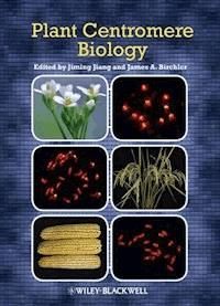
The most common feature associated with centromeres in both plants and animals is the presence of long arrays of satellite repeats (Henikoff et al., 2001; Jiang et al., 2003). This is also true for rice centromeres, which contain a 155-bp satellite repeat called CentO (originally RCS2; Dong et al., 1998; Figure 2.1). The CentO repeat arrays are frequently interrupted by CRR elements (Cheng et al., 2002). The amount of CentO repeat varies significantly among different centromeres, ranging from about 65 kb in the centromere of chromosome 8 (Cen8) to about 1,900 kb in Cen11 (Cheng et al., 2002; Figure 2.1). Association of the CentO repeats with the functional core of rice centromeres was initially supported by cytological evidence. On meiotic metaphase I chromosomes, the FISH signals derived from CentO were located at the most poleward positions of the bivalent chromosomes, suggesting that the CentO-containing chromosomal domains are associated with the kinetochore protein complex (Cheng et al., 2002). Rice telocentric chromosomes, which were derived from misdivisions of the centromeres, contain an average of 65% (ranging from 48% to 84%) of CentO compared with the normal centromeres, suggesting that the misdivisions occurred in the middle of the centromeric CentO arrays (Cheng et al., 2002).
Figure 2.1 Structure of rice Cen8. (A) FISH mapping of the CentO repeat (green) on rice pachytene chromosomes. Arrow points to chromosome 8, which contains the smallest CentO array (∼65 kb) among the 12 centromeres. (B) Digital separation of the FISH signals from (A). Arrow points to the CentO signal in Cen8 (Cheng et al., 2002. Copyright of the images owned by the American Society of Plant Biologists). (C) Pachytene chromosome 8 was digitally separated from (A) and was straightened. (D) Characterization of the ∼750-kb CENH3-binding domain of Cen8 by mapping of 454 sequence reads derived from ChIP against rice CENH3. Green bars represent relative abundance of the 454 sequence reads mapped to each location along Cen8. Each of the six CENH3-binding subdomains are shown by gray boxes. The horizontal red bars mark the locations of the CentO arrays within Cen8 (for details, see Yan et al., 2008). (E) Mapping of trimethylated H3 Lys 36 (H3K36me3), a euchromatic histone modification mark, within Cen8. Black bars represent relative enrichment of H3K36me3 across Cen8. The six CENH3-binding subdomains are shown by yellow boxes. Note, significantly H3K36me3-enriched regions are not identified in the CENH3-binding subdomains (Wu et al., 2011). (F) A diagrammed core domain of rice Cen8, consisting of interspersed blocks of CENH3 nucleosomes (red circles) and H3 nucleosomes (blue circles). (G) A model of a potential three-dimensional structure of rice Cen8. Coiling or folding of the nucleosome blocks within the centromeric core moves the CENH3 subdomains to the inner kinetochore and the centromeric H3 subdomains to an interior position (Wu et al., 2011). Copyright of the diagrams in E, F, and G owned by the American Society of Plant Biologists.
CentO was detected in the centromeres of wild Oryza species that are closely related to rice, but it was absent in several distantly related Oryza species, including O. brachyantha and O. granulata, which diverged from rice within the last 15 million years (Lee et al., 2005; Ammiraju et al., 2008). Thus, the CentO repeat evolved rapidly, which is typical of noncentromeric satellite repeat families. However, an 80-bp region within the CentO repeat shares sequence similarities with several centromeric satellite repeats reported in distantly related grass species, including maize and pearl millet (Pennisetum glaucum; Lee et al., 2005). Thus, the evolution of centromeric satellite repeats does not follow the pattern of other satellite repeats and may be constrained by centromere function.
Highly homogenous repetitive DNA sequences are difficult to clone, sequence, and assemble. Thus, long arrays of sequenced satellite repeats are rarely available even from the best sequenced plant genomes. Nevertheless, several long blocks of CentO arrays have been sequenced, including an approximately 65-kb array in Cen8 (Lee et al., 2006; Wu et al., 2009). Analysis of the CentO repeats located in rice Cen8 and Cen1 revealed local homogenization of the CentO sequences, because the repeats in the same centromeres were more similar to each other than those in different centromeres. Similar to the CRR elements, the CentO satellite repeats are also transcribed and some transcripts are processed into small RNAs (Lee et al., 2006). However, it is unclear if such transcripts and the small RNAs are associated with rice centromeric function.
Genome-wide mapping of CENH3-associated DNA sequences in rice centromeres
The functional core of a centromere is defined by the presence of a centromere-specific histone H3 variant, CENH3 (Henikoff et al., 2001). CENH3 has been found in every eukaryote analyzed and provides a molecular marker to track DNA and proteins associated with centromeres. Antibodies specific to CENH3 have been developed in a number of plant species. The association of a DNA sequence with centromeric nucleosomes can be determined by chromatin immunoprecipitation (ChIP) using the CENH3 antibodies followed by polymerase chain reaction (PCR) or gel blot hybridization (Zhong et al., 2002; Nagaki, Talbert, et al., 2003). ChIP analysis confirmed that both CRR and CentO sequences are associated with CENH3 (Nagaki et al., 2004).
Although the association of a specific DNA sequence with CENH3 can be determined by ChIP-PCR, characterization of the complete CENH3-binding domain of a centromere is a daunting task in most plant species because of the repetitive nature of centromeric DNA (Jiang et al., 2003). Rice represents a rare exception in which several centromeres contain only limited amounts of highly repetitive DNA sequences. At least four rice centromeres (Cen4, Cen5, Cen7, and Cen8) have been fully, or nearly fully, sequenced (Nagaki et al., 2004; Wu et al., 2004; Zhang et al., 2004; Matsumoto et al., 2005). These rice centromeres represent the best sequenced and characterized endogenous centromeres from any multicellular eukaryote.
Yan and colleagues (2008) conducted a 454 sequencing of DNA sample prepared from anti-CENH3 ChIP. This ChIP-seq effort produced a total of 325,298 sequence reads. Approximately 35% of the sequence reads were related to CentO repeats, confirming that CentO is the most dominant DNA component in rice centromeres (Yan et al., 2008). Mapping of the single- or low-copy 454 sequence reads on the rice reference genome revealed the boundaries of the CENH3 binding domains in 9 of the 12 rice chromosomes. The sizes of the CENH3-binding domains range from 390 kb in Cen12 to 1,210 kb in Cen3. The sizes of the CENH3-binding domains did not appear to be directly correlated with the sizes of the chromosomes. The CENH3-binding domain in each centromere was embedded in a much larger crossing over–suppressed chromosomal domain (Yan et al., 2008). For example, the CENH3-binding domain in Cen8 spans approximately 750 kb (Figure 2.1), which is embedded within a 2,312 kb of crossing over–suppressed domain (Yan et al., 2005).
The enrichment of ChIP-seq sequence reads was not uniform throughout individual rice centromeres. The CENH3-binding domain within a centromere consists of several CENH3-enriched subdomains separated by subdomains that lack CENH3 (Figure 2.1). These CENH3-lacking subdomains most likely contain H3-associated nucleosomes (Yan et al., 2008; Wu et al., 2011). A similar structure of alternating CENH3 and H3 subdomains was reported in a human neocentromere (Chueh et al., 2005).
Genes in rice centromeres
Rice Cen8 was the first fully sequenced centromere from a multicellular eukaryote (Nagaki et al., 2004). A total of 14 genes were identified within the approximately 750-kb CENH3 binding domain of Cen8


