87,99 €
Mehr erfahren.
- Herausgeber: John Wiley & Sons
- Kategorie: Wissenschaft und neue Technologien
- Sprache: Englisch
An updated guide to plant pathogens and their management
The impact of plant disease is far-reaching. Its effects are felt not only in the spheres of agriculture and horticulture, but also in human health and wellbeing. The challenges of population growth, climate change and global food security all increase the need to protect crops from disease and reduce the losses caused by plant pathogens. This requires ongoing research and novel solutions, making the detailed analysis offered by Plant Pathology and Plant Pathogens more relevant than ever.
Striking a balance between laboratory- and field-based aspects of its subject, this revised fourth edition of the text places plant disease in a wide biological context. Its contents cover causal agents and diagnosis, host–pathogen interactions, and disease management, including breeding for resistance, chemical, biological and integrated control. New to this edition are updated sections on molecular epidemiology, biosecurity, pathogenomics, and the biotechnological advances that are helping scientists make great strides in the fight against plant disease.
- Authored by a leading authority on plant pathology
- Offers new coverage of recent advances in molecular genetics and genomics, biotechnology, and plant breeding
- Places emphasis on interaction biology and biological concepts, such as immunity and comparisons with animal systems
- Includes access to a supplementary website featuring slides of all figures in the book
Plant Pathology and Plant Pathogens is an ideal textbook for graduate and upper-level undergraduate students in biology, botany, agricultural sciences, applied microbiology, plant-microbe interactions, and related subjects. It will also be a practical and enlightening resource for professionals in agricultural institutions, along with crop consultants seeking additional training or information.
Sie lesen das E-Book in den Legimi-Apps auf:
Seitenzahl: 792
Veröffentlichungsjahr: 2020
Ähnliche


Table of Contents
Cover
Preface
List of Abbreviations
About the Companion Website
Part I: Plant Disease
1 The Diseased Plant
Concepts of Disease
Symptoms of Disease
Causes of Disease
Significance of Disease
Further Reading
2 The Microbial Pathogens
Pathogens and Pathogenesis
Koch's Postulates
Host Resistance and Pathogen Virulence
Further Reading
3 Pathogen Biology
Fungi as Plant Pathogens
Oomycetes as Plant Pathogens
Bacteria as Plant Pathogens
Mycoplasma‐Like Organisms as Plant Pathogens
Protozoa as Plant Pathogens
Viruses as Plant Pathogens
Viroids as Plant Pathogens
Variation in Microbial Pathogen Populations
Dispersal of Pathogens
Pathogen Survival
Further Reading
4 Disease Assessment and Forecasting
Disease Detection
Disease Assessment in the Crop
Disease Forecasting
Further Reading
5 Plant Disease Epidemics
Epidemic Development
Global Pandemics
Relevance of Epidemiology to Control
Plant Biosecurity
Further Reading
Part II: Host–Pathogen Interactions
6 Entry and Colonization of the Host
The Infection Court
Direct Penetration
Penetration Through Natural Openings
Penetration Through Wounds
The Host–Pathogen Interface
Structure and Function of Haustoria
Development Following Infection
Further Reading
7 The Physiology of Plant Disease
Postinfectional Changes in Host Physiology
Conclusion
Further Reading
8 Microbial Pathogenicity
A Molecular Genetic Approach
Enzymes and Microbial Pathogenicity
Toxins
High Molecular Weight Compounds and Pathogenesis
Growth Disorders Caused by Bacterial Pathogens
Conclusion
Further Reading
9 Plant Defense
Types of Plant Defense
Preformed Inhibitors
Antimicrobial Proteins
Antiviral Proteins
Active Defense Mechanisms
Changes in Host Cell Walls
The Hypersensitive Response
The Oxidative Burst
Phytoalexins
Phytoalexins and Plant Defense
Biosynthesis of Defense Metabolites
Pathogenesis‐Related Proteins
Other Systemic Defense Responses
Defense Priming and the Immune Memory of Plants
Conclusion
Further Reading
10 Host–Pathogen Specificity
Types of Specificity
Microbial and Plant Elicitors of Defense
A Unifying Concept: Pathogen Effectors and Plant Immunity
Genes for Pathogen Recognition and Resistance
Conclusion
Further Reading
Part III: Disease Management
11 Disease Management by Chemicals
The Evolution of Fungicides
Mode of Action of Fungicides
Control by Gene Silencing
Fungicide Resistance
The Future of Fungicides
Further Reading
12 Disease Management by Host Resistance
Breeding for Disease Resistance
Resistance in the Field
Biotechnology and Breeding for Disease Resistance
Future Prospects for Crop Resistance to Pathogens
Conclusion
Further Reading
13 Biological Control of Plant Disease
Cultural Practices and Disease Control
Biological Control
Further Reading
14 An Integrated Approach to Disease Management
Integrated Control of Plant Disease
Integration of Fungicides with Host Resistance
Prolonging the Effectiveness of Host Resistance and Fungicides
Integration of Cultural and Biological Measures
Making Decisions
The Impact of New Technology – Digital Agriculture
The Impact of Biotechnology and Genomics
Designing Future Farms
The Technology Gap
Further Reading
Appendix 1: Annotated List of Pathogens and the Diseases they Cause
Appendix 2: Reference Sources for Figures
Index
End User License Agreement
List of Tables
Chapter 1
Table 1.1 Symptoms of disease in plants
Table 1.2 Angiosperms parasitic on other higher plants
Table 1.3 Natural and agricultural plant communities compared
Table 1.4 Some impacts of plant disease
Table 1.5 Some examples of the impacts of specific plant diseases
Table 1.6 Estimated losses (%) due to weeds, pests, and diseases in six major...
Table 1.7 Estimates of postharvest losses likely to occur in the absence of e...
Chapter 2
Table 2.1 Main characteristics of necrotrophic and biotrophic pathogens.
Table 2.2 Some parameters used to measure aggressiveness
Table 2.3 Hypothetical interaction between four host cultivars and four patho...
Chapter 3
Table 3.1 Higher plants and animals compared as hosts
Table 3.2 Some important oomycete plant pathogens
Table 3.3 Main genera of plant‐pathogenic bacteria and some example species a...
Table 3.4 Virus groups based on genome type (
International Committee on Taxon
...
Table 3.5 Mechanisms maintaining variation in pathogen populations. The numbe...
Table 3.6 Virus–vector relationships, based on insects in the order Hemiptera
Table 3.7 Survival of sclerotia of
Sclerotium cepivorum
buried at different de...
Chapter 4
Table 4.1 Properties of an ideal diagnostic test
Table 4.2 Molecular and biochemical diagnostic methods for pathogen detection...
Table 4.3 Effect of take‐all on yield components of winter wheat
Table 4.4 Factors important in the development of meteorological forecasting ...
Chapter 5
Table 5.1 Some features of monocyclic and polycyclic epidemics
Chapter 6
Table 6.1 Some modes of growth of pathogens in host plants
Chapter 8
Table 8.1 Some extracellular enzymes produced by plant pathogens
Table 8.2 Some toxins involved in plant disease
Table 8.3 Some pathogens which cause abnormal growth in the host plant
Chapter 9
Table 9.1 Some plant proteins that confer resistance to pests and pathogens
Table 9.2 Some cell wall changes occurring in response to infection
Table 9.3 Some features of phytoalexins
Table 9.4 Some families of
pathogenesis‐related
(
PR
) proteins
Table 9.5 Some characteristics of systemic acquired resistance (SAR)
Chapter 10
Table 10.1 Some examples of host–pathogen specificity
Table 10.2 Some elicitors of plant immune responses
Table 10.3 Some examples of pathogen effectors and their interactions with pl...
Table 10.4 Some examples of microbial polymers recognized by plants, and thei...
Table 10.5 Some cloned plant disease resistance genes
Chapter 11
Table 11.1 Examples of protectant, multisite fungicides
Table 11.2 Some examples of systemic, single‐site fungicides
Table 11.3 Specifications of the perfect fungicide
Table 11.4 Multisite and single‐site fungicides compared
Table 11.5 Some examples of plant defense activators
Table 11.6 Some advantages and disadvantages of plant defense activators
Table 11.7 Timeline of fungicide resistance
Table 11.8 Resistance management strategies
Chapter 12
Table 12.1 Sources of disease resistance
Table 12.2 Yellow rust (
Puccinia striiformis
f.sp.
tritici
) race changes in t...
Table 12.3 Strategies to counter the boom and bust cycle
Table 12.4 Race‐specific and race‐nonspecific resistance compared
Table 12.5 Some examples of genetically engineered plants with enhanced resis...
Chapter 13
Table 13.1 Approaches used for biological control of plant pathogens
Table 13.2 Some examples of commercial biocontrol products for plant diseases
Chapter 14
Table 14.1 Some applications of genomics and gene editing in disease manageme...
Part 1
Table 1 Benefits of healthy plants
Part 2
Table 1 Stages in host–pathogen interaction
Part 3
Table 1 Options for control of plant disease
List of Illustrations
Chapter 1
Figure 1.1 A plant life cycle and some effects of disease.
Figure 1.2 Some disease symptoms caused by pathogens infecting different pla...
Figure 1.3 Club root disease of brassicas. (a) Primary infection causes dist...
Figure 1.4 Rust of willows caused by
Melampsora
species. (a) Aerial view of ...
Figure 1.5 Agents responsible for plant disease, disorders, and damage. High...
Figure 1.6 Poplar tree infested by European mistletoe
Viscum album
.
Figure 1.7 Effect of infection by the rust fungus
Puccinia lagenophorae
on s...
Figure 1.8 Relationship between yield levels and crop loss, indicating econo...
Figure 1.9 Comparison of proportions of total production of major food and c...
Figure 1.10 A convergence of forces increasing the threat of plant disease....
Chapter 2
Figure 2.1 Symbiotic relationships: + positive effects on partner;
−
n...
Figure 2.2 Faba bean leaves infected by
Botrytis fabae
(
left
) showing necrot...
Figure 2.3 Apple scab disease caused by
Venturia inaequalis
. (
Left
) Scab les...
Figure 2.4 Nutritional modes in heterotrophic microorganisms.
Figure 2.5 Use of Koch’s postulates to establish the etiology of a new disea...
Figure 2.6 Relationships between host, pathogen, and disease reaction.
Figure 2.7 The quadratic check, showing interactions between alleles of a ho...
Chapter 3
Figure 3.1 (a)
Intercellular hypha
(
IH
) of the oomycete
Peronospora viciae
i...
Figure 3.2 Mycelial strands formed by a basidiomycete fungus. The fungus has...
Figure 3.3 Specialized parasitic structures, known as haustoria, formed by t...
Figure 3.4 Bacterial cell structure. (a) Electron micrograph of the bacteriu...
Figure 3.5 Structure of
tobacco mosaic virus
(
TMV
). (a) Electron micrograph ...
Figure 3.6 Viroid structures, showing (a) rod‐like secondary structure with ...
Figure 3.7 The processes involved in pathogen dispersal and the scale on whi...
Figure 3.8 Some routes by which pathogens are dispersed.
Figure 3.9 Active discharge of ascospores of
Sclerotinia sclerotiorum
from s...
Figure 3.10 Dispersal of spores or bacterial cells by rain drops from wet an...
Figure 3.11 Vectors exploited by plant pathogens.
Figure 3.12 The disease tetrahedron.
Figure 3.13 Different routes of plant viruses in their aphid vectors. The gu...
Figure 3.14 Survival strategies adopted by plant pathogens.
Figure 3.15 Electron micrograph sections of (a) a multicellular conidium of
Figure 3.16 Survival and germination of fungal sclerotia under natural condi...
Figure 3.17 Daily changes in populations of
Pseudomonas syringae
on bean lea...
Figure 3.18 Survival periods recorded for some pathogens in the field.
Chapter 4
Figure 4.1 Activities involved in disease assessment and prediction.
Figure 4.2 Serology‐based pathogen detection. (a) A sandwich ELISA assay. 1....
Figure 4.3 Coconut
lethal yellowing disease
(
LYD
) caused by a phytoplasma. (...
Figure 4.4
Next‐generation sequencing
(
NGS
) workflow based on RNA‐seq ...
Figure 4.5 Septoria tritici leaf blotch in England and Wales. (a) Risk map b...
Figure 4.6 Distribution of Asian soybean rust (
Phakopsora pachyrhizi
) in the...
Figure 4.7 Assessment of Septoria tritici leaf blotch of wheat (
Zymoseptoria
...
Figure 4.8 Sensor technologies used for automated detection and evaluation o...
Figure 4.9 Aerial photo of a winter wheat crop in the UK in June, showing di...
Figure 4.10 Imaging plant health. (a) Octocopter drone carrying sensors. (b)...
Figure 4.11 Some examples of equipment used to monitor the risk of disease. ...
Figure 4.12 Numbers of three aphid species (
Metopolophium dirhodum
,
Rhopalos
...
Figure 4.13 Contribution of the upper leaves and ear to final yield of wheat...
Figure 4.14 Effects of brown rust (
Puccinia triticina
) of cereals. (a) Wheat...
Figure 4.15 Relationship between late blight progress and yield loss of the ...
Figure 4.16 Fungal pathogens that can contaminate cereal grain. (a) Ergot on...
Figure 4.17 Incidence of black mold (
Pilgeriella anacardii
) in dwarf cashew ...
Chapter 5
Figure 5.1 Records of occurrence of southern corn leaf blight (
Bipolaris may
...
Figure 5.2 Epidemic growth curves for monocyclic (a,b) and polycylic (c,d) p...
Figure 5.3 Disease increase with time plotted on a logarithmic scale.
Figure 5.4 Disease models and their uses.
Figure 5.5 Diagrammatic representation of the HLIR disease model. Boxes show...
Figure 5.6 Example output from an HLIR model showing proportions of a host p...
Figure 5.7 Disease gradient for bean rust,
Uromyces phaseoli
, dispersing fro...
Figure 5.8 Annual incidence of
Phytophthora infestans
genotypes in UK popula...
Figure 5.9 Hedgerow elm infected by Dutch elm disease showing chlorosis (yel...
Figure 5.10 Global spread of yellow rust of wheat,
Puccinia striifomis
f.sp....
Figure 5.11 Genomic analysis of field isolates of wheat yellow rust,
Puccini
...
Figure 5.12 Progress of a potato late blight epidemic and the effects of two...
Figure 5.13 Timeline of first occurrence of invasive forest pathogens in the...
Figure 5.14 The five Ps of biosecurity, including national activities and in...
Figure 5.15 Border biosecurity. (
Top
) Sign at Guarulhos International Airpor...
Figure 5.16 Wart disease of potatoes, caused by
Synchytrium endobioticum
. In...
Chapter 6
Figure 6.1 Some entry routes for plant pathogens. PSTVd potato spindle tuber...
Figure 6.2 The external layers bounding herbaceous plant organs.
Figure 6.3 Early infection stages of the rice blast fungus
Magnaporthe oryza
...
Figure 6.4 Early development of fungal pathogens on host leaves viewed by sc...
Figure 6.5 Model for induction of cutinase synthesis in spores of
Fusarium s
...
Figure 6.6 Infection structures of stem‐ and root‐infecting fungi. (a)
Scann
...
Figure 6.7 Response of
Arabidopsis
stomata to exposure to (a) a plant‐pathog...
Figure 6.8 Pathogens that exploit wounds to infect the host. (a) Pink rot of...
Figure 6.9 Intracellular structures formed by biotrophic fungi and oomycetes...
Figure 6.10 The structure of haustoria. (a) Haustorium of flax rust
Melampso
...
Figure 6.11 Proton symport model for nutrient transport across the haustoriu...
Figure 6.12 Some patterns of pathogen invasion of plant tissues, based on cr...
Figure 6.13 Invasion of vascular tissues of kiwifruit by
Pseudomonas syringa
...
Chapter 7
Figure 7.1 Time course of respiration in two susceptible barley cultivars, W...
Figure 7.2 Theories concerning stimulation of respiration in infected plants...
Figure 7.3 Changes in photosynthesis of oak leaves following infection by th...
Figure 7.4 Effect of
Erysiphe polygoni
on the rate of photosynthetic
14
CO
2
a...
Figure 7.5 Chlorophyll fluorescence images of barley leaf tissues infected w...
Figure 7.6 Green island surrounding a lesion caused by the light leaf spot p...
Figure 7.7 Translocation of
14
C in healthy bean plants and plants infected b...
Figure 7.8 Kinetics of efflux of
14
C from healthy and rusted first leaves of...
Figure 7.9 Cell wall invertase activity in
Arabidopsis
leaves following inoc...
Figure 7.10 Vascular wilt syndrome. (a) Tomato plant infected by
Verticilliu
...
Figure 7.11 Transpiration rates over 24 hours of healthy barley plants and b...
Figure 7.12 Time course of symptom development on wheat leaves infected by
Z
...
Figure 7.13 (a) Maize cob infected by smut,
Ustilago maydis
. Infection induc...
Chapter 8
Figure 8.1 Experimental approaches to identifying pathogenicity genes. The e...
Figure 8.2 A heat map showing stage‐specific expression of 104 genes encodin...
Figure 8.3 Soft‐rot symptoms caused by
Pectobacterium carotovora
on potato (
Figure 8.4 Diagrammatic version of the organization of pectate lyase (
pel
) g...
Figure 8.5 Intramural growth of hypha of the cereal eyespot fungus
Oculimacu
...
Figure 8.6 Comparison of numbers of genes encoding cellulose‐ and hemicellul...
Figure 8.7 Original demonstration of effects of victorin on resistant (R) an...
Figure 8.8 Some host‐specific toxins involved in plant disease, showing dive...
Figure 8.9 Evidence for lateral transfer of a fungal virulence gene. An 11 k...
Figure 8.10 Structures of some nonhost‐specific toxins involved in plant dis...
Figure 8.11 Multiplication of the wildfire disease bacterium,
Pseudomonas sy
...
Figure 8.12 Colonization and biofilm formation by the fireblight pathogen
Er
...
Figure 8.13 Relationship between extracellular polysaccharide (EPS) producti...
Figure 8.14 Crown gall
Agrobacterium tumefaciens
and hairy root disease
A. r
...
Figure 8.15 Molecular processes underlying tumor induction in crown gall dis...
Chapter 9
Figure 9.1 Types of plant defense, based on existing anatomical or structura...
Figure 9.2 Some preformed antimicrobial compounds from plant tissues.
Figure 9.3 Relationship between production of the saponin‐detoxifying enzyme...
Figure 9.4 Examples of nontoxic glycosides converted to inhibitory breakdown...
Figure 9.5 (a) Inhibition of fungal growth by germinating radish seed and (b...
Figure 9.6 Cell wall reactions to penetration by fungal pathogens. (a) Stain...
Figure 9.7 Time course of accumulation of mRNA for hydroxyproline‐rich glyco...
Figure 9.8 The hypersensitive response. (a) Local lesions formed on a tobacc...
Figure 9.9 Diagrammatic overview of a plant cell undergoing the hypersensiti...
Figure 9.10 Origin and structure of some phytoalexins.
Figure 9.11 Bioassay of phytoalexins produced by broad bean (
Vicia faba
) lea...
Figure 9.12 Accumulation of phaseollin (μg per inoculated site) in beans ino...
Figure 9.13 Pathway for biosynthesis of phenylpropanoid defense metabolites ...
Figure 9.14 Timing of induction of phenylalanine ammonia lyase (PAL) mRNA an...
Figure 9.15 Induction of transcription of several defense‐related enzymes in...
Figure 9.16 Electrophoretic separation of proteins extracted from healthy to...
Figure 9.17 Plant and animal immunity compared.
Figure 9.18 (a) Local and (b) systemic acquired resistance in plants.
Figure 9.19 Basic model of systemic activation of plant defense. Inoculation...
Figure 9.20 Some signal molecules involved in systemic resistance in plants....
Figure 9.21 Induced resistance pathways in plants. Signal molecules implicat...
Figure 9.22 Effect of seed treatment with the defense activator
acibenzolar‐
...
Chapter 10
Figure 10.1 Genetic models of host–pathogen specificity. (a) Gene‐for‐gene s...
Figure 10.2 Cloning bacterial
avr
genes by function. Race 2 is incompatible ...
Figure 10.3 Procedure used to isolate a specific elicitor of host resistance...
Figure 10.4 (a) A cluster of
hrp
genes (
A–F
) from
Xanthomonas axonopod
...
Figure 10.5 Diagrammatic representation of the hypersensitive response and p...
Figure 10.6 A zig‐zag model of plant immunity. In the first phase, the prese...
Figure 10.7 Diagrammatic representation of suppression of PAMP‐triggered imm...
Figure 10.8 Delivery systems for cytoplasmic effectors. (a) Bacterial
type I
...
Figure 10.9 Oomycete effector delivery. (a) 3D projection of a confocal micr...
Figure 10.10 The classic gene‐for‐gene relationship (
left
), defined by the i...
Figure 10.11 Simplified scheme of processes in PAMP‐triggered immunity. A co...
Figure 10.12 Structure and cellular location of some proteins encoded by pla...
Figure 10.13 Structure and activation of NLR immune receptors. (a) NLRs are ...
Figure 10.14 Overview of the main steps in a plant virus life cycle. The vir...
Figure 10.15 Resistance to a virus due to lack of a compatible host suscepti...
Chapter 11
Figure 11.1 The evolution of fungicides. Main types available, origins, and ...
Figure 11.2 Activities involved in the discovery and development of new agro...
Figure 11.3 Effect of distribution on the efficiency of control of powdery m...
Figure 11.4 Proposed mechanism of
spray‐induced gene silencing
(
SIGS
)....
Figure 11.5 Some ways of applying pesticides to crops. (a) Tractor‐mounted b...
Figure 11.6 Fungicide treatment timings on winter wheat in the UK. GS + numb...
Figure 11.7 The dynamics of resistance development to fungicides. Discrete v...
Figure 11.8 Mechanisms of resistance to single‐site fungicides. 1. Alteratio...
Figure 11.9 Components of resistance management.
Figure 11.10 A risk matrix for estimating the likelihood of fungicide resist...
Figure 11.11 Courgette infected with gray mold
Botrytis cinerea
. This versat...
Figure 11.12 Quantification of fungicide resistance in ascospores of
Mycosph
...
Chapter 12
Figure 12.1 Selection for disease resistance in a population of plants. Nega...
Figure 12.2 Marker‐assisted selection for resistance to a soil‐borne mosaic ...
Figure 12.3 Flowchart for genomics‐assisted plant breeding.
Figure 12.4 The boom and bust cycle of crop resistance. Note that a similar ...
Figure 12.5 Annual changes in virulence frequencies (a) in yellow rust (
Pucc
...
Figure 12.6 Evolution of virulence in the yellow rust (
Puccinia graminis
) po...
Figure 12.7 Vertical and horizontal resistance compared. A cultivar with a s...
Figure 12.8 Options for the use and deployment of
R
genes in crops. Major ge...
Figure 12.9 Amounts of foliar infection by three pathogens of barley on thre...
Figure 12.10 Rate of disease development of frog‐eye of tobacco, a leaf spot...
Figure 12.11 Control of potato late blight (
Phytophthora infestans
) in field...
Figure 12.12 Aerial view of field trial of transgenic papaya resistant to pa...
Chapter 13
Figure 13.1 Effect of sowing date on the incidence of
barley yellow dwarf vi
...
Figure 13.2 Effect of rotation on the yield of two pea cultivars.
Figure 13.3 Relationship between inoculum density of
Thielaviopsis basicola
...
Figure 13.4 Effects of soil solarization and fumigation on (a) numbers of pr...
Figure 13.5 Effect of soil solarization on the disease progress curves of ga...
Figure 13.6 Severity of take‐all disease in continuous winter wheat crops at...
Figure 13.7 Some compounds produced by bacterial antagonists which play a ro...
Figure 13.8 Damage caused to conifer timber due to infection by butt rot,
He
...
Figure 13.9 Protection of pea seedlings against preemergence damping‐off cau...
Figure 13.10 Control of
Rhizoctonia
rot of tomatoes in the field by sprays o...
Figure 13.11 Modes of action of biocontrol agents.
Figure 13.12 An example of mycoparasitism. Infection of the grapevine downy ...
Figure 13.13 Severity of angular leaf spot, caused by the bacterium
Pseudomo
...
Chapter 14
Figure 14.1 Principles of integrated pest management, with sequential action...
Figure 14.2 Extending the durability of cultivar resistance with fungicides....
Figure 14.3 Avocado grove in southern California affected by
Phytophthora
ro...
Figure 14.4 Strategies for integrated control of
Phytophthora
root rot of av...
Figure 14.5 Effect of irrigation on severity of root rot of squash (
Cucurbit
...
Figure 14.6 Digital agriculture and potential applications in crop protectio...
Part 1
Figure 1 The disease triangle (a) incorporating the host–pathogen complex (b...
Part 2
Figure 1 Simplified model of host–pathogen interactions involving fungi, oom...
Part 3
Figure 1 The disease triangle showing options for intervention targeting the...
Figure 2 Farming systems compared.
Figure 3 Crop income versus control costs.
Guide
Cover
Table of Contents
Begin Reading
Pages
iii
iv
vii
viii
ix
xi
ix
1
2
3
4
5
7
8
9
10
11
12
13
14
15
16
17
18
19
20
21
22
23
24
25
26
27
28
29
31
32
33
34
35
36
37
38
39
40
41
42
43
44
45
46
47
48
49
50
51
52
53
54
55
56
57
58
59
60
61
62
63
64
65
66
67
68
69
70
71
72
73
74
75
76
77
78
79
80
81
83
84
85
86
87
88
89
90
91
92
93
94
95
96
97
98
99
100
101
102
103
104
105
106
107
108
109
110
111
112
113
114
115
116
117
118
119
120
121
122
123
124
125
126
127
128
129
130
131
132
133
134
135
137
138
139
140
141
142
143
144
145
146
147
148
149
150
151
152
153
154
155
156
157
158
159
160
161
162
163
164
165
166
167
168
169
170
171
172
173
174
175
176
177
178
179
180
181
182
183
184
185
186
187
188
189
190
191
192
193
194
195
196
197
198
199
200
201
202
203
204
205
206
207
208
209
210
211
212
213
214
215
216
217
218
219
220
221
222
223
224
225
226
227
228
229
230
231
232
233
234
235
236
237
238
239
240
241
242
243
244
245
246
247
248
249
250
251
252
253
254
255
256
257
258
259
260
261
262
263
264
265
266
267
268
269
270
271
272
273
274
275
276
277
279
280
281
282
283
284
285
286
287
288
289
290
291
292
293
294
295
296
297
298
299
300
301
302
303
304
305
306
307
308
309
310
311
312
313
314
315
316
317
318
319
320
321
322
323
324
325
326
327
328
329
330
331
332
333
334
335
336
337
338
339
340
341
342
343
344
345
346
347
348
349
350
351
352
353
354
355
356
357
359
360
361
362
363
364
365
366
367
368
369
370
371
372
373
375
376
377
378
379
380
381
382
383
384
385
387
388
389
390
391
392
393
394
395
396
397
398
399
400
401
402
403
404
405
406
407
408
409
410
411
412
413
414
415
416
417
Plant Pathology and Plant Pathogens
Fourth Edition
John A. Lucas
Rothamsted ResearchHarpendenHertfordshire, UK
This fourth edition first published 2020© 2020 John Wiley & Sons Ltd
First Edition 1977 by Blackwell Science LtdSecond Edition 1982Third Edition 1998Reprinted 2002
All rights reserved. No part of this publication may be reproduced, stored in a retrieval system, or transmitted, in any form or by any means, electronic, mechanical, photocopying, recording or otherwise, except as permitted by law. Advice on how to obtain permission to reuse material from this title is available at http://www.wiley.com/go/permissions.
The right of John A. Lucas to be identified as the author of this work has been asserted in accordance with law.
Registered Office(s)John Wiley & Sons, Inc., 111 River Street, Hoboken, NJ 07030, USAJohn Wiley & Sons Ltd, The Atrium, Southern Gate, Chichester, West Sussex, PO19 8SQ, UK
Editorial OfficeThe Atrium, Southern Gate, Chichester, West Sussex, PO19 8SQ, UK
For details of our global editorial offices, customer services, and more information about Wiley products visit us at www.wiley.com.
Wiley also publishes its books in a variety of electronic formats and by print‐on‐demand. Some content that appears in standard print versions of this book may not be available in other formats.
Limit of Liability/Disclaimer of WarrantyWhile the publisher and authors have used their best efforts in preparing this work, they make no representations or warranties with respect to the accuracy or completeness of the contents of this work and specifically disclaim all warranties, including without limitation any implied warranties of merchantability or fitness for a particular purpose. No warranty may be created or extended by sales representatives, written sales materials or promotional statements for this work. The fact that an organization, website, or product is referred to in this work as a citation and/or potential source of further information does not mean that the publisher and authors endorse the information or services the organization, website, or product may provide or recommendations it may make. This work is sold with the understanding that the publisher is not engaged in rendering professional services. The advice and strategies contained herein may not be suitable for your situation. You should consult with a specialist where appropriate. Further, readers should be aware that websites listed in this work may have changed or disappeared between when this work was written and when it is read. Neither the publisher nor authors shall be liable for any loss of profit or any other commercial damages, including but not limited to special, incidental, consequential, or other damages.
Library of Congress Cataloging‐in‐Publication Data
Names: Lucas, J. A. (John Alexander), 1948– author.Title: Plant pathology and plant pathogens / prof. John A. Lucas, Rothamsted Research, West Common, Harpenden, Hertfordshire, UK.Description: Fourth edition. | Hoboken : Wiley‐Blackwell, 2020. | Includes bibliographical references and index.Identifiers: LCCN 2019051991 (print) | LCCN 2019051992 (ebook) | ISBN 9781118893869 (paperback) | ISBN 9781118893821 (adobe pdf) | ISBN 9781118893852 (epub)Subjects: LCSH: Plant diseases. | Phytopathogenic microorganisms–Control. | Plant‐pathogen relationships.Classification: LCC SB731 .D5 2020 (print) | LCC SB731 (ebook) | DDC 632–dc23LC record available at https://lccn.loc.gov/2019051991LC ebook record available at https://lccn.loc.gov/2019051992
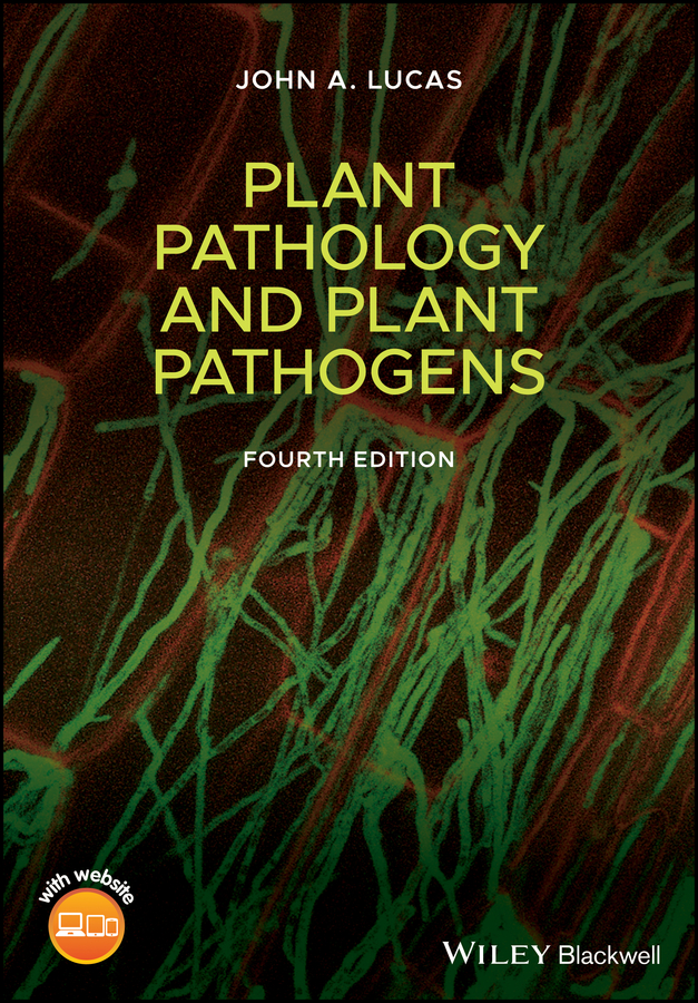
Cover Design: WileyCover Image: Confocal microscopy image of a GFP‐labelled strain of the cereal eyespot fungus Oculimacula colonizing a wheat coleoptile. Source: Bowyer et al. 2000.
Preface
Plant pathology is an applied science concerned primarily with practical solutions to disease problems in agriculture, horticulture, and forestry. The study of plant disease and the development of methods for its control continue to be vital elements in the drive to improve crop productivity and ensure global food security. Projected world population growth and the impacts of climate change make this a priority. From a more strategic perspective, the experimental analysis of the interactions between plants and pathogens has become fertile territory for scientists interested in emerging areas of plant biology, including recognition and response systems, signal pathways, and innate plant immunity. The use of molecular genetic techniques has provided new insights into how pathogens cause disease and how plants defend themselves against attack. In turn, this new understanding is suggesting novel ways in which plant disease might be controlled.
This book provides an introduction to the main elements of plant pathology, including the applied aspects of disease identification, assessment and control, and fundamental studies of host–pathogen interactions. The aim is to place the subject not only in the context of agricultural science but also in the wider spheres of ecology, population biology, cell biology, and genetics. The impact of molecular biology and genomics on the diagnosis and analysis of plant disease is a recurrent theme.
A large number of people have helped me in the production of this new edition. First, I should acknowledge the invaluable help of many of my colleagues at Rothamsted Research who have generously provided advice, assistance, and materials for use in the book. They include Kim Hammond‐Kosack, Jason Rudd, Kostya Kanyuka, Nicola Hawkins, Ana Machado, Bart Fraaije, Jon West, Stephanie Heard, Anastasia Sokolidi, Kevin King, John Jenkyn, Roger Plumb, James Bell, David Hughes, Mike Adams, Graham Shephard, and Sally Murdoch. Joe Helps advised on mathematical modeling as well as replotting data. The library staff, Tim Wales, Catherine Fernhead, and Chris Whitfield, helped with tracing publications and scanning figures. Special thanks are due to Lin Field and Sheila Bishop who supported me as a visiting worker in the Biointeractions and Crop Protection department. Numerous others in the plant pathology and crop protection community have also supplied images, articles, advice, and data for use in figures and tables, including Bruce McDonald, Mike Coffey, George Sundin, Pamela Gan, Richard O'Connell, Petra Boetink, Yaima Arocha‐Rosete, Geert Haesaert, David Cooke, Paul Birch, Diane Saunders, Judith Turner, Moray Taylor, Sarah Holdgate, Nicola Spence, Ana Lopez, Hans Cools, Mike Davey, Jurriaan Ton, Stephen Rolfe, Paul Bowyer, Clive Brasier, Joan Webber, John Mansfield, Lili Huang, Victoria Routledge, Lucy Carson‐Taylor, Jason Pollock, Dierdre Haines, David Whattoff, Graham Matthews, Haydn Beddows, Dennis Gonsalves, and Malcolm Briggs. Thanks go to Holly Regan‐Jones for her skilful copy editing and Dave Gardner who re‐drew many of the Figures. Vimali Joseph provided vital support during the final production process. My appreciation should also be extended to previous colleagues at the University of Nottingham, especially Alison Daniels and the late Brian Case, who took many of the studio photos featured in this edition. Space does not allow me to acknowledge the many other colleagues who agreed to the use of published results.
Finally, thanks are also due to my daughter Maria Gabriella, whose grasp of digital technology greatly exceeds my own, and my wife Adélia, not only for her forbearance throughout the time taken to complete this project but also for her active contribution searching out all manner of useful information on the internet.
John A. LucasJanuary 2020
List of Abbreviations
ABA
Abscisic acid
AUDPC
Area under disease progress curve
BCA
Biological control agent
CDA
Controlled droplet application
CDPK
Calcium‐dependent protein kinase
CK
Cytokinin
CWDE
Cell wall‐degrading enzyme
DAMP
Damage‐associated molecular pattern
DSS
Decision support system
EHM
Extrahaustorial membrane
ELISA
Enzyme‐linked immunosorbent assay
ETI
Effector‐triggered immunity
GA
Gibberellic acid
GLA
Green leaf area
GPS
Global positioning system
GWAS
Genome‐wide association studies
HGT
Horizontal gene transfer
HIGS
Host‐induced gene silencing
HLB
Huanglongbing disease of citrus, also known as citrus greening
HR
Hypersensitive response
HRGP
Hydroxyproline‐rich glycoprotein
Hrp
Hypersensitive response and pathogenicity
HST
Host‐specific toxin
IAA
Indole acetic acid
IPPC
International Plant Protection Convention
ISR
Induced systemic resistance
JA
Jasmonic acid
LAMP
Loop‐mediated isothermal amplification assay
LGT
Lateral gene transfer
LPS
Lipopolysaccharide
LYD
Lethal yellowing disease of coconuts
MAMP
Microbe‐associated molecular pattern
MAPK
Mitogen‐activated protein kinase
NDVI
Normalized difference vegetation index
NGS
Next‐generation sequencing, also known as high‐throughput sequencing
NLR
Family of receptor proteins characterized by nucleotide‐binding site and leucine‐rich repeat domains
NO
Nitric oxide
NRPS
Nonribosomal peptide synthase
PAL
Phenylalanine ammonia lyase
PAMP
Pathogen‐associated molecular pattern
PCD
Programmed cell death
PCR
Polymerase chain reaction
PG
Polygalacturonase
PGIP
Polygalacturonase‐inhibiting protein
PKS
Polyketide synthase
PRA
Pest risk analysis
PRR
Pattern recognition receptor
PTI
PAMP‐triggered immunity
QTL
Quantitative trait locus
REMI
Restriction enzyme‐mediated integration
RIP
Ribosome‐inactivating protein
RLK
Receptor‐like kinase
RLP
Receptor‐like protein
ROS
Reactive oxygen species
RT‐PCR
Real‐time polymerase chain reaction
RUE
Radiation use efficiency
SA
Salicylic acid
SAR
Systemic acquired resistance
SIGS
Spray‐induced gene silencing
SNP
Single nucleotide polymorphism
T3SS
Type 3 secretion system
UAV
Unmanned aerial vehicle
WRKY
(pronounced
worky
) Family of transcription factors containing a WRKY amino acid domain
About the Companion Website
This book is accompanied by a companion website:
The URL is www.wiley.com/go/Lucas_PlantPathology4
The website includes:
Figures from the book
Part IPlant Disease
We see our cattle fall and our plants wither without being able to render them assistance, lacking as we do understanding of their condition.
(J.C. Fabricius, 1745–1808)
The health of green plants is of vital importance to everyone, although few people may realize it. As the primary producers in the ecosystem, green plants provide the energy and carbon skeletons upon which almost all other organisms depend. The growth and productivity of plants determine the food supply of animal populations, including the human population. Factors affecting plant productivity, including disease, therefore affect the quantity, quality, and availability of staple foods throughout the world. Nowadays crop failure, due to adverse climate, pests, weeds, or diseases, is rare in developed agriculture, and instead there are surpluses of some foods. Nevertheless, disease still takes a toll, and much time, effort, and money are spent on protecting crops from harmful agents. In developing countries, the consequences of plant disease may be more serious, and crop failure can damage local or national economies, and lead directly to famine and hardship. Improvements in the diagnosis and management of plant disease are a priority in such instances. Furthermore, the pressures on plant productivity are increasing. The area of cultivated land available per person on the planet declined from around 0.4 ha in the 1960s to less than 0.3 by the year 2000 (FAO data), and as the human population continues to multiply the area will further decrease.
As well as supplying staple foods, plants provide many other vital commodities such as timber, fibers, oils, spices, and drugs. The use of plants as alternative renewable sources of energy and chemical feedstocks is becoming more and more important, as other resources such as fossil fuels are depleted and the need to mitigate climate change becomes a priority. Finally, the quality of the natural environment, from wilderness areas to urban parks, sports fields, and gardens, also depends to a large extent on the health of plants.
Healthy plants provide a series of benefits for the farmer, food chain, and the environment (Table 1). The yield and quality of crop products are ensured, and healthy plants are more efficient at using precious resources such as water and nutrients. In doing so, they also prevent losses of nitrogen and other nutrients to the wider environment, and reduce pollution of rivers and ground water, which in many areas is a potential problem for drinking supplies. Vigorous, healthy plants are more competitive with weeds, and are easier to harvest than crops that are stunted or collapsed. Plant root systems play an important role in reducing soil erosion, and thereby help to conserve another precious resource. Finally, given the mounting concerns about climate change, it should be noted that healthy crops have a lower carbon footprint than diseased crops, due to their greater productivity and more efficient use of inputs per area of cultivated land.
Table 1 Benefits of healthy plants
Greater yield
Superior quality
More competitive with weeds
Easier harvesting
Fewer residual nutrients, reduced pollution
Improved control of soil erosion
Lower carbon footprint
The science of plant pathology is the study of all aspects of disease in plants, including causal agents, their diagnosis, physiological effects, population dynamics and control. It is a science of synthesis, using data and techniques from fields as diverse as agriculture, microbiology, meteorology, engineering, genetics, genomics, and biochemistry. But first and foremost, plant pathology is an applied science, concerned with practical solutions to the problem of plant disease. Part of the appeal of the subject is to be found in this mixture of pure and applied aspects of biology.
The scope of plant pathology is difficult to define. On a practical level, any shortcoming in the performance of a crop is a problem for the plant pathologist. In the field, he or she may well be regarded in the same way as the family doctor – expected to provide advice on all aspects of plant health! A distinction is often drawn between disease caused by infectious agents and disorders due to noninfectious agents such as mineral deficiency, chemical pollutants, or adverse climatic factors. The main emphasis of this book is on disease caused by plant pathogenic microorganisms such as fungi, oomycetes, bacteria, and viruses. Under favorable conditions, these pathogens can multiply and spread rapidly through plant populations to cause destructive disease epidemics. Many of the principles discussed apply equally well, however, to other damaging agents such as insect pests and nematodes.
A fundamental concept in plant pathology is the disease triangle (Figure 1) which shows that disease results from an interaction between the host plant, the pathogen, and the environment. This can be enlarged to include a further component, the host–pathogen complex (Figure 1), which is not simply the sum of the two partners, as the properties of each are changed by the presence of the other.
A comprehensive analysis of plant disease must take all four components into account. Obviously, one needs to be familiar with the characteristics of the host and the pathogen in isolation. The successful establishment of a pathogen in its host gives rise to the host–pathogen complex. Unraveling the dynamic sequence of events during infection, the molecular “cross‐talk” taking place between the partners, is one of the most challenging problems in experimental biology. In addition, the effects of the environment on each of other components must be understood. This includes not only physical and chemical factors but also macro‐ and microbiological agents. The two‐way arrow between the host and the environment in Figure 1b should not be overlooked, as populations of crop plants often have important effects on their surrounding microclimate. For example, the relative humidity within a crop canopy is higher than that outside and this will favor the development of some microorganisms. Plants attacked by pests and pathogens often produce volatile compounds that can act as signals sensed by neighboring plants. Effects of pathogens on the environment are more subtle but may be significant; some fungi, for instance, produce the volatile hormone ethylene, which can in turn affect the development of adjacent host plants.
Figure 1 The disease triangle (a) incorporating the host–pathogen complex (b).
This book is intended to provide an outline of the main elements of modern plant pathology. The approach is designed to achieve a balance between laboratory and field aspects of the subject, and to place the phenomenon of plant disease in a wider biological context. Research in plant pathology can be broadly divided into tactical and strategic aspects. The former is concerned with providing solutions to disease problems by identifying causal agents and evaluating the most cost‐effective options for their control. The latter is longer term and aims to understand fundamental aspects of plant disease such as pathogen ecology, population biology, host–pathogen interactions, and plant immunity. This knowledge can then be applied to devise improved methods of disease control.
The book is divided into three parts. The first focuses on the problem of plant disease, the causal agents and their significance, disease diagnosis, and the development of epidemics in plant populations. This account highlights the influence of environmental factors on the multiplication and spread of pathogens, and the use of climate data in disease prediction. The second part deals with host–pathogen interactions: how pathogens gain entry to the host, how their growth and development in the plant lead to disease symptoms, and how the plant responds. The outcome of any host–pathogen confrontation depends on a dynamic interplay between factors determining microbial pathogenicity and the active mechanisms of plant immunity. This interaction ultimately determines host–pathogen specificity, whereby any pathogen is able to cause disease in only a restricted range of host plants. Plant and molecular biologists are studying host–pathogen systems as experimental models to probe the mechanisms of gene expression and regulation, and to reveal details of the evolutionary “arms race” taking place between plants and pathogens. Part III deals with the practical business of disease control, often described as crop protection. This covers the management of disease by means of chemicals, breeding for resistance, and alternative biological approaches. Finally, the combined use of cultural practices and all these other measures to provide sustainable, integrated systems for disease control is described.
A comprehensive treatment of individual diseases and the methods used in their control is beyond the scope of a text of this length. For the sake of brevity, specific pathogens or the diseases they cause are often mentioned without further explanation. This approach may be likened to that adopted in many ecology texts, in which the reader is expected to be familiar with most of the higher plants or animals discussed therein. There is, however, an appendix listing all the pathogens and diseases mentioned in the book, together with brief details which will enable the reader to obtain further information about particular diseases. More detail concerning specific aspects of pathology may be obtained by consulting the recommended further reading.
Further Reading
Books
Agrios, G. (2005).
Plant Pathology
, 5e. Amsterdam: Elsevier Academic Press.
Schumann, G.L. and D'Arcy, C.J. (2009).
Essential Plant Pathology
, 2e. St Paul, Minnesota: American Phytopathological Society Press.
Strange, R.N. (2003).
Introduction to Plant Pathology
. Chichester: Wiley.
Reviews and Papers
Foley, J.A., Ramankutty, N., Brauman, K.A. et al. (2011). Solutions for a cultivated planet.
Nature
478: 337–342.
Godfray, H.C.J., Beddington, J.R., Crute, I.R. et al. (2010). Food security: the challenge of feeding 9 billion people.
Science
327: 812–818.
Savary, S., Bragaglio, S., Willocquet, L. et al. (2017). Crop health and its global impacts on the components of food security.
Food Security
9: 311–327.
https://doi.org/10.1007/s12571‐017‐659‐1
.
Scientific Journals
Many scientific journals contain reviews and research papers relevant to plant pathology. One especially useful source is the Annual Review of Phytopathology. Others include:
Advances in Botanical Research
Annals of Applied Biology
Crop Protection
European Journal of Plant Pathology
Fungal Genetics and Biology
Journal of Phytopathology
Molecular Plant–Microbe Interactions
Molecular Plant Pathology
Mycological Research
New Phytologist
Pest Management Science
Plant Disease
The Plant Cell
The Plant Journal
Physiological and Molecular Plant Pathology
Phytopathology
Plant Pathology
PLOS Pathogens
American Phytopathological Society: www.apsnet.org
British Society of Plant Pathology: www.bspp.org.uk
European Foundation for Plant Pathology: www.efpp.net
Review of Plant Pathology, an abstracts database of plant pathology research: www.cabi.org/publishing‐products/online‐information‐resources/review‐of‐plant‐pathology


