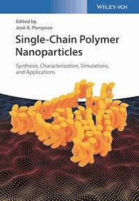
Single-Chain Polymer Nanoparticles E-Book
138,99 €
Mehr erfahren.
- Herausgeber: Wiley-VCH Verlag GmbH & Co. KGaA
- Kategorie: Wissenschaft und neue Technologien
- Sprache: Englisch
This first book on this important and emerging topic presents an overview of the very latest results obtained in single-chain polymer nanoparticles obtained by folding synthetic single polymer chains, painting a complete picture from synthesis via characterization to everyday applications.
The initial chapters describe the synthetics methods as well as the molecular simulation of these nanoparticles, while subsequent chapters discuss the analytical techniques that are applied to characterize them, including size and structural characterization as well as scattering techniques. The final chapters are then devoted to the practical applications in nanomedicine, sensing, catalysis and several other uses, concluding with a look at the future for such nanoparticles.
Essential reading for polymer and materials scientists, materials engineers, biochemists as well as environmental chemists.
Sie lesen das E-Book in den Legimi-Apps auf:
Seitenzahl: 749
Veröffentlichungsjahr: 2017
Ähnliche


Table of Contents
Cover
Title Page
Copyright
List of Contributors
Preface
Chapter 1: Synthetic Methods Toward Single-Chain Polymer Nanoparticles
1.1 Introduction
1.2 Single-Chain Rings via Irreversible and Reversible Bonds
1.3 Single-Chain Nanoparticles via Irreversible Bonds
1.4 Single-Chain Nanoparticles via Supramolecular Chemistry
1.5 Single-Chain Nanoparticles Based on Dynamic Covalent Chemistry
1.6 Conclusions and Outlook
Acknowledgments
References
Chapter 2: Computer Simulations of Single-Chain Nanoparticles
2.1 Computer Simulations in Soft Matter Science
2.2 Simulation of Single-Chain Nanoparticles: Antecedents
2.3 A Bead–Spring Model for Single-Chain Nanoparticles
2.4 Conventional Routes in Good Solvent: Sparse Single-Chain Nanoparticles
2.5 Routes to Globular Single-Chain Nanoparticles
2.6 Sparse SCNPs: Analogies with Intrinsically Disordered Proteins
2.7 Globular SCNPs: A New Class of Soft Colloids
2.8 Conclusions and Outlook
Acknowledgments
References
Chapter 3: Characterization of Single-Chain Polymer Nanoparticles: Analytical Techniques
3.1 Introduction
3.2 Single-Chain Polymer Nanoparticle Characterization via Size Exclusion Chromatography (SEC)
3.3 Spectroscopic Characterization of Single-Chain Polymer Nanoparticles
3.4 Characterization of Single-Chain Polymer Nanoparticle Morphology
3.5 Conclusions and Outlook
References
Chapter 4: Structure and Dynamics of Systems Based on Single-Chain Polymer Nano-Particles Investigated by Scattering Techniques
4.1 Introduction
4.2 Scattering Experiments
4.3 Sources and Instrumentation
4.4 Application of Scattering Techniques to Polymeric Systems
4.5 SCNPs in Dilute Solution
4.6 SCNPs in Bulk
4.7 All-Polymer Nano-Composites: SCNPs Dispersed in a Linear Polymer Matrix
4.8 SCNPs as Confining Medium of Linear Chains
4.9 Conclusions
Acknowledgments
References
Chapter 5: Dynamically Folded Single-Chain Polymeric Nanoparticles
5.1 Introduction
5.2 Single-Chain Polymeric Nanoparticles versus Conventional Nanoparticles
5.3 Preparation of Dynamically Folded Single-Chain Polymeric Nanoparticles
5.4 Characterization of Dynamically Folded Single-Chain Polymer Nanoparticles
5.5 Conclusions and Future Outlook
References
Chapter 6: Metal Containing Single-Chain Nanoparticles
6.1 Introduction
6.2 Palladium
6.3 Iron
6.4 Copper
6.5 Other Metals
6.6 Conclusions and Outlook
References
Chapter 7: Colloidal Unimolecular Polymer Particles: CUP
7.1 Introduction
7.2 Synthesis
7.3 Theory of the Formation of CUP Particles
7.4 Conformation of the CUP Particles
7.5 Electrokinetic Behavior in CUPs
7.6 Electroviscous Effect in CUPs
7.7 Gel Point Behavior
7.8 Surface Tension Behavior
7.9 Cup Surface Water
7.10 Study of Core Environment of CUPs
7.11 Applications: Use of CUPs in Coatings
References
Chapter 8: Single-Chain Nanoparticles via Self-Folding Amphiphilic Copolymers in Water
8.1 Introduction
8.2 Single-Chain Folding Amphiphilic Random Copolymers
8.3 Precision Self-Assembly and Self-Sorting of Amphiphilic Random Copolymers
8.4 Single-Chain Crosslinked Star Polymers
8.5 Conclusions and Future Directions
References
Chapter 9: Applications of Single-Chain Polymer Nanoparticles
9.1 Introduction
9.2 Nanomedicine
9.3 Catalysis
9.4 Sensing
9.5 Other Uses
9.6 Conclusions and Outlook
Acknowledgments
References
Index
End User License Agreement
Pages
xi
xii
xiii
xiv
xv
1
2
3
4
5
6
7
8
9
10
11
12
13
14
15
16
17
18
19
20
21
22
23
24
25
26
27
28
29
30
31
32
33
34
35
36
37
38
39
40
41
42
43
44
45
47
48
49
50
51
52
53
54
55
56
57
58
59
60
61
62
63
64
65
66
67
68
69
70
71
72
73
74
75
76
77
78
79
80
81
82
83
84
85
86
87
88
89
91
92
93
94
95
96
97
98
99
100
101
102
103
104
105
106
107
108
109
110
111
112
113
114
115
116
117
118
119
120
121
122
123
124
125
126
127
128
129
130
131
132
133
134
135
136
137
138
139
140
141
142
143
144
145
146
147
148
149
150
151
152
153
154
155
156
157
158
159
160
161
162
163
164
165
166
167
168
169
170
171
172
173
174
175
176
177
178
179
180
181
183
184
185
186
187
188
189
190
191
192
193
194
195
196
197
198
199
200
201
202
203
204
205
206
207
208
209
210
211
212
213
214
215
216
217
218
219
220
221
222
223
224
225
226
227
228
229
230
231
232
233
234
235
236
237
238
239
240
241
242
243
244
245
246
247
248
249
250
251
252
253
254
255
256
257
259
261
262
263
264
265
266
267
268
269
270
271
272
273
274
275
276
277
278
279
280
281
282
283
284
285
286
287
288
289
290
291
292
293
294
295
296
297
298
299
300
301
302
303
304
305
306
307
308
309
310
311
312
313
314
315
316
317
318
319
320
321
322
323
324
325
326
327
328
329
330
331
332
333
334
335
336
337
338
339
341
342
343
344
345
346
347
348
349
350
351
352
353
354
355
356
357
358
359
360
361
362
363
364
365
366
367
368
369
370
371
372
373
374
375
376
377
378
379
380
381
382
383
384
385
386
387
388
389
390
391
392
393
394
395
396
397
398
399
400
Guide
Cover
Table of Contents
Preface
Begin Reading
List of Illustrations
Chapter 1: Synthetic Methods Toward Single-Chain Polymer Nanoparticles
Figure 1.1 Synthetic route for the preparation of well-defined cyclic polystyrene via the combination of ATRP and CuAAC reaction. (i) NaN
3
, DMF, room temperature (r.t.). (ii) CuBr/Bipy, in degassed DMF, 120 °C.
Figure 1.2 Synthetic route for the preparation of cyclic poly(ϵ-caprolactone). (i) 1,5,7 Triazabicyclo[4.4.0]dec-5-ene (TBD), CH
2
Cl
2
, r.t. (ii) Acryloyl chloride, triethylamine, tetrahydrofuran (THF), r.t. (iii) hν (λ
max
= 320 nm), acetonitrile, r.t.
Figure 1.3 Combined RAFT and thiolactone approach toward functionalized cyclic polymers. (i) RAFT polymerization. (ii) Propylamine or ethanolamine, dichloromethane (DCM). (iii) DCM, 2 days.
Figure 1.4 General strategy for preparing α,ω hydrogen donor/acceptor functional polymers and their subsequent single-chain self-assembly. (i) CuBr, PMDETA, styrene, anisole, 90 °C. (ii) NaN
3
, DMF, r.t. (iii) CuSO
4
× 5 H
2
O, sodium ascorbate, DMF, alkyne functional HW. (iv) High dilution in DCM.
Figure 1.5 Synthesis of benzocyclobutene-functionalized linear polystyrene and schematic representation of the intramolecular collapse of the linear polymer. (i) 120 °C. (ii) 250 °C.
Figure 1.6 Single-chain nanoparticle construction at 25 °C under normal air atmosphere from naked P(MMA-
co
-PgA) precursor copolymers via the Glaser–Hay alkyne coupling reaction. (i) CPDB, BPO/NNDMANIL, THF, 25 °C. (ii) CuI, TMEDA, Et
3
N, THF, air, 25 °C.
Figure 1.7 Synthetic strategy for the preparation of SCNPs through photoactivated thiol–ene and thiol–yne reactions. (i) THF, BPO/NNDMANIL, CPBD, r.t., 17 h. (ii) 3,6-dioxa-1,8-octane-dithiol, DMPA, THF, UV light irradiation at 300–400 nm, r.t., 90 min.
Figure 1.8 Synthetic strategy for the preparation of monofunctional single-chain polymeric nanoparticles. (i) 125 °C, 6 h. (ii) DMF, K
2
CO
3
, 70 °C, 15 h. (iii) DMF, K
2
CO
3
, r.t., 24 h. (iv) Toluene, 100 °C, 15 h. (v) DCM, hν (λ
max
= 320 nm), 30 min.
Figure 1.9 Collapse of single polymer chains to form monodisperse single-chain nanoparticles via intramolecular cross-linking mediated by ring-opening polymerization. (i) AIBN, RAFT agent, 80 °C. (ii) Benzyl alcohol as an initiator, methanesulfonic acid as an organocatalyst.
Figure 1.10 Chemical structures of polymers that can be intramolecularly self-assembled via triple hydrogen bonds under selected conditions generating helical structures employing ATRP and the CuAAC reaction.
Figure 1.11 (a) Design of a triblock copolymer with orthogonal complementary motifs (HW, BTA, and CA) alongside the polymer chain, which is designed to fold into a compartmentalized structure via orthogonal self-assembly. (b) Chemical structure of the functionalized triblock copolymers. (c) Self-assembly of BTA units via threefold symmetric hydrogen bonding. (d) Self-assembly of HW and CA via multiple hydrogen bonding.
Figure 1.12 (a) Design of the initial diblock copolymer system bearing the host–guest folding motif (B21C7/AS), followed by the folding/unfolding of the resulting SCNP. (b) Design of the tetrablock copolymer systems featuring the orthogonal complementary folding motifs (HW/CA and B21C7/AS), followed by folding and the orthogonal stepwise unfolding. (c) Self-assembly of HW and CA via multiple hydrogen bonds. (d) Self-assembly of B21C7 and AS units via host–guest complexation.
Figure 1.13 Decoration of a poly(
N
-hydroxyethyl) acrylamide homopolymer with bipyridine and naphthyl moieties via isocyanate coupling and subsequent SCNP formation through host–guest complexation after the addition of cucurbit[8]uril (CB[8]) cross-linker in high dilution (0.1 mg mL
−1
).
Figure 1.14 Single-chain folding of the PS
30
-
b
-PDMAA
20
-
b
-PPFS
30
block copolymer into a β-hairpin-folded SCNP via π–π-interactions.
Figure 1.15 Synthesis of the polystyrene-
co
-poly[4-((4-vinylbenzyl)oxy)phthalonitrile] via RAFT polymerization and the subsequent formation of SCNPs by cyclotetramerization. (i) AIBN, DMF, CPBD, 65 °C. (ii) Phthalonitrile, CuCl, DBU, benzyl alcohol, 150 °C, 29 h.
Figure 1.16 Synthetic strategy for the preparation of palladium(II)-cross-linked single-chain nanoparticles (Pd-SCNPs). (i) Bulk, 125 °C, 5 h. (ii) 4-(Diphenylphosphino)benzoic acid, K
2
CO
3
, DMF (dry), 50 °C, 16 h. (iii) Pd(COD)Cl
2
, DCM (dry), r.t., 21 h.
Figure 1.17 Synthesis of different poly(PEGMA-
co
-RMA) amphiphilic random copolymers via ruthenium-catalyzed living radical polymerization and schematic illustration of the single-chain folding of these amphiphilic copolymers in water.
Chapter 2: Computer Simulations of Single-Chain Nanoparticles
Figure 2.1 Snapshots of polystyrene SCNPs obtained by simulations of two similar united atom models, in Refs. [7] (top) and [10] (bottom). In both cases 40 % of the monomers have reactive units. In the top panel the reactive (BCB) groups are depicted in gray (light and dark for unreacted and reacted units, respectively).
Figure 2.2 Temperature dependence of the macromolecular radius of gyration, , and of the mean chemical distance, , for polystyrene precursors and for the obtained SCNPs. The mean chemical distance is the mean contour distance, measured in a number of monomers along the precursor backbone, between two cross-linked groups.
Figure 2.3 Typical equilibrium conformation of a bead–spring precursor. The backbone beads are depicted in white. The unreactive and reactive beads in the side groups are depicted in gray and black, respectively.
Figure 2.4 -dependence of the average squared radius of gyration of the precursors (big symbols) and the SCNPs (small symbols) for the simple monofunctional case. The different data sets correspond to the different fractions of reactive groups, . Lines are fits to the scaling law . The data sets are normalized by the respective values of (displayed in the inset), in order to highlight the common scaling with and 0.56 for the precursors and the SCNPs, respectively.
Figure 2.5 Typical snapshots of three different SCNPs obtained from homofunctional precursors in good solvent, with and . Unreactive and reactive beads are depicted in white and black, respectively.
Figure 2.6 Hydrodynamic radii of SCNPs versus their respective precursors. The dashed lines correspond to the theoretical expectations for the scaling of the SCNPs, , with the exponents and . The solid line is the best fit of the whole set of data, yielding .
Figure 2.7 Typical snapshots of different SCNPs obtained through the SP routes, with and . Top: SP4. Bottom: SP6. The unreactive beads are depicted in blue. The reactive beads are represented with other colors (a different color for each chemical species; note the pairs of bonded beads).
Figure 2.8 Radius of gyration of the SCNPs (main panel) obtained through the SP routes, with being the number of different species of the reactive groups. Symbols are simulation values versus the backbone length . All data correspond to the case . Different data sets (see legend) correspond to different -values. The -exponents, as obtained from fits (lines) to power laws , are represented versus in the inset.
Figure 2.9 For , , and several -values, distributions of the time-averaged radii of gyration (top) and asphericity (bottom) of the SCNPs.
Figure 2.10 Distribution of contour distances between bonded side groups for SCNPs with identical values of and , at different -values.
Figure 2.11 Distributions of time-averaged values for the radius of gyration (a) and the asphericity (b) of SCNPs obtained by the route SP3, with and . Filled and empty symbols correspond to the SCNPs obtained by the simultaneous and sequential version of the SP3 route, respectively.
Figure 2.12 Typical snapshots of two different SCNPs obtained by the model EC2. White and gray beads correspond to the precursor and the bridges, respectively.
Figure 2.13 Distributions of the time-averaged asphericities for SCNPs with and obtained from the routes SP1 (triangles) and EC2 (circles). The value for the corresponding precursor is indicated by the arrow.
Figure 2.14 For the Model I, (a) average squared radius of gyration of the swollen SCNPs versus the backbone length for different fractions of reactive groups. For comparison, the Figure includes representative results for the collapsed precursor in bad solvent (empty circles). Symbols are simulation data. Solid and dashed lines are fits to power-laws . The exponents are indicated. (b) Distribution for and several values of . The corresponding results obtained for the system SP2 (synthesis in good solvent), with , are included as inverted triangles for comparison.
Figure 2.15 Typical snapshots of swollen SCNPs synthesized in bad solvent (Model I), for monomers per backbone and different fractions of reactive groups.
Figure 2.16 Distributions of the time-averaged radius of gyration (a) and asphericity (b) for the swollen SCNPs of Model I ( and several values of ). Results for the SP2-SCNPs synthesized in good solvent (, ) are included for comparison.
Figure 2.17 Typical snapshots from simulations of the Model II (RAN case), for precursors with and . (a): Collapsed precursor forming the core–shell structure. (b): The obtained SCNP in the swollen state. White beads represent the solvophilic units. Gray and black beads represent the unreactive and reactive solvophobic groups, respectively.
Figure 2.18 Typical snapshots of SCNPs synthesized by Protocols I (a) and II (c). Backbone beads are depicted in gray. Side group beads are depicted in white (unreactive) and black (reactive). The snapshots (b) and (d) correspond to the same configurations of (a) and (c), respectively, but only the cross-linked sites are displayed. (e) Radial distribution function of the cross-linked sites (centers of the bonds) for Protocols I and II. (Basasoro
et al.
2016 [16]. Reproduced with permission of John Wiley and Sons.)
Figure 2.19 Hydrodynamic radii of denatured proteins (circles), IDPs (diamonds), and folded proteins (squares). The curves are fits to , with being the number of residues. The obtained scaling exponents are , and 0.29 for denatured, intrinsically disordered, and folded proteins, respectively.
Figure 2.20 (a): Snapshots of two typical SCNPs. The domains are represented in different gray tones. The monomers not belonging to domains are depicted in white. (b): Snapshot of the HSPB6 protein [50]. Only the -carbons are represented. Coils and turns are depicted in white. The -domain is depicted in dark gray.
Figure 2.21 Density dependence of the scaling exponent for linear chains and SCNPs (average). Data are also shown for the 10% most ordered and most disordered SCNPs. The arrows indicate the overlap density .
Figure 2.22 Snapshots of a concentrated solution of linear chains (a) and SCNPs (b), both at a monomer density . Both panels show a selected macromolecule and its 12 nearest neighbors (in terms of the distance between the macromolecular centers of mass). Different macromolecules are depicted in different colors.
Figure 2.23 Radial distribution function of the centers of mass at different densities, for the systems P72 (lines) and M72 (symbols).
Figure 2.24 (a): Normalized MSD of the centers of mass for the M72 solution at different densities. Top and bottom dashed lines indicate diffusive and subdiffusive behavior, respectively. (b): Normalized diffusivity versus density for the three investigated systems of globular SCNPs. For comparison, results for sparse SCNPs synthesized in good solvent (GS) are included. Unless explicitly indicated, error bars are smaller than the symbol size.
Figure 2.25 Normalized product versus for P72, M72, and P30 and for the hard-sphere (HS) system. is the relaxation time of the coherent scattering function and is proportional to the viscosity.
Chapter 3: Characterization of Single-Chain Polymer Nanoparticles: Analytical Techniques
Figure 3.1 Schematic representation of the intramolecular collapse of linear polymer
8
to give the single-chain polymer nanoparticle
9
.
Figure 3.2 Overlay of SEC traces for (a) the starting linear polymer, and (b)–(e) single-chain polymer nanoparticles with 5, 10, 20, and 25 mol% BCB incorporation, respectively.
Figure 3.3 (a) Synthetic route and graphical representation of the oxidative polymerization of pendent proDOT groups of poly(ProDOT-Sty) yielding conjugated polymeric nanoparticles. (b) SEC traces during oxidative polymerization of poly(ProDOT-Sty) at 0 h (solid blue), 1 h (dotted red), 3 h (dashed purple) and 5 h (dashed orange).
Figure 3.4 Apparent polydispersity index (PDI
app
) of intramolecular cross-linked nanoparticles versus polydispersity index (PDI) of the corresponding polymeric prescursors for: (a) PS single-chain nanoparticles. (b) PMMA single-chain nanoparticles.
Figure 3.5 Reversible single-chain nanoparticle formations via intramolecular disulfide bridges.
Figure 3.6 SEC-MALS traces of parent polymer (
P2
:
M
w
= 50.1 kDa,
R
h
= 4.5 nm) and SCNPs (
N2
:
M
w
= 59.3 kDa,
R
h
= 3.9 nm).
Figure 3.7 MALS (double peak) and RI (single peak) traces of SCNPs.
Figure 3.8 Schematic representation of the intramolecular collapse of poly(2-isocyanatoethyl methacrylate-
co
-methyl methacrylate) copolymer
1
, to give single-chain nanoparticle
3
by reaction with 2,2′-(ethylenedioxy)diethylamine (bottom), and reaction of
1
with methoxyethylamine to form the control linear copolymer
2
(top).
Figure 3.9 Plot of the reduced viscosity versus concentration for control copolymers (▪, 150 kDa; ▴, 100 kDa) and their analogous cross-linked single-chain polymer nanoparticles (□, 150 kDa; 4, ▵, 100 kDa) in THF.
Figure 3.10
1
H NMR spectra in CDCl
3
of coumarin-containing copolymers before (bottom) and after (top) photo-cross-linking to single-chain polymer nanoparticles.
Figure 3.11 (a)
T
2
decay curves of –CH
2
O– groups on the side chain of coumarin-containing copolymers in CDCl
3
with various photodimerization degrees: 0%, 11%, 23%, 36%, 49%, 60% and 71%, respectively. (b)
T
2
of fast component of –CH
2
O– groups versus dimerization degree. (c) Fraction of fast component of –CH
2
O– groups versus dimerization degree.
Figure 3.12 (a) Different terpolymers (P3, P4, and P5) derived from a single perfluoro-activated ester homopolymer, poly(PFPA)
100
. (b)
19
F NMR spectra in CDCl
3
of poly(PFPA)
100
(A), and its modification with BTA-NH
2
(B), Bipy-NH
2
(C), and Jeffamine (D), as well as a terpolymer containing no
19
F after dialysis (E).
Figure 3.13 The Hahn stimulated echo (STE) with pulsed field gradients.
Figure 3.14 Stacked DOSY spectra illustrating the reaction progress of polystyrene through ATRP. DOSY spectra of 0, 2, 4, 8, 20, and 45 h are stacked.
X
-axis shows all the
1
H resonances of the components in solution.
Y
-axis is the diffusion dimension. Olefinic peaks of styrene were used as internal references to monitor the viscosity change of the dilute crude solution. Because of the heavy overlap of the aromatic regions of styrene and polystyrene, they are not shown in the stacked spectra.
Figure 3.15 DOSY spectra of the parent polymer (
4a
) and subsequent single-chain polymer nanoparticle (
5a
).
Figure 3.16 (a) Full IR spectrum of polymer (red) and single-site [Fe–Fe] Hydrogenase mimic (blue). (b) Region of the spectrum corresponding to the energies of the iron-bound CO stretches. For these representative data, polystyrene was used as the polymer scaffold.
Figure 3.17 (a) Kinetic investigation of the intramolecular cross-linking of a precursor polymer (
c
precursor
= 0.017 mg ml
−1
) in THF via fluorescence spectroscopy (λ
exc
= 315 nm, detection at λ
em
= 535 nm). The normalized fluorescence is plotted against time and reaches its maximum after approximately 7 min. (b) Fluorescence spectra of SCNPs recorded consecutively with continuous irradiation (λ
exc
= 315 nm).
Figure 3.18 TEM image of single-chain polymer nanoparticles. The samples were stained by RuO
4
for 40 min.
Figure 3.19 TEM images of: (a) Neat silica spheres before polymer grafting; (b) PEO-
b
-PCEMA grafted silica spheres after photo-cross-linking of the PCEMA block stained by RuO
4
.
Figure 3.20 TEM image of oblong SCNP fabricated by the photodimerization of anthracene-functionalized polymers.
Figure 3.21 TEM images of different single-chain PDMAEMA-
b
-PS nanoparticles: (a) NP0.1-PDMAEMA74-
b
-PS297, (b) NP0.2-PDMAEMA74-
b
-PS297, and (d) NP0.34-PDMAEMA15-
b
-PS151 prepared by casting from THF solutions; (c) dynamic light scattering curves of NP0.1-PDMAEMA74-
b
-PS297 (denoted as 10%) and NP0.2-PDMAEMA74-
b
-PS297 (denoted as 20%), and (e) tapping mode AFM image of NP0.1-PDMAEMA74-
b
-PS297 adsorbed on the surface of mica.
Figure 3.22 Characterization of different single-chain polystyrene nanoparticles containing copper phthalocyanine (a–d, e–h, and i–l): AFM images of a mica surface (a, e, i) and the corresponding size distribution histograms (b, f, j); 3D AFM images of the corresponding nanoparticles (c, g, k); SEC traces of precursor copolymers (dot) and SCNPs (solid) (d, h, l).
Figure 3.23 A soft spherical particle drop-cast onto a surface takes on a hemi-ellipsoidal shape.
Figure 3.24 AFM images of single-chain polymer nanoparticle aggregates prepared by slowed evaporation.
Figure 3.25 Schematic representations of folding polymers with: (a) UPy modules, and (b) BTA modules, which self-assemble into dimer and helical columnar aggregates, respectively, resulting in single-chain nanoparticle formation. (c) Schematic illustrations of the mechanical unfolding experiment on a single-chain nanoparticle.
Figure 3.26 General synthetic scheme for the preparation of monofunctional single-chain polymeric nanoparticles via intramolecular UV-induced Diels–Alder cross-linking.
Figure 3.27 DLS results showing the hydrodynamic radii (
D
h
) of the linear precursor polymers and their respective SCNPs.
Figure 3.28 (a) SAXS data and fitting curves (black solid lines) of different precursors (P2[−−−], P2[−B−]) and SCNPs (P2[UBU], P2[UBU]UV). The SAXS data were recorded in 1 mg ml
−1
solutions in 75/25 (v/v) methylcyclohexane/1,4-dioxane at 20 °C and are drawn here on the same scale but offset vertically for clarity. (b)
R
g
values for P2[−−−], P2[−B−], P2[UBU], and P2[UBU]UV obtained from fitting analysis of the SAXS data. The insets schematically illustrate the triblock copolymer in a partly folded state, denoted as P2[UBU], and the fully folded state after UV irradiation, denoted as P2[UBU]UV.
Chapter 4: Structure and Dynamics of Systems Based on Single-Chain Polymer Nano-Particles Investigated by Scattering Techniques
Figure 4.1 Schematic representation of a scattering experiment.
Figure 4.2 Illustration of small-angle scattering.
Figure 4.3 Schematic representation of (a) a direct time-of-flight spectrometer on a reactor and (b) an inverted geometry time-of-flight (BS-ToF) spectrometer (in this case, only one set of analyzer/detector has been represented).
Figure 4.4 Compilation of hydrodynamic radii reported in the literature for SCNPs obtained from DLS as a function of those of their precursors. Data for different systems are represented with different symbols. Continuous line represents the case (=0.59); dotted line, the globular case (=1/3); and dashed line, the Gaussian coil case (=0.5).
Figure 4.5 SANS results on linear precursor chains (circles) and the SCNPs obtained from them by Michael addition (diamonds) and Cu complexation (squares): (a) measured intensity and fit with generalized Gaussian coil form factors (Eq. 4.32) (lines); (b) Kratky representation of the SCNPs data, compared with the cases of a random coil ( = 0.5, dashed line) and a compact globule ( = 1/3, dotted line).
Figure 4.6 (a) SAXS form factors for the unlinked precursor (circles) and for the SCNPs obtained by Michael addition (squares) and by sequential Michael reaction and metal complexation (diamonds). Data have been shifted in the vertical axes for clarity. Lines are fits to the power law with the -values indicated. Experimental data extracted from Ref. [66]. (b) Kratky plot for solutions of Michael SCNPs synthesized with cross-linkers of functionality = 2 (empty symbols) and = 3 (filled symbols). These experimental data, obtained by SANS, are extracted from Ref. [70]; similar results were obtained by SAXS [71].
Figure 4.7 Kratky plot of the SAXS results on SCNPs obtained by photoactivated synthesis via Thiol–Yne coupling reaction (filled circles) and the corresponding precursor (empty circles).
Figure 4.8 (a) DLS results on solutions of a PMMA-AEMA precursor (diamonds) and SCNPs obtained from it by Cu-complexation (circles). Lines are fits of single exponential functions. (b) Distribution function of diffusion coefficients deduced from a CONTIN analysis of the experimental data (same symbols as in (a); lines are just connecting the points).
Figure 4.9 NSE results on the Cu-SCNPs (closed symbols) and precursor (open symbols) solutions at the different -values indicated in . Lines are fits of single exponentials to the experimental data (solid lines to Cu-SCNPs and dotted lines to precursors).
Figure 4.10 Fits of the Zimm model (continuous lines) and of the Zimm model with internal friction (dashed lines) to the NSE results on the precursor (a) and the Cu-SCNPs (b) solutions at the different -values denoted in Figure 4.9. Symbol code for the different -values as in Figure 4.9.
Figure 4.11 Characteristic times of the Zimm modes as a function of the scaling variable deduced for the precursor (circles) and the Cu-SCNPs (diamonds) in solution. Only modes with mode number below or equal to 4 (precursor) and 2 (Cu-SCNPs) highlighted as filled symbols would substantially contribute. Dashed-dotted and solid lines represent the characteristic times for the ZIF model for the precursor and the Cu-SCNPs, respectively. Dotted arrows mark the value of and the location of the crossover from solvent- to internal-friction-dominated relaxation for the precursor. (Arbe 2016 [65]. Reproduced with permission of Elsevier.)
Figure 4.12 XR structure factors measured at room temperature on SCNPs obtained by Cu-complexation (circles) and on the corresponding linear precursors (diamonds) (a). In (b), the results on PMMA (pluses) and PAEMA (crosses) are shown for comparison. The dashed line represents their weighed sum according to the molar content of MMA and AEMA units in the precursors.
Figure 4.13 Relaxation map of PMMA: inverse-temperature-dependence of the characteristic times obtained for different dynamic processes: -relaxation from dielectric spectroscopy [97] (solid line), coherent scattering (NSE) at [51] (circles) and incoherent scattering (BS) [51] (diamonds); -relaxation from dielectric spectroscopy [97] (dotted line); methyl-group rotations: ester MG [88] and MG [51].
Figure 4.14 WAXS patterns obtained at 100 C for PEO (circles), the SCNPs (diamonds), and the nano-composite (squares). Solid line represents the result of combining the patterns corresponding to the neat components weighed by the respective volume fractions in the mixture.
Figure 4.15 Normalized form factor of SCNPs in different environments determined by SANS [98] and represented against the reduced variable . Filled squares represent results on SCNPs in good solvent at high dilution, and empty circles in a linear polymeric matrix. Solid lines are fits of generalized Gaussian coils (Eq. 4.32) with the values of the scaling exponent indicated.
Figure 4.16 Temperature-dependence of the derivative of the reversible heat flow with respect to temperature in the nano-composite (solid circles) and the neat components: bulk PEO (empty squares) and bulk SCNPs (empty circles). Arrows mark the locations of the glass-transition temperatures.
Figure 4.17 (a) Ratio of the incoherent contribution to the total intensity scattered by the nano-composites where one component is protonated and the other is deuterated, measured by diffraction with polarization analysis. (b) Absolute value of the ratio between the coherent and the incoherent contributions to the NSE signal of the nano-composite sample (filled circles) and the blend with the precursor (empty squares). Solid line shows a -power law.
Figure 4.18 Scattering function measured on the 75hPEO/25dSCNPs sample at 400 K and the different -values indicated (in ). Dotted line shows the instrumental resolution function.
Figure 4.19 Intermediate scattering functions obtained after Fourier transformation and deconvolution of spectra obtained for the 75hPEO/25dSCNPs nano-composite at 350 K (a) and 400 K (b) and the different -values indicated in .
Figure 4.20 KWW characteristic time for the incoherent scattering function () as a function of momentum transfer at the different temperatures investigated for (a) bulk PEO [89] and (b) PEO in the nano-composite. For better visibility 350 and 375 K data are multiplied by 100 and 10 respectively. Dashed lines show fits of the low- results by -laws. Solid lines are fits in the whole accessed -range to the anomalous jump diffusion model.
Figure 4.21 (a) -dependence of the effective Rouse variable obtained from the incoherent scattering function of PEO in the nano-composite (filled circles) [53] and bulk PEO (empty symbols). Results at 375 and 350K have been divided by 10 and 100 for clarity. Arrows show the low- asymptotic limits. (b) Inverse-temperature-dependence of the Rouse variable reported from incoherent scattering (circles: experiments; squares: MD-simulations) and NSE experiments on the single-chain dynamic structure factor (triangles) for bulk PEO (empty symbols) [89, 102] and for PEO in the nano-composite (filled symbols) [53, 87]. Lines are VF laws: solid lines are fits to the PEO sets of data and dotted lines the expectations for the PMMA component of the mixtures.
Figure 4.22 Normalized of PEO chains in (a) the nano-composite and (b) the blend with the linear precursor at the -values indicated. Solid lines are descriptions with the Rouse model (/ns).
Figure 4.23 Normalized of PEO chains in (a) the nano-composite and (b) the blend with the linear precursor at the -values indicated. Lines are fits of the reptation model (Eqs. 4.23 –4.25) to the experimental data for 35 ns in (a) and 20 ns in (b), fixing /ns. For the nano-composite = 9.5 nm was obtained (a); for the blend, the value of the tube diameter was fixed to that of PEO in bulk, 5.3 nm.
Chapter 5: Dynamically Folded Single-Chain Polymeric Nanoparticles
Figure 5.1 (a) Self-assembly of amphiphilic block copolymers into micelles and polymersomes. (Blanazs
et al
. 2009 [23]. Reproduced with permission of John Wiley and Sons.) (b) Folding single polymer chains into nanoparticles.
Figure 5.2 Different approaches to prepare random copolymers either by direct copolymerization of functional monomers or by post-functionalization of a polymer backbone with reactive side groups.
Figure 5.3 (a) Folding UPy-containing polymers into SCPNs. (Mes
et al
. 2011 [69]. Reproduced with permission of John Wiley and Sons.) (b) Folding polymers stepwise into SCPNs by BTA-mediated self-assembly.
Figure 5.4 Orthogonal self-assembly of BTA and UPy or HW-CA and BTA in triblock copolymers.
Figure 5.5 Single-chain folding of BTA-containing polymers in water affording a compartmentalized catalyst for the transfer hydrogenation of ketones.
Figure 5.6 SCPNs formed by the self-complementary hydrogen-bonding motif U-DPy.
Figure 5.7 Folding of single polymer chains by CB[8]-based host–guest interactions.
Figure 5.8 Copper cross-linked SCPNs as efficient catalysts toward alkyne–azide “click” reactions in water and in cells.
Figure 5.9 Folding amphiphilic copolymers in water by hydrophobic effect.
Figure 5.10 Disulfide bonds in preparing dynamic SCPNs.
Figure 5.11 Hydrazone bond-based SCPNs and their reversible transformation into hydrogel.
Figure 5.12 Preparation of SCPNs through photo-dimerization of coumarin moieties.
Figure 5.13 BTA-containing triblock copolymers and the proposed structure of SCPNs.
Figure 5.14 Schematic representation of the sensing function of the BiPy-BTA-based SCPNs.
Figure 5.15 Characterization of polymer folding by SEC.
Figure 5.16 Folding of BTA-containing copolymers into elongated SCPNs.
Figure 5.17 AFM height micrographs and schematic representation of the polymer structures.
Figure 5.18 Schematic representations of mechanical unfolding UPy- or BTA-based SCPNs.
Figure 5.19 The TEMPO-containing polymers.
Chapter 6: Metal Containing Single-Chain Nanoparticles
Scheme 6.1 Formation of large cyclic complexes by Pd complexation.
Scheme 6.2 Preparation and cross-linking of Pd-SCNPs.
Scheme 6.3 Preparation of Pd(II)-SCNPs through
o
-CH activation.
Scheme 6.4 Reversible formation of SCNPs via external voltage stimuli.
Scheme 6.5 Formation of Fe-SCNPs by terpyridine complexation.
Scheme 6.6 Synthesis of polymer-bound Fe
2
-H
2
ase model complex.
Scheme 6.7 Preparation of porphyrin-cored single-chain polymer nanoparticles.
Scheme 6.8 Formation of Cu-SCNPs by imidazole complexation.
Scheme 6.9 (a) Preparation of Cu-SCNPs through complexation to β-ketoester moieties (b) Catalytic activity of Cu-SCNPs in oxidative coupling of terminal acetylenes.
Scheme 6.10 Preparation of water-soluble globular Cu-SCNPs.
Scheme 6.11 The effect of selective and nonselective solvents on the spatial distribution of catalytic sites within Cu-SCNPs.
Scheme 6.12 Synthetic route toward copper phtalocyanine SCNPs.
Scheme 6.13 (a) Preparation of water-soluble Cu-ONPs (b) The catalytic activity of Cu-ONPs in benchmark Cu(I)-catalyzed “click” reactions.
Scheme 6.14 Postpolymerization approach for the preparation of amphiphilic catalytically functional collapsed polymers.
Scheme 6.15 Preparation and schematic representation of the sensing function of BiPy-BTA functional polymers.
Scheme 6.16 Synthetic route toward Rh(I)-ONPs.
Scheme 6.17 Synthesis of Ni(0)−SCNPs (a) and organobimetallic SCNPs (b) with Rhodium(I) and Iridium(I).
Scheme 6.18 Preparation of Rh(I)-ONPs from polybutadiene and reaction with PCy
3.
Scheme 6.19 Supramolecular single-chain folding of polymer
P8
in water affording a compartmentalized catalyst for the transfer hydrogenation of ketones.
Scheme 6.20 Structure of catalytically active SCNPs for the oxidation of alcohols in water; structures of the substrates and products.
Scheme 6.21 The synthetic route to ZnS nanocrystals.
Scheme 6.22 Schematic illustration of zein-pyridine-gold interactions in pyridine-functionalized SCNPs.
Scheme 6.23 Preparation of coumarin SCNPs to tune the formation of AuNPs.
Scheme 6.24 Synthetic route to Gd(III)-loaded SCNPs.
Scheme 6.25 Synthetic route toward
67
Ga-loaded SCNPs.
Chapter 7: Colloidal Unimolecular Polymer Particles: CUP
Figure 7.1 Process of forming CUP particles from poly(ethyl methacrylate-
co
-methacrylic acid) copolymers: (I) random coil configuration in tetrahydrofuran (THF), (II) random coil intimate ion pair, (III) extended coil solvent separated ion pair, (IV) collapsed coil, and (V) hard sphere.
Figure 7.2 Viscosity of polymer
32
(Table 7.1) during water reduction of 20 g polymer in 80 g in THF with 160 g water being added.
Figure 7.3 Particle size measured by dynamic light scattering (DLS) and calculated from SEC/GPC data.
Figure 7.4 Solution clarity of a 4.2 nm CUP solution versus a 32 nm polyurethane dispersion, PUD, and a 100 nm latex resin.
Figure 7.5 Electrophoretic mobility (μ) and conductivity (σ) versus number density.
Figure 7.6 Effective charge (
Z
eff
) measured by Hessinger's model and predicted by Belloni's model.
Figure 7.7 Specific viscosity of CUPs at different concentrations and different levels of NaCl.
Figure 7.8 Calculated volume fraction at gel point as function of particle size and thickness of bound water layer (
n
is the number of water layers, RCP is random closed packing).
Figure 7.9 Relative viscosities at different volume fraction.
Figure 7.10 Equilibrium surface tension behavior of different CUPs versus concentration.
Figure 7.11 Dynamic surface tension behavior of different CUPs versus surface age at a concentration of 0.5 mol m
−3
.
Figure 7.12 Effect of concentration on dynamic surface tension for SO
3
−
CUPs-28K.
Figure 7.13 The heat of fusion of water (continuous line) and CUPs from polymer
1
(dotted line).
Figure 7.14 The specific enthalpy of CUPs from polymer
1
at different concentrations: 5, 10, 15 and 20%, respectively.
Figure 7.15 Comparison of weight fraction of nonfreezable water versus percent solid for CUPs prepared from different polymers (see Table 7.1).
Figure 7.16 Spin–lattice relaxation time at 18 °C for high and low molecular weight CUPs at different concentration.
Figure 7.17 Spin–lattice relaxation time for low molecular weight CUPs at different concentration and at different temperatures.
Figure 7.19 Latex of 100 nm, dispersion of 25 nm, and CUP of 3–8 nm in size.
Figure 7.18
T
2
relaxation of CUPs plotted in the temperature range 25–70 °C.
Figure 7.20 Steps involved in the cross-linking of the acrylic–melamine resin.
Figure 7.21 Functionalization of EA–AA copolymer with 2-methylazidirine to give an amino functional copolymer.
Chapter 8: Single-Chain Nanoparticles via Self-Folding Amphiphilic Copolymers in Water
Scheme 8.1 Amphiphilic PEGMA-based random copolymers: self-folding, self-assembly, and self-sorting polymers with thermoresponsive properties and unique functions.
Scheme 8.2 Single-chain folding of PEGMA-based amphiphilic random copolymers in aqueous, organic, and fluorinated media.
Scheme 8.3 Self-folding amphiphilic random copolymers via hydrophobic interaction in water: Synthesis of PEGMA/RMA amphiphilic random copolymers via ruthenium-catalyzed living radical copolymerization of PEGMA and RMA.
Figure 8.1 DLS intensity size distribution of PEGMA/DMA (a) random or (b) block copolymers (20 mol% DMA) in water or CH
2
Cl
2
at 25 °C; [polymer] = 10 mg ml
−1
.
Figure 8.2 Self-folding and self-assembly of (a) PEGMA/DMA or (b) PEGMA/RMA random copolymers (DP = 200) in water: (a) Effect of DMA content (0–60 mol%) on
R
h
(by DLS) and aggregation number [
N
agg
= (MALLS)/
M
w,DMF
(MALLS)]; (b) Effect of the carbon number of RMA on the compactness of micelles [
M
p
(H
2
O)/
M
p
(DMF)]. DLS: [polymer] = 10 mg ml
−1
in water or DMF or CH
2
Cl
2
at 25 °C.
Figure 8.3 (a) Turbidity measurements of the aqueous solutions of PEGMA/RMA random copolymers (20 mol% RMA, DP = 200) at the temperature range between 70 and 95 °C: [polymer] = 4 mg ml
−1
; hearting = 1 °C min
−1
. (b) Cloud point temperature of the aqueous solutions against the carbon number of the RMA pendants [R: (CH
2
)
n
H; n = 1–18].
Figure 8.4 Self-folding PEGMA/DMA star random copolymers in water: (a) Synthesis of the star polymers via ruthenium-catalyzed living radical copolymerization of PEGMA and DMA with a trifunctional initiator, and (b) self-folding of three amphiphilic arm chains in water.
Scheme 8.4 Amphiphilic random copolymers with hydrogen-bonding urea pendants as self-folding polymers in aqueous and organic media [35]. Synthesis of PEGMA/BPUMA random, gradient, and block copolymers and PEGMA-based random copolymers with urethane or ester pendants via ruthenium-catalyzed living radical polymerization.
Figure 8.5 Single-chain folding and self-assembly of PEGMA/BPUMA random copolymers and PEGMA-based random copolymers with urethane or ester-pendants in water: (a–c) SEC curves of PEGMA/BPUMA random copolymers (BPUMA: 5, 20, and 40 mol%, DP = 200) in water or DMF. (d) M
p
(H
2
O)/M
p
(DMF) of their random copolymers as a function of BPUMA or RMA content.
Scheme 8.5 (a) Helical self-assembly of BTA derivatives. (b) PEGMA/BTAMA random copolymers for single-chain folding polymers via the helical self-assembly of the chiral BTA pendants in water. (c) A ruthenium-bearing PEGMA/BTAMA random terpolymer for a single-chain folding polymer catalyst in water.
Figure 8.6 Temperature-dependent CD spectra of a PEGMA/BTAMA random copolymer (
l
/
m
/
n
= 8.5/90/10, DP = 100) in water at different temperatures between 273 and 363 K (10 K interval): [BTA] = 50 µM.
Scheme 8.6 Single-chain folding polymer Ru catalysts for transfer hydrogenation of ketones in water.
Scheme 8.7 Synthesis of PEGMA/R
F
MA random copolymers via ruthenium-catalyzed living radical copolymerization of PEGMA and R
F
MA (13FOMA, 17FDeMA) (top) and multi-mode self-folding polymers via the reversible folding of amphiphilic/fluorous random copolymers in aqueous, organic, and fluorinated media (bottom).
Scheme 8.8 Conjugation of a disulfide pyridine-bearing amphiphilic/fluorous random copolymer and a thiolated lysozyme (Lyz-SH) (Lyz structure PDB: 2LYZ).
Scheme 8.9 Storage of proteins (lysozyme or α-chymotrypsin) within perfluorinated PEG compartments of amphiphilic/fluorous random copolymers in 2H,3H-perfluoropentane (protein structures, PDB: 2LYZ, 1YPH).
Figure 8.7 Effects of DP and DMA composition on the intermolecular (filled circles) or intramolecular (open circles) self-assembly of the random copolymers in water.
Figure 8.8 Precision intermolecular self-assembly of PEGMA/DMA random copolymers into uniform nanoparticles in water: (a) SEC curves (by refractive index detector) of the 40 mol% DMA copolymers with different DP (44, 52, 69, 102, 190, and 424) by living radical polymerization and that by free radical polymerization (FRP) in DMF and H
2
O. (b, c) Absolute weight-average molecular weight [
M
w
(MALLS)] of 40 mol% (b) or 50 mol% (c) DMA copolymers in DMF and H
2
O.
Figure 8.9 Orthogonal self-assembly and self-sorting of PEGMA/DMA random copolymers into discrete nanocompartments with different hydrophobicity in water. (a, d) SEC curves of nanoaggregates [50 mol% DMA,
N
agg
= 4.6, 30 mol% DMA,
N
agg
= 1.0, and 30 mol% DMA,
N
agg
= 2.0] in water. (b, e) SEC curves of the aqueous mixtures of different nanoparticles. (c, f) SEC curves of the aqueous solutions of blended copolymers with different DMA content.
Scheme 8.10 Synthesis of single-chain star polymers via the intramolecular crosslinking of self-folding amphiphilic random copolymers with an azo initiator or a ruthenium catalyst in water.
Figure 8.10 Intramolecular crosslinking of olefin-bearing amphiphilic random copolymers (a, c) with 2,2′-azobis(2-methylpropionamidine)dihydrochloride (V-50) in water or (b, d) with 2,2-azobis(isobutyronitrile) in toluene: [polymer] = 10 mg ml
−1
. SEC curves of precursors (dash lines) and products (solid lines) in DMF.
Chapter 9: Applications of Single-Chain Polymer Nanoparticles
Figure 9.1 Illustration of dynamic single-ring (a), “alpha” letter-shaped single-ring (b), compositionally unsymmetrical single-ring (c) and complex multi-ring system (d).
Figure 9.2 Schematic illustration of sparse single-chain polymer nanoparticle (a) and globular single-chain polymer nanoparticle (b).
Figure 9.3 Analogy of the morphology of sparse (type I) and globular (type II) single-chain nanoparticles (SCNPs) with intrinsically disordered proteins (IDPs) and globular proteins, respectively.
Figure 9.4 Single-chain nanoparticles of type I (a) show the presence of multiple locally compact, but accessible, sites/cavities/zones, so-called “local pockets,” whereas SCNPs of type II (b) show a single pocket of larger size.
Figure 9.5 Illustration of (a) tadpole, (b) dumbbell, and (c) hairpin morphologies.
Figure 9.6 Illustration of different nanostructures resulting from the self-assembly of individual amphiphilic tadpoles.
Figure 9.7 Main applications of single-chain polymer nanoparticles.
Figure 9.8 (a) Percentage of living HEK293T cells after exposure to different concentrations of biodegradable single-chain polymer nanoparticles. (b) Percentage of HeLa cell viability upon exposure for 24 h to fluorescein-containing poly(norbornene)-based single-chain polymer nanoparticles of different molecular weight. (c) Cell viability after 72 h of single-chain polymer nanoparticle incubation with six pancreatic adenocarcinoma cell lines (PANC-1, BXPC3, Su86.86, Colo-357, ASPC-1, and T3M4).
Figure 9.9 l-φAA and d-φAA sorption isotherms for Type I- and Type II-imprinted single-chain polymer nanoparticles. Data represented by dashed and solid lines were obtained from the Langmuir model by assuming the presence of one (specific) or two (specific and non-specific) types of binding sites.
Figure 9.10 Schematic illustration of the conjugation of peptide therapeutics to a single-chain polymer nanoparticle decorated with dendritic molecular transporter molecules and fluorescent moieties via thiol–disulfide exchange reaction under aqueous reaction conditions.
Figure 9.11 Delivery curve in water at neutral pH from vitamin B
9
-loaded “Michael” single-chain polymer nanoparticles and best-fit of the experimental data to the power law model:
C
t
/C
f
=
K t
n
,
C
t
= concentration of drug released at time
t
,
C
f
= total concentration of drug released,
K
= constant,
n
= release exponent.
Figure 9.12 Simultaneous delivery data in water at pH = 6 (circles) and pH = 8 (triangles) of hinokitiol (solid symbols) and vitamin B
9
(open symbols) from “Michael” single-chain polymer nanocarriers.
Figure 9.13 Drug release-time profiles for FU-loaded POEGMA-U-DPy single-chain polymer nanoparticles in PBS buffer under different environmental conditions.
Figure 9.14 SPECT-TC images of a mouse injected with
67
Ga-loaded/peptide-decorated single-chain polymer nanoparticles acquired 48 h after administration, showing, in color, the accumulation of radioactivity in major organs and tissues: (a) Pancreatic tumor (indicated by a red arrow). (b) Liver. (c) Bone junctions and bladder.
Figure 9.15 (a) Photobleaching study using 470 nm LED with fluorescein (1 μM, left vial) and fluorescein-loaded single-chain polynorbornene nanoparticles (1 μM, right vial) before and after irradiation in pH 7.4 phosphate buffer. (b) Three-dimensional reconstruction of a confocal image stack showing the fluorescence of maleimide functional polymeric microspheres (average diameter: 11 µm) upon reaction with non-fluorescent tetrazole-decorated polystyrene nanoparticles (average diameter: 3 nm). (c) Illustration of the bright fluorescence exhibited by dansylhydrazine-conjugated polystyrene nanoparticles (right vial) when compared to neat polystyrene nanoparticles (left vial). (d) Fluorescence image of bulk polystyrene nanoparticles prepared by a synthetic route leading to fluorescent single-chain polymer nanoparticles upon direct intra-chain cross-linking.
Figure 9.16 Illustration of the catalytic selectivity of Cu(II)-containing single-chain polymer nanoparticles when compared to a classical catalyst during alkyne homocoupling experiments.
Figure 9.17 Tentative mechanism proposed for the biphenyl production catalyzed by the Rh(I)-containing nanoparticles.
Figure 9.18 Schematic illustration of the formation of organocatalytic single-chain nanoparticles containing entrapped B(C
6
F
5
)
3
molecules endowed with “polymerase-like” activity toward tetrahydrofuran (THF) in the presence of small amounts of glycidyl phenyl ether (GPE).
Figure 9.19 Schematic illustration of the bioinspired construction of copper-containing globular single-chain nanoparticles endowed with metalloenzyme-mimicking characteristics toward controlled synthesis of water-soluble polymers and thermoresponsive hydrogels.
Figure 9.20 TEM image of gold nanoparticles synthesized by using single-chain polymer nanoparticles as nanoreactors. Average gold nanoparticle size: 6–9 nm.
Figure 9.21 TEM images of ZnS (a) and CdS (b) quantum dots synthesized by using single-chain polyacrylic acid nanoparticles as nanoreactors.
Figure 9.22 TEM images of carbon nanodots with different average diameters: (a) 4.5 nm; (b) 2.1 nm, synthesized by using single-chain polymer nanoparticles of different size as sacrificial nanoreactors.
Figure 9.23 Illustration of the response of fluorescent bipyridine-containing single-chain nanoparticles toward different metal ions. The degree of fluorescence quenching is expressed as
I
0
/
I
,
I
0
and
I
being the fluorescence intensity at 520 nm in the absence and presence of the metal ion, respectively.
Figure 9.24 Photographs of the sensing system based on pyridine–gold–zein interactions involving pyridine-functionalized single-chain nanoparticles in the presence of decreasing concentrations of zein.
Figure 9.25 Field emission scanning electron microscopy cross-section image of a nanoporous MSSQ thin film with 20% porosity prepared by using single-chain polymer nanoparticles as porogens.
Figure 9.26 Schematic illustration of single-chain nanoparticle formation via intramolecular “click” (Cu(I)-catalyzed azide-alkyne) cycloaddition, and subsequent nanoparticle functionalization by means of a second “click” reaction.
Figure 9.27 Illustration of the dependence of reduced viscosity, η
red
, on concentration,
c
, for two linear precursors (black circles and diamonds) and their corresponding single-chain polymer nanoparticles (gray circles and diamonds), respectively. The value of intrinsic viscosity, [η], can be obtained by extrapolating to
c
= 0 using, for example, the Huggins equation η
red
= [η] +
k
([η])
2
c
. The single-chain polymer nanoparticles show lower values of [η] when compared to the corresponding values for the linear precursors. (Perez-Baena
et al
. 2014 [131]. Reproduced with permission of Royal Society of Chemistry.)
List of Tables
Chapter 4: Structure and Dynamics of Systems Based on Single-Chain Polymer Nano-Particles Investigated by Scattering Techniques
Table 4.1 Values of the average NS lengths , their squares , and their deviations for different isotopes
Chapter 7: Colloidal Unimolecular Polymer Particles: CUP
Table 7.1 List of polymers synthesized for CUP study.
Table 7.2 Solubility parameters of individual solvents and their blends with water at collapse point [20]
Table 7.3 Thermodynamic parameters of micelle formation in some common surfactants [21–24]
Table 7.4 Associated water fraction, β, and surface water thickness, δ, for CUPs with different functional groups
Table 7.5 Relaxation time (τ
k
) for three sulfonate CUPs at various concentrations [8]
Table 7.6 Bound water layer thickness calculated for high and low molecular weight CUPs at different temperatures and different concentrations [7]
Table 7.7 Gloss, flexibility, impact resistance, dry and wet adhesion resistance, minimum film forming temperature (MFFT), pencil hardness, and indentation hardness of the epoxy clear coats [18]
Chapter 9: Applications of Single-Chain Polymer Nanoparticles
Table 9.1 Different stimuli used for the disassembly of a variety of single-chain polymer nanoparticles with reversible interactions
Table 9.2 Properties of different fluorescent single-chain polymer nanoparticles
Table 9.3 Reactions catalyzed by different single-chain polymer nanoparticles


