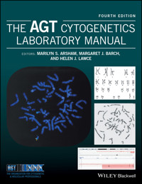
The AGT Cytogenetics Laboratory Manual E-Book
209,99 €
Mehr erfahren.
- Herausgeber: John Wiley & Sons
- Kategorie: Wissenschaft und neue Technologien
- Sprache: Englisch
Cytogenetics is the study of chromosome morphology, structure, pathology, function, and behavior. The field has evolved to embrace molecular cytogenetic changes, now termed cytogenomics. Cytogeneticists utilize an assortment of procedures to investigate the full complement of chromosomes and/or a targeted region within a specific chromosome in metaphase or interphase. Tools include routine analysis of G-banded chromosomes, specialized stains that address specific chromosomal structures, and molecular probes, such as fluorescence in situ hybridization (FISH) and chromosome microarray analysis, which employ a variety of methods to highlight a region as small as a single, specific genetic sequence under investigation. The AGT Cytogenetics Laboratory Manual, Fourth Edition offers a comprehensive description of the diagnostic tests offered by the clinical laboratory and explains the science behind them. One of the most valuable assets is its rich compilation of laboratory-tested protocols currently being used in leading laboratories, along with practical advice for nearly every area of interest to cytogeneticists. In addition to covering essential topics that have been the backbone of cytogenetics for over 60 years, such as the basic components of a cell, use of a microscope, human tissue processing for cytogenetic analysis (prenatal, constitutional, and neoplastic), laboratory safety, and the mechanisms behind chromosome rearrangement and aneuploidy, this edition introduces new and expanded chapters by experts in the field. Some of these new topics include a unique collection of chromosome heteromorphisms; clinical examples of genomic imprinting; an example-driven overview of chromosomal microarray; mathematics specifically geared for the cytogeneticist; usage of ISCN's cytogenetic language to describe chromosome changes; tips for laboratory management; examples of laboratory information systems; a collection of internet and library resources; and a special chapter on animal chromosomes for the research and zoo cytogeneticist. The range of topics is thus broad yet comprehensive, offering the student a resource that teaches the procedures performed in the cytogenetics laboratory environment, and the laboratory professional with a peer-reviewed reference that explores the basis of each of these procedures. This makes it a useful resource for researchers, clinicians, and lab professionals, as well as students in a university or medical school setting.
Sie lesen das E-Book in den Legimi-Apps auf:
Seitenzahl: 3028
Veröffentlichungsjahr: 2017
Ähnliche


Table of Contents
Cover
Title Page
Contributing authors
Preface
Acknowledgments
CHAPTER 1: The cell and cell division
1.1 The cell
1.2 The cell cycle
1.3 Recombinant DNA techniques
1.4 The human genome
References
CHAPTER 2: Cytogenetics: an overview
2.1 Introduction
2.2 History of human cytogenetics
2.3 Cytogenetics methods
2.4 Slide‐making
2.5 Chromosome staining
2.6 Chromosome microscopy/analysis
2.7 Laboratory procedure manual
References
Contributed protocols
Protocol 2.1 Slide‐making
Protocol 2.2 Slide‐making
Protocol 2.3 Making wet slides for chromosome analysis
Protocol 2.4 Slide‐making
Protocol 2.5 Slide preparation
Protocol 2.6 Slide preparation procedure
CHAPTER 3: Peripheral blood cytogenetic methods
3.1 Using peripheral blood for cytogenetic analysis
3.2 Special uses of peripheral blood cultures
3.3 Peripheral blood constituents
3.4 Specimen handling
3.5 Cell culture equipment and supplies
3.6 Harvesting peripheral blood cultures
3.7 Chromosome analysis of peripheral blood
3.8 Storage of fixed specimens
Acknowledgments
References
Contributed protocols
Protocol 3.1 Blood culture and harvest procedure
Protocol 3.2 High‐resolution peripheral blood method
Protocol 3.3 Constitutional cytogenetic studies on peripheral blood
Protocol 3.4 Blood culture and harvest procedure for microarray confirmation studies
CHAPTER 4: General cell culture principles and fibroblast culture
4.1 Definitions of a culture
4.2 Basic considerations in cell culture
4.3 Fibroblast culture
4.4 Lymphoblastoid cell lines
Glossary
Reference
Additional readings
Contributed protocols section
Protocol 4.1 Solid tissue collection for establishing cultures
Protocol 4.2 Solid tissue transport and sendout media
Protocol 4.3 Tissue culture reagents
Protocol 4.4 Phosphate buffer solution deficient in Ca
2+
and Mg
2+
Protocol 4.5 Solid tissue and fibroblast culture setup
Protocol 4.6 Solid tissue setup and processing
Protocol 4.7 Flask and coverslip setup for POC/fibroblast cultures
Protocol 4.8 Coverslip setup for solid tissue biopsy specimens
Protocol 4.9 Solid tissue (fibroblast) culturing and harvesting
Protocol 4.10 Fibroblast culture maintenance: media feeding and changing
Protocol 4.11 Routine subculture of fibroblast cultures
Protocol 4.12 Manual harvest for flasks
Protocol 4.13 Treated media for contamination
Protocol 4.14 Fungizone–mycostatin solution for treatment of fungus/yeast contaminated cultures
Protocol 4.15 Mycoplasma testing
Protocol 4.16 Plating efficiency of serum
Protocol 4.17 Routine replication plating for human diploid cells
Protocol 4.18 Cell counting chamber method
Protocol 4.19 Cell viability by dye exclusion
Protocol 4.20 Mitotic index
Protocol 4.21 Growth rate‐estimation of mean population doubling time during logarithmic growth
Protocol 4.22 Maintenance of fibroblast cultures as non‐mitotic population
Protocol 4.23 Synchronization at S‐phase with BrdU
Protocol 4.24 Making direct FISH preparations from abortus tissue
Protocol 4.25 Cryopreservation
Protocol 4.26 Cryopreservation with Nalgene cryogenic container
Protocol 4.27 Lymphoblastoid lines
Protocol 4.28 Freezing tissue cultures (cryopreservation)
CHAPTER 5: Prenatal chromosome diagnosis
5.1 Introduction
5.2 Amniotic fluid
5.3 Culture of amniotic fluid
5.4 Analysis of amniotic fluid
5.5 Chorionic villus sampling
5.6 Analysis of chorionic villi
References
Contributed protocols section
Protocol 5.1 Amniotic fluid culture setup and routine maintenance
Protocol 5.2 Coverslip (in situ) harvest procedure for chromosome preparations from amniotic fluid, CVS, or tissues (manual method)
Protocol 5.3 Harvest of flask amniocyte cultures
Protocol 5.4 Amniotic fluid culturing, subculturing, and harvesting (flask method)
Protocol 5.5 Criteria for interpreting mosaic amniotic fluid cultures
Protocol 5.6 Chorionic villi sampling – setup, direct harvest, and culture
Protocol 5.7 Chorionic villus sampling
Protocol 5.8 G‐Banding with Leishman’s stain (GTL)
Protocol 5.9 Cystic hygroma fluid protocol
CHAPTER 6: Chromosome stains
6.1 Introduction
6.2 Chromosome banding methods
6.3 5‐bromo‐2′‐deoxyuridine methodologies
6.4 T‐banding/CT‐banding
6.5 Antibody banding and restriction endonuclease banding
6.6 Destaining slides
6.7 FISH DAPI bands
6.8 Sequential staining
Acknowledgments
References
Contributed protocols section
Protocol 6.1 Conventional Giemsa staining (unbanded)
Protocol 6.2 Leishman’s stain
Protocol 6.3 Quinacrine mustard chromosome staining (Q‐bands)
Protocol 6.4 C‐banding
Protocol 6.5 C‐banding
Protocol 6.6 C‐banding
Protocol 6.7 C‐banding of blood slides
Protocol 6.8 Giemsa‐11 staining technique
Protocol 6.9 Distamycin A/DAPI staining
Protocol 6.10 Chromomycin/methyl green and chromomycin/distamycin fluorescent r‐banding method
Protocol 6.11 Bone marrow and cancer blood G‐banding
Protocol 6.12 Trypsin G‐banding
Protocol 6.13 Giemsa‐trypsin banding with Wright stain (GTW) for suspension culture slides and in situ culture coverslips
Protocol 6.14 G‐banding blood lymphocyte slides
Protocol 6.15 Cd staining
Protocol 6.16 CREST/CENP antibody staining
Protocol 6.17 AgNOR (silver staining)
Protocol 6.18 Sister chromatid exchange blood culture and staining
Protocol 6.19 Sister chromatid exchange fibroblast culture and staining
Protocol 6.20 T‐banding by thermal denaturation
Protocol 6.21 CT‐banding
Protocol 6.22 lymphocyte culture and staining procedures for late replication analysis
Protocol 6.23 Destaining and sequential staining of slides
Protocol 6.24 Restaining permanently mounted slides
CHAPTER 7: Human chromosomes: identification and variations
7.1 Understanding the basics
7.2 Description of human chromosome shapes
7.3 Determination of G‐banded chromosome resolution
Acknowledgments
Glossary
References
CHAPTER 8: ISCN: the universal language of cytogenetics
8.1 Introduction
8.2 Language
8.3 Karyotype
8.4 Numerical events
8.5 Structural events
8.6 Derivative chromosomes (der)
8.7 Symbols of uncertainty
8.8 Random versus reportable
8.9 Multiple cell lines and clones
8.10 Fluorescence in situ hybridization
8.11 Microarray (arr) and region‐specific assay (rsa)
8.12 Conclusion
Acknowledgments
Addendum for
ISCN 2016
updates
References
CHAPTER 9: Constitutional chromosome abnormalities
9.1 Numerical abnormalities
9.2 Structural rearrangements
References
CHAPTER 10: Genomic imprinting
10.1 Introduction
10.2 Human genomic disease and imprinting
10.3 Germ cell tumors – UPD and imprinting
Glossary
References
CHAPTER 11: Cytogenetic analysis of hematologic malignant diseases
11.1 Introduction
11.2 Myeloid leukemias
11.3 Myelodysplastic syndromes
11.4 Myeloproliferative neoplasms
11.5 B‐ and T‐cell lymphoid neoplasms
11.6 Lymphomas
11.7 Laboratory practices
Acknowledgments
Glossary of hematopoietic malignancies
References
Contributed protocols section
Protocol 11.1 Cancer cytogenetics procedure
Protocol 11.2 Bone marrow/leukemic peripheral blood setup and harvest procedure
Protocol 11.3 Bone marrow and leukemic blood culture and harvest procedure using DSP30 CPG oligonucleotide/interleukin‐2 for b‐cell mitogenic stimulation
Protocol 11.4 Culture of CpG‐stimulated peripheral blood and bone marrow in chronic lymphocytic leukemia
Protocol 11.5 Plasma cell separation and harvest procedure for FISH analysis
Protocol 11.6 Plasma cell separation and harvest procedure for FISH
Protocol 11.7 Bone marrow Gtg‐banding
Protocol 11.8 Gtw banding procedure (G‐bands by trypsin using Wright stain)
CHAPTER 12: Cytogenetic methods and findings in human solid tumors
12.1 Introduction
12.2 Processing tumor specimens
12.3 Recurrent cytogenetic abnormalities
12.4 Molecular genetic and cytogenetic techniques
12.5 Conclusion
Glossary
References
Contributed protocol section
Protocol 12.1 Solid tumor cell culture and harvest
Protocol 12.2 Solid tumor cell culture and harvest
Protocol 12.3 Solid tumor culture
Protocol 12.4 Solid tumor harvest: monolayer and flask methods
Protocol 12.5 Solid tumor culturing and harvesting
CHAPTER 13: Chromosome instability syndromes
13.1 Introduction
13.2 Fanconi anemia
13.3 Bloom syndrome
13.4 Ataxia–telangiectasia
13.5 Nijmegen breakage syndrome
13.6 Immunodeficiency, centromeric instability, and facial anomalies syndrome
13.7 Roberts syndrome
13.8 Werner syndrome
13.9 Rothmund–Thomson syndrome
13.10 Proficiency testing
Glossary
References
Contributed protocol section
Protocol 13.1 Fanconi anemia chromosome breakage procedure for whole blood
Protocol 13.2 Supplemental procedure; ficoll separation of whole blood
Protocol 13.3 Fanconi anemia fibroblast set up, culture, subculture, and harvest procedure
Protocol 13.4 Fanconi anemia chromosome breakage analysis policy
Protocol 13.5 Table for breakage studies result interpretation
Protocol 13.6 Fanconi anemia
CHAPTER 14: Microscopy and imaging
14.1 The standard microscope
14.2 Brightfield microscopy
14.3 Fluorescence microscopy
14.4 Specialized microscopy
14.5 Capturing the microscopic image
References
CHAPTER 15: Computer imaging
15.1 Introduction
15.2 Techniques to improve karyogram image quality
15.3 Metaphase preparation
15.4 Microscopy
15.5 Image capture
15.6 Enhancement
15.7 Advanced contrast
15.8 Macro programming
15.9 FISH imaging
15.10 Printing
15.11 Quality control
15.12 Archiving
Acknowledgments
References
CHAPTER 16: Fluorescence in situ hybridization (FISH)
16.1 Introduction
16.2 Clinical applications of FISH probes
16.3 Deletion/duplication probes for constitutional abnormalities
16.4 Hematology/oncology and solid tumor probes
16.5 Sources and characteristics of probes available to the clinical cytogenetics laboratory
16.6 Special uses of probes
16.7 Important FISH probe adjuvants
16.8 Principles of FISH
16.9 FISH methods – an overview
16.10 FISH analysis and reporting
16.11 FISH probe testing and validation
16.12 FISH for special investigation
16.13 Preimplantation genetic FISH
16.14 Other applications
16.15 Variants in FISH signal patterns
16.16 Conclusion
Acknowledgments
Glossary
References
Contributed protocols
Protocol 16.1 Fish (fluorescence in situ hybridization) methods
Protocol 16.2 Lsi, cep, and paint probe protocol
Protocol 16.3 Fish protocol for multiprobe® fish panels
Protocol 16.4 Slide pretreatment with pepsin for fish
Protocol 16.5 Interphase fish for amniotic fluid specimen aneuploidy
Protocol 16.6 Fish on direct preparations from abortus tissue
Protocol 16.7 Fish on cultured non‐mitotic abortus tissue
Protocol 16.8 Fish on smears
Protocol 16.9 Fish on very small samples
Protocol 16.10 Paraffin‐embedded tissue fish method
Protocol 16.11 VP2000 automated slide processor method for ffpe fish
Protocol 16.12 Plasma cell targeted fish
Protocol 16.13 Plasma cell separation for interphase fish using easy sep magnet method
Protocol 16.14 Preimplantation genetic testing (pgd) for aneuploidy
Protocol 16.15 Preimplantation genetic testing (pgd) fish for translocations
Protocol 16.16 Post‐fish brdu antibody detection
Protocol 16.17 Same‐day her2 iq‐fish pharmDx™ for breast tissue
CHAPTER 17: Multicolor FISH (SKY and M‐FISH) and CGH
17.1 Introduction
17.2 Multicolor FISH (SKY/M‐FISH)
17.3 Comparative genomic hybridization
17.4 Conclusion
Acknowledgments
References
Contributed protocols section
Protocol 17.1 Spectral karyotyping (SKY)
Protocol 17.2 Spectral karyotyping (SKY)
Protocol 17.3 DNA spectral karyotyping
Protocol 17.4 Multicolor‐fish method (M‐FISH) I
Protocol 17.5 Multicolor fish (M‐FISH) or 24‐color fish II
Protocol 17.6 Multicolor FISH (M‐FISH) III
Protocol 17.7 comparative genomic hybridization I
Protocol 17.8 comparative genomic hybridization ii
CHAPTER 18: Genomic microarray technologies for the cytogenetics laboratory
18.1 Introduction
18.2 Applications
18.3 Genomic microarray in a cytogenetics laboratory
18.4 Conclusion
Acknowledgment
Authors’ note
References
CHAPTER 19: Mathematics for the cytogenetic technologist
19.1 General concepts
19.2 Solutions
19.3 Statistical tools
19.4 Using a hemacytometer
19.5 Quantification and purity determination of DNA using spectroscopy
Reference
Additional readings
CHAPTER 20: Selected topics on safety, equipment maintenance, and compliance for the cytogenetics laboratory
20.1 Introduction
20.2 Biological hazard safety
20.3 Chemical safety
20.4 Fire safety
20.5 Electrical safety
20.6 Disaster plan
20.7 Equipment operation, maintenance, and safety
20.8 Ergonomics
20.9 Regulatory considerations
Acknowledgments
References
Contributed protocols section
Protocol 20.1 Autoclave sterilization, liquid nitrogen, pro‐par
Protocol 20.2 Dishwashing procedure
Protocol 20.3 Eppendorf pipette calibration
Protocol 20.4 NIST thermometer calibration
Protocol 20.5 Thermometer calibration
Protocol 20.6 Timer calibration
CHAPTER 21: A system approach to quality
21.1 Quality system
21.2 Process management
21.3 Documents and records
21.4 Assessments
21.5 Continual improvement
21.6 Summary
References
Contributed protocols section
Protocol 21.1 Quality control overview document
Protocol 21.2 Monitoring specimen quality from off‐hill sites
CHAPTER 22: Laboratory management
22.1 Introduction
22.2 Management concepts and functions
22.3 Personnel management
22.4 Quality management and control
22.5 Budget development and monitoring
22.6 Conclusion
References
Suggested reading
CHAPTER 23: Laboratory information system
23.1 Historical perspective
23.2 General description of LIS
23.3 LIS in cytogenetics laboratories
23.4 Trends for the future LIS
Acknowledgments
References
CHAPTER 24: Animal cytogenetics
24.1 Introduction
24.2 Domestic animal fertility
24.3 Captive management
24.4 Wildlife conservation
24.5 General sample collection considerations
24.6 Fibroblast cell culture
24.7 Peripheral blood culture
24.8 Chromosome analysis
24.9 Molecular and comparative cytogenetics
Acknowledgments
Glossary
References
Contributed protocol section
Protocol 24.1 Blood feather collection
Protocol 24.2 Avian lymphocyte culture (for large birds)
Protocol 24.3 Lymphocyte culture using whole blood
Protocol 24.4 Lymphocyte culture using autologous plasma/buffy coat (ap/bc)
Protocol 24.5 Horse lymphocyte culture method
Protocol 24.6 Rhino blood culture
Protocol 24.7 Organ tissue collection protocol from carcass
Protocol 24.8 Skin biopsy procedure
Protocol 24.9 Placenta biopsy procedure
Protocol 24.10 Freezing of fibroblast cell cultures
Protocol 24.11 Freezing tissue biopsy samples for later initiation of cell culture (tissue piecing)
Protocol 24.12 Preparation of primary cultures from feather pulp
Protocol 24.13 Preparation of primary cultures from solid tissue (explants)
Protocol 24.14 Preparation of primary cultures using enzyme digestion
Protocol 24.15 Harvesting of fibroblast cell cultures
Protocol 24.16 Preparation of competitor dna for FISH hybridization
Protocol 24.17 In situ hybridization of BAC clones labeled with spectrum fluorochromes: probe and slide preparation
Protocol 24.18 Labeling DNA with spectrum fluorochromes
CHAPTER 25: Online genetic resources and references
25.1 Introduction
25.2 Resource information
Index
End User License Agreement
List of Tables
Chapter 01
Table 1.1 Genetic code
Table 1.2 Characteristics of satellite DNA
Chapter 02
Table 2.1 Major discoveries and landmarks in cytogenetics, 1865–2001
Table 2.2 Tissue culture media commonly used for cytogenetics
Table 2.3 Colcemid® concentrations
Table 2.4 Formulas for hypotonic solutions
Table 2.5 Chemical agents used to produce cell synchrony
Table 2.6 Chemical additives used to produce elongated chromosomes
Table 2.7 Comparison of bands on four chromosomes at two different band levels
Chapter 05
Table 5.1 Comparison of direct preparations and cultured cells from CVS for chromosome analysis
Table 5.2 Determining culture number based on specimen size
Chapter 06
Table 6.1 Examples of variation in staining of specific bands with various techniques
Table 6.2 Spectral characteristics of commonly used fluorescent dyes when complexed with calf thymus DNA
Table 6.3 Suggested filter combinations for fluorescent microscopy
Table 6.4 Slide‐aging methods. Aging slides will improve banding quality for most methods by partially removing water from the chromatin.
Chapter 07
Table 7.1 Haploid band resolution determination by the Vancouver method
Table 7.2 Haploid band resolution determination using chromosome 10 by the Johnson and Stallard method
Chapter 08
Table 8.1 Alphabetic symbols. This table provides a list of selected symbols that are being used in this chapter
Table 8.2 Nonalphabetic symbols. This table is a partial list of nonalphabetic symbols used in this chapter. The list does not include every symbol; therefore, the reader should refer to Chapter 3 in
ISCN 2016
[
1
] for a more complete list
Table 8.3 Near‐ploid guidelines. Near‐ploid guidelines have been established for writing a karyotype when the ploidy range is unclear due to aneuploid complications. The difference between each ploidy level is
n
±11. Using the diploid range, for example, a chromosome count that falls between 35 and 45 is considered a hypodiploid number, and count ranges that fall between 47 and 57 are hyperdiploid
Table 8.4 Polymorphisms
Table 8.5 Karyotype formulae. A review of some of the intrachromosomal and interchromosomal karyotypes discussed in this chapter
Chapter 09
Table 9.1 Some of the more common recognized chromosomal abnormalities by chromosome number and a brief description of the syndrome’s predominant phenotypic characteristics. Not all listed characteristics, however, will be seen in every individual diagnosed with these chromosomal anomalies
Chapter 10
Table 10.1 List of currently characterized and localized human imprinted genes
Table 10.2 Clinical features seen in Prader–Willi and Angelman syndromes
Chapter 11
Table 11.1 Common recurring aberrations
–
(bold ISCN indicates that an example is in Figure 11.2)
Table 11.2 Incidences of Leukemia
Table 11.3 General presenting characteristics of leukemia
Table 11.4 Types and incidences of lymphoma
Table 11.5 Summary of cytogenetic methods from some laboratories. All methods require incubation at 37 °C, and all laboratories use 3 : 1 methanol–acetic acid for fixation
Table 11.6 Mitogens used in leukemias/lymphomas
Table 11.7 ICD groupings for hematological diseases
Table 11.8 Routine bone marrow and peripheral blood setup (GCT)
Table 11.9 Setting guidelines for low–adequate count routine cancer specimens
Table 11.10 Mitotic arrest and harvest
Table 11.11 Specimen inoculation chart 5‐mL culture tubes
Chapter 12
Table 12.1 Recommended culture methods for specific solid neoplasms
Table 12.2 Recurrent cytogenetic abnormalities of mesenchymal tumors
Table 12.3 Recurrent cytogenetic abnormalities of non‐mesenchymal tumors
Table 12.4 DNA fluorescence in situ hybridization probes available for the study of human solid malignancies
Chapter 13
Table 13.1 List of diseases discussed in this chapter: their genes, protein products, chromosomal locations, and cytogenetic manifestations
Chapter 16
Table 16.1 Commonly used probes for hematological malignancies and solid tumors
Table 16.2 Some common cross‐hybridizations between human alpha satellite DNA Sequences. Experienced cytogenetic technologists should keep in mind that small, extra signals may be due to cross hybridization from similar pericentromeric sequences between chromosomes
Table 16.3 Constitutional deletions. Some deletions are common enough to have commercial probes available for their detection
Table 16.4 Commonly used fluors and their excitation and emission wavelengths. Some of these fluors work well with direct labeling to nucleotides, while others work best when conjugated to antibodies or avidin
Table 16.5 Segregation products of a 3;13 translocation using two telomere probes and a centromere probe
Chapter 17
Table 17.1 Comparison of G‐banding analysis, FISH, SKY/M‐FISH, and CGH
Table 17.2 Clinical and research applications of SKY/M‐FISH
Table 17.3 Outline of SKY/M‐FISH and CGH procedures
Table 17.4 Common fluors and haptens currently used in SKY and M‐FISH
Table 17.5 Probe combinatorial labeling schemes for SKY/M‐FISH
Table 17.6 Slide Pretreatment for SKY/M‐FISH and CGH
Table 17.7 Factors contributing to poor hybridization in SKY/M‐FISH
Table 17.8 Collection of nearly 8000 tumors examined by CGH grouped according to their entity and ranked on the average number of copy number alterations (CNAs)
1
,
2
Table 17.9 Steps in CGH image capture, karyotyping, and analysis
Table 17.10 Troubleshooting in CGH
Table 17.11 Master Mix
Chapter 18
Table 18.1 Genomic microarray platforms available for use in genome‐wide CMA
Table 18.2 Identification of various abnormalities by different cytogenetic and molecular cytogenetic techniques
Chapter 19
Table 19.1 The powers of 10. Understanding the symbolic representation for macro and micro weights and measures is critical to all scientific fields
Table 19.2 Excel spreadsheet with probe validation data
Chapter 20
Table 20.1 Equipment maintenance schedule
Table 20.2 Take ergonomics personally
Table 20.3 Weights of water in air
Chapter 21
Table 21.1 Test method validation requirements
Table 21.2 Elements for policies, processes, procedures and forms. The elements that should be incorporated into laboratory documents are listed in the table
Table 21.3 Quality plan
Chapter 23
Table 23.1 An overview of various cytogenetics LIS
Chapter 24
Table 24.1 Recommended growth temperatures. This table shows the incubation temperatures for optimal cell growth for each class
Table 24.2 Suggested media. This table provides the media most commonly used for optimal cell growth for each classification
Table 24.3 Supply list. Purchasing information (item, part or catalog #, and supplier) for materials described in the protocol section of this chapter are listed in this table for quick reference. To locate a supplier’s contact information, refer to Table 24.4. This list is not considered an endorsement for any product or supplier
Table 24.4 Supplier contact information. Contact information for the suppliers of the material listed in Table 24.3, and within the protocol section of this chapter, is provided in this table for a quick reference. This list is not considered an endorsement for any supplier
List of Illustrations
Chapter 01
Figure 1.1 An electron micrograph showing the various components of a eukaryotic (human) cell.
Figure 1.2 Chemical structure where A is adenine, T is thymidine, G is guanine, C is cytosine, S is sugar, and P is phosphate. The left strand polarity is from the 5′ base to the 3′ base, and the right strand has a 3′ to 5′ opposite polarity.
Figure 1.3 This diagram illustrates two new helices being replicated semiconservatively.
Figure 1.4 DNA transcription involves synthesis of a RNA molecule complementary to one strand of the DNA (top). RNA translation refers to the synthesis of protein molecules specified by the RNA sequence (bottom). Watson 1983 [28].
Figure 1.5 Hypothetical relationship between DNA, nucleosomes, chromomeres, G‐bands, and spiralized chromosomes
Figure 1.6 This pie graph demonstrates the four major stages of the human cell cycle and each stage’s relative timeframe, e.g., Gap 1 (9 hours), Synthesis (5 hours), Gap 2 (3 hours), and Mitosis (Prophase, Metaphase, Anaphase, Telophase) (1 hour).
Figure 1.7 During the 1 hour that mitosis typically takes, replicated chromatin condenses in prophase to form identifiable chromosomes, and the nuclear envelope breaks down. Chromosomes that line up at the metaphase plate are attached at their kinetochores with spindle fibers that are connected at the other end to the centrioles at each of the poles. At anaphase, the spindle fibers pull the duplicated chromosome arms to opposite poles to form the two daughter cells in telophase, when the nuclear envelope is re‐formed. Cytokinesis is the formation of new cell membranes between the daughter cells, each with a complete, identical set of genetic information.
Figure 1.8 Meiosis, which takes place in the reproductive organs, starts with diploid cells and ends with haploid germ cells, i.e., spermatids and ova. Genetic crossing over, critical for genetic variation, occurs in prophase I. A second cell division, meiosis II, is responsible for the reduction of the diploid count to two haploid sets of chromosomes.
Figure 1.9 DNA fragments are separated according to size by agarose gel electrophoresis. They are then transferred to a nitrocellulose filter where they are exposed to radioactive probes which hybridize with complementary sequences. The radioactive signals are detected by autoradiography using X‐ray film. Watson 1983 [28].
Chapter 02
Figure 2.1 Schematic of chromosome preparation methods. Cutting chromosomes from prints has largely been replaced by computerized karyotyping systems. At the “Stain slide” step, the slide may be used for FISH instead of banded chromosome preparations. FISH studies can be performed on interphase cells at almost any stage, including on fresh, unharvested tissues (e.g., formalin‐fixed, paraffin‐embedded tissue sections or cerebrospinal fluid dropped onto slides and fixed in situ), or on fixed tissues and/or cells, whether harvested or not. Metaphase FISH can be performed on unstained cells from harvested cultures, or sequentially after staining chromosomes with banding methods or solid stain.
Figure 2.2 Colchicine and Colcemid®. Colcemid® is deacetylmethylcolchicine: the acetyl group of colchicine (upper circle) is replaced by a methyl group (lower circle).
Figure 2.3 Comparing banding methods. A human chromosome 1, stained with (left to right) solid Giemsa, trypsin G‐bands, C‐bands, Q‐bands, and R‐bands.
Figure 2.4 Fluorescence in situ hybridization on metaphase and interphase cells. FISH may be used on metaphase or interphase cells. This metaphase has been hybridized to three probes with three different colored fluorophores. The chromosome 4 long arm telomere is in red, chromosome 9 centromeric, alpha‐satellite DNA is in aqua, and the chromosome 9 long arm is in green. Note that the signals can be enumerated in an interphase cell (right), as well as in the metaphase chromosomes. The small aqua on the B‐group chromosome is due to similarities between the alpha satellite DNA between chromosomes 4 and 9.
Figure 2.5 Tissue culture plasticware. Common tissue culture plasticware used by cytogenetic culture methods include the T‐75 flask (a) and T‐25 flask (f), which are appropriate for growing attached or suspension cultures, as are the chamber slide (c) and Petri dishes (b,d). The smaller Petri dish (d) also comes with a coverslip inside for in situ culture and harvesting. The 15‐mL conical centrifuge tube (e) is used for growing (in culture) and harvesting blood, bone marrow, and other suspension cultures, and for harvesting attached cells that have been brought into suspension for a harvest.
Figure 2.6 Colcemid® concentration effect on blood chromosome quality. The following examples show representative blood cells from a Colcemid® experiment using various concentrations of Colcemid® for 30 minutes. (a) No Colcemid® added. Mitotic index was very low, because mitoses were not accumulated. Metaphase chromosomes are bent, with chromosomes unable to spread out due to the effect of the spindle; (b) 0.02 μg/mL Colcemid®. Mitotic index on the slide was better, less chromosome bending is evident, and spreading is improved due to spindle poisoning; (c) 0.05 μg/mL Colcemid®. Chromosomes show better spreading and are straight, but are beginning to shorten and show fewer bands; (d) 0.1 μg/mL Colcemid®. Mitotic index was very high (not shown) because there are enough Colcemid® molecules to poison all of the microtubules of the spindles of all cells in the culture. Also, chromosomes are well spread and have few crossed chromosomes. However, chromosomes are highly contracted, and many small abnormalities, such as the Prader–Willi and DiGeorge deletions, would not be detectable at this band level.
Figure 2.7 Colcemid® and ethidium bromide (EB) concentration effect on melanoma cells. Representative cells from an experiment on melanoma cells. (a) Harvested with 0.02 μg/mL Colcemid® and no EB. (b) Harvested with 0.02 μg/mL of Colcemid® and 5 μg/mL of EB. Chromosomes are longer than without EB, but tend to clump together and cross each other. (c) Harvested with 0.05 μg/mL of Colcemid® and 5 μg/mL of EB. This concentration of Colcemid® mitigates the clumping effect of the EB and reveals better band resolution for abnormal chromosomes than without EB.
Figure 2.8 Hypotonic action on tumor cells in 0.075 M KCl. (a) Within 3 minutes of addition of hypotonic solution, cells have not begun to swell. (b) At 10 minutes of hypotonic duration, cells are double in volume, and membrane is stretched but still strong. (c,d) After 30 minutes and 45 minutes of hypotonic, respectively, cells have begun to lose integrity and collapse under the weight of the coverslip. A hole has opened in this cell (d) and cytoplasmic contents are streaming out. If this were a metaphase cell, with the nuclear envelope disassembled, some of the chromosomes could leak from the hole, leading to hypodiploidy or lowered mitotic index.
Figure 2.9 Forming metaphase bands. G‐banded chromosome 11 at various band levels to illustrate how prophase and prometaphase bands (right) coalesce to form metaphase bands (left).
Figure 2.10 Two uses of anticontraction chemicals. (a) To detect microdeletions and other small aberrations: two patients with Prader–Willi (deletion of chromosome 15) were used. Patient 1 (left‐hand pair): Band‐level resolution is not high enough to determine the presence or extent of the deletion for chromosome 15. Patient 1 (right‐hand pair): Chromosomes 15 from the same patient are at a high enough band level to determine the presence of a deletion at band 15q11.2. Patient 2: This deletion is from band 15q11.2 through the proximal q13 band (called q13.1). Note: Deleted chromosomes for both patients are placed on the right. Both deletions were confirmed with FISH.
(
b) A second use of anticontraction chemicals is used to obtain a higher quality preparation in samples that are normally poor, such as bone marrow and tumor specimens. This abnormal bone marrow was harvest with ethidium bromide and shows improved quality.
Figure 2.11 Advantages of short chromosomes. Relatively short chromosomes are also helpful in certain situations. (a) A pericentric inversion of this chromosome 6 (left) at the 700 band level is difficult to visualize because the centromere is not strongly constricted; whereas at the 550 band level (right), it is much easier to visualize. (b) Smith Magenis syndrome is caused by a small deletion in the proximal short arm of chromosome 17. At higher band levels (left), the abnormality seems to be more subtle than at shorter levels (right). This deletion was confirmed with FISH.
Figure 2.12 Metaphase cells at various band levels. (a) About 400–550 bands per haploid set (BPHS). This length is acceptable for certain types of studies, such as malignancies. (b) About 550–700 BPHS. This is a level most people feel comfortable with for analyzing cells at the microscope, and most abnormalities will be evident at this stage. (c,d) 700–850 BPHS is generally suitable for microdeletions greater than 3~5 Mb, and other small aberrations.
Figure 2.13 Chromosome spreading and drying time. Metaphases spread as a function of the duration of drying time. This figure illustrates a side view where the top of the fixative solution is represented by a single line, and the slide surface is represented by a double line. Time is represented from top to bottom. (a) Metaphases that dry too fast are often tight with many overlapping chromosomes. (b) Metaphases that dry at the optimum rate (for a top view, see Figure 2.14) have few overlaps and are not broken. (c) Metaphases that dry too slowly are characterized by both broken metaphases and (d) by tight, “rolled” metaphases.
Figure 2.14 An amniocyte cell during the drying process. Videotape images of an amniocyte during the drying process at early (a), intermediate (b), and near‐optimum (c) stages.
Figure 2.15 G‐banded metaphase from poor slide‐making. This picture demonstrates a G‐banded metaphase that dried too fast and is encapsulated in the cytoplasmic membrane. Chromosomes did not have time to spread out completely, and trypsin digestion is uneven due to interference by proteins in the thick membrane over the chromosomes.
Figure 2.16 Differential drying. One side of the cell has dried at an optimal rate, and the other side has dried too fast, creating more contraction of chromosomes on that side. Note, for example, the two chromosomes 2 and the two 8s (arrows).
Figure 2.17 G‐band quality comparison. Three cells are shown with different phase contrast images and their subsequent G‐banding quality. (a) Too gray on phase contrast image. (b) Subsequent G‐bands also lack contrast. (c) Too dark on phase contrast. (d) Subsequent G‐bands are harsh, with high contrast. (e) Nice, medium dark cell on phase contrast. (f) Subsequent cell shows good contrast and chromosome morphology.
Figure 2.18 Chromosome shapes. The size and shape of a chromosome is an integral part of identifying a chromosome, along with its banding pattern (not shown).
Figure 2.19 Normal and abnormal chromatid separation at mitosis. (a) Chromosomes at metaphase plate between centrioles. (b) Normal disjunction at anaphase – one chromosome at each centriole. (c) Nondisjunction at anaphase – two chromatids travel to one centriole, resulting in a cell with an extra chromosome and a cell with a missing chromosome. (d) Anaphase lag – one chromatid fails to attach to the spindle and is usually excluded from the nuclei and lost from both daughter cells.
Figure 2.20 The development of mosaicism in the early zygote. (a) Normal cell division. (b) Unbalanced segregation at the first mitotic event caused by nondisjunction. This results in cell lines with 45, 46, and 47 chromosomes, and is often present in all tissue types examined. (c) Unbalanced segregation at the second mitotic event caused by nondisjunction. This also results in cell lines with 45, 46, and 47 chromosomes; however, if tissues have begun to form into separate germ layers, one or more of the abnormal cell lines could be confined to a single tissue type, such as skin, which is derived from the particular germ cell layer involved.
Figure 2.21 Genealogy of the organs of the body. In checking for mosaicism, tissue from different groups should be examined. Note, for example, that blood is mesodermal in origin, and skin is ectodermal.
Figure 2.22 Tilted vertical slide method. Tilt wet, cold slide at a 30–60° angle, rinse with fixative, and allow two drops of cell suspension to run from label to lower edge. Tap moderately on counter edge, about 1 second between taps, until slide is dry.
Chapter 03
Figure 3.1 (a) A metaphase cell from peripheral blood stimulated with phytohemagglutinin, synchronized with methotrexate, released with thymidine, and arrested with Colcemid
®
. The quality that can be attained from lymphocyte cultures, combined with the ease of specimen procurement, makes this the tissue type of choice for most constitutional cytogenetic studies. (b) One field showing an interphase, prophase, prometaphase, and metaphase cell.
Figure 3.2 Interphase and metaphase nuclei in peripheral blood cultures. (a) Lymphocyte cultures after 24 hours. Notice the multilobed polymorphonuclear cells (segmented neutrophils) from the myeloid series that do not respond to mitogens. (b) Lymphocyte cultures after 48 hours. Note the presence of transformed (large) and nontransformed (small, dense) nuclei. (c) Lymphocytes from a 72‐hour culture. Most polymorphonuclear cells are absent, and the lymphocyte nuclei are large, with open, decondensed chromatin. It is the action of the mitogens that transforms the nuclei.
Figure 3.3 Peripheral blood preparation for chromosome analysis.
Chapter 04
Figure 4.1 Amniotic fluid cell types. (a) AF‐type cells from amniotic fluid in colony formation. (b) Confluent colony of AF‐type cells from amniotic fluid. (c) E‐type cells from amniotic fluid. These cells generally have a low mitotic rate and rarely yield metaphase chromosome preparations at harvest. (d) Fibroblast cells from skin biopsy. These cells have a spindle appearance. Mitotically active cells are round with a pale circle around them (examples indicated by arrows).
Figure 4.2 Sterile technique. Being aware of the various aspects of proper sterile technique is critical when working with patient specimens. Invitrogen provides a video that demonstrates proper sterile technique, which can be viewed at the following Web address: http://media.invitrogen.com.edgesuite.net/Cell‐Culture/videos/SterileTechnique.html?CID=ccbvid2. The Laboratory Response Network at New York State Department of Health Wadsworth Center (http://www.wadsworth.org/testing/lrn/) also provides a video, “Essentials in Biosafety,” for the proper use of a biological safety cabinet. http://www.wadsworth.org/testing/lrn/resources.html. Select Biosafety Video at the bottom of the page.
Figure 4.3 Distinguishing a villus frond from maternal decidua. Villi (top) appear as branching “fronds” attached to a base. Maternal decidua (bottom) is amorphous, without the branching effect.
Figure 4.4
Reagent labeling
(as defined by CAP checklist COM.30300 Phase II revision 7/28/2015). The College of American Pathologists (CAP) has developed accreditation standards that have been adopted by over 7000 laboratories worldwide. This figure demonstrates the five essential elements required either on each reagent label or in a paper or electronic log which can be traced to every associated container. Neither “Date Received” or “Date Opened” is required by CAP, but upon opening a reagent, the new expiration date, if it changes, must be recorded, along with any other affected parameter, such as storage requirements, etc. The CAP program provides a checklist that defines detailed and focused laboratory standards, and it uses professionals within their specialized areas to perform onsite inspections of associated laboratories. Medicare and Medicaid Services (CMS), the Joint Commission (TJC), and many state certification requirements have all recognized this program as a reliable and effective overall assessment for the management and operation of laboratories within their specialties. The standards demonstrated in this figure have been based on 2015 revisions and may become overridden or changed in subsequent releases. Adapted from College of American Pathologists.
Chapter 05
Figure 5.1 Characteristic fern‐shaped crystals from amniotic fluid.
Figure 5.2 Diagram of first trimester chorionic villi within placenta. Diagram shows chorionic villi within the placenta at the time of first trimester prenatal diagnosis. The fetal side of the placenta is at the bottom, and the maternal side is at the top of the diagram.
Figure 5.3 Removing maternal decidua from villus frond. White amorphous segments of maternal decidua that are attached to the branching villi must be carefully removed before processing or maternal contamination (MCC) may interfere with fetal analysis.
Figure 5.4 Cleaned villi. First trimester chorionic villi pieces that have been cleaned of maternal decidua. Note extensive branching. Although not shown in this figure, blood vessels may be visible within the villi.
Figure 5.5 Determining CVS weight in milligrams (mg). Visual estimation of villi volume is critical not just to the technologist setting up the culture, but to the physician performing the procedure. Having a visual guide can help both parties more accurately estimate the success of the sampling. Petri dishes used in this photo are 60 mm in diameter.
Chapter 06
Figure 6.1 Solid staining with Wright stain. A metaphase spread and karyogram using Wright stain to visualize chromosomes. Note that it does not differentiate all of the chromosomes. They are placed by size and arm ratio. If there is a structural abnormality, it will be difficult to observe using this type of solid stain.
Figure 6.2 G‐banded karyogram using Wright stain. A metaphase spread and karyogram that have been G‐banded using trypsin and Wright stain. This procedure allows positive identification of each chromosome and each arm so that abnormalities are much easier to identify.
Figure 6.3 Quinacrine banding of a normal male cell. Q‐banding patterns are similar to G‐banding patterns with the Q‐band bright bands corresponding to the G‐band dark bands. The exceptions are that the heterochromatin regions of chromosome 16 and Y are bright with Q‐banding but do not exhibit dark G‐bands.
Figure 6.4 Structure of quinacrine. Note that quinacrine dihydrochloride differs from quinacrine mustard by the substitution of a hydrogen molecule for a chlorine molecule in the R position.
Figure 6.5 Schematic diagram of a nucleosome core particle. The DNA molecule is wound 1‐3/4 turns around a histone octamer (two molecules each of histones H2A, H2B, H3, and H4). Histone H1 (not shown) is bound to the linker DNA. Note that the two linker units point in the same direction.
Figure 6.6 Various stages in the condensation of DNA and chromatin to form a metaphase chromosome. The dimensions indicate known sizes of intermediates, but the detailed structures are postulated.
Figure 6.7 Structural formulas of the thiazin dyes, eosin Y, and toluidine blue. Giemsa stain components are mixtures of the basic thiazine dyes, which differ in the number of methyl groups attached to a core of two benzene rings bound together by a sulfur and a nitrogen molecule.
Figure 6.8 Example of an undertrypsinized metaphase. There are few bands and the contrast is very low between bands so small chromosome abnormalities would possibly be missed. Note that the trypsinization is uneven across the cell. This effect can be due to inconsistent slide drying during cell spreading. There are many causes for this phenomenon, including poor quality glass slides.
Figure 6.9 Example of an overtrypsinized G‐banded metaphase. The chromosomes are still recognizable, but ghost‐like with poor morphology. Telomere regions tend to be so light that they can appear deleted.
Figure 6.10 Example of a G‐banded metaphase that are appropriately trypsinized. The chromosomes show good morphology, and the contrast is good but the telomere regions are still showing nicely.
Figure 6.11 C‐banding of a normal male cell. This cell was obtained following barium hydroxide incubation. Note large blocks of heterochromatin on chromosomes 1, 9, 16, and Y.
Figure 6.12 Karyogram of a G‐11‐stained metaphase. G‐11 karyotypes show very distinct heterochromatin regions while retaining some G‐bands for better identification of each homologue.
Figure 6.13 Diagrammatic representation of the mammalian centromere. (a) This figure shows the domains of the mammalian centromeres and their diagnostic antigens. The kinetochore plates have been shown as flat in this diagram for the purpose of simplicity. In reality, the kinetochore is slightly curved so that the borders of the outer plate contact the centromeric heterochromatin. (b) Diagram of metaphase chromosome with kinetochore.
Figure 6.14 CENP‐C antibody staining. Chromosomes are treated with anticentromere antibodies (ANA) from a patient's serum with CREST syndrome (calcinosis cutis, Raynaud's phenomenon, biliary cirrhosis). Then chromosomes are treated with fluorescence‐conjugated goat antihuman IgG and counterstained with ethidium bromide.
Figure 6.15 Reverse banding of a 46,XX cell by chromomycin A3 and methyl green. R‐bands are the mirror image of Q‐ and G‐bands, and are strong for showing the G‐band light telomeric regions of the chromosomes that carry many genes but are difficult to stain well with GTG bands.
Figure 6.16 DA‐DAPI staining. DA‐DAPI (DAPl/distamycin A) stains show the heterochromatin of the 1, 9, 15p, 16, and Y very nicely.
Figure 6.17 Cells from a FISH slide (TUPLE1/ARSA probes) are stained with DAPI to show a deletion of the TUPLE1 region. (a) In true colors. (b) Displayed in black and white to show bands and FISH. (c) Reversed DAPI to show as G‐banding patterns.
Figure 6.18 AgNOR staining. The nucleolus organizing regions are located on the short arms of the human acrocentric chromosomes. Arrowheads indicate NOR‐positive D‐group chromosomes and arrows indicate NOR‐positive G‐group.
Figure 6.19 Chemical structures of thymidine and 5‐bromo‐2′‐deoxyuridine. Note the only difference between these two structures is that a methyl group (CH
3
) has been substituted with a bromine molecule (Br) in the 5‐bromo‐2′‐deoxyuridine (from ref. 320), making BrdU ideal for substitution into synthesizing chromatin. BrdU has the capability of quenching the fluorescence of the Hoechst 33258 stain, and makes it possible to visualize where synthesis has taken place.
Figure 6.20 Incorporation of BrdU (B) into DNA during synthesis. The BrdU will substitute in the part of the strand that was synthesizing at the time it was added (orange section) but not in the section that was not synthesizing (gold). The orange sections will be quenched and represent the late replicating (labeling) chromatin if the BrdU was added late in DNA synthesis and the early replicating chromatin if the BrdU was added at the beginning of DNA synthesis and then rinsed out of the culture (Figure 6.21).
Figure 6.21 Incorporation of BrdU (B*) into early replicating DNA, followed by a terminal thymidine (T) pulse. BrdU substitutes for thymidine only in the regions that were replicating early in the S period so the early replicating sections will be quenched. The BrdU is removed from the culture, and the rest of the DNA synthesizes with thymidine during the last part of S. All BrdU substituted DNA will be quenched (dimly stained) when stained with Hoechst 33258, acridine orange, or propidium iodide, giving rise to the replication band patterns (inset).
Figure 6.22 Replication banding. The images represent replication banding using a 6‐hour terminal pulse with BrdU (final concentration 10 µg/mL) and stained with acridine orange (0.01%). (a). Normal cell. Note: early (bright, upper right) and late (dull, lower left) replicating X (arrows). (b). Late replication banding pattern of a pseudo‐isodicentric X chromosome with break and fusion points at Xq28; psu idic(X)(q28).
Figure 6.23 Sister chromatid exchange (SCE). Diagrammatic representation of BrdU substitution for two cell cycles make sister chromatid exchanges apparent. The two original chromatids (shown in green) of a chromosome, each with two DNA strands (a), replicate in the presence of BrdU to make two new chromatids (b), each of which has a DNA strand with BrdU substitution (shown in gold) at the thymidine bases. Each of these unifilary substituted chromatids will partially quench Hoechst stain equally to its sister chromatid, so there will not yet be any difference in the way the two chromatids appear on a fluorescence microscope. Then each of these chromatids goes through a second division in the presence of BrdU (c), so for each of these green/gold chromatids (e.g., the one in the box) there will be two daughter chromatids, one daughter chromatid with one native, green strand plus a BrdU substituted gold strand, and another daughter chromatid with two substituted (gold) strands. The double‐substituted chromatid will quench the Hoechst dye much more than the single‐substituted chromatid, so the eye will perceive one dark (quenched) and one bright chromatid on each chromosome in the cell. If there is an exchange between the chromatids of a chromosome, it will be visible as a chromosome with quenched and non‐quenched sections that alternate between chromosome arms (d). The singly‐substituted chromatid sections (orange‐green) will be bright, and the doubly‐substituted sections (orange‐orange) will be dull.
Figure 6.24 Sister chromatid exchange. (a) Sister chromatid exchanges in a metaphase cell from a normal individual. (b) Sister chromatid exchanges in a metaphase cell from a patient with Bloom syndrome.
Figure 6.25 C‐Banding. Male C‐banded metaphase showing a large heterochromatic region on the long arm of chromosome 1 (1qh+).
Chapter 07
Figure 7.1 ISCN regions and bands – numbering system. This chromosome has one landmark band in the q arm, dividing the arm into two regions. Bands are numbered outward from the centromere. A two‐digit code defines the region number, followed by its band number within that region. This numbering system is used when specifying breakpoints. If a break is between bands, the higher numbered band, farther from the centromere, is used. The ISCN numbering system provides a unique language that has for decades provided geneticists with the ability to accurately describe chromosome rearrangements verbally, without requiring photographs.
Figure 7.2 ISCN regions, bands, and sub‐bands – numbering system. As shown in Figure 7.1, this chromosome is divided into regions that are defined by landmarks, and then further divided into bands. Sub‐bands are noted by numbers following the decimal point: for example, 16q12.2 indicates chromosome 16 long arm, region 1, band 2, sub‐band 2.
Figure 7.3 (a) ISCN diagrams of chromosome 1 at the 300‐, 400‐, 550‐, 700‐, and 850‐band levels.
Figure 7.4 (a) ISCN diagrams of chromosome 2 at the 300‐, 400‐, 550‐, 700‐, and 850‐band levels.
Figure 7.5 (a) ISCN diagrams of chromosome 3 at the 300‐, 400‐, 550‐, 700‐, and 850‐band levels.
Figure 7.6 (a) ISCN diagrams of chromosome 4 at the 300‐, 400‐, 550‐, 700‐, and 850‐band levels.
Figure 7.7 (a) ISCN diagrams of chromosome 5 at the 300‐, 400‐, 550‐, 700‐, and 850‐band levels.
Figure 7.8 (a) ISCN diagrams of chromosome 6 at the 300‐, 400‐, 550‐, 700‐, and 850‐band levels.
Figure 7.9 (a) ISCN diagrams of chromosome 7 at the 300‐, 400‐, 550‐, 700‐, and 850‐band levels.
Figure 7.10 (a) ISCN diagrams of chromosome 8 at the 300‐, 400‐, 550‐, 700‐, and 850‐band levels.
Figure 7.11 (a) ISCN diagrams of chromosome 9 at the 300‐, 400‐, 550‐, 700‐, and 850‐band levels.
Figure 7.12 (a) ISCN diagrams of chromosome 10 at the 300‐, 400‐, 550‐, 700‐, and 850‐band levels.
Figure 7.13 (a) ISCN diagrams of chromosome 11 at the 300‐, 400‐, 550‐, 700‐, and 850‐band levels.
Figure 7.14 (a) ISCN diagrams of chromosome 12 at the 300‐, 400‐, 550‐, 700‐, and 850‐band levels.
Figure 7.15 Features of autosomal acrocentric chromosomes, demonstrated on a Q‐banded chromosome 13. The acrocentric chromosomes (13, 14, 15, 21, and 22) all have variable regions involving the centromeric and short arm regions. This Q‐banded chromosome 13 demonstrates the satellite, stalk, short arm, and centromere region, all of which may show variation in presence or absence, size, staining intensity, and in some cases, position. The variable stalk lengths of acrocentric chromosomes correspond with variably sized NOR staining regions.
Figure 7.16 Acrocentric chromosome variations. (a) Acrocentric chromosomes 13, 14, 15, 21, and 22 may have no visible short arm (left), or the short arm may be small (middle) or large (right), with or without stalks and satellites. (b) Stalks may be very short (left), moderate length (middle), or long (right). (c) Satellites may be very small (left), medium‐sized or large (middle), or absent (right). (d) Satellite staining intensity may vary from light (left) to dark (middle), and may be of uniform staining intensity or show some variation (right). (e) Some acrocentric chromosomes may have more than one satellite.
Figure 7.17 (a) ISCN diagrams of chromosome 13 at the 300‐, 400‐, 550‐, 700‐, and 850‐band levels.
Figure 7.18 (a) ISCN diagrams of chromosome 14 at the 300‐, 400‐, 550‐, 700‐, and 850‐band levels.
Figure 7.19 (a) ISCN diagrams of chromosome 15 at the 300‐, 400‐, 550‐, 700‐, and 850‐band levels.
Figure 7.20 (a) ISCN diagrams of chromosome 16 at the 300‐, 400‐, 550‐, 700‐, and 850‐band levels.
Figure 7.21 (a) ISCN diagrams of chromosome 17 at the 300‐, 400‐, 550‐, 700‐, and 850‐band levels.
Figure 7.22 (a) ISCN diagrams of chromosome 18 at the 300‐, 400‐, 550‐, 700‐, and 850‐band levels.
Figure 7.23 (a) ISCN diagrams of chromosome 19 at the 300‐, 400‐, 550‐, 700‐, and 850‐band levels.
Figure 7.24 (a) ISCN diagrams of chromosome 20 at the 300‐, 400‐, 550‐, 700‐, and 850‐band levels.
Figure 7.25 (a) ISCN diagrams of chromosome 21 at the 300‐, 400‐, 550‐, 700‐, and 850‐band levels.
Figure 7.26 (a) ISCN diagrams of chromosome 22 at the 300‐, 400‐, 550‐, 700‐, and 850‐band levels.
Figure 7.27 (a) ISCN diagrams of the X chromosome at the 300‐, 400‐, 550‐, 700‐, and 850‐band levels.
Figure 7.27 (c) A common banding variation for the X chromosome. Pericentromeric variants are rare but may include an increased or decreased size; FISH probes for the DXZ1 region may also exhibit a range of sizes from nearly invisible to very large.
Figure 7.28 (a) ISCN diagrams of the Y chromosome at the 300‐, 400‐, 550‐, 700‐, and 850‐band levels.
Figure 7.29 Human male karyogram at the 650–670 band level (G‐banded).
Chapter 08
Figure 8.1 Human centromere positions. Duplicated sister chromatids are held together by a centromere. (a)
Metacentric:
centromere is centrally positioned; arms are of near equal length. (b)
Submetacentric:
centromere off‐center, creating upper and lower arms of different lengths. The shorter arm (in normal chromosomes) is the upper or p arm, and the longer arm is the q arm. (c)
Acrocentric:
centromere near the end, with only a small p arm. The inherited stalk (pstk) that connects the satellite (ps) to the p arm is a nucleolus organizing region (NOR).
Figure 8.2 Distal vs. proximal. Bands are sequentially numbered in a
distal
direction; therefore, numbers will increase as they move farther from the centromere and closer to the terminal end of that same arm. Band 1p22 is proximal to band 1p32, because it is closer to the centromere, but it is distal to band 1p13, which is closer to the centromere.
Figure 8.3 Band derivation. Early geneticists used G‐ and Q‐band patterns to identify 86 staining regions, 66 of which were marked by intensely stained bands. These intense segments subdivided a chromosome arm into “landmark” regions, which is still today the first digit of the band number after arm designation. In this figure chromosome 7 has one intensely staining region in the short arm, which will divide the p arm into p1 and p2 regions. The long arm shows two intense bands, thus forming three regions (q1, q2, q3) [1, Table 3]. Band 7q11.2 demonstrates the breakdown of a sub‐band into three further band levels: q11.21, q11.22, and q11.23 at the 550‐band level for this chromosome.
Figure 8.4 Band notation. When reading a band notation, each digit after the chromosome number (or letter) is pronounced separately in order to emphasize its numerical independence, e.g., “seventeen, p, one, one, point, two”. Chromosome arm is always included in a band notation; chromosome number is used when clarification is needed.
Figure 8.5 Determining band levels. To determine band level for analysis, sub‐bands are compared with the idiogram. Chromosome 10 must show band q22.2 to verify that the 550‐band level was achieved, and band p12.32 for reaching the 850‐band (high resolution) level. Additional shade definition throughout this chromosome at the 850‐band level demonstrates that a higher level of resolution can sometimes be achieved.
Figure 8.6 Abnormal karyogram. Chromosomes from a cell are paired in a karyogram by banding patterns into 22 autosomal groups (1–22) and two sex chromosomes (X and Y). Generally speaking, chromosomes are aligned at the centromere; however, some pericentric inversions may be more easily compared if aligned by their terminal ends. An arrow is used to point to any abnormal structure, which in this karyogram is an additional isochromosome for the short arms of chromosome 12.
Figure 8.7 Karyogram placement with whole arm translocations. Because either partner (or both) could potentially have contributed its centromere in a whole arm translocation (see 8.6.7, Nonacrocentric whole arm derivatives), the chromosome with the short arms of highest karyotype order priority, i.e., X before Y before autosomes in ascending numerical order, will be placed first in the karyogram. (a) In this p‐to‐q arm rearrangement between chromosomes 17 and 20, the chromosome holding 17p (attached to 20q) is placed alongside the normal homologue 17, and its 20p/17q partner is placed alongside the normal homologue for chromosome 20. (b) If a p‐to‐p arm rearrangement occurs, 17p (attached to 20p) will be positioned alongside the normal homologue 17. Its 17q/20q partner will be placed alongside the normal 20 homologue, with alignment being with the two long arms of chromosome 20. (c) If one of the two translocation partners is lost, creating an unbalanced situation, the rearrangement becomes a derivative chromosome and is placed in the karyogram following highest karyotype order priority, irrespective of which arm is present. For example, if only the 17q/20p derivative chromosome is present, it will be placed next to the normal 17 homologue, parallel with the 17q arms. Arrows will point to both the derivative
and
the empty position. The resulting imbalance in this karyogram is a loss of one copy of 17p and 20q.
Figure 8.8 Chromatid versus chromosome break. A chromatid break (chtb) shows full discontinuity of a section of the arm, but its sister chromatid remains intact. A chromosome break (chrb) involves both sister chromatids. Because the fragment has no centromere (ace), it will become lost, leaving chromosome 16 deleted in subsequent generations.
Figure 8.9 Short form karyotype formulae. Most karyotypes are written with one of these four basic formulae, based upon how many chromosomes and breakpoints are involved. There are no spaces in these formulae.
Figure 8.10 Terminal deletion. This idiogram of a one‐break terminal deletion demonstrates how the loss of a small terminal segment from the long arm of chromosome 2 is written by both the short and detailed karyotype methods.
Figure 8.11 Karyotype order priority. The placement of events being described in a karyotype follows a specific priority order.
Figure 8.12 Endoreduplication. Endoreduplication (
end
) is a polyploid derivative that results after DNA replication, when sister chromatids fail to separate and the cell fails to divide (cytokinesis), giving rise to four‐stranded chromosomes at subsequent prophase and metaphase stages.
Figure 8.13 Microdeletions. Because routine cytogenetics cannot detect abnormalities less than 3~5 Mb, microdeletions are not always detected by standard cytogenetic analysis.
Figure 8.14 Dicentric chromosome. A true dicentric will show two centromeric constrictions. This neoplastic rearrangement involving chromosomes 17 and 20 is seen in patients with AML and MDS.
Figure 8.15 Triplication.
46,XY,trp(1)(q21q32)
46,XY,trp(1)(pter→q32::q21→q32::q21→qter)Three copies of the long arm segment between q21 and q32 are present on chromosome 1.
Figure 8.16 Pericentric inversion. When breaks occur on both arms (p and q) of one chromosome, and the broken edges reattach to opposite arms, a pericentric inversion has occurred. The breakpoints for pericentric inversions surround and thus involve the centromere, which will also make it easier to detect if the chromosome’s arm ratio becomes distorted from this displacement. The short form karyotype will describe the event as an inversion (inv), and the reader can infer by the involved arms whether it is pericentric (different arms) or paracentric (same arm). The detailed karyotype describes the chromosome as it appears, from the end of the short arm (pter) to the end of the long arm (qter), using double colons where each break and union occurred.
Figure 8.17 Isochromosome comparison, 21q and 12p. (a)
i(21)(q10)
Two duplicate q arms in this 21q isochromosome gives three copies of the Down syndrome critical region, making the patient positive for Down syndrome. (b)
i(12)(p10)
This extra isochromosome may look like (a), but it is actually an isochromosome for the short arms of chromosome 12, resulting in tetrasomy for that arm. The full karyogram for this cell can be seen in Figure 8.6. Mosaic tetrasomy 12p is found in patients with Pallister–Killian syndrome.
Figure 8.18 Isodicentrics and pseudoisodicentrics
. 46,X,idic(X)(q13)
(Left): Although this chromosome is a mirror image of the p arms, the break occurred at band q13, not at the centromere, leaving the proximal segment of the q arm also duplicated. Both centromeres remain active, making this structure an isodicentric, with the duplicated p arm segment embedded between the two centromeres.
46,X,psu idic(X)(p11.2)
(Right): This mirror image of the q arms of the X sex chromosome includes duplication, not just of the long arm, but also of the proximal p11.2 region of the short arm. Because there is only one visual primary constriction, the chromosome is called a pseudoisodicentric. If one looks carefully, the two arms of this pseudodicentric are not equal in length.
Figure 8.19 Forming a recombinant chromosome.
46,XY,rec(7)dup(7p)inv(7)(p15q32)mat
46,XY,rec(7)(pter→q32::p15→pter)inv(7)(pter→p15::q32→p15::q32→qter)matThe mother (mat) of this proband has a pericentric inversion of chromosome 7, in which the entire segment between 7p15 and 7q32 is inverted. As a result of crossover during prophase I of
meiosis
, the p arm segment from 7p15 to its 7p terminal is now present at both ends of the recombinant chromosome 7 (hence, duplicated), and the q arm segment from 7q32 to its 7q terminal end is deleted.
Figure 8.20 Ring chromosome.
46,XY,r(18)(p11.3q23)
46,XY,r(18)(::p11.3→q23::)This monocentric ring was formed as a result of breaks occurring on opposite arms. The new structure retains the centromere, but has lost genetic material distal to the two breakpoints. Ring structures may show structural instability during mitosis, which can lead to a variety of altered appearances and numerical variations.
Figure 8.21 Reciprocal translocation.
Figure 8.22 Four‐break translocation.
46,XY,t(5;12
;
8;21)(q31;p13;q22;q22.1)
When writing a translocation involving more than two chromosomes, the first‐listed chromosome will follow standard karyotype order, e.g., X before Y before lowest autosome number. The remaining order will depend upon which chromosome is the receiver of the previous chromosome’s segment. The last chromosome will be the one donating the segment to the first chromosome.
Figure 8.23 Rearrangement between homologues.
46,XY,der(
1
)t(
1
;1)(p36.2;q21)
Homologous exchange in this example resulted in partial trisomy for the long arm of chromosome 1 from q21 to the q terminal end. The extra segment is attached to the p arm of chromosome 1 at p36.2.
Figure 8.24 Whole arm derivative karyotype.
46,XX,+9,der(9;18)(p10;q10)
46,XX,+9,der(9;18)(9pter→9p10::18q10→18qter)The derivation of this neoplastic karyotype requires three steps for completion. (a) In order for the end karyotype to have three chromosome 9 centromeres, trisomy for chromosome 9 probably occurred first, e.g., 47,XX,+9. (b) A whole arm translocation occurs between the short (p) arm of the chromosome 9 that had been duplicated, and the long arm of chromosome 18. Their reciprocal arms (long arm of chromosome 9 and short arm of chromosome 18) are lost, leaving the unbalanced cell line with a pseudodiploid 46‐chromosome count, a net gain of 9p, and a net loss for 18p. (c) Because centromere origin within the new derivative chromosome is unclear, both derivative partners are described in the first parentheses. This eliminates the need to include ‐18; however, the extra chromosome 9 must be described. Numerical abnormalities precede structural abnormalities that involve the same first chromosome; therefore, the karyotype becomes 46,XX,+9,der(9;18)(p10;q10). If this was the final result, total cell count in square brackets would be added to the end of this neoplastic karyotype, e.g., 46,XX,+9,der(9;18)(p10;q10)[20]. The end result for this cell is a net
GAIN
for the short arm of chromosome 9, where the
JAK2
or
Janus‐kinase 2
gene that regulates protein kinase activity is located, as well as a net
LOSS
for the short arm of chromosome 18.
Figure 8.25 Homogeneously staining region (hsr). Chromosome 9 shows an insertion of an unidentifiable, homogeneously staining region.
Figure 8.26 Clonal evolvement. In a neoplastic karyotype, each evolving clone is described independently, starting with the most basic (stemline) event(s) and continuing in increasing complexity. The number of cells counted in each clone is enclosed in square brackets at the end of each cell line, and forward slashes separate cell lines. The most basic stemline is described first, followed by its sidelines in increasing complexity.
Figure 8.27 Karyotype string of cytogenetic tests.
46,XY,del(22)(q11.2q11.2).ish del(22)(D22S75–,N85A3+).nuc ish(D22S75×1,N85A3×2)
When multiple tests are performed on the same specimen, a period is used to separate each test. These tests are described in the following order: classical cytogenetics, metaphase FISH, interphase/nuclear FISH, and chromosome microarray analysis. Neoplastic karyotypes will include cell count in brackets at the end of each test.
Figure 8.28 Metaphase detection for gene locus deletion.
ish del(D22S75–,N85A3+)
Positive results show a deletion for the
DGCR
region on chromosome 22, using both the
D22S75
locus‐specific probe (1R) for this region and the control subtelomeric locus
N85A3
in green (2G).
Figure 8.29 Fusion (F) probe strategy. Fusion probes detect gene juxtapositioning which most commonly results from a translocation or inversion.
nuc ish(ABL1,BCR)×3(ABL1 con BCR×2)
Karyotype represents interphase FISH results only.
Figure 8.30 Break‐apart strategy.
nuc ish(CBFB×2)(5′CBFB sep 3′CBFB×1)
(Left) Normal pattern for this strategy is two fusions (2F).(Middle and right) The separation of one yellow fusion signal (1R1G1F) into separate red (1R) and green (1G) signals indicates either a translocation or inversion of those loci.
Note
: Distance between symbols must also exceed a specific distance (e.g., 1 or 2 signal widths) in order to distinguish potentially normal chromatin duplication from an abnormal rearrangement.
Figure 8.31 Centromeric and gene locus strategy.
nuc ish(DXZ1×1,DYZ3x1,D18Z1×2),(RB1,D21S259/D21S341/D21S342)×2
(Left) Normal male pattern for centromeric probes targeting chromosome 18 (2A), one X (1G) and one Y (1R). If the result is female, the pattern will show no red Y signal and two X green signals (2G). Note: Centromeric probes may show a size difference due to normal heteromorphic variation of that repetitive DNA. (Right) Normal pattern for locus specific targets for chromosomes 13 (2G) and 21 (2R). Note: Variation in locus‐specific probe sizes could indicate a deletion or duplication and may require further investigation.
Figure 8.32 Paraffin‐embedded breast tissue.
nuc ish(D17Z1×2)(ERBB2 amp)
(a) Amplification of the
ERBB2
locus with two centromeric D17Z1 signals (green) of chromosome 17. (b) Invasive breast tumor with amplified ERBB2 signals alongside nonamplified tissue.
Figure 8.33 Microarray identifies a 3.3 Mb deletion of the subtelomeric region of chromosome 21 at q22.3. Harrison, Rikki. Brain Tickler.
JAGT
, 2
nd
Quarter 2016; 42[2], p.18.
Chapter 09
Figure 9.1 Mapping recurrent constitutional gains and losses. For the clinical description of these syndromes, see Table 9.1. Idiograms have been resized and are not to scale. ISCN diagram of human idiograms at the 850‐band level (©2009 Nicole Chia) used by permission of the artist.
Figure 9.2 First aneuploidies identified. Karyograms (a) and selected clinical features (b) are demonstrated for four of the earliest aneuploid syndromes identified: trisomy 13, 18, 21, and monosomy X.
Figure 9.3 Meiosis I occurs solely in germ cells and involves a single replication event, multiple recombination events in prophase of meiosis I (X), and two division events – the first in meiosis I and the second in meiosis II. Four unique sperm are formed from each male meiosis, while female meiosis produces a single egg and multiple polar bodies.
Figure 9.4 Homologous chromosomes fail to segregate in a classic meiosis I nondisjunction event, producing both disomic and nullisomic gametes. Note the chromosomes within the disomic gametes are different or heterozygous along their entire length, because recombination has been omitted for simplicity.
Figure 9.5 Sister chromatids fail to segregate in a classic meiosis II nondisjunction event, producing both disomic and nullisomic gametes. Note the chromosomes within the disomic gametes are identical or homozygous along their entire length, because recombination has been omitted for simplicity.
Figure 9.6 Nondisjunction secondary to chromatid predivision, like classic nondisjunction, produces both disomic and nullisomic gametes. Only one of the multiple ways that the prematurely separated sister chromatids can segregate during meiosis I is shown.
Figure 9.7 Anaphase lag is shown during meiosis I but can also occur during meiosis II. The hatched chromosome fails to attach to the spindle apparatus and is lost from the cell. Nullisomic gametes, but not disomic gametes, result.


