Veterinary Anatomy of Domestic Animals E-Book
259,99 €
Mehr erfahren.
- Herausgeber: Thieme
- Kategorie: Fachliteratur
- Sprache: Englisch
Put yourself in the box seat at exam time …
The days of cramming dry anatomical facts are over. It's time to look at anatomy as an opportunity to appreciate a fascinating world of relationships and interconnections.
Featuring:
- The complete spectrum of systematic and topographic anatomy: clearly structured and vividly presented, featuring superb high-quality images
- A combined text and atlas: takes into account a variety of species, an ideal resource for developing a comprehensive understanding of anatomical structures and relationships
- Integrated sectional anatomy and contemporary diagnostic imaging: a window into the application of anatomy in diagnostics
Highlights you won't want to miss:
- A new chapter on avian anatomy: the fundamental structural features of birds, at a glance
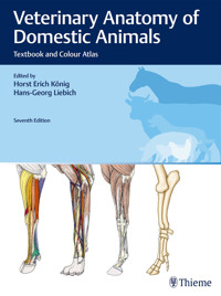
- Over 1100 exceptional images: anatomical specimens and histological images, thin slice plastinations, colour schematics, diagnostic imaging, sectional anatomy
- Numerous references to clinical and applied anatomy: including equine endoscopy, arthrocentesis, examination of the udder, rectal examination and laparotomy
- A unique bonus: CT, MRI and ultrasonographic images
Bringing anatomy to life!
Das E-Book können Sie in Legimi-Apps oder einer beliebigen App lesen, die das folgende Format unterstützen:
Seitenzahl: 1975
Veröffentlichungsjahr: 2020
Ähnliche


Veterinary Anatomy of Domestic Animals
Textbook and Colour Atlas
Horst Erich König, Hans-Georg Liebich
Christine Aurich, Hermann Bragulla, Klaus-Dieter Budras, Gerhard Forstenpointner, Jenny Hagen, Sibylle Kneissl, Horst Erich König, Rafael Latorre, Hans-Georg Liebich, Eberhard Ludewig, Johann Maierl, Christoph Mülling, Peter Paulsen, Christian Peham, William Pérez, Sven Reese, Jesús Ruberte, Hanna Schöpper, Johannes Seeger, Mircea-Constantin Sora, Péter Sótonyi, Gerald Weissengruber, Kirsti Witter
7th, updated and extended edition
1147 figures figures
Foreword to the 7th edition
In each of the six editions published since 1999, it has been our goal to provide readers with the latest scientific information, and to illustrate this with outstanding colour photographs of high-quality anatomical preparations, pictures derived from contemporary imaging modalities and digitally coloured schematic representations. The same approach has been rigorously pursued in this, the seventh, edition. Numerous suggestions from students and practising veterinary colleagues have been incorporated into this new version. Throughout this process, the overwhelmingly positive responses to our earlier work inspired and challenged us in equal measure to maintain our previously established standards.
The addition of a chapter on avian anatomy has transformed the book into a comprehensive volume covering the anatomy of domestic animals. Based on our colour text and atlas “Anatomie der Vögel” (2009; published in English as “Avian Anatomy” in 2016), this new chapter focuses on the anatomical features that are particular to the class Aves. More detailed descriptions and additional illustrations pertaining to the fundamental and clinical/applied anatomy of birds can be found in “Avian Anatomy”.
To acknowledge the lasting contribution that our book has made to veterinary anatomy, a further priority in developing the seventh edition was to preserve its essential purpose. Thus, the newly integrated learning and teaching materials are intended not only to satisfy the increasing demands of anatomical study and clinical practice, but also, ideally, to inspire enthusiasm in our readers for a discipline that is often regarded as “dry”. If we make any progress toward reaching this desired goal, we will have achieved a great deal.
The new edition covers essential aspects of systemic, topographic and clinical anatomy and aims to promote understanding of the complex relationships between anatomical structures and their associated functions. The contemporary approach of combining a veterinary anatomy textbook and colour atlas is further enhanced by the inclusion of imaging and pictures of anatomical sections. In this way, we seek to address the challenges of modern-day veterinary practice by providing practitioners with a tool for interpreting sectional representations obtained using diagnostic imaging modalities.
This edition has also been designed to facilitate modular interdisciplinary education in organ structure and function. Far more than ever before, veterinary curricula require students to acquaint themselves with the fundamentals of anatomy through independent study and critical evaluation. However, this can result in knowledge deficits that are often quite profound. This 7th edition strongly supports independent learning by organising information based on systems and topography. Presentation of fundamental anatomical content in this discipline-oriented manner facilitates understanding of applied anatomy in clinical contexts, as required in modular learning and teaching settings. To further ensure scientific rigour, anatomical terms used in the 7th edition are taken consistently from the most recent version of the Nomina Anatomica Veterinaria (2017).
Our combined text and colour atlas has in recent years been recognised internationally, earning acclaim from students and colleagues around the world. This is reflected in the appearance of licensed editions in 11 additional languages. Of particular note are the translations into Portuguese, Spanish, Italian, Polish, Turkish, Japanese and Chinese. Further translations are currently in preparation. Arguably the world’s most widely translated veterinary textbook, it serves as a valuable tool for students, helping them to prepare for and successfully complete their veterinary examinations. Moreover, as a companion reference book, it has an established role in facilitating proficiency in clinical practice. These are pleasing outcomes in which we take a certain degree of pride.
Updating of the educational content in this edition would not have been possible without the assistance of numerous colleagues. We have, in the past, had opportunity to express our deep appreciation to co-authors who contributed written content and images to earlier editions, to colleagues responsible for preparing anatomical specimens and to co-workers who brought their technical expertise to bear in ensuring that the various editions have been so aesthetically pleasing. In particular, we thank Dr. Polsterer (Vienna) for the schematic illustrations and Ms Schura (Munich) for her expertise in digital colouring.
Several colleagues in the scientific community have provided helpful suggestions and contributions to this latest edition, in the form of both text and images. We acknowledge and express our thanks to Professor Latorre (Murcia, Spain) and Dr. Hartmann (Stephanskirchen) for providing clear and informative arthroscopic images and pictures of the guttural pouch of the horse. Dr. Witter (Vienna) and Dr. Schöpper (Vienna) also deserve particular thanks for their contributions to the chapter on the common integument, and to the description of the teeth. Sincere thanks are due to Professor Kneissl (Vienna) and Professor Ludewig (Vienna) for revising and updating the section on diagnostic imaging. A further debt of gratitude is owed to Associate Professor Paulsen (Vienna) for contributing recent findings to the chapter describing the lymphatic organs. Professor Budras (Berlin) supplied important suggestions regarding the common integument. Selected material relating to hoof trimming, provided by Dr. Hagen (Leipzig) for the online component of the 6th German edition, has been incorporated into this new edition. With reference to the chapter on avian anatomy, we extend our thanks to Dr. Donoso (Universidad de Concepción, Chile) for furnishing us with a valuable image of a claw on the wing of a Chilean bird.
Particular thanks are due to Dr. Corinna Klupiec for her scholarly and technically competent translation of the additional text and image labels appearing in the 7th English edition. Her profound bilingual and anatomical skills have once again been demonstrated in this publication. Our thanks also go to Professor Simoens (Ghent) for his expert suggestions and for his critical appraisal and revisions of the text and image labels.In closing, we wish to thank Dr. Schäfer, Ms Schwarz and Ms Wallstein who, on behalf of the publisher, have provided active and helpful stewardship throughout the production of this 7th edition.
Vienna and Munich, in autumn 2019
Horst Erich König and Hans-Georg Liebich
Contents
Titelei
Foreword to the 7th edition
1 Introduction and general anatomy
1.1 History of veterinary anatomy
1.2 Directional terms and planes of the animal body
1.3 Division of the animal body in organs and organ systems
1.4 Locomotor apparatus (apparatus locomotorius)
1.4.1 Skeletal system (systema skeletale)
1.4.2 Muscular system (systema musculare)
1.5 General anatomy of angiology (angiologia)
1.5.1 Organisation of the cardiovascular system
1.5.2 Vessels (vasa)
1.5.3 Lymphatic system (systema lymphaticum)
1.6 General anatomy of the nervous system (systema nervosum)
1.6.1 Functions of the nervous system
1.6.2 Architecture and structure of the nervous system
1.6.3 Nerve tissue (textus nervosus)
1.6.4 Central nervous system (systema nervosum centrale, CNS)
1.6.5 Peripheral nervous system (systema nervosum periphericum, PNS)
1.6.6 Nerve transmission of information
1.6.7 Barriers in the nervous system
1.7 General anatomy of the viscera
1.7.1 Visceral mucosa
1.7.2 Visceral connective tissue
1.7.3 Functions of the viscera
1.7.4 Body cavities and their serous lining
2 Axial skeleton (skeleton axiale)
2.1 Overview of the skull
2.2 Overview of the vertebral column or spine
2.3 Overview of the thorax
2.4 Skeleton of the head
2.4.1 Skull, neural part (cranium, neurocranium)
2.4.2 Skull, facial part (facies, viscerocranium)
2.5 The skull as a whole
2.5.1 The skull of carnivores
2.5.2 Cavities of the skull of carnivores
2.5.3 The skull of the horse
2.5.4 Cavities of the equine skull
2.6 Vertebral column or spine (columna vertebralis)
2.6.1 Cervical vertebrae (vertebrae cervicales)
2.6.2 Thoracic vertebrae (vertebrae thoracicae)
2.6.3 Lumbar vertebrae (vertebrae lumbales)
2.6.4 Sacral vertebrae (vertebrae sacrales)
2.6.5 Caudal or coccygeal vertebrae (vertebrae caudales)
2.7 Thoracic skeleton (skeleton thoracis)
2.7.1 Ribs (costae)
2.7.2 Sternum
2.8 Joints of the skull and trunk (suturae capitis, articulationes columnae vertebralis et thoracis)
2.8.1 Joints of the skull (synchondroses cranii)
2.8.2 Joints of the vertebral column, the thorax and the skull (articulationes columnae vertebralis, thoracis et cranii)
2.8.3 Intervertebral articulations (articulationes columnae vertebralis)
2.8.4 Articulations of the ribs with the vertebral column (articulationes costovertebrales)
2.8.5 Joints of the thoracic wall (articulationes thoracis)
2.9 The vertebral column as a whole
3 Fasciae and muscles of the head, neck and trunk
3.1 Fasciae
3.1.1 Superficial fasciae of the head, neck and trunk
3.1.2 Deep fasciae of the head, neck and trunk
3.2 Cutaneous muscles (musculi cutanei)
3.2.1 Cutaneous muscles of the head (musculi cutanei capitis)
3.2.2 Cutaneous muscles of the neck (musculi cutanei colli)
3.2.3 Cutaneous muscles of the trunk (musculi cutanei trunci)
3.3 Muscles of the head (musculi capitis)
3.3.1 Facial muscles (musculi faciales)
3.3.2 Muscles of the lips and cheeks (musculi labiorum et buccarum)
3.3.3 Muscles of the nose (musculi nasi)
3.3.4 Extraorbital muscles of the eyelids (musculi extraorbitales)
3.3.5 Muscles of the external ear (musculi auriculares)
3.3.6 Mandibular muscles
3.3.7 Specific muscles of the head
3.4 Muscles of the trunk (musculi trunci)
3.4.1 Muscles of the neck (musculi colli)
3.4.2 Muscles of the back (musculi dorsi)
3.4.3 Muscles of the thoracic wall (musculi thoracis)
3.4.4 Muscles of the abdominal wall (musculi abdominis)
3.4.5 Muscles of the tail (musculi caudae)
4 Forelimbs or thoracic limbs (membra thoracica)
4.1 Skeleton of the thoracic limb (ossa membri thoracici)
4.1.1 Pectoral girdle (cingulum membri thoracici)
4.1.2 Skeleton of the arm (brachium)
4.1.3 Skeleton of the forearm (skeleton antebrachii)
4.1.4 Skeleton of the manus (skeleton manus)
4.1.5 Skeleton of the manus (forepaw) in carnivores
4.1.6 Skeleton of the manus in the horse
4.2 Joints of the thoracic limb (articulationes membri thoracici)
4.2.1 Articulations of the thoracic limb to the trunk
4.2.2 Shoulder or humeral joint (articulatio humeri)
4.2.3 Elbow joint (articulatio cubiti)
4.2.4 Articulations of the manus (articulationes manus)
4.2.5 Intermetacarpal joints (articulationes intermetacarpeae)
4.2.6 Phalangeal joints
4.3 Muscles of the thoracic limb (musculi membri thoracici)
4.3.1 Deep fasciae of the thoracic limb
4.3.2 Girdle or extrinsic musculature of the thoracic limb
4.3.3 Intrinsic musculature of the thoracic limb
5 Hindlimbs or pelvic limbs (membra pelvina)
5.1 Skeleton of the pelvic limb (ossa membri pelvini)
5.1.1 Pelvic girdle (cingulum membri pelvini)
5.1.2 Pelvic cavity
5.1.3 Skeleton of the thigh (skeleton femoris)
5.1.4 Kneecap (patella)
5.1.5 Skeleton of the leg (skeleton cruris)
5.1.6 Skeleton of the hindfoot (skeleton pedis)
5.2 Joints of the pelvic limb (articulationes membri pelvini)
5.2.1 Sacroiliac joint (articulatio sacroiliaca)
5.2.2 Coxofemoral or hip joint (articulatio coxae)
5.2.3 Stifle joint (articulatio genus)
5.2.4 Tibiofibular joints
5.2.5 Pedal joints (articulationes pedis)
5.3 Muscles of the pelvic limb (musculi membri pelvini)
5.3.1 Fasciae of the pelvis and the pelvic limb
5.3.2 Girdle or extrinsic musculature of the pelvic limb
5.3.3 Intrinsic musculature of the pelvic limb
5.3.4 Muscles of the crus
5.3.5 Short digital muscles
6 Statics and dynamics
6.1 Architecture of the trunk
6.2 Thoracic limb
6.3 Pelvic limb
6.4 Gaits
7 Body cavities
7.1 Positional stability of the organs in the body cavities
7.1.1 Thoracic cavity (cavum thoracis)
7.1.2 Abdominal and pelvic cavity (cavum abdominalis et pelvis)
8 Digestive system (apparatus digestorius)
8.1 Mouth and pharynx
8.1.1 Oral cavity (cavum oris)
8.1.2 Palate (palatum)
8.1.3 Tongue (lingua, glossa)
8.1.4 Sublingual floor of the oral cavity
8.1.5 Salivary glands (glandulae salivariae)
8.1.6 Masticatory apparatus
8.1.7 Pharynx (cavum pharyngis)
8.1.8 Muscles of the hyoid apparatus
8.2 Cranial part of the alimentary canal (oesophagus and stomach)
8.2.1 Oesophagus
8.2.2 Stomach (gaster, ventriculus)
8.3 Intestine
8.3.1 Structure of the intestinal wall
8.3.2 Innervation of the intestine
8.3.3 Blood supply of the intestine
8.3.4 Small intestine (intestinum tenue)
8.3.5 Large intestine (intestinum crassum)
8.4 Glands associated with the alimentary canal
8.4.1 Liver (hepar)
8.4.2 Gallbladder (vesica fellea)
8.4.3 Pancreas
9 Respiratory system (apparatus respiratorius)
9.1 Functions of the respiratory system
9.2 Upper respiratory tract
9.2.1 Nose (rhin, nasus)
9.2.2 Nasal cavities (cava nasi)
9.2.3 Paranasal sinuses (sinus paranasales)
9.3 Lower respiratory tract
9.3.1 Larynx
9.3.2 Trachea (trachea)
9.3.3 Lung (pulmo)
10 Urinary system (organa urinaria)
10.1 Kidney (nephros, ren)
10.1.1 Location of the kidneys
10.1.2 Shape of the kidneys
10.1.3 Structure of the kidneys
10.1.4 Functional units of the kidneys
10.1.5 Blood supply
10.1.6 Lymphatics
10.1.7 Innervation
10.2 Renal pelvis (pelvis renalis)
10.3 Ureter (ureter)
10.3.1 Blood supply
10.3.2 Lymphatics
10.3.3 Innervation
10.4 Urinary bladder (vesica urinaria)
10.4.1 Blood supply
10.4.2 Lymphatics
10.4.3 Innervation
10.5 Urethra (urethra)
11 Male genital organs (organa genitalia masculina)
11.1 Testis (orchis)
11.1.1 Structure of the testis
11.2 Epididymis
11.3 Deferent duct (ductus deferens)
11.4 Investments of the testis
11.4.1 Vaginal process (processus vaginalis) and spermatic cord (funiculus spermaticus)
11.4.2 Position of the scrotum
11.5 Blood supply, lymphatic drainage and innervation of the testis and its investments
11.6 Urethra
11.7 Accessory genital glands (glandulae genitales accessoriae)
11.7.1 Vesicular gland (glandula vesicularis)
11.7.2 Prostate gland (prostata)
11.7.3 Bulbourethral gland (glandula bulbourethralis)
11.8 Penis
11.8.1 Prepuce (preputium)
11.8.2 Muscles of the penis
11.8.3 Blood supply, lymphatic drainage and innervation of the urethra and the penis
11.8.4 Erection and ejaculation
12 Female genital organs (organa genitalia feminina)
12.1 Ovary (ovarium)
12.1.1 Position, form and size of the ovaries
12.1.2 Structure of the ovaries
12.2 Uterine tube (tuba uterina)
12.2.1 Mesovarium, mesosalpinx and ovarian bursa
12.3 Uterus (metra, hystera)
12.3.1 Structure of the uterine wall
12.4 Vagina
12.4.1 Vestibule of the vagina (vestibulum vaginae)
12.5 Vulva
12.6 Ligaments (adnexa)
12.7 Muscles of the female genital organs
12.8 Blood supply, lymphatic drainage and innervation
13 Organs of the cardiovascular system (systema cardiovasculare)
13.1 Heart (cor)
13.1.1 Pericardium
13.1.2 Position and size of the heart
13.1.3 Shape and surface topography of the heart
13.1.4 Compartments of the heart
13.1.5 Structure of the cardiac wall
13.1.6 Blood supply
13.1.7 Conducting system of the heart
13.1.8 Innervation
13.1.9 Lymphatics
13.1.10 Functions of the heart
13.2 Vessels (vasa)
13.2.1 Arteries of the pulmonary circulation
13.2.2 Arteries of the systemic circulation
13.2.3 Veins (venae)
14 Immune system and lymphatic organs (organa lymphopoetica)
14.1 Lymph vessels (vasa lymphatica)
14.2 Lymph nodes (lymphonodi, nodi lymphatici)
14.2.1 Lymph nodes of the head
14.2.2 Lymph nodes of the neck
14.2.3 Lymph nodes of the thoracic limb
14.2.4 Lymph nodes of the thorax
14.2.5 Lymph nodes of the abdomen
14.2.6 Lymph nodes of the pelvic cavity and the pelvic limb
14.3 Lymph-collecting ducts
14.4 Thymus (thymus)
14.4.1 Function of the thymus
14.4.2 Position and form of the thymus
14.5 Spleen (lien, splen)
14.5.1 Function of the spleen
14.5.2 Position and form of the spleen
14.5.3 Blood supply, lymphatics and innervation
15 Nervous system (systema nervosum)
15.1 Central nervous system (systema nervosum centrale)
15.1.1 Spinal cord (medulla spinalis)
15.1.2 Brain (encephalon)
15.1.3 Pathways of the central nervous system
15.1.4 Central autonomic nervous system
15.1.5 Meninges of the central nervous system
15.1.6 Ventricles and cerebrospinal fluid
15.1.7 Blood vessels of the central nervous system
15.2 Peripheral nervous system (systema nervosum periphericum)
15.2.1 Cerebrospinal nerves and ganglia
15.2.2 Cranial nerves (nervi craniales)
15.2.3 Spinal nerves (nervi spinales)
15.3 Peripheral autonomic nervous system (systema nervosum autonomicum)
15.3.1 Structure of the autonomic nervous system
15.3.2 Sympathetic system
15.3.3 Parasympathetic system
15.3.4 Intramural system
16 Endocrine glands (glandulae endocrinae)
16.1 Pituitary gland (hypophysis or glandula pituitaria)
16.1.1 Position and form of the pituitary gland
16.1.2 Function of the pituitary gland
16.2 Pineal gland (epiphysis cerebri or corpus pineale, glandula pinealis)
16.2.1 Position and form of the pineal gland
16.2.2 Function of the pineal gland
16.3 Thyroid gland (glandula thyroidea)
16.3.1 Position and form of the thyroid gland
16.3.2 Function of the thyroid gland
16.3.3 Blood supply, lymphatics and innervation
16.4 Parathyroid glands (glandulae parathyroideae)
16.4.1 Position and form of the parathyroid glands
16.4.2 Function of the parathyroid glands
16.4.3 Blood supply, lymphatics and innervation
16.5 Adrenal glands (glandulae adrenales or suprarenales)
16.5.1 Position and form of the adrenal glands
16.5.2 Function of the adrenal glands
16.5.3 Blood supply, lymphatics and innervation
16.6 Paraganglia
16.7 Pancreatic islets (insulae pancreatici)
16.8 Gonads as endocrine glands
17 Eye (organum visus)
17.1 Eyeball (bulbus oculi)
17.1.1 Shape and size of the eyeball
17.1.2 Directional terms and planes of the eyeball
17.1.3 Structure of the eyeball
17.2 Adnexa of the eye (organa oculi accessoria)
17.2.1 Orbit (orbita)
17.2.2 Fasciae and extrinsic muscles of the eyeball
17.2.3 Eyelids (palpebrae)
17.2.4 Lacrimal apparatus (apparatus lacrimalis)
17.3 Blood supply and innervation
17.3.1 Blood vessels of the eye
17.3.2 Innervation of the eye and its adnexa
17.4 Visual pathways and optic reflexes
18 Vestibulocochlear organ (organum vestibulocochleare)
18.1 External ear (auris externa)
18.1.1 Auricle (auricula)
18.1.2 External acoustic meatus (meatus acusticus externus)
18.1.3 Tympanic membrane (membrana tympani)
18.2 Middle ear (auris media)
18.2.1 Tympanic cavity (cavum tympani)
18.2.2 Auditory ossicles (ossicula auditus)
18.2.3 Auditory tube (tuba auditiva, Eustachian tube)
18.3 Internal ear (auris interna)
18.3.1 Vestibular labyrinth (pars statica labyrinthi)
18.3.2 Cochlear labyrinth (pars auditiva labyrinthi)
19 Common integument (integumentum commune)
19.1 Subcutaneous layer (subcutis, tela subcutanea)
19.2 Skin (cutis)
19.2.1 Dermis (corium)
19.2.2 Epidermis (epidermis)
19.2.3 Blood supply
19.2.4 Nerves and sense organs of the skin
19.3 Hairs (pili)
19.3.1 Hair types
19.3.2 Patterns of hair
19.3.3 Shedding
19.4 Skin glands (glandulae cutis)
19.4.1 Specialised forms of skin glands
19.5 Mammary gland (mamma, uber, mastos)
19.5.1 Suspensory apparatus of the mammary glands
19.5.2 Structure of the mammary glands
19.5.3 Development of the mammary gland
19.5.4 Lactation
19.5.5 Mammary glands (mamma) of carnivores
19.5.6 Mammary glands (mamma) of the pig
19.5.7 Udder (uber) of small ruminants
19.5.8 Bovine udder (uber)
19.5.9 Equine udder (uber)
19.6 Footpads (tori)
19.7 Digit (organum digitale)
19.7.1 Function
19.7.2 Segmentation
19.7.3 Horny enclosure of the distal phalanx (capsula ungularis)
19.7.4 Deciduous horn shoe (capsula ungulae decidua)
19.7.5 Modification of the different segments
19.7.6 Claw (unguicula)
19.7.7 Hooves (ungula) of ruminants and pigs
19.7.8 Equine hoof (ungula)
19.8 Horn (cornu)
19.8.1 Bovine horn (cornu)
19.8.2 Horn (cornu) of small ruminants
20 Topographical-clinical anatomy
20.1 Head (caput)
20.1.1 Stratigraphy
20.1.2 Regions
20.1.3 Clinical applications
20.2 Neck (collum)
20.2.1 Stratigraphy
20.2.2 Regions
20.2.3 Clinical applications
20.3 Thorax
20.3.1 Visible and palpable bony structures
20.3.2 Superficial thoracic and muscular grooves
20.3.3 Stratigraphy
20.3.4 Regions
20.3.5 Clinical applications
20.4 Abdomen
20.4.1 Visible and palpable bony structures
20.4.2 Superficially visible pathways
20.4.3 Stratigraphy
20.4.4 Regions
20.4.5 Clinical applications
20.5 Forelimb or thoracic limb (membra thoracica)
20.5.1 Regions
20.5.2 Clinical applications
20.6 Hindlimb or pelvic limb (membra pelvina)
20.6.1 Regions
20.6.2 Clinical applications
20.7 Palpable bony structures
20.7.1 Palpable bony structures of the head
20.7.2 Palpable bony structures of the neck and the back
20.7.3 Palpable bony structures of the thorax
20.7.4 Palpable bony structures of the thoracic limbs
20.7.5 Palpable bony structures of the pelvic limbs
20.8 Projection of the organs onto the body surface(1)
20.8.1 Organs of the abdominal cavity
20.8.2 Organs of the thoracic cavity
21 Avian anatomy
21.1 Introduction
21.2 Musculoskeletal system
21.2.1 Pectoral limbs (wings)
21.2.2 Pelvic limbs
21.3 Body cavities
21.4 Digestive system (systema digestorium)
21.4.1 Proximal digestive tract
21.4.2 Gastrointestinal tract
21.4.3 Accessory organs of the intestinal tract
21.5 Respiratory system (systema respiratorium)
21.5.1 Nasal cavity (cavum nasi)
21.5.2 Larynx
21.5.3 Trachea
21.5.4 Syrinx
21.5.5 Lung (pulmo)
21.5.6 Bronchial system and gas exchange
21.5.7 Air sacs (sacci pneumatici, sacci aerophori)
21.6 Urinary system (systema urinarium)
21.7 Male genital organs (organa genitalia masculina)
21.7.1 Testis (orchis)
21.7.2 Epididymis
21.7.3 Deferent duct (ductus deferens)
21.7.4 Phallus (penis, phallus masculinus)
21.8 Female genital organs (organa genitalia feminina)
21.8.1 Ovary (ovarium)
21.8.2 Oviduct (oviductus)
21.8.3 Uterus (metra)
21.8.4 Vagina
21.9 Structure of the avian egg
21.10 Cardiovascular system (systema cardiovasculare)
21.11 Immune system and lymphatic organs (organa lymphopoetica)
21.11.1 Lymphatic vessels (systema lymphovasculare)
21.11.2 Lymph heart (cor lymphaticum)
21.11.3 Lymphoreticular formations
21.11.4 Lymphatic organs (thymus, bursa cloacalis and spleen)
21.12 Central nervous system (systema nervosum centrale, CNS)
21.12.1 Brain (encephalon)
21.12.2 Spinal cord (medulla spinalis)
21.13 Endocrine glands (glandulae endocrinae)
21.14 Eye (organum visus)
21.14.1 Sclera
21.14.2 Middle layer (tunica media)
21.14.3 Inner layer (tunica interna bulbi, retina)
21.15 Ear (organum vestibulocochleare)
21.15.1 External ear (auris externa)
21.15.2 Middle ear (auris media)
21.15.3 Internal ear (auris interna)
21.16 Common integument (integumentum commune)
21.16.1 Skin and skin appendages
21.16.2 Featherless body regions
21.16.3 Feathered body regions
22 Sectional anatomy and imaging processes
22.1 Plastination in science
22.2 Diagnostic imaging
22.2.1 Imaging modalities
22.2.2 Endoscopy in the horse
Section II Appendix
23 Literature
24 Glossary of terms
About the Authors
Contact Information
Index
Imprint
1 Introduction and general anatomy
H.-G. Liebich, G. Forstenpointner and H. E. König
1.1 History of veterinary anatomy
G. Forstenpointner
The doctrine of morphology as the scientific study of the form and structure of organisms was founded by Aristotle. He defined morphology as the search for a common construction plan for all structures, while adhering to a strict methodological process. Where similarities can be found, the relation between form and function requires further clarification. This scientific approach set Plato’s best student apart from the early Greek natural philosophers and to this day is the principle method employed in all areas of basic research.
Presumably Aristotle performed anatomical research through dissections. References found in his work Historia Animalium indicate that he published another treatise, Parts of Animals, which sadly did not survive. This work dealt mainly with the digestive and reproductive systems. Here Aristotle recorded his impressions with schematic illustrations. Many of his observations were naturally incomplete, which often led him to erroneous conclusions. However, many of his thoughts on function remain worth reading: for example, his explanation of quadruped locomotion recorded in The Gait of Animals. Being a teacher, Aristotle’s greatest motivation for research was gaining knowledge for knowledge’s sake. This motivation was carried on by his student, Theophrastos of Eresos, and by Roman researchers in natural science such as Plinius and Aelian.
Almost two thousand years passed before the humanists of the 15th and 16th centuries renewed the Aristotelan access to comperative morphology. Especially in Italy, the study of animal and human bodies led to many new discoveries in this field. These findings were meticulously recorded in a demandingly artistic manner and are today famous works of art.
Leonardo de Vinci, by far the most famous artist of this period, embodied this new quest for knowledge and understanding, as is evidenced in his multifaceted research.
Other excellent researchers were Fabrizio d’Acquapendente, who completed the first work in comparative embryology (De formatu foetu, 1600), and Marcello Malpighi, who studied the development of the chick embryo (Opera omnia, 1687). Even though initial progress was impeded by an unstable political situation as well as by the religious establishment, these scientists were forerunners in their fields and heralded in a golden age of comparative anatomy. This trend continued until the end of the 19th century and was characterised by a period of extraordinary productivity of many notable naturalists.
Richard Owen, a renowned English anatomist, and the Germans Johann Friedrich Meckel and Caspar Friedrich Wolff played dominant roles in the resurgence of comparative anatomy as a discipline in Europe. Since the turn of the 20th century, the field of zoological research has been subject to constant redirection. This has led to the development of new disciplines which abandoned the orginal intentions of comparative anatomy as intended by its founders.
Anatomical knowledge is not achied for its own sake, but is a prerequisite for successful medical practice. Throughout the ages, due to religious and ethical boundaries, human dissections were highly restricted or forbidden outright. Recorded exceptions were seldom, one being from the Hellenic school of Alexandria under the leadership of Herophilus and Erasistratos. These vivisections on criminal convicted contributed to a greater understanding of neuroanatomy. Furthermore, Aristotle’s singular work on human anatomy was drawn from the dissection of a miscarried foetus.
Since animal dissections were the only possibility of studying the principles of form and function, these findings were extrapolated to human anatomy. Claudius Galenus, who served under the Emperors Marcus Aurelius and Commodus, became the most famous and influential doctor in Rome.
The results and interpretation which he gained through consequent research laid the unchallenged foundation of anatomical knowledge that lasted throughout the following 1500 years. Galen considered himself a physician by trade, however his understanding of anatomy and physiology was based upon Aristotle’s publications, such as The nature of Things. He ardently pursued research in these two disciplines Aristotle’s methodology. Because Galen’s teachings were intelligible and rational, they endured as the unchallenged foundation of anatomical and physiological knowledge for over 1500 years.
Even though his anatomical conclusions were sound, Galen’s interpretations of some systems, such as the heart and large vessels, were erroneous. Due to the lack of human autopsies, Galen’s extrapolations of animal dissection results were often misguided. For example, he suspected that the Rete mirabile epidurale would also be found in humans even though now we know it is a typical structure of ruminants. Further, he concluded that humans must have a caecum built like that of herbivores or a uterus with cotyledons.
In the tradition of Galen, an anatomical atlas of the pig (Anatomia porci, written by Copho or Kopho) was published in Salerno around 1100–1150. This was not the first anatomy book in veterinary medicine, but rather was meant as an anatomical teaching tool for students of human medicine. The generally accepted myth then and today – that the pig resembles the human more than any other animal – relies greatly on the similar eating habits and the availability of subject material in those days.
During the Renaissance, anatomical studies on human corpses were no longer taboo. With his monumental work on human anatomy (De humani corporis fabrica, 1543), Andreas Vesalius marked the hesitant beginning of a revolutionary new attitude toward the human body. Early anatomists still considered themselves naturalists, compiling many fundamental discoveries of morphology through continuing studies of animal anatomy. Vesalius was the first to realize that the Rete mirabile epidurale represented a typical structure of ruminants. Studies of ruminant digestion were advanced by Johann Conrad Peyer through his magnificent publication in 1685, Merycologia sive de ruminantibus et ruminatione commentarius ( ▶ Fig. 1.1). His discovery of lymphatic tissue (Lymphonoduli aggregati) in the intestinal mucosa resulted in the name Peyer’s patches. From the beginning, the study of comparative anatomy has remained a domain for research institutes specialising in human anatomy, even more so as zoological research turned away from the study of morphology.
Fig. 1.1 Cover of Merycologia.
(Johann Konrad Peyer, Basel, 1685)
Fig. 1.2 Early illustration of the blood vessels of the horse.
(Seifert von Tennecker, pseudonym Valentin Trichter, 1757)
In the last decades of the 20th century, the use of laboratory animals led to the optimizing of therapeutic approaches. The implementation of experimental concepts has been possible only through the application of necessary basic animal morphology, which has largely been provided by physicians. Interestingly, still today animals are chosen as models, not because of their morphological comparability, but rather for their availability.
Veterinary anatomy as a prerequisite for practicing veterinary medicine has developed only in the last few centuries as an independent teaching and research subject. It is evident through ancient and medieval texts for animal caretakers that the anatomical knowledge, especially of horses, was more or less precise ( ▶ Fig. 1.2). However, the systematic portrayal of the basic morphological associations was nonexistent.
Equerry handbooks created in the tradition of Jordanus Ruffus in the late middle ages and early modern age were not systematically organised. They did contain information on equine anatomy, which was often accompanied by ineffectual illustrations. In 1598, Carlo Ruini published an at that time exceptional handbook, Dell’ Anatomia e dell’Infirmita del Cavallo ( ▶ Fig. 1.3). Seeming to appear without a forerunner, this textbook was undoubtedly inspired by Vesalius.
Fig. 1.3 Illustration of the equine musculature.
(source: Dell’Anatomia e dell’ Infirmita del Cavallo; Carlo Ruini, Venice, 1598)
Ruini was born into a prosperous family from Bologna and neither worked as an equerry nor was a member of the university. Through excellent private tutors, he developed a passionate interest in the natural sciences and was an enthusiastic equestrian. Although incomplete and sometimes flawed, his seminal work was nevertheless the first comprehensive and systematic portrayal of equine anatomy. The second half of the book concerning equine diseases was largely an indiscriminate recapitulation of much older literature. The magnificence of this publication lies in its illustrative quality, which rivals that of Leonardo da Vinci or Vesalius. Ruini’s textbook was to be republished, plagiarised and translated many times ( ▶ Fig. 1.5).
At the beginning of the 17th century, veterinary anatomy was slowly beginning to enter a renaissance. However, it was not to be until about 150 years later that a veterinary academy was created where Ruini’s textbook could be used to train animal doctors.
Considered to be the father of veterinary anatomy, Philippe Etienne Lafosse opened at his own expense a private veterinary school in Paris in 1767. This endeavour proved unsuccessful, and the school was closed in 1770. Two years later, he published his most successful work, Cours d’Hippiatrique. (A Course on Hippiatry or A Complete Treatise on the Medicine of the Horse). This work was organised according to organ systems, fundamentally resembling the form used today in modern anatomy textbooks. The clinical relevance of a topographical approach was soon integrated into the teaching of anatomy.
One of the earliest topographical illustrations of the horse ( ▶ Fig. 1.4) is found in the lecture notes recorded and published in 1770 by Ludwig Scotti, the first director of the School for Horse Cures and Operations in Vienna. The development of anatomy as an independent discipline at the newly founded European veterinary schools was tentative at best. Consequently, it was not until 1822 that the first comprehensive anatomy text- or handbook was published. The first german comprehensive veterinary anatomy reference was Konrad Ludwig Schwab’s Anatomy Textbook of Domestic Animals of 1821 ( ▶ Fig. 1.6), followed closely by Ernst Friedrich Gurlt’s Handbook of Comparative Anatomy of Domestic Mammals in 1822 ( ▶ Fig. 1.7). These works represented the beginning of a long German tradition of veterinary anatomical research that quickly gained international recognition and lasted far into the 20th century. Eighteen editions of Gurlt’s work were published, with each new edition being revised and improved until the final one was print in 1943. Wilhelm Ellenberger and Hermann Baum were responsible for the 9th to the 17th editions, creating the style that can still be observed today in this readily available book ( ▶ Fig. 1.8 and ▶ Fig. 1.9). The sheer volume of publications in veterinary anatomy originating from Germany, Switzerland and Austria in the middle and late 19th century was overwhelming. This reflects the field’s significance and the high esteem in which veterinary anatomy was held in those days.
Fig. 1.4 Original drawing showing the body regions of the horse.
(from the lecture notes of Ludwig Scotti, School of Horse Cures and Operations, Vienna, 1770)
Fig. 1.5 Topography of the horse abdomen.
(William Gibson, London, 1754)
Fig. 1.6 Title page of the first german language Anatomy Textbook of Domestic Animals.
(Konrad Ludwig Schwab, Munich, 1821)
Fig. 1.7 First edition title page of Handbook of Comparative Anatomy of Domestic Mammals.
(Ernst Friedrich Gurlt, Berlin, 1822)
Fig. 1.8 Coloured illustration of the horse thorax.
(from the Atlas to the Handbook of Comparative Anatomy of Domestic Mammals; Ernst Friedrich Gurlt, Berlin, 1860)
Fig. 1.9 Vascularization of the equine hoof.
(from Leisering’s Atlas on the Anatomy of Horses and other Domestic Animals; Wilhelm Ellenberger in cooperation with Hermann Baum, Leipzig, 1899)
A landmark decision during the modern era of veterinary anatomy was the establishment of the International Committee for Nomenclature in Veterinary Anatomy. Emulating the human medical publication, Nomina Anatomica, the first edition of Nomina Anatomica Veterinaria was published in 1968. This work standardises worldwide anatomical terminology in veterinary medicine, thus providing a useful instrument to maintain the importance of anatomy in a steadily changing medical landscape.
Anatomy is the branch of morphology dealing with the form, structure, topography and the functional interaction of the tissues and organs that comprise the body. The dissection of dead animals is still the most important and efficient method to study and comprehend anatomy. With the advancement of classical anatomy, histology including microscopic anatomy and embryology developed into independent disciplines. Although inseparable as a whole, this division facilitates a more structured and thus easier approach to gaining anatomical knowledge.
Systematic anatomy has to do with “sytem”, in other words with structures and organs that fulfil a common function. The respiratory system for example is responsible for the gaseous exchange, whereas the nervous system receives, translates, transmits and responds to stimuli. Differences between the individual species can be compared, so that from an anatomical point of view “systematic anatomy” in teaching also represents a comparative anatomy, preferably limited to the domestic animals and poultry.
It is of great importance that students acquire a profound knowledge of systematic anatomy, from which they can then derive the overall connection of the structure and function of the animal body. Knowledge of systematic anatomy is the essential foundation for topographical anatomy, which describes the relative position and functional interaction of organs and structures of the various regions of the body. It presupposes a thorough working knowledge of systematic anatomy. Both systematic and topographic anatomy constitute the foundations of clinical practice.
Modern technologies, such as x-ray, ultrasound, computer tomography or magnetic resonance imaging, demand from the clinician greater knowledge of topographic anatomy, which is gained through studying sectional views of the body. Sectional anatomy represents a new direction in teaching and research in veterinary anatomy; a modern textbook would be incomplete without it.
1.2 Directional terms and planes of the animal body
H.-G. Liebich and H. E. König
Certain descriptive terms are employed to indicate precisely and unambiguously the position or direction of body parts. The most important anatomical terms are illustrated in ▶ Fig. 1.10 and the organ systems are listed with a short explanation in ▶ Table 1.1 .
Fig. 1.10 Directional terms and planes of the animal body (schematic); fig. based on data from Dyce, Sack and Wensing, 2002.
Fig. 1.11 Distal limb of a young cat during chondral ossification (histological section, Goldner staining).
Table 1.1
Directional terms and virtual planes of the animal body.
Term
Meaning
Usage
Cranial
Towards the head, trunk and tail
Trunk and tail, limbs proximal to the carpus and tarsus
Rostral
Towards the tip of the nose
Head
Caudal
Towards the tail
Head and trunk, limbs proximal to the carpus and tarsus
Dorsal
Towards the back
Trunk, head and the front of the limbs distal of carpus and tarsus
Ventral
Towards the belly
Underside of the trunk, head
Medial
Towards the centre
Head, trunk and limbs
Lateral
Towards the side
Head, trunk and limbs
Median
In the middle
Trunk, head and limbs
Proximal
Towards the trunk
Limbs and other body parts located close to the trunk or projecting away from the trunk
Distal
Away from the trunk
Limbs and other body parts located at a distance form the trunk or projecting away from the trunk
Palmar
Towards the palm of the hand
Forelimbs distal of the carpal joint
Plantar
Towards the sole of the foot
Hindlimbs distal of the tarsal joint
Axial
Towards the axis of the digits
Digits
Abaxial
Away from the axis of the digits
Digits
External
Located outside
Body parts and organs
Internal
Located inside
Body parts and organs
Superficial
Located near the surface
Body parts and organs
Deep
Located in the depth
Body parts and organs
Temporal
Towards the temporal bone
Eye
Nasal
Towards the nose
Eye
Superior
Above
Eyelid
Inferior
Below
Eyelid
Apical
Towards the tip
Nose, digits and tail
Oral
Towards the mouth
Head
Virtual planes of the animal body
Median plane
Plane dividing the body in two equal parts
Paramedian plane
Any plane parallel and close to the median plane
Sagittal plane
Any plane parallel to the median plane but located further lateral
Dorsal plane
Any plane parallel to the dorsal surface
Transverse plane
Any plane perpendicular to the long axis
The body of an animal has major divisions which are clearly distinguishable externally: the head (caput), the neck (collum), the trunk (truncus), the tail (cauda) and the limbs (membra). Each one of these sections is in turn divided into regions, which function as descriptive motifs for the topographic anatomy; for more information see Chapter 20 ▶ “Topographical-clinical anatomy”.
1.3 Division of the animal body in organs and organ systems
H.-G. Liebich and H. E. König
Cells and tissues similar in structure and function are joined together to form individual organs or organ systems. These act synergistically to fulfill functions that define the organism and ensure survival. The individual organ systems are composed of different types of tissue. An individual organ is made up of two types of tissue:
parenchyma and
interstitial tissue.
The cells of the parenchyma are responsible for the function of the organ (e.g. hepatic cells of the liver, renal cells of the kidneys, glandular cells of the salivary glands). The interstitial tissue builds the connective tissue that, for example, either encloses a small functional unit or separates larger areas of an organ into lobules (lobuli) or lobes (lobi). Connective tissue also supports metabolic transport to and from the organs, enclosing not only blood and lymph vessels but also peripheral nerves from the nervous system, all of which supply the organ. Together these structures form an overriding system that greatly influences the structural and functional character of an organ. Systematic anatomy examines in detail individual organ systems of the body, which are listed in ▶ Table 1.2 .
Table 1.2
Organ systems.
Name
Primary function
Outer skin
Protective covering of the animal body
Skeleton and joints
Supporting framework of the body
Musculature of the skeleton
Locomotion
Digestive system
Food intake, mastication, chemical digestion, excretion and absorption
Respiratory system
Oxygen supply, elimination of carbon dioxide and production of sound
Urogenital system
Excretion and reproduction
Circulatory system
Transport and exchange of substances
Nervous system
Regulation, transmission, reaction in response to external stimuli
Organs of sense
Reception of external stimuli
Endocrine glands
Regulation of cell functions by hormones
Immune system
Response to infection
Veterinary anatomy deals mainly with domestic mammals. These are taxonomically classified as Canis lupius f. familiaris (dog), Felis sylvestris f. catus (cat), Sus scrofa f. domestica (pig), Bos primigenius f. taurus (cattle), Ovis ammon f. aries (sheep), Capra aegagrus f. hircus (goat) and Equus przewalskii f. caballus (horse). Also included in the study of veterinary anatomy is poultry, whereby Gallus gallus f. domestica (chicken) is the most common specimen. Poultry is an important field in veterinary medicine and is therefore extensively covered in a separate textbook containing a revised and updated introduction to avian propedeutic and clinical medicine (König HE, Korbel R, Liebich H-G. Anatomie der Vögel. 2. Aufl. Stuttgart: Schattauer; 2009).
1.4 Locomotor apparatus (apparatus locomotorius)
H.-G. Liebich and H. E. König
The locomotor apparatus is a complex organ system whose primary function is mechanical. The skeleton and the muscles are the major elements comprising this system, forming and maintaining the individual body shape and providing for the locomotion of body parts or the whole organism.
The skeleton is composed of individual elements: thebones (ossa), cartilage (cartilagines), ligaments (ligamenta) and the joints (articulationes) that together create the body’s framework, the skeletal system (systema skeletale).
The skeletal system constitutes the passive part of the locomotor system, whereas the musculature (systema musculare) represents the active part. Both parts form a functional unit that is integrated into the body’s circulatory, lymphatic and nervous systems.
The system performs many metabolic functions at a cellular level. Hormones regulate a constant process of growth, modification and breakdown. The term “locomotor system” does not do justice to this many-facetted system; therefore this system is more appropriately referred to as the system of motion, stability and support.
Malfunctions and diseases of this system are among the most common diagnoses made in clinical veterinary medicine. The importance of basic anatomical knowledge is often greatly underestimated.
1.4.1 Skeletal system (systema skeletale)
1.4.1.1 Osteology (osteologia)
Osteology is the study of bones (ossa) that combine to form the skeletons of diverse animal species. Bones are composed of:
bone tissue which is sheathed inside and outside by the
endosteum and periosteum, respectively, and the
bone marrow (medulla ossium), as well as the
blood vessels and nerves supplying these structures.
These components classify bone as an organ.
The individual form of each bone is genetically determined and remains even though forces of traction and compression subject the bone to continual adaptation processes. Because of their high mineral content (60–70%), bones do not undergo post-mortem changes and are thus useful objects of archaeological study. Maceration of bone is the process of removing organic components through the use of weak lye. Bones used as teaching objects have usually undergone such treatment. Treating the bones with an acid removes the inorganic or mineralised components.
Skeletal design
Connective tissue precursor of bone
All components of the skeletal system develop from the middle embryonic germ layer (mesoderm). Early in embryonic development, the mesoderm differentiates into three types of connective tissue: embryonic, reticular and fibrous. These tissues consist of:
cells (e.g. fibrocytes),
fluid-filled intercellular spaces and
fibrous components (collagen and elastin).
These tissues become more abundant as development progresses, and at genetically determined locations these tissues transform into tendons, ligaments and fasciae. Developmental processes in the embryonic regions of the trunk and limbs begin early in development and lead to structural and functional specialisation of the embryonic tissue. The two elements of support tissue, cartilage and bone, develop from this primordial loose connective tissue (textus connecticus collagenosus laxus).
Both bone and cartilage originate from mesenchymal precursor cells, the chondroblasts andosteoblasts, respectively, which mature into the chondrocytes and osteocytes. These cells synthesise a matrix of collagen fibres and matrix.
Development and growth of cartilage
Cartilage is characterised by the structure of its formless matrix, the intercellular substance, which consists primarily of glycosaminoglycans. Embedded in the intercellular substance are collagenous fibres, the structural element of cartilage. This unique construction lends cartilage strength and flexibility. Due to their chemical structure, glycosaminoglycans are able to bind water, resulting in the increased elasticity and pliability of cartilage.
Blood vessels and nerves are absent in cartilage. Nutrients must diffuse through the matrix from blood vessels located in surrounding connective tissue, synovia or subchondral bone.
There are three types of cartilage classified according to the quality of the embedded fibres: hyaline, elastic or fibrous cartilage.
In adults, hyaline cartilage persists at the articular ends of long bones (cartilagines articulares), at the tips of the ribs (cartilago costae), and in parts of the laryngeal (cartilago laryngis), tracheal (cartilago trachealis), and bronchial (cartilago bronchialis) walls. Elastic cartilage forms the internal support for the epiglottis and the ear. Fibrocartilage forms the intervertebral discs, the menisci and the articular disc in the jaw joint. With increasing age, cartilage may ossify, to become invested with calcium salts. This occurs frequently in cats, for example, in the costal cartilage or the menisci.
The formation of cartilage (chondrogenesis, ▶ Fig. 1.11) originates in the mesenchymal (embryonal) connective tissue (see above), remnants of which still surround the cartilage in later stages of development. These remnants are the perichondrium, cells of which the fibroblasts, differentiate into chondroblasts, which produce the cartilage matrix containing water (70%), collagenous or elastic fibres and glycosaminoglycans.
Cartilage growth occurs through proliferation of chondroblasts in the perichondrium. This continual process leads to the appositional expansion of cartilage where new cartilage is created on the bone perimeter, directly beneath the surrounding perichondrium. Conversely, interstitial growth involves the proliferation of differentiated chondroblasts within the cartilage matrix, which continue to divide and form new matrix substance from the inside.
Forms of bony tissues
Bones differ greatly in form, size and strength, not only between species but also within the same individual. These characteristics of bone are determined greatly through genetics, but also static-dynamic influences and structural changes due to nutrition during the juvenile and adult phases play an important role. Broad muscles or cordlike tendons create mechanical influences at their insertion points on the bone, leading to the development of a process, depression, tuberosity/protuberance, unevenness, ridge, or an edge. Blood vessels, nerves or organs (i.e. the brain, the eye, the cochlea of the inner ear) can also influence the surface structure of bone.
Despite the great variety of bones, they can be grouped according to common structural characteristics. These are as follows:
long bones (ossa longa),
short bones (ossa brevia),
flat bones (ossa plana),
pneumatic bones (ossa pneumatica) and
irregular bones (ossa irregularia).
Long bones are characterised by a shaft or diaphysis, formed from a thick, outer layer of compact bone (substantia compacta), and an inner medullary cavity (cavum medullare, ▶ Fig. 1.12). Long bones have two ends, the proximal epiphysis and the distal epiphysis, which are covered by a thin layer ofcortical substance (substantia corticalis). Both extremities contain spongy bone, which, as the name implies, resembles an ossified sponge with delicate pores (substantia spongiosa, ▶ Fig. 1.13 and ▶ Fig. 1.14). Long bones form the basis of the limbs, i.e. upper arm (humerus), shin bone (tibia), or the metacarpal bones (ossa metacarpalia).
Fig. 1.12 Sagittal section of a long bone after maceration (A), and sagittal section of an untreated long bone including joint cartilage and red bone marrow(B).
Fig. 1.13 Architecture of the wall of a long bone showing compact and trabecular bone.
Fig. 1.14 Cross section of lamellar bone.
Short bones can have different forms: cylindrical, cubic or round. Such bones contain an extensive latticework of spongiosa in which haemoreticular tissue is present. Examples of short bones are those of the vertebral column and the hock joint.
Flat and wide bones consist of two layers of compact bone (tabulae) surrounding either spongy bone (diploe) or air (sinus). This group contains for example the scapula, the iliac bone or the ribs. Some bones of the skull are flat bones surrounding cavities of air (ossa pneumatica). These bones have formed through the subsequent resorption of bone substance and are lined with mucosa. Examples are the maxilla or the ethmoid bone.
Examples of irregular bones are the wedge-shaped bones of the skull: the sphenoid, presphenoid and basisphenoid bones. Sesamoid bones (ossa sesamoidea) are found close to the joints (i.e. the foot joints) and either lie beneath or are embedded in (i.e. patella) a tendon ( ▶ Fig. 1.33).
An apophysis is a bony protuberance that developed from an independent ossification centre. These structures provide attachment sites for muscles and ligaments. An example is a vertebral spinous process or the trochanter major on the femur. Bones of the organs are not related to the locomotor system. Such bones are found in the penis of male cats and dogs or in the bovine heart.
▶ Fig. 1.25, ▶ Fig. 1.26, ▶ Fig. 1.27, ▶ Fig. 1.28 and ▶ Fig. 1.29 schematically show the skeletons of the domestic animals covered in this textbook: cat, dog, pig, ox and horse. These illustrations provide a general overview of the topography of the bones, enabling a comparison between the species. The individual bones are described in detail in later chapters.
Architecture of bone
The high stability of bone is created through the bone tissue. Bone tissue is not massive and homogenous, but rather each individual bone has a specific architecture which is influenced by the:
structure of the compact bone (substantia compacta),
arrangement of the spongy bone (substantia spongiosa),
form of the central medullary cavity (cavum medullare),
principles of tensile (traction) and compressive stress (pressure),
formation of stress trajectories and
flexure (shear stress) demands on the entire bone.
The bone surface is constructed of compact lamellae which form the basis of the compact substance of bone. This solid layer surrounds the spongiosa, a delicate latticework of bone trabeculae and lamellae. Trabeculae and lamellae are arranged in a pattern of stress lines that have formed in response to external mechanical factors, the maximal tensile and compressive forces acting on the bone. The stress lines are either tensile or compressive trajectories. The family of curves that are tensile trajectories run parallel to one another just as the compressive trajectories run parallel to each other. These two types of stress trajectories always cross at right angles to each other (trajectorial construction). One can distinguish between:
tubules of bone (substantia tubulosa),
trabeculae of bone (substantia trabeculosa) and
lamellae of bone (substantia lamellosa, ▶ Fig. 1.13 and ▶ Fig. 1.14).
Stress occurring in the diaphysis of long bones does not affect stabilty but leads to tensile forces on the convex side of the bone and compressive forces on the concave side of the bone. In the centre, these two forces cancel out one another, and the net force is negligible. Thus the bone does not have to contain force-bearing structures in its centre: the ideal shape of bone is a long, hollow tube with reinforced walls, such as the diaphysis.
Instead of spongiosa as in the epiphyses, the diaphysis encloses themedullary cavity (cavum medullare). Here the compact substance is reinforced through thickened layers of lamellar bone ( ▶ Fig. 1.12). The medullary cavity is filled with red bone marrow where blood cells are produced (hematopoiesis), thus classifying bone as a hemopoietic organ.
Bone is built to maximise strength and stability while simultaneously minimizing material and weight. The architecture of bone creates the optimal prerequisites: the hollow tube creates strong resistance to stress, and the spongiosa economises material and is very light. The thickness of the diaphysis adapts to the maximal strain excercised on the bone. The medial walls of the limb bones carry a greater load and thus are thicker than the outer walls. Flat bones such as the scapula are denser on the edges and therefore thinner in the centre. Inorganic substances account for approximately two-thirds of a bone’s dry weight. The remaining one-third is the organic substance of the bone, comprised mainly of collagen structural proteins and lipids (5–10%). Bone decalcification with acid removes the inorganic substances in bone, leaving the treated bone soft and pliable. Burning a bone destroys the organic substances; what remains are the bone ashes.
Endosteum and periosteum
Bones are sheathed on the inside and outside surfaces by a connective tissue membrane called the endosteum and periosteum, respectively. The endostium lines the medullary cavity and covers the spongiosa, thus creating a border between bone or spongiosa and bone marrow ( ▶ Fig. 1.17). The periosteum covers the external surface of bone, but is absent from the articular surfaces and where tendons and ligaments attach. Close to the joints, the periosteum detaches from the surface of the bone and consolidates with the joint capsule. On the opposite side of the joint, the periosteum leaves the capsule and attaches again to the surface of the adjacent bone. At bone/cartilage intersections, for example on the ribs, the periosteum continues over the cartilage as the perichondrium.
The periosteum is necessary not only for the blood supply, growth, regeneration, and fracture repair of bones, but also for the transfer of muscle power to the bone. The periosteum is composed of two layers, the:
inner cellular,osteogenic layer (stratum osteogenicum, previously stratum cambium) and
outer protective fibrous layer (stratum fibrosum).
The stratum osteogenicum ( ▶ Fig. 1.16 and ▶ Fig. 1.17) lies directly on the bone and produces bone tissue (i.e. is osteogenic). A great number of sensory nerve fibres as well as a network of blood and lymphatic vessels supplying the bone are enclosed in this layer. This layer also contains progenitor cells, the preosteoblasts, which can differentiate into osteoblasts, which produce bone and are responsible for appositional growth. This layer retains lifelong the ability to build bone tissue, which is important for physiological bone remodeling and fracture healing. The stratum osteogenicum forms the cartilage callusand bone callus, and prolonged mechanical stimulus of the periosteum can lead to the formation of osseous bulges (exostoses or splints).
Fig. 1.15 Middle phalanx of a horse embryo (histological section, Azan staining).
Fig. 1.16 Embryonic vertebrae (histological section, Azan staining).
Fig. 1.17 Section of compact bone from the diaphysis (schematic).
Dense connective tissue interwoven with elastic fibers makes up the outer layer, the stratum fibrosum ( ▶ Fig. 1.16 and ▶ Fig. 1.17), which is very resistant to tensile forces. Collagen fibre bundles originating in this layer (perforating fibres) bind it to the surface lamella of the bone matrix (Sharpey fibres). These fibres firmly anchor the periosteum to the bone surface. The stratum fibrosum is also responsible for the attachment of muscles, tendons and ligaments to bone. At the site of attachment, fibres from the tendon or ligament branch out into the stratum fibrosum and, continuing as Sharpey fibres, strongly attach to the bone.
The endosteum ( ▶ Fig. 1.17) consists of a single layer of flattened, inactive osteoprogenitor (bone-lining) cells. They can differentiate into either bone-forming cells (osteoblasts) or bone-resorbing cells (osteoclasts). The endosteum borders the capillary network of the bone marrow and like the periosteum, is capable of producing bone tissue (osteogenic potential).
Bone regeneration
Osteoprogenitor cells in the periosteum and endosteum are responsible for regeneration processes of the bone tissue. Regeneration is only possible when two conditions are met: 1. mesenchyme cells are available and 2. osteoblast precursor cells can proliferate. New tissue bridges the gap in the bone resulting from fracture.
Primary fracture healing occurs when motion between the two fracture pieces is negligible and they are separated by only small gaps. Lamellar bone forms directly in the fracture gap, reuniting the two ends of the bone. When the edges are too far apart, then secondary fracture healing occurs. Fibrous connective tissue initially bridges the fracture gap forming a soft callus. The callus ossifiesthrough mineralisation until, after a long reorganisation process, compact bone is formed.
Supply of blood vessels and nerves to the bone
Bone is an extremely well vascularised tissue, and this underscores its metabolic importance. A dense network of blood vessels supplies not only bone tissue, but also bone marrow, the periosteum and the endosteum. Bone trauma or fracture can interrupt the vascularisation, leading in extreme cases to tissue death (bone necrosis).
The vascularisation of bone is achieved through a unique, systematic distribution of blood vessels. Nutrient arteries (aa. nutriciae) branch off from the larger limb arteries and enter the long bones through openings (foramina nutritia) in the diaphysis. These vessels reach the medullary cavity after passing through the stratum compactum. Here they divide into several ascending and descending branches supplying the proximal and distal epi- and metaphyses ( ▶ Fig. 1.23). At the epiphyses, the vessels form looped-ending arteriesthat reach through the epiphyseal subchondral bone to supply the calcified zone of the joint cartilage. From the medullary cavity, the blood vessels supply the compact substance of bone through the Volkmann’s canals (see below). The spongy bone does not contain blood vessels but is supplied through diffusion from the bone marrow. The venous return occurs through the axial system of the bone marrow.
Bone tissue does not contain lymphatic vessels. A dense network of lymphatic vessels is present only in the periosteum. Bone tissue itself is not sensitive to pain. Alone vegetative nerve fibres follow the path of the blood vessels within the Haversian canals.
Bone as an organ
Bone constitutes an organ-like system composed of:
ossified elements,
joint cartilage (when present),
periosteum and endosteum,
bone marrow and
nerve tracts.
Bone architecture and its extracellular matrix (organic and inorganic material) provide the stabilising components of the passive system of motion, stability and support. The organization of collagen I fibres, the mineralised interfibrillary matrix and the structure of bone tissue all play a large role in stabilisation.
A bone can withstand the application of mechanical stress, body weight, muscle strength or acceleration. These forces work as compression, traction, loading, torque and shear, and do not result in fracture when they are within certain limits. As opposed to the intermittent application of force, bone that experiences a continuous loading force atrophies. On the other hand, bone experiencing constant tensile force hypertrophies.
The architecture of bone tissue is governed lifelong by functional demand. Compact and spongy bone structures are continuously adapting to changes in biomechanical forces. The endosteum is responsible for inducing these structural changes which occur following the physiological principles of bone formation and resorption, for more information see Chapter ▶ “Osteogenesis”.
Osteogenesis
During foetal development, a precursor skeleton of cartilage is formed, providing support and shape (primordial skeleton)for the growing foetus. Until ossification, this primordial skeleton undergoes quick successions of mitotic division, eventually determining the growth and form of the entire organism. In most cases, each piece of the primordial skeleton acts as a space saver for the bone tissue that will eventually replace the cartilage. Positively influencing bone tissue formation are inductive mediators (i.e. bone morphogenetic protein, mitogenic factors). At a certain developmental stage, the cartilage of the primordial skeleton slowly undergoes remodelling. The cartilage is slowly resorbed and eventually replaced by bone. This process is chondral orindirect ossification. New, foetal bone is referred to as immature or woven bonedue to the random honeycomb architecture of the trabeculae. Eventually, resorbed and woven bone is replaced with mature lamellar bone. The majority of adult bones (i.e. the vertebrae and limb bones) are formed through chondral ossification.


