20,99 €
Mehr erfahren.
- Herausgeber: Crowood
- Kategorie: Lebensstil
- Sprache: Englisch
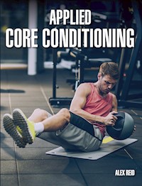
Applied Core Conditioning contains all the information necessary to help design, prescribe and programme core conditioning strategies for those who wish to remain well conditioned and to prevent injury, and for those undergoing rehabilitation. The knowledge that eighty percent of us all will experience low back pain at some point in our lives should be motivation enough to take conditioning and preventative steps via an effective applied core conditioning programme. The book presents sport specific solutions for exercise selection, with programme design, sets, repetitions and loading all discussed in detail, in addition to a chapter presenting six different case studies that reflect the challenges of rehabilitating debilitating injuries like prolapsed disc, pars defect and hamstring tendinopathy, amongst others. Effective rehabilitation strategies are presented in detail to provide an insight into recovery and strengthening concepts. Aimed at serious sports people and coaches it presents six different case studies that reflect the challenges of rehabilitating debilitating injuries. Fully illustrated with colour photographs and diagrams.
Das E-Book können Sie in Legimi-Apps oder einer beliebigen App lesen, die das folgende Format unterstützen:
Veröffentlichungsjahr: 2019
Ähnliche


APPLIEDCORE CONDITIONING
ALEX REID
THE CROWOOD PRESS
First published in 2018 byThe Crowood Press LtdRamsbury, MarlboroughWiltshire SN8 2HR
www.crowood.com
This e-book first published in 2019
© Alex Reid 2018
All rights reserved. This e-book is copyright material and must not be copied, reproduced, transferred, distributed, leased, licensed or publicly performed or used in any way except as specifically permitted in writing by the publishers, as allowed under the terms and conditions under which it was purchased or as strictly permitted by applicable copyright law. Any unauthorised distribution or use of this text may be a direct infringement of the author's and publisher's rights, and those responsible may be liable in law accordingly.
British Library Cataloguing-in-Publication Data
A catalogue record for this book is available from the British Library.
ISBN 978 0 78500 522 0
Dedication
For Malc. For Mum. For Dad. Thank you.
CONTENTS
Acknowledgements
Getting Through This
1 Introduction: What is the Core? Why is it Important?
2 Musculoskeletal Disorders: The Financial Cost and Possible Solutions
3 Case Studies: The Practical Application of Core Conditioning, Prescription and Exercise Selection
4 Applied Core Conditioning: Programming, Prescription and Sports-Specific Training
5 Summary
Glossary
References
Index
ACKNOWLEDGEMENTS
Thank you to the following contributors within this book. Your efforts and the time given to help bring things together are greatly appreciated:
Core, anatomy and muscular images: Visualcoaching Pro (visualcoaching.com); Photography: Peter Court Media Services
The case studies in this book are anonymous, but I would like to thank my clients, who have provided me with insight and experience, and inspired me to provide optimal conditioning, rehabilitation and reconditioning programmes for them.
I would also like to thank my family, particularly Isla and Machrie, as well as my friends who have encouraged and supported me during the writing and creation of this book.
GETTING THROUGH THIS
I was asked to write this foreword as a fairly average person who was fit and healthy and worked hard at a lot of different things. I started running in my early thirties and quickly challenged myself to get faster and faster. I pushed myself quite hard and was fairly happy with my personal bests at 10k and half-marathon distances. Then, when I was thirty-five I ruptured my anterior cruciate ligament playing a bounce game of 5-a-side football. I went home after the game thinking I had done something bad. It was diagnosed by the physio the next day and all was confirmed by a MRI scan a couple of days later. I had little knowledge of the severity of this injury until I started my rehabilitation. Notwithstanding a second operation due to receiving an infection during my first operation, the journey back to running was an arduous, yearlong, pain-threshold-extending challenge. I’ve never recovered fully from this and, with the exception of a burst of activity to beat my 10k personal best, I’ve never got near my old cadence.
In 2017, six years after my ACL repair, I suffered a prolapsed disc in my lower back. Anyone who has ever experienced this (and you may well have if you’re taking the time to read this book) will know the pain a body can go through. The ACL injury was nothing in comparison. Prolapsed discs don’t usually happen because of a one-off episode. Instead, they usually build up over a prolonged period of time. I put mine down to a few years of imbalance after losing the ability to bend my right knee properly following the two ACL operations. Not being strong enough in my lower back, having poor posture and indulging in some rather savage sessions removing bamboo from our front drive, all conspired to create the perfect storm.
Six months after the prolapsed disc diagnosis, I am now able to run again, but those six months have been torture. It affects everything. Even trying to move an inch in bed is difficult. Your head thinks about your posture the whole day. It gets to you. You can’t sit with your family and have a bit of fun. You can’t lie on the floor, sit on a chair, cross the road fast, sit at your PC. You walk along the road and spasm. You stare at seats on the train trying to work out how to sit on them.
The only way you can recover is by getting stronger and preventing it from happening again. The core is the key. Every exercise that strengthens your trunk brings you one step closer to shaking off vulnerability. From my ACL recovery and strengthening everything around the knee, my focus has now shifted to strengthening my core. If only this book had already been written, and I had started earlier!
Anonymous
CHAPTER 1
INTRODUCTION:WHAT IS THE CORE?WHY IS IT IMPORTANT?
As you sit reading this, you will be subconsciously activating your abdominal muscles and trunk, commonly referred to as your ‘core’. Without this activation, our skeleton would be floppy, our spine unsupported and our overall function compromised. The muscles around our mid-section or trunk are constantly working to allow locomotion, smooth movement, control and function, and the activities that we do constantly demand this activation from the muscles involved.
Beth West, one of my good friends, posted the following on social media whilst on her journey to work in London one day in 2017:
Commuting on Southwest Trains of a morning is a tough core workout. Too crowded to be able to reach anything to hold on to, you must use your core to ensure that you are not toppling on to fellow passengers. They should market that. Maybe put some personal trainers on carriages and make it a proper workout!
A true reflection of the day-to-day challenges placed upon us all, it made me laugh out loud. Our core, lumbar spine, glutes, pelvis and associated limbs are constantly in use during our working day, with numerous actions requiring strength and control. When standing on a busy commuter train, we must stabilize and control our body all the time, in order not to fall on to the person next to us. It is a good example of a chaotic environment that we need to control, just like sport or lifting or working in a physical job. Every day, we place many demands on our body and expect it to cope. Sometimes we forget that, although the human body is made to work and move, if it is not properly conditioned, then injury is likely.
The body works as a global, combined unit. Despite a limb moving, it may appear, independently, on the contrary the movement is as a result of a number of combined, coordinated actions within the body: a neural, muscular, metabolic, conscious and subconscious decision to complete a movement made by a reaction to a stimulus, by choice, or sometimes involuntary, like reacting to a sudden move on that busy train, or reaching instinctively to catch something as it falls, or playing catch with your kids. It is very complex, but it works seamlessly on most occasions and allows us to be the mobile, skilled, dexterous beings that we are.
But what happens if we get injured? What if we are not strong enough to cope with these demands or have bad posture, which leads to pain and immobility? What if there is a leg-length discrepancy, which may lead to poor biomechanics and resultant back pain, or an over-use injury as a result of a muscular imbalance? How in those cases do we address the day-to-day issues and challenges? The solution lies in becoming stronger and better conditioned, ensuring good mobility, retraining our movement patterns, and reintroducing effective and efficient activation of the trunk and core musculature.
The muscles around the abdominal region, known widely as the core musculature, originate and insert within the axial skeletal system and link through to the appendicular skeleton. They are the link in the chain that coordinates our movement. Without them, movement would be clumsy and uncoordinated. The body works well as a unit, but sometimes it needs to be retrained and reeducated to take care of itself. It is very easy for it to become lazy, to adopt bad habits and become deactivated, which can cause problems. Often the larger, more dominant muscles are recruited before the more appropriate, synergistic muscles. This poor muscle recruitment can lead to an imbalance and over-use.
The lifestyle choices that many people make in this time of high technology – sitting for hours playing games, using social media or messaging on their devices – can cause a deconditioning of the body and, potentially, long-term health issues. Prolonged use of and addiction to mobile phones and hand-held devices lead to poor posture and bad habits, which can cause muscular over-use and pain.
Prolonged use of smart phones and hand-held devices often lead to poor posture and resultant neck pain.
Similarly, drivers who commute to work by car, or drive for a living, can also suffer health problems, as a result of sitting in a flexed position for many hours, often with poor posture and with the neck tilted forward. We have all got out a car after a long journey and felt stiff and sore because of the position we have been in for the past few hours. The effect is exacerbated for those who drive day after day, for lengthy periods. They can experience all sorts of issues, including a reduction in muscle length and postural imbalances, as well as weight gain, which can of course lead to other health issues, such as obesity, high blood pressure and diabetes.
The body likes to work. It is happier being active and also healthier. Detraining often causes problems in a body that craves action. Inactivity and poor posture and biomechanics can cause long-term problems, which lead to pain and immobility. However, good posture and positioning, and a core and trunk that activate well on demand and in sequence, allow the body to move effectively and without pain. If an individual’s capacity to control the body’s movements is compromised by injury, illness, detraining, bad habits, bad posture or extreme overload, things may well start to break down and cause pain.
There is a high incidence of low back pain in a number of groups, including the obese, lowincome, elderly and middle-aged populations, as well as those in occupations such as firefighting, baggage-handling, health care, long-haul and taxi driving and office work, who are sitting for prolonged periods of time, or lifting and rotating, pushing and pulling as part of their daily activities. Chronic pain may lead to further problems, such as depression and mental health issues, as well as the inability to work, which affects not only the individual in pain, but the whole family. It is a massive worldwide problem.
The link between pain and depression and depression and pain has been the subject of much research. In a 2004 paper, Madhukar H. Trivedi, MD, discussed the incidence of pain and depression and the similarities. Major depression is also associated with painful physical symptoms such as headache, backache, stomach ache, joint ache and muscle ache. Because depression and pain share a common neuro-chemical pathway, in that they are both influenced by serotonin and norepinephrine, depression and associated painful physical symptoms must be treated together in order to achieve remission (Madhukar H. Trivedi, M.D, 2004).
Depression can present itself as pain in the body and it also seems to be the case that chronic pain can lead to depression or mental health issues, creating a vicious cycle. Research by Denniger et al. (2002) has also shown that physical symptom improvement was correlated with the improvement of other depression symptoms. This suggests that the patient’s ability to achieve depression remission may be directly related to the reduction of painful physical symptoms. Therefore, it appears to be a cyclical problem:
If pain can be reduced, mental health issues can hopefully be affected in a positive way. The challenge is the worry that any remission may be temporary and that the problem may come back with another painful debilitating onset. This is where an effective and efficient conditioning programme can help, in removing any concerns about future pain, inhibition or loss of function.
The trunk and the core protect and stabilize the axial skeleton, to allow it to function free from pain; if this is compromised at all, problems may occur. The aim of this book is to provide a useful resource that will help you to become more applied in the prescription and programming of appropriate core-conditioning strategies, to allow you to stay fit and have a healthy spine, prevent injury by ensuring appropriate conditioning of the core. It should be suitable for performance athletes, sedentary populations, as well as those who have had the unfortunate experience of low back pain and associated injury.
There are many other resources available, with exercises and guidelines to follow, but the approach here is slightly different, in that it is more applied. Chapter 3 presents a number of case studies, relating to some of the most common injuries, including prolapsed disc, pars defect, groin strain, piriformis syndrome, hamstring tendinopathy and diastasis recti. In each case study, an applied conditioning plan has been devised, based upon real challenges and issues, and this can be used to identify appropriate exercises to address common and often debilitating problems in an applied setting.
The case studies, along with the sports-specific conditioning explained in Chapter 4, will help with exercise selection, prescription and programme design. The sports-specific exercises in Chapter 4 will help ensure a strong, robust core, which should reduce the potential for injury and enhance performance. They cover the whole range, from foundation, isometric exercises to more functional, whole-body demands. Sets, repetitions and loading are all discussed in detail.
THE SKELETAL AND MUSCULAR SYSTEM
The skeletal system in an adult body is made up of 206 individual bones. These bones are arranged into two major areas within the skeletal system: the axial skeleton and the appendicular skeleton.
Skeletal system.
The axial skeleton (the bones of the head and trunk) runs along the body’s midline axis and is made up of 80 bones in the following regions:
■ Skull
■ Hyoid
■ Auditory ossicles
■ Ribs
■ Sternum
■ Vertebral column
The appendicular skeleton is made up of 126 bones in the following regions:
■ Upper limbs
■ Lower limbs
■ Pelvic girdle
■ Pectoral (shoulder) girdle
The axial skeleton and the appendicular skeleton are connected via connective tissue, such as fascia, ligaments, tendons and muscles, making up the global unit within the body that allows coordinated movement. For reference within this book, the core and trunk musculature includes both the axial skeleton, specifically the vertebral column, and the appendicular skeleton, specifically the pelvic girdle and lower limbs. These work in unison to allow functional and specific movement patterns for effective locomotion and stability.
The bony framework of 206 bones – 80 axial or trunk bones and 126 bones of the limbs (appendicular) – does not include teeth or sesamoid bones other than the patella. The location of these bones is presented below.
Axial (80 bones)
Appendicular (126 bones)
Head
Trunk
Upper extremities
Lower extremities
(29 bones)
(51 bones)
(64 bones)
(62 bones)
Cranial 8
Frontal 1 Parietal 2 Occipital 1 Temporal 2 Sphenoid 1 Ethmoid 1
Facial 14
Maxilla 2 Mandible 1 Zygoma 2 Lacrimal 2 Nasal 2 Turbinate 2 Vomer 1 Palatine 2
Hyoid 1 Auditory ossicles 6
Malleus 2 Incus 2 Stapes 2
Vertebrae 26
Cervical 7 Thoracic 12 Lumbar 5 Sacrum 1 Coccyx 1
Ribs 24
True rib 14 False rib 6 Floating rib 4
Sternum 1
Arms and shoulders 10
Clavicle 2 Scapula 2 Humerus 2 Radius 2 Ulna 2
Wrists 16
Scaphoid 2 Lunate 2 Triquetrum 2 Pisiform 2 Trapezium 2 Trapezoid 2 Capitate 2 Hamate 2
Hands 38
Metacarpal 10 Phalanx (finger bones) 28
Legs and hips 10
Innominate or hip bone (fusion of the ilium, ischium, and pubis) 2 Femur 2 Tibia 2 Fibula 2 Patella (kneecap) 2
Ankles 14
Talus 2 Calcaneus (heel bone) 2 Navicular 2 Cuboid 2 Cuneiform, internal 2 Cuneiform, middle 2 Cuneiform, external 2
Feet 38
Metatarsal 10 Phalanx (toe bones) 28
(http://medical-dictionary.thefreedictionary.com/axial+skeleton)
As the case studies and sport-specific exercises will show, many muscles in the lower limbs cross over between the appendicular and axial skeleton and are therefore vital in establishing a stable, strong body and core. The exercises within the conditioning programmes have been selected often both for core function and for their overall benefit to functional movement and performance. Many whole-body exercises are also included, which challenge the core and create a more functional outcome.
Movement within the skeletal system is achieved by the use of levers. In the human body, the joints act as a fulcrum and the bones as the levers. As one muscle shortens, the opposite muscle will lengthen with concentric or eccentric movement. This is the basic concept of a lever within the human body, using agonist and antagonist muscles.
There are three types of lever used within the human body: first class, second class and third class. They are defined by the relative position of three elements of the lever: the effort (E), the position of the fulcrum (F) and the load or resistance (R).
Example of a lever: biceps curl.
The muscles attach to the skeleton via tendons and as a muscle shortens through a contraction it will cause movement along the lever or a contraction of the muscle. It is necessary to have a fulcrum to work with the lever to allow for movement. The core musculature, around the spine and vertebral column and pelvic girdle, generally works as a stabilizer without an obvious single lever in place. Unlike a biceps curl, for example, which involves an agonist and antagonist muscle action during the flexion of the arm, the core musculature acts to stabilize in a constant manner, based upon its muscle fibre type, to allow multi-directional, controlled movement. It is like a cylinder that allows free-flowing rotation, flexion, lateral flexion and extension around the pelvic girdle, lower limbs and vertebral column. This concept of a cylinder around the spine explains why this area of the body is known as ‘the core’.
In order to plan and programme different exercises, it is important to recognize the different types of muscles and tissues around the core and to consider their function. Based upon certain structural and functional characteristics, muscle tissue is classified into three types: cardiac, smooth and skeletal.
■ Skeletal muscle (or striated muscle) is responsible for locomotion and general movement. Skeletal muscle tissue can be made to contract or relax by conscious control so its engagement is generally voluntary.
■ Cardiac muscle (relating to the heart): contraction of this type is completed without thinking about the muscular action and is therefore involuntary.
■ Smooth muscle (also an involuntary muscle) lines the walls of the arteries to control blood pressure, and controls the digestion of food by causing movement of the intestine and the urinary bladder, for example.
Types of muscle: cardiac, skeletal and smooth muscle.
Fascia: the depicted white areas are fascia within the skeletal system.
Fascia is the soft-tissue component of the connective tissue system and is another important structure within the skeletal system, particularly around the core musculature. It interpenetrates and surrounds muscles, bones, organs, nerves, blood vessels and other structures. It is an uninterrupted, three-dimensional web of tissue that extends from head to toe, from front to back, from interior to exterior (Fascia Research Congress, 2009).
Fascia is responsible for maintaining the structural integrity of the skeleton and for providing support and protection. It also acts as a shock absorber. There is a significant amount of fascia around the core area within the axial and appendicular skeleton.
Skeletal muscles contain thousands of muscle cells, or muscle fibres, which run from one tendon to the other. They have a capacity to contract and extend, which allows for movement. There are three main types of muscle fibre – Type I, Type IIA and Type IIB – and each one has a specific role within muscular function.
Muscles of the shoulders and arms are not constantly active but are used intermittently, usually for short periods, to produce large amounts of tension, as in lifting and throwing. These muscles have a higher proportion of Type I and Type IIB fibres. Most skeletal muscles within the body however are a mixture of all three types of fibre, with the proportion varying according to the action of the muscle. For example, the postural muscles of the neck, back and legs have a higher proportion of Type I fibres, which means they are aerobic in nature and have a high resistance to fatigue. This allows them to function at a constant activity level; they are often referred to as tonic muscles.
The core musculature is dominant in this category of muscles that have a high proportion of slow-contracting Type I fibres. It is capable of remaining tonic to support the spine and lumbo-pelvic region as the body moves, and has a high resistance to fatigue with a low force production. It is only when an increased demand is placed upon the region that the Type IIA and Type IIB fibres kick in, to increase strength and activation. As the Type IIA and IIB fibres have a lower resistance to fatigue, when those muscles are called into action with specific core exercises, the body will feel the effect – like the ‘burn’ experienced after completing a large number of sit-ups!
Even though most skeletal muscle has a mixture of all three types of muscle fibre, all the skeletal muscle fibres of any one motor unit are all the same. In addition, the different skeletal muscle fibres in a muscle may be used in different ways, depending on the requirement or demands placed upon it. For example, if a task demands only a weak contraction, only Type I fibres will be activated by their motor units. If a stronger contraction is needed, the motor units of Type IIA fibres will be activated. If a maximal contraction is required, motor units of Type IIB fibres will be activated as well. It depends on supply and demand. Activation of various motor units is determined in the brain and in the spinal cord. Although the number of the different skeletal muscle fibres does not change, the characteristics of those present can be altered by training and load. Their condition can be improved, so that they become better able to cope with any increased load placed upon them. The capacity of the muscles can be increased via conditioning and exercise, and the risk of injury can be reduced, as regular exercise and appropriate loading will prove to be protective to the body.
Fibre Type:
Type I fibres
Type IIA fibres
Type IIB fibres
Contraction time
Slow
Fast
Very fast
Size of motor neuron
Small
Large
Very large
Resistance to fatigue
High
Intermediate
Low
Activity Used for
Aerobic
Long-term anaerobic
Short-term anaerobic
Force production
Low
High
Very high
Mitochondrial density
High
High
Low
Capillary density
High
Intermediate
Low
Oxidative capacity
High
High
Low
Glycolytic capacity
Low
High
High
Major storage fuel
Triglycerides
CP, Glycogen
CP, Glycogen
(BrianMac Sports Coach: http://www.brianmac.co.uk)
Within the movement of the core, the origin and insertion of key muscles is important so we know what muscles may be activated by certain movements, for example, the flexion and extension of the spine. This is important when considering strength training and conditioning. We need to know that we are strengthening the target muscles by specific movements. The key is to ensure that it is not just the big, dominant muscles that perform a movement, but that the smaller, synergistic, control muscles are also doing their job. If the big muscles take over and dominate a movement, that is when muscle imbalances occur and there is a risk of injury, as the smaller muscles ‘shut off’ and become lazy and inactive.
Consideration of the type of muscle contraction required is important when planning a conditioning programme. An individual’s objectives, as well as their training experience or injury status, will all affect any decision to include specific exercises. The muscle action can be manipulated by selecting a particular tempo or speed of movement of the contraction. Sometimes, the contraction needs to be quick and explosive, sometimes it should be more endurance-based and regular, and sometimes you want to encourage the muscle to lengthen while it is under tension or loading. There are three main types of muscle contraction:
Isometric exercise: plank.
■Isometric: the muscle is activated, but, instead of being allowed to lengthen or shorten, it is held at a constant length or position and contracted.
Concentric exercise: loaded sit-up.
■Concentric: the muscle shortens as it contracts. For example, a sit-up, from a flat position to a flexed position, causes a concentric contraction of the abdominal muscles.
Sit-up.
■Eccentric: the opposite of concentric, with the muscle lengthening as it contracts under load or gravity, for example, in the downward phase of a sit-up as you extend and return to the floor or bench. To experience this as eccentric load, the tempo or speed of movement would be slow. It may be 2-1-4, which means a count of 2 to flex upward in the sit-up, a count of 1 at the top end position of the sit-up and a count of 4 in the lowering/extension phase of the sit-up to elicit eccentric demands within the muscles.
Eccentric exercise: lowering phase of a medicine ball sit-up.
Another example of eccentric muscle action would be the Romanian dead lift, in which the barbell is slowly lowered with control as the hamstrings lengthen whilst loaded. Eccentric exercise is more likely to produce delayed onset muscle soreness (DOMS) 24 to 48 hours after the session.
KEY MUSCLES AND ACTIONS OF THE TRUNK AND SPINE
The Hip and Pelvic Girdle
These muscles, although predominately in the lower limb, are bi-articular muscles, around the pelvis and hip. Their origin is around the pelvis or lumbar spine and insertion is along the long bone shaft in most cases, so movement will be affected by actions from core activation, pelvic tilt, stability and balance demands, locomotion, movement and function. Looking at the origin (marked ‘O’ in the images) and insertion (marked ‘I’ on the images), it is clear why the muscles associated with the hips and pelvic girdle are so involved with spinal stability, and why it is so important to consider these during training and rehabilitation when conditioning the trunk. The arrows indicate the action that the muscle creates when activated – abduction, adduction, rotation, flexion, extension, compression and stabilization.
The Trunk and Spinal Column
This is a very complex system within the body, consisting of twenty-four intricate articulating vertebrae that protect the spinal column with its thirty-one pairs of spinal nerves. The anterior portion of the trunk contains the abdominal muscles, where some sections are linked by fascia rather than bony joints, which adds to its complexity. These intrinsic, stabilizer muscles around the spinal column and trunk are very important in spinal stability and function and need to be trained and stimulated with good activation, functional movement patterns and conditioning.
Deep lateral rotator muscles.
Iliopsoas muscles.
Psoas major
Psoas minor
Iliacus
Sartorius
Rectus femoris
Tensor fasciae latae
Gluteus medius
Gluteus minimus
Gluteus maximus
Piriformis
Obturator externus
Obturator internus
Quadratus femoris
Biceps femoris
Semitendinosus
Semimembranosus
Pectineus
Adductor brevis
Adductor longus
Adductor magnus
Gracilis
The planes of movement that the spinal column perform are as follows:
■ Spinal flexion
■ Spinal extension
■ Lateral flexion: left or right
■ Spinal rotation: left or right
Spinal column: cervical, thoracic and lumbar spine.
Rectus abdominis
External oblique


