Atlas of Endoscopic Ultrasonography E-Book
156,99 €
Mehr erfahren.
- Herausgeber: John Wiley & Sons
- Kategorie: Fachliteratur
- Sprache: Englisch
Atlas of Endoscopic Ultrasonography
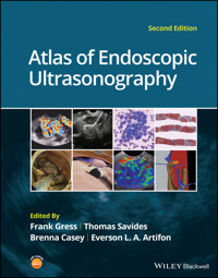
Atlas of Endoscopic Ultrasonography, Second Edition offers an outstanding visual guide to this very common diagnostic and therapeutic endoscopic tool. With contributions from noted experts in the field, the Atlas contains 400 high-quality color and black and white images obtained from real cases, each accompanied by detailed annotation to aid readers in their understanding of this popular technical procedure. In addition, there is a companion website featuring 50 video clips of real-life procedures in action, as well as the entire collection of images from within the book.
- Updated throughout to include the most recent advances in interventional Endoscopic Ultrasound (EUS) guided therapies
- Contains a large collection of color images obtained from both diagnostic and therapeutic procedures, also available on the companion website image bank
- Provides a highly integrated and accessible multimedia introduction to endoscopic ultrasonography
- Includes a companion website offering insightful videos
Written for gastroenterologists, students, residents, and radiologists, Atlas of Endoscopic Ultrasonography, Second Edition is an essential introduction to endoscopic ultrasonography.
Sie lesen das E-Book in den Legimi-Apps auf:
Seitenzahl: 539
Veröffentlichungsjahr: 2021
Ähnliche


Table of Contents
Cover
Title Page
Copyright Page
Contributors
Preface
About the Companion Website
1 Normal EUS Anatomy
1 Normal Human Anatomy
Introduction
Normal EUS anatomy from the esophagus
Normal EUS anatomy from the stomach
Normal EUS anatomy from the duodenum
Normal EUS anatomy from the rectum
Vascular videos
2 Esophagus
Layers of the esophageal wall
Normal radial extraesophageal anatomy (Video 2.1)
Normal linear thoracic anatomy
3 Normal Mediastinal Anatomy by EUS and EBUS
Introduction
Anatomical definitions
Equipment
Endoscopic ultrasound technique
Endobronchial ultrasound
Complications and safety
Conclusions
4 Stomach
5 Bile Duct
Normal bile duct anatomy
Normal anatomy of the bile duct and gallbladder with radial echoendoscope
Normal anatomy of the bile duct and gallbladder with linear echoendoscope
6 EUS of the Normal Pancreas
Radial examination of the pancreas
Linear examination of the pancreas
Endosonographic appearance of the normal pancreatic parenchyma
7 Liver, Spleen, and Kidneys
Introduction
Liver
Spleen
Kidney
Adrenal glands
8 Anatomy of the Anorectum
Introduction
Examination technique
Normal anatomy
2 Upper and Lower GI EUS
9 Esophageal Cancer
Introduction
Updated American Joint Committee on Cancer staging guidelines for esophageal cancer 2017 and implications for endosonographers
Role of EUS in staging of esophageal cancer
Limitations
Impact of EUS staging on management
Technique
References
10 EUS for Achalasia
Introduction
Clinical presentation and diagnosis
Role of EUS in achalasia
11 Malignant Mediastinal Lesions
12 Benign Mediastinal Lesions
13 Gastric Cancer
14 Gastric and Esophageal Subepithelial Masses
Introduction
Lipoma
Carcinoid tumors
Granular cell tumor
Duplication cyst
Pancreatic rest
Varices
Gastrointestinal stromal cell tumors and leiomyomas
Glomus tumor
Gastritis cystica profunda
Extrinsic compression lesions
15 Anorectal Neoplasia
Colorectal cancer staging by EUS
Endoscopic ultrasound for local recurrence of colorectal carcinoma
Submucosal tumors of the colorectal wall
16 Anal Sphincter Disease
Introduction
Fecal incontinence
Perianal fistula
17 Endometriosis
Introduction
Definition and location
Epidemiology and risk factors
Clinical picture
Diagnosis
Classification
Imaging methods
18 Vascular Anomalies and Abnormalities
Introduction
Aortic arch anomalies
Vascular calcification and plaques
Aneurysms and pseudoaneurysms
Venous thrombosis
Dieulafoy lesions
Neoplasms
Miscellaneous aberrancies
3 Pancreatico‐biliary
19 Duodenal and Ampullary Neoplasia
20 Biliary Tract Pathology
21 Gallbladder Pathology
Introduction
Gallbladder stones
Gallbladder polyps
Gallbladder carcinoma
22 Pancreatic Adenocarcinoma
Introduction
Tumor identification and diagnosis via fine needle aspiration or fine needle biopsy
Evaluation of vascular invasion
Evaluation of peripancreatic lymphadenopathy
Limitations and complications of EUS in patients with pancreatic cancer
Conclusion
References
23 Pancreatic Malignancy (Non‐adenocarcinoma)
Introduction
Endocrine pancreatic tumors (Figures 23.1 and 23.2)
Primary pancreatic lymphoma (Figure 23.3)
Solid pseudopapillary tumors (Figure 23.4)
Acinar cell carcinoma (Figure 23.5)
Secondary metastatic tumors (Figure 23.6)
Summary
24 Autoimmune Pancreatitis
Introduction
Endoscopic ultrasound imaging (Video 24.1)
Image‐enhancing techniques during EUS
EUS‐FNA and EUS‐FNB
Histologic features
Summary
25 Pancreatic Cystic Lesions
Introduction
Pseudocyst
Serous lesions
Mucinous lesions
Other cystic neoplasms
26 Intraductal Papillary Mucinous Neoplasms
Introduction
Clinical features
Role of imaging
Cross‐sectional imaging
Endoscopic ultrasound evaluation
Management of small IPMN (≤3 cm)
27 Chronic Pancreatitis
Introduction
Clinical overview of chronic pancreatitis
Endoscopic ultrasound imaging of the normal pancreas
EUS imaging in chronic pancreatitis: historical perspectives
EUS imaging in chronic pancreatitis: the Rosemont Criteria
Endoscopic ultrasound imaging in chronic pancreatitis: the future
28 Liver Pathology
Introduction
Cirrhosis
Fatty liver disease
Hepatic cysts
Neoplasms
Dilated intrahepatic ducts
4 How to Section
29 How to Interpret EUS‐FNA Cytology
Introduction
Technical quality of EUS biopsy material
Quality of the interpretation
Integration of pathologic and clinical information
30 How to do Mediastinal FNA
31 How to do Pancreatic Mass FNA
Introduction
The technique
32 How to do Pancreatic Cyst FNA
Introduction
Technique (Video 32.1)
Summary
33 How to do Pancreatic Pseudocyst Drainage
Introduction
Patient selection
Requisite instruments and accessories
Assessment of the pseudocyst by EUS prior to drainage
Technique for placement of plastic endoprosthesis
Technique for lumen‐apposing metal stent placement
Post‐procedure follow‐up
34 How to do EUS‐guided Pancreatic Cyst Chemoablation
Background
Pretreatment evaluation
Patient selection
Technical aspects of the procedure
Postoperative care and follow‐up
Conclusions
References
35 How to do Celiac Plexus Block
Introduction
Technique (Video 35.1)
Complications
36 How to Place Fiducials for Radiation Therapy
Introduction
Equipment
Techniques
Periprocedural care
37 How to Inject Chemotherapeutic Agents
38 How to do EUS‐guided Pelvic Abscess Drainage
Introduction
Patient preparation
Devices and accessories
Procedural technique
Clinical outcomes
Technical limitations
Conclusions
39 How to do Doppler Probe EUS for Bleeding
Background and equipment
Practical application of DopUS probe
Conclusions
40 How to do Endoscopic Ultrasound‐guided Portal Pressure Gradient Measurement
Introduction
Endoscopic ultrasound‐guided PPGM technique
References
41 How to do Endoscopic Ultrasound‐guided Liver Biopsy
Indications and contraindications
EUS‐LB technique
Postprocedure recovery after EUS‐LB
Adverse effects
Conclusions
References
42 How to do EUS‐guided Treatment of Gastric Varices
Introduction
Technique (Video 42.1)
Complicatons
43 How to do EUS‐guided Arterial Embolization
Background
Technique
Postprocedural management and complications
References
44 How to do EUS‐guided Radiofrequency Ablation of Pancreatic Neuroendocrine Tumors
Introduction
Methods for EUS‐guided RFA
Indications
Results for EUS‐guided RFA studies
Conclusion
References
45 How to do EUS Pancreatic Duct Access and Drainage
Introduction
Indications
Contraindications
Before the procedure
Techniques for drainage
Outcomes
Algorithm
Controversies and future directions
46 How to do EUS Gallbladder Drainage
Introduction
Background concept
Choice of stents: metal stents versus plastic stents
EUS‐GBD with LAMS
Postprocedural management
Long‐term management
Conclusion
References
47 How to do an EUS‐guided Gastrojejunostomy
Introduction
Technique (Video 47.1)
Complications
References
48 How to do EUS Elastography
Introduction
Technique
Indications
Complications
Further reading
49 How to do Contrast‐enhanced EUS
Introduction
Principle of CH‐EUS
Critical points for performing CH‐EUS
Advantages of CH‐EUS compared to other contrast‐enhanced modalities
Advantages of CH‐EUS compared to conventional EUS
Evaluation of CH‐EUS image for lesions in different organs
50 How to do EUS‐guided Ablation of Pancreatic Neurendocrine Tumors
Introduction and indications
Techniques
Conclusion and future perspectives
51 How to do EUS‐guided Needle Confocal Laser Endomicroscopy of Pancreatic Cysts
Confocal laser endomicroscopy technique
52 How to use ex vivo Models in Teaching Therapeutic Endoscopic Ultrasound
Introduction
Ex vivo models
Conclusion
53 How to do Endoscopic Necrosectomy
Introduction
Preprocedure assessment
EUS evaluation of walled‐off pancreatic necrosis
Access creation: cystgastrostomy or cystenterostomy
Necrosectomy tools and technique
Complication management
Conclusion
References
54 How to Perform Pancreatic Mass Fine Needle Biopsy
Introduction
Technique (Video 54.1)
Summary
55 How to Perform Endoscopic Ultrasound‐directed Transgastric Endoscopic Retrograde Cholangiopancreatography (EDGE)
Introduction
Technique
Post‐EDGE fistula management
Summary
Index
End User License Agreement
List of Tables
Chapter 3
Table 3.1 Mediastinal lymph node stations with their anatomical correlations...
Chapter 8
Table 8.1 Anatomical structures and abbreviations.
Table 8.2 Normal sphincter values. This is highly dependent on age, sex, and...
Chapter 9
Table 9.1 American Joint Committee on Cancer (AJCC) staging of esophageal ca...
Chapter 13
Table 13.1 American Joint Committee on Cancer (AJCC) Staging: TNM classifica...
Table 13.2 Ann Arbor staging system for gastrointestinal lymphomas.
Chapter 14
Table 14.1 Endoscopic ultrasound (EUS) characteristics of gastric and esopha...
Table 14.2 Extrinsic compression of the esophagus and stomach.
Chapter 25
Table 25.1 Demographics and characteristics of pancreatic cystic neoplasms.
Chapter 27
Table 27.1 The Rosemont Criteria.
Table 27.2 The Rosemont diagnostic stratification.
Chapter 30
Table 30.1 TNM staging system for lung cancer (7th edition).
Chapter 34
Table 34.1 Summary of EUS‐guided ablation trials (only studies with a prospe...
Chapter 39
Table 39.1 Correlation of endoscopic appearance of peptic or anastomotic ulc...
Chapter 45
Table 45.1 Outcomes of EUS‐PDD.
Chapter 46
Table 46.1 Currently available LAMS on the market.
Chapter 48
Table 48.1 Elastographic pattern classification.
Table 48.2 Elastographic quantitative evaluation.
Chapter 50
Table 50.1 EUS‐guided radiofrequency ablation for treatment of pNETs: litera...
Table 50.2 EUS‐guided ethanol injection for treatment of pNETs: literature r...
List of Illustrations
Chapter 1
Figure 1.1 Visible Human Model of esophagus, stomach, and duodenum. The gree...
Figure 1.2 Visible Human Model of esophagus, stomach, and duodenum. The red ...
Figure 1.3 Transaxial cross‐section of digital anatomy taken at the level of...
Figure 1.4 Sagittal cross‐section of digital anatomy at the level of the gas...
Figure 1.5 Visible Human Model of an image plane that is in the location in ...
Figure 1.6 The cross‐sectional anatomy within the plane shown in Figure 1.3....
Figure 1.7 Visible Human Model with a plane that is in a location similar to...
Figure 1.8 Cross‐sectional anatomy generated within the plane shown in Figur...
Figure 1.9 Endobronchial view of the carina, showing the right (RMB) and lef...
Figure 1.10 Endobronchial view of the first branch of the right mainstem bro...
Figure 1.11 Endobronchial view of the bifurcation of the bronchus intermediu...
Figure 1.12 Endobronchial view of bifurcation of the left mainstem bronchus ...
Figure 1.13 A Visible Human Model of the bronchial tree.
Chapter 2
Figure 2.1 (a) Radial array image of esophageal wall with small echolayer II...
Figure 2.2 Radial array image at gastroesophageal (GE) junction. IVC, inferi...
Figure 2.3 Radial array image at the level of the left atrium. PV, pulmonary...
Figure 2.4 Radial array image at the level of the mitral valve.
Figure 2.5 Radial array image at the level of the aortic root.
Figure 2.6 Radial array image at the level of the azygos arch.
Figure 2.7 Radial array image at the mid aortic arch.
Figure 2.8 Radial array image at the level of the left carotid and subclavia...
Figure 2.9 Radial array image at the level of the thyroid.
Figure 2.10 Linear array image at the mid aorta.
Figure 2.11 Linear array image at the level of the celiac artery.
Figure 2.12 Linear array image at the aortic root.
Figure 2.13 Linear array image at the aortopulmonary window (APW). PA, pulmo...
Chapter 3
Figure 3.1 Mediastinal lymph node stations.
Figure 3.2 Types of echoendoscopes: (a) linear probe; (b) endobronchial prob...
Figure 3.3 Lymph node at station 8 (between calipers).
Figure 3.4 Subcarinal station (station 7).
Figure 3.5 Aortopulmonary window station (stations 4L, 5 and 6). AO, aorta; ...
Figure 3.6 Endobronchial ultrasound (EBUS) images of common mediastinal stat...
Chapter 4
Figure 4.1 Circumferential image of the gastric wall after filling and diste...
Figure 4.2 Five‐layer structure of the gastric wall demonstrating a relative...
Figure 4.3 Five‐layer structure of the gastric wall obtained with an electro...
Figure 4.4 Nine‐layer structure of the gastric wall obtained with a 20 MHz c...
Chapter 5
Figure 5.1 Bile duct as visualized from the duodenal bulb (radial echoendosc...
Figure 5.2 Bile duct followed towards the head of the pancreas from the duod...
Figure 5.3 Gallbladder from the duodenal bulb (radial echoendoscope). CBD, c...
Figure 5.4 Bile duct and pancreatic duct from the second portion of the duod...
Figure 5.5 Common hepatic duct and cystic duct as visualized from the duoden...
Figure 5.6 Bile duct and pancreatic duct from the second portion of the duod...
Chapter 6
Figure 6.1 Radial EUS: pancreatic body and portal/splenic vein confluence. P...
Figure 6.2 Radial EUS: portal/splenic vein confluence. PV, portal vein; SMA,...
Figure 6.3 Radial EUS: pancreas tail. PD, pancreatic duct; SA, splenic arter...
Figure 6.4 Radial EUS: pancreas tail.
Figure 6.5 Radial EUS: pancreas neck. PD, pancreatic duct; PV, portal vein; ...
Figure 6.6 Radial EUS: head of pancreas. DP, dorsal pancreas; VP, ventral pa...
Figure 6.7 Radial EUS: ampulla.
Figure 6.8 Radial EUS: head of pancreas. CBD, common bile duct; PD, pancreat...
Figure 6.9 Radial EUS: head of pancreas, vasculature. CBD, common bile duct;...
Figure 6.10 Radial EUS: head of pancreas. CBD, common bile duct; PD, pancrea...
Figure 6.11 Linear EUS: pancreas body. PD, pancreatic duct; SA, splenic arte...
Figure 6.12 Linear EUS: pancreas tail. PD, pancreatic duct; SV, splenic vein...
Figure 6.13 Linear EUS: ampulla.
Figure 6.14 Linear EUS: head of pancreas. CBD, common bile duct; PD, pancrea...
Chapter 7
Figure 7.1 Image of the liver scanned by radial ultrasound.
Figure 7.2 Celiac artery imaging by linear ultrasound.
Figure 7.3 Image of liver by linear ultrasound.
Figure 7.4 Image of spleen by radial ultrasound.
Figure 7.5 Image of spleen by linear ultrasound.
Figure 7.6 Image of left kidney by radial ultrasound.
Figure 7.7 Image of left kidney by linear ultrasound.
Figure 7.8 Image of left adrenal gland by radial ultrasound.
Figure 7.9 Image of left adrenal gland by linear ultrasound.
Chapter 8
Figure 8.1 (a) Levator ani muscle with the puborectal part (PR) in between m...
Figure 8.2 Internal anal sphincter (echo‐poor) and external anal sphincter (...
Chapter 9
Figure 9.1 Distal esophageal adenocarcinoma shown to be T1a on pathology. (a...
Figure 9.2 T1b adenocarcinoma in a background of Barrett’s esophagus at the ...
Figure 9.3 (a) T1b adenocarcinoma at the gastroesophageal junction with cent...
Figure 9.4 (a) T2N1Mx lesion, squamous cell cancer with ulceration 24 cm fro...
Figure 9.5 (a) T2N0Mx lesion, adenocarcinoma of lower thoracic esophagus inv...
Figure 9.6 T3N3Mx lesion, adenocarcinoma involving the lower thoracic esopha...
Figure 9.7 (a) T3N1Mx adenocarcinoma with friable mass at the gastroesophage...
Figure 9.8 T4a lesion, adenocarcinoma in lower thoracic esophagus invading t...
Figure 9.9 T4b lesion, squamous cell cancer in upper thoracic esophagus inva...
Chapter 10
Figure 10.1 The muscularis propria is divided into two layers, the outer lon...
Figure 10.2 Normal esophageal wall layer pattern. Normal thickness of muscul...
Figure 10.3 Thickening of the circular muscle wall layer to 3 mm in a patien...
Figure 10.4 Thickening of the circular muscle wall layer to an average of 2....
Figure 10.5 Thickening of the circular muscle wall layer to an average of 3....
Figure 10.6 Thickening of the circular muscle wall layer to an average of 3....
Figure 10.7 Uniform thickening of the circular muscle wall layer to an avera...
Chapter 11
Figure 11.1 American Thoracic Society staging diagram for posterior mediasti...
Figure 11.2 Metastatic posterior mediastinal melanoma. Endoscopic ultrasound...
Figure 11.3 Renal cell cancer metastasis. The lesion was identified slightly...
Figure 11.4 Lung cancer mass. Endoscopic ultrasound‐guided fine needle aspir...
Figure 11.5 Malignant lymph node. The needle is within the rounded, well‐dem...
Chapter 12
Figure 12.1 Benign subcarinal lymph nodes. Note the oval and triangular‐shap...
Figure 12.2 Sarcoid lymph nodes. Note the matted together, draping nodes wit...
Figure 12.3 Cryptococcal lymph node. These were incidental lymph nodes found...
Figure 12.4 Duplication cyst. Note the anechoic appearance with acoustic enh...
Figure 12.5 Enlarged lymph node due to
Blastocystis hominis
infection.
Chapter 13
Figure 13.1 T1 gastric cancer. This is a 2.2 × 0.5 cm thick tumor confined t...
Figure 13.2 T2 gastric cancer. This large 4.2 × 3.6 cm exophytic tumor invad...
Figure 13.3 T3 gastric cancer. This 2.5 × 1.6 cm tumor invades through all l...
Figure 13.4 T4 gastric cancer. This invasive poorly differentiated cancer in...
Figure 13.5 Diffuse gastric cancer. The stomach is thickened to 0.8 cm (norm...
Figure 13.6 Pseudo‐linitis plastica. A patient with breast cancer has metast...
Figure 13.7 Gastric lymphoma causing a linitis plastica appearance. This pat...
Figure 13.8 Gastric lymphoma. The five‐layer wall pattern is obliterated and...
Figure 13.9 Perigastric lymphadenopathy in a patient with primary gastric ly...
Figure 13.10 Ascites. A large pocket of hypoechoic material is seen next to ...
Figure 13.11 Endoscopic ultrasound‐guided fine needle aspiration (EUS‐FNA) o...
Chapter 14
Figure 14.1 Lipoma. (a) Endoscopic view of lipoma with smooth overlying muco...
Figure 14.2 Carcinoid tumor. (a) Endoscopic view of a carcinoid tumor. (b) E...
Figure 14.3 Granular cell tumor. (a) Endoscopic view of esophageal granular ...
Figure 14.4 Esophageal duplication cysts. (a) Endoscopic view of several eso...
Figure 14.5 Pancreatic rest. (a) Endoscopic view of a gastric antral pancrea...
Figure 14.6 Esophageal varices. (a) Endoscopic view of grade 3 esophageal va...
Figure 14.7 Gastric gastrointestinal stromal tumor (GIST). (a) Endoscopic vi...
Figure 14.8 Leiomyoma. (a) Endoscopic view of an esophageal leiomyoma. (b) E...
Figure 14.9 Glomus tumor. (a) Endoscopic view of a gastric antral glomus tum...
Figure 14.10 Gastritis cystica profunda (GCP). (a) Endoscopic view showing G...
Figure 14.11 Extrinsic compression. (a) Endoscopic and EUS view of extrinsic...
Chapter 15
Figure 15.1 (a) A T1 rectal adenocarcinoma (by radial EUS) arising in a vill...
Figure 15.2 (a) A rectal adenocarcinoma that appears to be T2 (penetration i...
Figure 15.3 Radial EUS of a T3 lesion showing the primary rectal tumor penet...
Figure 15.4 Radial EUS of the patient in Figure 15.3 showing a 7‐mm round, p...
Figure 15.5 EUS‐guided fine needle aspiration of a perirectal lymph node. Th...
Figure 15.6 Recurrent rectal cancer. During surveillance colonoscopy in a pa...
Figure 15.7 (a) A subepithelial bulge in the rectum from a large intramural ...
Figure 15.8 Rectal carcinoid. (a) A rectal bulge was noted during routine co...
Chapter 16
Figure 16.1 Schematic diagram of the anal canal. EAS, external anal sphincte...
Figure 16.2 Normal anal sphincter complex. The internal anal sphincter (IAS)...
Figure 16.3 Normal anal sphincter complex. The internal anal sphincter (IAS)...
Figure 16.4 Forty percent defect in both the internal hypoechoic anal sphinc...
Figure 16.5 Forty percent defect in external anal sphincter (EAS), likely du...
Figure 16.6 Thirty percent defect in both the internal anal sphincter (IAS) ...
Figure 16.7 Transperineal three‐dimensional ultrasound images showing the an...
Figure 16.8 Transverse (cross‐sectional) 1‐mm multislice imaging of the norm...
Figure 16.9 Perianal fistula. Note that the anechoic tubular structure is ou...
Figure 16.10 Perianal fistula with small perianal abscess measuring 11 × 7 m...
Figure 16.11 Perianal abscess and fistula.
Figure 16.12 Post‐hydrogen peroxide injection image of the perianal fistula ...
Figure 16.13 Perianal fistula. A hyperechoic path is visible, starting on th...
Figure 16.14 The path of a complex perianal fistula is identified by inserti...
Figure 16.15 Perirectal abscess. A 36‐year‐old woman with history of Crohn’s...
Figure 16.16 Anastomotic abscess. A patient with history of sigmoidectomy fo...
Chapter 17
Figure 17.1 (a, b) Classification of intestinal endometriosis according to R...
Figure 17.2 Endometriosis sites with alteration of the colonic wall of the r...
Figure 17.3 (a, b) Endoscopic view of the area of sigmoid substenosis. (c) P...
Figure 17.4 Radial scanning with two foci in different locations: (a, b) rec...
Figure 17.5 (a) Bulging of the upper rectum wall. (b–d) Radial exploration o...
Figure 17.6 (a, b) Radial exploration of sites of hypoechoic, homogeneous, s...
Figure 17.7 (a, b) Radial exploration of a heterogeneous hypoechoic focus wi...
Figure 17.8 (a, b) Endoscopic view of the upper rectum showing bulging of th...
Figure 17.9 (a, b) Extrinsic sigmoid compression of a left ovary endometriom...
Figure 17.10 (a) Colonoscopy showing a subepithelial lesion as sigmoid bulgi...
Figure 17.11 (a) Bulging of the rectosigmoid junction. (b, c) Sectoral explo...
Figure 17.12 Endometrial gland and endometrial stroma and superficial coloni...
Figure 17.13 Radial scanning EUS of two foci of endometriosis located in two...
Figure 17.14 Glandular cells aspirated from endometrial lining.
Chapter 18
Figure 18.1 A radial array endoscopic ultrasound image of a calcific aortic ...
Figure 18.2 A linear array image of the aortopulmonary window with an isoech...
Figure 18.3 A linear array image showing the aortic root with clot due to an...
Figure 18.4 Radial array Doppler image of a clot in the splenic vein.
Figure 18.5 A linear array image of a metastasis (T) from breast cancer to t...
Figure 18.6 A linear array image of a rhabdomyosarcoma infiltrating into the...
Chapter 19
Figure 19.1 The linear array echoendoscope is in the duodenum directly adjac...
Figure 19.2 A hypoechoic mass is seen here arising from the ampulla. The mal...
Figure 19.3 An ampullary lesion is noted endoscopically. On EUS, water is in...
Chapter 20
Figure 20.1 In this example of a small bile duct malignancy, a focal hypoech...
Figure 20.2 The linear array echoendoscope is positioned in the second porti...
Figure 20.3 This 78‐year‐old woman has unresectable pancreatic cancer. Endos...
Figure 20.4 This 46‐year‐old woman presented with elevated liver function te...
Chapter 21
Figure 21.1 Gallbladder stone (arrow) with posterior shadowing.
Figure 21.2 Gallbladder stone (arrow) with posterior shadowing.
Figure 21.3 Gallbladder sludge (arrow).
Figure 21.4 Gallbladder microlithiasis (arrows).
Figure 21.5 Gallbladder polyps (arrows). Notice the lack of shadowing.
Figure 21.6 Gallbladder polyps (arrows). Notice the lack of shadowing.
Figure 21.7 Gallbladder carcinoma (arrow).
Figure 21.8 Thickened gallbladder wall (arrow).
Chapter 22
Figure 22.1 (a) Representative EUS image of a large pancreatic adenocarcinom...
Figure 22.2 EUS‐guided FNA of a pancreatic adenocarcinoma with a 22‐gauge ne...
Figure 22.3 EUS image of a pancreatic adenocarcinoma in the body of the panc...
Figure 22.4 A 2‐cm pancreatic head mass seen to abut and compress the common...
Figure 22.5 Large pancreatic cancer in the head of the pancreas seen to enca...
Figure 22.6 A 15‐mm peripancreatic lymph node. Node is hypoechoic, round, we...
Figure 22.7 EUS‐guided FNA of a large, 4‐cm peripancreatic node in a patient...
Chapter 23
Figure 23.1 Images obtained from a patient with a gastrinoma following helic...
Figure 23.2 Following a negative computed tomography (CT) to search for a pr...
Figure 23.3 Linear endoscopic ultrasound imaging of a patient with a pancrea...
Figure 23.4 Endoscopic ultrasound revealed a large well‐circumscribed hypoec...
Figure 23.5 A pancreatic head acinar cell tumor is identified by endoscopic ...
Figure 23.6 The varied appearance of metastatic tumors to the pancreas is de...
Chapter 24
Figure 24.1 Classic endoscopic ultrasound appearance of autoimmune pancreati...
Figure 24.2 Almost classic endoscopic ultrasound appearance of autoimmune pa...
Figure 24.3 Almost classic endoscopic ultrasound appearance of autoimmune pa...
Figure 24.4 Endoscopic ultrasound finding of a mass‐like lesion in a patient...
Figure 24.5 Endoscopic ultrasound features of nonspecific chronic pancreatit...
Figure 24.6 Histologic examination of endoscopic ultrasound‐guided Tru‐cut b...
Figure 24.7 IgG
4
immunostaining of the Tru‐cut biopsy specimen reveals the f...
Chapter 25
Figure 25.1 Thick‐walled cystic lesion undergoing fine needle aspiration (FN...
Figure 25.2 Serous cystadenoma. Multicystic lesion in the body of the pancre...
Figure 25.3 (a) Mucinous cystic neoplasm with multiple septations. The lesio...
Figure 25.4 (a) Thinly septated intraductal papillary mucinous neoplasm (IPM...
Figure 25.5 (a) Pancreatic cystic endocrine tumor. Note the presence of the ...
Chapter 26
Figure 26.1 Reformatted computed tomography scan demonstrating a side‐branch...
Figure 26.2 Magnetic resonance cholangiopancreatography demonstrating a side...
Figure 26.3 Linear endoscopic ultrasound image demonstrating a dilated main ...
Figure 26.4 Linear endoscopic ultrasound image revealing a small cystic lesi...
Figure 26.5 Linear endoscopic ultrasound image of a malignant‐appearing 2.9‐...
Figure 26.6 Fine needle aspiration of a malignant cystic mass.
Figure 26.7 International Association of Pancreatology (IAP) algorithm for m...
Chapter 27
Figure 27.1 Endoscopic ultrasound (linear) showing features of chronic pancr...
Figure 27.2 Endoscopic ultrasound showing a hyperechoic duct wall (crossmark...
Chapter 28
Figure 28.1 (a) Cirrhosis. The cirrhotic liver is characterized by diffuse, ...
Figure 28.2 Fatty liver is described as diffuse or as foci of hyperechoic or...
Figure 28.3 Simple hepatic cyst can be seen here as an anechoic fluid‐filled...
Figure 28.4 (a, b) Metastatic liver disease. Lesions found within the hepati...
Figure 28.5 Dilated intrahepatic ducts (arrows) are visualized here and are ...
Chapter 29
Figure 29.1 (a) Core biopsies demonstrate intact tissue architecture and str...
Figure 29.2 Diagnostic features of autoimmune pancreatitis rely on the demon...
Figure 29.3 Aspirate smears demonstrate fine cellular detail such as squamou...
Figure 29.4 Cell blocks, prepared from fine needle aspirate material and pla...
Figure 29.5 Clotted blood within the needle obscures lesional tissue (a). Op...
Figure 29.6 Air‐dried slides stained with a modified Romanowsky stain demons...
Figure 29.7 Distinctive or commonly encountered in endoscopic ultrasound‐gui...
Chapter 30
Figure 30.1 The International Association for the Study of Lung Cancer (IASL...
Chapter 31
Figure 31.1 (a) Hypoechoic, irregular, 3‐cm adenocarcinoma in the head of pa...
Figure 31.2 (a) Small adenocarcinoma in the head of pancreas adjacent to the...
Figure 31.3 (a) Large 5‐cm adenocarcinoma in the tail of pancreas. (b) Mass ...
Figure 31.4 (a) A 2‐cm mass in the head of pancreas with acoustic interferen...
Figure 31.5 (a) Small 1‐cm hypoechoic neuroendocrine tumor in the head of pa...
Figure 31.6 (a) Cystic neoplasm with malignant degeneration in the head of p...
Figure 31.7 Hepatic metastases visualized during EUS examination for a pancr...
Chapter 32
Figure 32.1 EUS of left lobe of liver.
Figure 32.2 EUS of aorta, celiac artery (CA), and superior mesenteric artery...
Figure 32.3 Transgastric EUS examination of pancreas (genu, body, tail).
Figure 32.4 Transduodenal EUS examination of pancreas (head and uncinate pro...
Figure 32.5 (a) EUS of anechoic septated lobular cyst in tail of pancreas. (...
Figure 32.6 Position lesion at 6 o’clock for FNA/FNB.
Figure 32.7 EUS with Doppler showing blood vessels (purple) in path of needl...
Figure 32.8 EUS needle handle.
Figure 32.9 FNA needle within pancreatic cyst (yellow arrow indicates tip of...
Figure 32.10 Vacuum syringe.
Figure 32.11 Serous fluid aspirated from large pancreatic cyst.
Figure 32.12 String sign consistent with mucinous cyst.
Chapter 33
Figure 33.1 A large unilocular pseudocyst with clear contents as visualized ...
Figure 33.2 Two pseudocysts are seen at EUS: the promixal lesion on FNA anal...
Figure 33.3 Presence of intervening vasculature between the EUS transducer a...
Figure 33.4 Endoscopic ultrasound guidance enables access to the pseudocyst ...
Figure 33.5 Pseudocyst accessed with a 19‐gauge FNA needle.
Figure 33.6 (a) Acute angulation encountered when the echoendoscope is locat...
Figure 33.7 A 0.035‐inch guidewire is coiled within the pseudocyst under flu...
Figure 33.8 The transmural tract is dilated using a 5‐Fr endoscopic retrogra...
Figure 33.9 The transmural tract is subsequently dilated using an 8 mm over‐...
Figure 33.10 Multiple 7‐Fr transmural stents are deployed (fluoroscopy view)...
Figure 33.11 (a) Endoscopic view of the proximal flange of the LAMS in the s...
Chapter 34
Figure 34.1 The alcohol free EUS‐guided cyst ablation process. The FNA needl...
Figure 34.2 A high‐pressure gun, syringe, and short connector tubing assembl...
Figure 34.3 (a) Magnetic resonance image showing a 76‐year‐old female with a...
Figure 34.4 (a) MRI (
top
) and magnetic resonance cholangiopancreatography (M...
Chapter 35
Figure 35.1 Hypoechoic pancreatic mass as seen by EUS.
Figure 35.2 Endoscopic ultrasound demonstration of lobularity, one of the ma...
Figure 35.3 Celiac axis (CA) as seen by EUS. AO, aorta.
Figure 35.4 Celiac axis confirmed by the aorta, celiac artery (CA) and super...
Figure 35.5 Insertion of the EUS‐guided fine needle aspiration needle into t...
Chapter 36
Figure 36.1 Gold coil fiducial marker is (a) flexible and (b) has a coiled d...
Figure 36.2 Gold coil fiducial preloaded on needle‐carrier delivery device....
Figure 36.3 Endoscopic ultrasound image (linear array) of fiducial within an...
Figure 36.4 Abdominal radiograph (anteroposterior view) demonstrating three ...
Chapter 37
Figure 37.1 Initial assessment of tumor size by endoscopic ultrasound (EUS) ...
Figure 37.2 Endoscopic ultrasound (EUS)‐guided fine needle injection (FNI) o...
Figure 37.3 Tumor assessment during weekly injections of chemotherapeutic ag...
Figure 37.4 Gross specimen of the resected pancreas in Figure 37.3 revealing...
Figure 37.5 Histology from the specimen in Figure 37.4 demonstrating a compl...
Chapter 38
Figure 38.1 Computed tomography (CT) scan of the pelvis revealing an 8 × 7 c...
Figure 38.2 Passage of a 19‐gauge fine needle aspiration (FNA) needle into t...
Figure 38.3 A 0.035‐inch guidewire is then coiled within the abscess cavity:...
Figure 38.4 Fluoroscopy view revealing dilation of the tract using an endosc...
Figure 38.5 Fluoroscopic view revealing the presence of a stent within the p...
Chapter 39
Figure 39.1 VTI endoscopic Doppler system. (a) A 20‐MHz pulsed‐wave Doppler ...
Figure 39.2 Endo‐Dop, 16‐MHz pulsed‐wave, multigated Doppler ultrasound (Dop...
Figure 39.3 Illustration of a bleeding peptic ulcer with a non‐bleeding visi...
Figure 39.4 Doppler ultrasound (DopUS) in acute peptic ulcer bleeding. (a) E...
Figure 39.5 Doppler ultrasound‐guided hemostasis in acute peptic ulcer bleed...
Figure 39.6 Atypical peptic ulcer bleeding: recurrent major bleeding from a ...
Figure 39.7 Doppler‐negative non‐bleeding visible vessel. Endoscopic image o...
Figure 39.8 Atypical appearance of a bleeding gastric varix. Mild active ooz...
Figure 39.9 Doppler ultrasound examination of a gastrointestinal stromal tum...
Figure 39.10 Dieulafoy lesion in gastric cardia. (a) Active bleeding. (b) Do...
Figure 39.11 Dieulafoy lesion in rectum, Doppler with strong arterial signal...
Chapter 40
Figure 40.1 Compact manometer for EUS‐guided portal pressure measurement....
Figure 40.2 EUS‐guided portal pressure measurement apparatus showing noncomp...
Figure 40.3 EUS image (a) and schematic (b) of needle puncture of the middle...
Figure 40.4 EUS Doppler flow image of the middle hepatic vein demonstrating ...
Figure 40.5 EUS image (a) and schematic (b) of needle puncture of the left p...
Figure 40.6 EUS Doppler flow image of the left portal vein demonstrating typ...
Chapter 41
Figure 41.1 Visualization of the left hepatic lobe from the proximal stomach...
Figure 41.2 View of liver (on the left) and spleen (on the right) from the p...
Figure 41.3 Right lobe biopsies are obtained with the echoendoscope tip plac...
Figure 41.4 Close‐up of the tips of the core needles used for EUS‐LB: (
left
)...
Figure 41.5 Comparison of a 19G FNB needle to 19G FNA needle in terms of (a...
Figure 41.6 Excellent core of cirrhotic liver obtained with the 19G FNB nee...
Figure 41.7 Priming the needle with heparin for wet suction technique.
Figure 41.8 (a) Use of tissue sieve (CoreCatcher; ProAct Ltd, Center Hall, P...
Figure 41.9 Long liver core is placed in formalin jar without excessive hand...
Chapter 42
Figure 42.1 Endoscopic imaging of gastric varix.
Figure 42.2 Gastric varix seen by endoscopic ultrasonography.
Figure 42.3 Gastric varix seen with color Doppler.
Figure 42.4 Needle inside the varix.
Figure 42.5 Obliterated varix right after treatment.
Figure 42.6 Radiographic image of the coil inside the varix.
Chapter 43
Figure 43.1 Patient 1 with refractory bleeding due to gastric cancer. (a) Bl...
Figure 43.2 Patient 2 with bleeding pseudoaneurysm located to the pancreatic...
Chapter 44
Figure 44.1 EUS‐guided RFA with contrast enhancement control for NET located...
Chapter 45
Figure 45.1 Access points and routes for EUS‐PDD: 1, transgastric rendezvous...
Figure 45.2 Transgastric EUS‐guided rendezvous for MPD drainage. (a) Dilated...
Figure 45.3 Transgastric EUS‐PDD. (a) Dilated MPD at the level of the pancre...
Figure 45.4 The novel wire‐guided fine‐gauge electrocautery dilator, with a ...
Figure 45.5 Algorithm for EUS‐PDD. *, depending on the anatomic conditions a...
Chapter 46
Figure 46.1 Puncture of gallbladder with 19‐gauge needle with linear echoend...
Figure 46.2 Contrast injection within the gallbladder.
Figure 46.3 Puncturing of gallbladder with forward‐viewing echoendoscope.
Figure 46.4 Looping of guidewire within the gallbladder.
Figure 46.5 Tract dilatation with 4‐mm biliary balloon (EUS view).
Figure 46.6 (a) Distal flange of AXIOS stent deployed under EUS guidance. (b...
Figure 46.7 (a) Luminal view of “black mark” with the AXIOS stent. Once the ...
Figure 46.8 (a) Luminal view with SPAXUS stent using the deployment‐in‐chann...
Figure 46.9 Direct puncture with the Hot AXIOS into the gallbladder.
Chapter 47
Figure 47.1 Puncture of the targeted bowel under endosonography.
Figure 47.2 Deployment of the lumen‐apposing metal stent into the small bowe...
Figure 47.3 Balloon dilatation of the lumen‐apposing metal stent.
Figure 47.4 Visualization of the bowel through the fully distended lumen‐app...
Chapter 48
Figure 48.1 Standard EUS elastographic image. A ROI is used to define the ar...
Figure 48.2 EUS‐guided elastography of a malignant lymph node. This image em...
Figure 48.3 Pancreatic adenocarcinoma displaying a predominantly heterogeneo...
Figure 48.4 Mass forming chronic pancreatitis displaying a predominantly het...
Figure 48.5 EUS‐guided elastography from normal pancreas showing a predomina...
Figure 48.6 EUS‐guided elastography displaying a blue pattern in a metastati...
Figure 48.7 EUS‐guided elastography of a typical benign lymph node.
Figure 48.8 Elastographic evaluation of a subepithelial lesion showing a het...
Figure 48.9 Metastatic lesion on left adrenal gland, from a lung cancer, dis...
Chapter 49
Figure 49.1 Principle of contrast harmonic imaging.
Figure 49.2 Behavior of microbubbles when exposed to ultrasound beams based ...
Figure 49.3 Contrast‐enhanced endoscopic ultrasonography (CH‐EUS) procedures...
Figure 49.4 Contrast‐enhanced pattern compared to the surrounding tissue: (a...
Figure 49.5 Comparison of contrast‐enhanced patterns in the lesion: (a) homo...
Figure 49.6 Detecting a lesion via contrast‐enhanced endoscopic ultrasonogra...
Figure 49.7 Distinguishing a mural nodule from a mucous clot using contrast‐...
Figure 49.8 Typical contrast‐enhanced endoscopic ultrasonography (CH‐EUS) fi...
Figure 49.9 Contrast‐enhanced endoscopic ultrasonography (CH‐EUS) for gastro...
Figure 49.10 Distinguishing malignant from benign lymph nodes in contrast‐en...
Chapter 50
Figure 50.1 EUS‐RFA radiofrequency system. a) EUSRA (STARmed, Goyang, South ...
Figure 50.2 EUS‐guided radiofrequency ablation of a pancreatic tail symptoma...
Figure 50.3 EUS‐guided radiofrequency ablation of a pancreatic tail symptoma...
Figure 50.4 EUS‐guided radiofrequency ablation of a pancreatic tail symptoma...
Chapter 51
Figure 51.1 Moray micro forceps.
Figure 51.2 Overview of nCLE technique.
Chapter 52
Figure 52.1 (a) Dissected specimen: esophagus, stomach, duodenum, pancreas, ...
Figure 52.2 Vascular simulation of aorta and use of the color Doppler featur...
Figure 52.3 Simulation of hepatic nodules and lymph nodes. (a) Incision in t...
Figure 52.4 Simulation of subepithelial lesions. (a) Serous and muscle layer...
Figure 52.5 Ex vivo model for EUS‐FNA of cystic lesions. The cysts were crea...
Figure 52.6 Ex vivo model for pseudocyst drainage. The pseudocysts were crea...
Figure 52.7 Ex vivo model for dilated hepatocholedochus evaluation. The hepa...
Figure 52.8 Ex vivo model for EUS biliary drainage. The dilated biliary trac...
Chapter 53
Figure 53.1 There is a perigastric varix present within the wall of a necrot...
Figure 53.2 (a, b) Hyperechoic and isoechoic solid necrosis within necrotic ...
Figure 53.3 (a, b) Clots and blood products within a peripancreatic collecti...
Figure 53.4 Lumen‐apposing metal stent placed for cystgastrostomy.
Figure 53.5 CT scan demonstrates two LAMS stents from the stomach and duoden...
Figure 53.6 (a, b) Endoscopic necrosectomy with rat tooth forceps and retrie...
Figure 53.7 CT scan shows local pneumoperitoneum around the cystastrostomy s...
Figure 53.8 Over‐the‐scope clip placement for closure of cystgasatrostomy si...
Figure 53.9 Purulence and infection as a result of occlusion of LAMS cystgas...
Chapter 54
Figure 54.1 Fine needle aspiration versus fine needle biopsy needles.
Figure 54.2 EUS of aorta, celiac artery (CA) and superior mesenteric artery ...
Figure 54.3 Tail of pancreas (yellow arrow) and spleen.
Figure 54.4 EUS‐guided (a) transgastric and (b) transduodenal pancreatic tis...
Figure 54.5 Large heterogeneous well‐defined pancreatic head mass.
Figure 54.6 Vessels (yellow arrow) in path of FNB needle to pancreatic mass....
Figure 54.7 Fine needle biopsy handle.
Figure 54.8 EUS‐FNB of pancreatic mass. Tip of FNB needle seen within mass (...
Figure 54.9 Fanning to increase tissue acquisition.
Figure 54.10 Macroscopically visible strands of fine needle biopsy tissue....
Chapter 55
Figure 55.1 View of the excluded stomach from the gastric pouch using a line...
Figure 55.2 A 19‐gauge needle is used to puncture the excluded stomach under...
Figure 55.3 Fluoroscopic view of the excluded stomach after injection of con...
Figure 55.4 View of lumen‐apposing metal stent after deploying the proximal ...
Figure 55.5 Fluoroscopic view after successful creation of a gastro‐gastric ...
Figure 55.6 Dilation of the lumen‐apposing metal stent channel during the in...
Figure 55.7 Careful advancement of a duodenoscope through a freshly placed l...
Figure 55.8 Fluoroscopic view of a double pigtail stent placed within the lu...
Figure 55.9 Gastro‐gastric fistula seen endoscopically following removal of ...
Guide
Cover Page
Title Page
Copyright Page
Contributors
Preface
About the Companion Website
Table of Contents
Begin Reading
Index
Wiley End User License Agreement
Pages
iii
iv
vii
viii
ix
x
xi
xii
1
3
4
5
6
7
8
9
10
11
12
13
14
15
16
17
18
19
20
21
22
23
24
25
26
27
28
29
30
31
32
33
34
35
37
39
40
41
42
43
44
45
46
47
48
49
50
51
52
53
54
55
56
57
58
59
60
61
62
63
64
65
66
67
68
69
70
71
72
73
74
75
76
77
78
79
80
81
82
83
84
85
86
87
88
89
90
91
93
95
96
97
98
99
100
101
102
103
104
105
106
107
108
109
110
111
112
113
114
115
116
117
118
119
120
121
122
123
124
125
126
127
128
129
130
131
132
133
135
137
138
139
140
141
142
143
144
145
146
147
148
149
150
151
152
153
154
155
156
157
158
159
160
161
162
163
164
165
166
167
168
169
170
171
172
173
174
175
176
177
178
179
180
181
182
183
184
185
186
187
188
189
190
191
192
193
194
195
196
197
198
199
200
201
202
203
204
205
206
207
208
209
210
211
212
213
214
215
216
217
218
219
220
221
222
223
224
225
226
227
228
229
230
231
232
233
234
235
236
237
238
239
240
241
242
243
244
245
246
247
248
249
250
251
252
253
254
255
256
257
258
259
260
261
262
263
264
265
266
267
268
269
270
271
272
273
274
275
276
277
278
279
280
281
282
283
284
285
286
287
Atlas of Endoscopic Ultrasonography
SECOND EDITION
EDITED BY
Frank Gress, MD
Professor of Medicine
Icahn School of Medicine
Division of Gastroenterology and Hepatology
Mount Sinai Hospital
New York, USA
Thomas Savides, MD
Professor of Clinical Medicine
Division of Gastroenterology
University of California
San Diego, California, USA
Brenna Casey, MD
Assistant Professor of Medicine
Division of Gastroenterology
Massachusetts General Hospital
Harvard Medical School
Boston, Massachusetts, USA
Everson L. A. Artifon, MD
Associate Professor of Surgery
Department of Surgery
University of São Paulo
São Paulo, Brazil
This edition first published 2022© 2022 John Wiley & Sons Ltd
Edition HistoryBlackwell Publishing Ltd (1e, 2012)
All rights reserved. No part of this publication may be reproduced, stored in a retrieval system, or transmitted, in any form or by any means, electronic, mechanical, photocopying, recording or otherwise, except as permitted by law. Advice on how to obtain permission to reuse material from this title is available at http://www.wiley.com/go/permissions.
The right of Frank Gress, Thomas Savides, Brenna Casey, and Everson L. A. Artifon to be identified as the authors of the editorial material in this work has been asserted in accordance with law.
Registered OfficesJohn Wiley & Sons, Inc., 111 River Street, Hoboken, NJ 07030, USAJohn Wiley & Sons Ltd, The Atrium, Southern Gate, Chichester, West Sussex, PO19 8SQ, UK
Editorial Office9600 Garsington Road, Oxford, OX4 2DQ, UK
For details of our global editorial offices, customer services, and more information about Wiley products visit us at www.wiley.com.
Wiley also publishes its books in a variety of electronic formats and by print‐on‐demand. Some content that appears in standard print versions of this book may not be available in other formats.
Limit of Liability/Disclaimer of WarrantyThe contents of this work are intended to further general scientific research, understanding, and discussion only and are not intended and should not be relied upon as recommending or promoting scientific method, diagnosis, or treatment by physicians for any particular patient. In view of ongoing research, equipment modifications, changes in governmental regulations, and the constant flow of information relating to the use of medicines, equipment, and devices, the reader is urged to review and evaluate the information provided in the package insert or instructions for each medicine, equipment, or device for, among other things, any changes in the instructions or indication of usage and for added warnings and precautions. While the publisher and authors have used their best efforts in preparing this work, they make no representations or warranties with respect to the accuracy or completeness of the contents of this work and specifically disclaim all warranties, including without limitation any implied warranties of merchantability or fitness for a particular purpose. No warranty may be created or extended by sales representatives, written sales materials or promotional statements for this work. The fact that an organization, website, or product is referred to in this work as a citation and/or potential source of further information does not mean that the publisher and authors endorse the information or services the organization, website, or product may provide or recommendations it may make. This work is sold with the understanding that the publisher is not engaged in rendering professional services. The advice and strategies contained herein may not be suitable for your situation. You should consult with a specialist where appropriate. Further, readers should be aware that websites listed in this work may have changed or disappeared between when this work was written and when it is read. Neither the publisher nor authors shall be liable for any loss of profit or any other commercial damages, including but not limited to special, incidental, consequential, or other damages.
Library of Congress Cataloging‐in‐Publication Data
Names: Gress, Frank G., editor. | Savides, Thomas J., editor. | Casey, Brenna, editor. | Artifon, Everson L. A., editor.Title: Atlas of endoscopic ultrasonography / edited by Frank G. Gress, Thomas John Savides, Brenna Casey, Everson L. A. Artifon.Description: Second edition. | Hoboken, NJ : Wiley‐Blackwell, 2022. | Includes bibliographical references and index.Identifiers: LCCN 2021015783 (print) | LCCN 2021015784 (ebook) | ISBN 9781119523000 (hardback) | ISBN 9781119523093 (adobe pdf) | ISBN 9781119523031 (epub)Subjects: MESH: Endoscopy, Digestive System | Digestive System Diseases–diagnostic imaging | Digestive System–diagnostic imaging | Ultrasonography | AtlasClassification: LCC RC801 (print) | LCC RC801 (ebook) | NLM WI 17 | DDC 616.3/07545–dc23LC record available at https://lccn.loc.gov/2021015783LC ebook record available at https://lccn.loc.gov/2021015784
Cover Design: WileyCover Images: Courtesy of Dalton Chaves, Courtesy of Anthony Teoh, Courtesy of Cynthia Behling, Courtesy of David L. Diehl, MD, Courtesy of Wilson Kwong, Courtesy of Spencer Cheng, Courtesy of Julio Iglesias Garcia, Courtesy of Toltech
Contributors
Douglas G. Adler, MD, FACG, FASGEPeak Gastroenterology AssociatesColorado Springs, CO, USA
Livia Archibugi, MDPancreas Translational and Clinical Research CenterDivision of Pancreato‐Biliary Endoscopy and EndosonographySan Raffaele Scientific Institute IRCCSVita‐Salute San Raffaele UniversityMilan, Italy
Paolo Giorgio Arcidiacono, MD, FASGEPancreas Translational and Clinical Research CenterDivision of Pancreato‐Biliary Endoscopy and EndosonographySan Raffaele Scientific Institute IRCCSVita‐Salute San Raffaele UniversityMilan, Italy
José Celso Ardengh, md, phd, fasgeProfessor of Surgery and AnatomyRibeirão Preto Medical SchoolUniversity São Paulo, Ribeirão PretoSão Paulo, BrazilHead in the Endoscopy Service Hospital 9 de JulhoSao Paulo, Brazil
Everson L.A. Artifon, MD, MBA, PhD, FASGEAssociate Professor of SurgeryDepartment of SurgeryUniversity of Sao PauloSao Paulo, BrazilAna Costa HospitalSantos, Brazil
Andrew J. Bain, MDRoswell Park Comprehensive Cancer CenterBuffalo, NY, USA
Marc Barthet, MD, PHDHead of Gastroenterology DepartmentDigestive endoscopy and gastroenterology departmentNorth Hospital, Marseille, France
Ahmad Najdat Bazarbashi, MDDivision of Gastroenterology, Hepatology and EndoscopyBrigham and Women’s HospitalHarvard Medical SchoolBoston, MA, USA
Cynthia Behling, MD, PHDPacific Rim Pathology GroupSharp Memorial HospitalSan Diego, CA, USA
Prashant Bhenswala, MD, MSCRDepartment of MedicineMount Sinai South Nassau HospitalOceanside, NY, USA
Manoop S. Bhutani, MD, FASGE, FACG, FACP, AGAFProfessor of Medicine, Experimental Diagnostic Imaging and Biomedical EngineeringDirector, Endoscopic Research and DevelopmentUniversity of Texas MD Anderson Cancer CenterHouston, TX, USA
Brenna Casey, MD, FASGEInterventional GastroenterologyDirector of Interventional EndoscopyMassachusetts General HospitalHarvard Medical SchoolBoston, MA, USA
William R. Brugge, MDDirector, Gastrointestinal EndoscopyMassachusetts General HospitalProfessor of MedicineHarvard Medical SchoolBoston, MA, USA
Jonathan M. Buscaglia, MDDirector of Advanced EndoscopyAssistant Professor of MedicineStony Brook University HospitalRenaissance School of Medicine at Stony Brook UniversityStony Brook, NY, USA
Marc F. Catalano, MDClinical Associate Professor of MedicineMedical College of WisconsinPancreatobiliary ServicesSt. Luke’s Medical CenterMilwaukee, WI, USA
Indraneel Chakrabarty, MD, MAClinical Associate of MedicineTufts University School of MedicineDivision of GastroenterologyLahey Clinic Medical CenterBurlington, MA, USA
Shannon Melissa Chan, MBCHB, FRCSEd, FHKAM (SURGERY)Department of SurgeryPrince of Wales HospitalThe Chinese University of Hong KongShatin, Hong Kong
Kenneth J. Chang, MDProfessor of Clinical MedicineDivision Chief, GastroenterologyUniversity of CaliforniaIrvine, CA, USA
Michael Chang, MDAssistant ProfessorDepartment of MedicineUniversity of California San DiegoLa Jolla, CA, USA
Suresh T. Chari, MDMD Anderson Cancer HospitalHouston, TX, USA
Dalton Marques ChavesGastrointestinal Endoscopy UnitUniversity of Sao PauloSao Paulo‐Brazil
Filipe Tomishige ChavesGastrointestinal Endoscopy UnitUniversity of Sao PauloSao Paulo‐Brazil
Antonio R. Cheesman, MDDivision of GastroenterologyIcahn School of Medicine at Mount SinaiNew York, NY, USA
Spencer Cheng, MD, PHDDepartment of GastroenterologyGastrointestinal Endoscopy UnitUniversity of São PauloSão Paulo, Brazil
Juan Corral, MDDivision of Gastroenterology and HepatologyMayo Clinic College of MedicineJacksonville, FL, USA
Juliana Silveira Lima de Castro, MDStaff of Endoscopy Service Hospital Nove de JulhoSao Paulo, Brazil
Daniel de la Iglesia‐García, MDDepartment of Gastroenterology and HepatologyHealth Research InstituteUniversity Hospital of Santiago de CompostelaSantiago de Compostela, Spain
John C. Deutsch, MDEssentia Health Care SystemsDuluth, MN, USA
John M. DeWitt, MD, FACG, FACP, FASGEAssociate Professor of MedicineCo‐Director, Endoscopic Ultrasound Clinical ProgramDivision of Gastroenterology and HepatologyIndiana University Medical CenterIndianapolis, IN, USA
J. Enrique Dominguez‐Muñoz, MD, PHDDepartment of Gastroenterology and HepatologyHealth Research InstituteUniversity Hospital of Santiago de CompostelaSantiago de Compostela, Spain
Vinay Dhir, MD, DNBDirector Clinical Research and Chief of EndosonographyInstitute of Advanced EndoscopyMumbai, India
David L. Diehl, MD, FASGEProfessor of MedicineGeisinger Medical Center and Geisinger Commonwealth School of MedicineDanville, PA, USA
Christoph F. Dietrich, MDProfessor, Second Department of Internal MedicineCaritas‐KrankenhausBad Mergentheim, Germany
Christopher J. DiMaio, MDDirector of Therapeutic EndoscopyIcahn School of Medicine at Mount SinaiNew York, NY, USA
Mohamad A. Eloubeidi, MD, MHS, FASGE, FACP, FACG, AGAFProfessor of MedicineAmerican University of Beirut School of MedicineBeirut, Lebanon
Richard A. Erickson, MD, FACP, FACG, AGAFDirector, Division of GastroenterologyScott and White Clinic and HospitalProfessor of MedicineTexas A&M Health Science CenterTemple, TX, USA
Armen Eskandari, MDDivision of Gastroenterology and HepatologyUniversity of California San DiegoLa Jolla, CA, USA
Douglas O. Faigel, MD, FACG, FASGE, AGAFProfessor of MedicineMayo Clinic College of MedicineScottsdale, AZ, USA
Syed M. Abbas Fehmi, MDClinical Assistant Professor of MedicineDivision of Gastroenterology and HepatologyUniversity of California San DiegoLa Jolla, CA, USA
M. Phillip Fejleh, MDDivision of GastroenterologyUniversity of California San Diego Health SciencesLa Jolla, CA, USA
Sebastian Fernandez‐Bussy, MDDivision of Pulmonary Medicine and Critical CareMayo Clinic College of MedicineJacksonville, FL, USA
David G. Forcione, MDAssociate Director of Interventional EndoscopyMassachusetts General HospitalHarvard Medical SchoolBoston, MA, USA
Larissa Fujii‐Lau, MDUniversity of HawaiiHonolulu, HI, USA
Carlos K. Furuya JR., PHD, MDAssistant Professor of MedicineDepartment of GastroenterologyGastrointestinal Endoscopy UnitUniversity of São PauloSão Paulo, Brazil
Mohamed Gasmi, MDDigestive endoscopy and gastroenterology departmentNorth Hospital, Marseille, France
Jean‐Michel Gonzalez, MD, PHDHead of Endoscopy UnitDigestive endoscopy and gastroenterology departmentNorth Hospital, Marseille, France
Adam J. Goodman, MDAssociate Professor of MedicineNYU Langone HealthNew York, NY, USA
Frank G. Gress, MD, MBADepartment of MedicineDivision of Gastroenterology and HepatologyIcahn School of Medicine at MountSinai and Mount Sinai South Nassau HospitalOceanside, NY, USA
Nalini M. Guda, MD, FASGEClinical Associate Professor of MedicineUniversity of Wisconsin, School of Medicine and Public HealthPancreatobiliary ServicesSt. Luke’s Medical CenterMilwaukee, WI, USA
Kapil Gupta, MD, MPHAssociate Director, Pancreatic and Biliary DiseasesInterventional EndoscopyDivision of GastroenterologyCedars‐Sinai Medical CenterLos Angeles, CA, USA
Rintaro Hashimoto, MD, PHDDepartment of GastroenterologyUniversity of CaliforniaIrvine, CA, USA
Sammy Ho, MDAssistant Professor of MedicineDirector of Pancreaticobiliary Services and Endoscopic UltrasoundDivision of GastroenterologyMontefiore Medical Center/AECOMBronx, NY, USA
Joo Ha Hwang, MD, PHDStanford UniversityPalo Alto, CA, USA
Edson Ide, PHD, MDDepartment of GastroenterologyGastrointestinal Endoscopy UnitUniversity of São PauloSão Paulo, Brazil
Julio Iglesias‐Garcia, MD, PHDDepartment of Gastroenterology and HepatologyHealth Research InstituteUniversity Hospital of Santiago de CompostelaSantiago de Compostela, Spain
Ann Marie Joyce, MDAssistant Professor of MedicineTufts University School of MedicineBurlington, MA, USA
Michel Kahaleh, MD, AGAF, FACG, FASGEDistinguished Professor of MedicineClinical Director of GastroenterologyChief of EndoscopyDirector Pancreas ProgramRutgers Robert Wood Johnson Medical SchoolNew Brunswick, NJ, USA
Masayuki Kitano, MD, PHDSecond Department of Internal MedicineWakayama Medical UniversityWakayama, Japan
Kumar Krishnan, MDInterventional EndoscopyHarvard Medical SchoolMassachusetts General HospitalBoston, MA, USA
Wilson T. Kwong, MD, MSAssistant Professor of MedicineDivision of GastroenterologyUniversity of California San Diego Health SciencesLa Jolla, CA, USA
Sandeep Lakhtakia, MD, MNAMS, DMConsultantAsian Institute of GastroenterologyHyderabad, India
Alberto Larghi, MD, PHDDigestive Endoscopy UnitFondazione Policlinico A. Gemelli IRCCSRome, Italy
Jose Lariño‐Noia, MDDepartment of Gastroenterology and HepatologyHealth Research InstituteUniversity Hospital of Santiago de CompostelaSantiago de Compostela, Spain
Linda S. Lee, MDDivision of Gastroenterology, Hepatology and EndoscopyBrigham and Women’s HospitalHarvard Medical SchoolBoston, MA, USA
Michael J. Levy, MDConsultantMayo ClinicRochester, MN, USA
Mauricio K. Minata, MSC, MDDigestive Endoscopy UnitUniversity of São PauloSP, Brazil
Matthew T. Moyer, MD, MS, FASGEAssociate Professor of MedicineDivision of Gastroenterology and HepatologyPenn State Hershey Medical CenterHershey, PA, USA
V. Raman Muthusamy, MD, FACG, FASGEDirector, Gastroenterology Fellowship ProgramHealth Sciences Associate Clinical Professor of MedicineDivision of GastroenterologyDepartment of MedicineUniversity of CaliforniaIrvine, CA, USA
Satish Nagula, MDDirector of Endoscopic UltrasoundIcahn School of Medicine at Mount SinaiNew York, NY, USA
Yunseok Namn, MDStony Brook University HospitalRenaissance School of Medicine at Stony Brook UniversityStony Brook, NY, USA
M. Babitha Reddy, DO, MPHGastroenterology FellowLenox Hill HospitalNew York, NY, USA
Mihai Rimbaș, MD, PHDDepartment of GastroenterologyColentina Clinical Hospital;Internal Medicine DepartmentCarol Davila University of MedicineBucharest, Romania
David H. Robbins, MD, MSCAssociate DirectorLenox Hill HospitalNew York, NY, USA
Sarah A. Rodriguez, MDAssistant Professor of MedicineOregon Health and Science UniversityPortland, OR, USA
Samuel Galante Romanini, MDStaff of Endoscopy Service Hospital Nove de JulhoSao Paulo, Brazil
Gemma Rossi, MDPancreas Translational and Clinical Research CenterDivision of Pancreato‐Biliary Endoscopy and EndosonographySan Raffaele Scientific Institute IRCCSVita‐Salute San Raffaele UniversityMilan, Italy
Sohini Sameera, MDRutgers Robert Wood Johnson Medical SchoolNew Brunswick, NJ, USA
Thomas J. Savides, MDDivision of GastroenterologyUniversity of California San DiegoLa Jolla, CA, USA
Sam M. Serouya, MDAssistant Professor of MedicineNYU Langone Grossman School of MedicineNew York, NY, USA
Juan Pablo Román Serrano, MDStaff of Endoscopy Service Hospital Nove de JulhoSao Paulo, Brazil
James T. Sing JR., DO, FACG, AGAFAssistant Professor of MedicineTexas A&M University Health Science CenterDirector, EndoscopyDepartment of MedicineScott and White Clinic and HospitalTexas A&M University Health Science CenterTemple, TX, USA
Muhammad Tahir, MDRoswell Park Comprehensive Cancer CenterBuffalo, NY, USA
Raymond S. Tang, MDClinical Professional ConsultantInstitute of Digestive DiseaseThe Chinese University of Hong KongPrince of Wales HospitalShatin, New TerritoriesHong Kong, China
Anthony Yuen Bun Teoh, MBCHB, FRCSED, FHKAM (SURGERY)Department of SurgeryPrince of Wales HospitalThe Chinese University of Hong KongShatin, Hong Kong
Sabrina Gloria Giulia Testoni, MDPancreato‐Biliary Endoscopy and Endosonography DivisionPancreas Translational and Clinical Research CenterSan Raffaele Scientific Institute IRCCSVita‐Salute San Raffaele UniversityMilan, Italy
Isabela Trindade Torres, MDStaff of Endoscopy Service Hospital Nove de JulhoSao Paulo, Brazil
Shyam Varadarajulu, MDDirector of EndoscopyUniversity of Alabama at Birmingham School of MedicineBirmingham, AL, USA
Michael B. Wallace, MD, MPHDivision of Gastroenterology and HepatologyMayo Clinic College of MedicineJacksonville, FL, USA
James L. Wise, MDEssentia Health Care SystemsDuluth, MN, USA
Richard C.K. Wong, MD, FASGE, FACG, AGAF, FACPProfessor of MedicineCase Western Reserve University;Medical Director, Digestive Health InstituteEndoscopy UnitUniversity Hospitals Case Medical CenterCleveland, OH, USA
Yasunobu Yamashita, MD, PHDSecond Department of Internal MedicineWakayama Medical UniversityWakayama, Japan
Sam Yoselevitz, MDClinical Associate of MedicineTufts University School of MedicineBurlington, MA, USA
Preface
Learning to perform and interpret endoscopic ultrasound (EUS) requires both didactic learning and repetitive exposure to images usually accomplished through procedural volume. We provided detailed aspects of the didactic portion of learning in the Gress and Savides textbook Endoscopic Ultrasonography. We then created the Gress, Savides, Bounds and Deutsch Atlas of Endoscopic Ultrasonography to provide aspiring endosonographers access to numerous images and videos to assist them with improving their pattern recognition of pathologic conditions.
In this second edition of the Atlas, we are grateful that the renowned Brazilian endoscopist Everson Artifon has joined our team, along with our previous editor Brenna Casey who has continued with the Atlas. Our previous editor, John Deutsch, has retired and fortunately his timeless and superb chapters related to learning EUS anatomy are retained.
In this edition, we are excited to have expanded our international panel of world class endosonographers as contributing authors to provide a variety of styles and approaches to EUS. Our authors include some of the “first‐generation” pioneers of endoscopic ultrasound as well as the next generation of interventional EUS pioneers who are improving the imaging abilities of new and enhanced EUS technology and expanding the breadth of interventional techniques. We are especially pleased to offer many new sections on “How to do” aspects of interventional and therapeutic EUS procedures.
We hope this Atlas will appeal to a wide spectrum of endosonographers, from those who are beginning their training to those who are looking to expand their horizons with therapeutic techniques.
Finally, we want to thank our families, colleagues, editors, authors, and especially Jenny Seward from our publisher, Wiley, for all their support without whom this Atlas could not be possible.
Frank Gress MD
Thomas Savides MD
About the Companion Website
This book is accompanied by a companion website:
www.wiley.com/go/gress/atlas
Videos showing procedures described in the book. (All videos are referenced in the text at the end of each chapter.)
All figures from the book available for downloading
1Normal EUS Anatomy
1 Normal Human Anatomy
John C. Deutsch


