79,99 €
Mehr erfahren.
- Herausgeber: Thieme
- Kategorie: Fachliteratur
- Serie: Tabar Mammo
- Sprache: Englisch
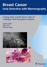
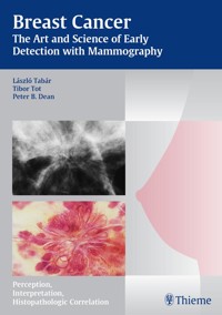
In "Breast Cancer: The Art and Science of Early Detection with Mammography-Perception, Interpretation, Histopathologic Correlation," László Tabár, one of the world's most renowned mammographers, has shared his decades of experience in presenting the fundamentals of perception and interpretation of mammographic images. This is the second volume in a series of books written by the team of Tabár, Tot, and Dean describing breast cancer in its earliest phase according to the imaging findings and correlating these findings with sophisticated histopathologic images and patient outcome. This volume covers a particularly troublesome subtype of breast cancer characterized by casting type calcifications.
Highlights:
- Extensive coverage of the morphology and outcome of this deceptive breast cancer subtype
- Nearly 1000 illustrations of stunning quality showing the full range of manifestations
- Photomicrographs of large, thin-section pathology slides and unique 3D pathology images which are carefully correlated with mammographic images to explain why mammograms appear as they do
- Stereoscopic images that demonstrate normal breast structures and the distortion caused by this unique malignant process
- Presentation of an original theory of neoductgenesis to explain the surprising disease outcome all too frequently observed
- Scientific rationale for using individualized treatment methods which include the mammographic prognostic features
Das E-Book können Sie in Legimi-Apps oder einer beliebigen App lesen, die das folgende Format unterstützen:
Seitenzahl: 162
Veröffentlichungsjahr: 2007
Ähnliche


Library of Congress Cataloging-in-Publication Data is available from the publisher.
©2007 Georg Thieme Verlag, Rüdigerstrasse 14, 70469 Stuttgart, Germanyhttp://www.thieme.de Thieme New York, 333 Seventh Avenue, New York, NY 10001, USAhttp://www.thieme.com
Typesetting by primustype Hurler GmbH, NotzingenPrinted in Germany by Grammlich, Pliezhausen
ISBN 978-3-13-135391-7 (TPS, Rest of World)ISBN 978-1-58890-580-2 (TPN, The Americas)
1 2 3 4 5 6
Important note: Medicine is an ever-changing science undergoing continual development. Research and clinical experience are continually expanding our knowledge, in particular our knowledge of proper treatment and drug therapy. Insofar as this book mentions any dosage or application, readers may rest assured that the authors, editors, and publishers have made every effort to ensure that such references are in accordance with the state of knowledge at the time of production of the book.
Nevertheless, this does not involve, imply, or express any guarantee or responsibility on the part of the publishers in respect to any dosage instructions and forms of applications stated in the book. Every user is requested to examine carefully the manufacturers' leaflets accompanying each drug and to check, if necessary in consultation with a physician or specialist, whether the dosage schedules mentioned therein or the contraindications stated by the manufacturers differ from the statements made in the present book. Such examination is particularly important with drugs that are either rarely used or have been newly released on the market. Every dosage schedule or every form of application used is entirely at the user's own risk and responsibility. The authors and publishers request every user to report to the publishers any discrepancies or inaccuracies noticed. If errors in this work are found after publication, errata will be posted at www.thieme.com on the product description page.
Some of the product names, patents, and registered designs referred to in this book are in fact registered trademarks or proprietary names even though specific reference to this fact is not always made in the text. Therefore, the appearance of a name without designation as proprietary is not to be construed as a representation by the publisher that it is in the public domain.
This book, including all parts thereof, is legally protected by copyright. Any use, exploitation, or commercialization outside the narrow limits set by copyright legislation, without the publisher's consent, is illegal and liable to prosecution. This applies in particular to photostat reproduction, copying, mimeographing, preparation of microfilms, and electronic data processing and storage.
This book is dedicated to those physicians who believe that greater knowledge of the many subtypes of breast cancer will lead to more individualized diagnosis and therapy to the ultimate benefit of their patients.
Preface
Breast cancer is a heterogeneous disease in terms of histology, imaging, and outcome. Mammography as a screening examination has brought to light a new spectrum of breast cancer cases dominated by nonpalpable, preclinical tumors. The outcome of women with early stage disease is considerably better than that of women with late stage disease. The histologic and imaging findings in the early phase consist of a wide spectrum of minimal changes caused by early invasive and in situ disease, but the spectrum of imaging findings narrows as the disease advances. Our series of volumes on the earliest manifestations of breast cancer is being written to demonstrate and teach the importance of this phenomenal heterogeneity, which should affect the planning of surgical and adjuvant therapy.
This volume seeks to uncover a deviant subtype of breast cancer, easily recognizable by its mammographic and histologic appearance, one that has yet to receive sufficient attention despite its uniquely unpredictable nature. This subtype is generally considered to be a form of in situ disease or one that is associated with minimal invasion, although it may behave like a very advanced cancer, much to the surprise of all concerned.
Failure to recognize and single out this subtype, as well as separate it from the other, truly early cases, has led to undertreatment of some women and overtreatment of many women with mammographically detected breast cancer. We sincerely hope that greater knowledge and increased awareness of the breast cancer subtype presenting with casting type calcifications on the mammogram will serve two distinct purposes: to enable physicians to prevent an even greater number of early deaths from breast cancer, while at the same time minimizing the need for grueling and fatiguing therapy of most women with early breast cancer.
László TabárTibor TotPeter B. Dean
List of contributors
Hsiu-Hsi Chen, DDS, PhD Professor Institute of Preventive Medicine Division of Biostatistics Centre of Biostatistics Consultation College of Public Health National Taiwan University Taiwan
Stephen W. Duffy, MSc Professor of Cancer Screening Cancer Research Centre for Epidemiology, Mathematics and Statistics Wolfson Institute of Preventive Medicine London, UK
Ming-Fang Yen, PhD Institute of Preventive Medicine College of Public Health National Taiwan University Taiwan
Yueh-Hsia Chiu, PhD Institute of Preventive Medicine College of Public Health National Taiwan University Taiwan
Contents
Chapter 1: Description of Calcifications Localized within the Ducts
General Introduction
Description of Calcifications localized within the Ducts
Anatomical Background
“Secretory Disease” Type, Plasma Cell Mastitis Type Calcifications
Casting Type Calcifications
Fragmented and Dotted Casting Type Calcifications
Fragmented Casting Type Calcifications on the Mammogram
Dotted Casting Type Calcifications on the Mammogram
Fragmented Casting Type Calcifications
Mammographic Display of Widespread Fragmented Casting Type Calcifications in Five Separate Cases
Macroscopic and Subgross Histological Correlation When Fragmented Casting Type Calcifications Fill an Entire Lobe
Characteristic Mammographic and Histological Presentation of Fragmented Casting Type Calcifications on the Mammogram
Mammographic–Histological Correlation When Both Fragmented Casting Type and Dotted Casting Type Calcifications Are Present
Casting Type Calcifications—Differential Diagnostic Problems
Dotted Casting Type Calcifications
Mammographic Display of Widespread Dotted Casting Type Calcifications in Five Separate Cases
Mammographic–Histological Correlation of Dotted Casting Type Calcifications on the Mammogram
Mammographic–Histological Correlation of Dotted Casting Type Calcifications Caused by Grade 1 DCIS, a Rare Event
Chapter 2: The Evolution of Casting Type Calcifications
Introduction
No Apparent Abnormality on the Previous Mammogram
Cluster of Calcifications as the Earliest Sign
Subtle Casting Type Calcifications as the Earliest Sign
Chapter 3: The Theory of Neoductgenesis
Introduction
Comparison of Benign and Malignant Intraductal Calcifications
The Theory of Ductoneogenesis
Proposal of the Theory of Neoductgenesis
Morphological Demonstration of Neoductgenesis
(A) Unnaturally High Concentration of Ductlike Structures within a Limited Area
(B) Desmoplastic Reaction and Extensive Lymphocytic Infiltration
(C) Disorganized Architecture
(D) Pathologic Ducts: Contorted and Crowded Closely Together
Histological Demonstration of Multiple Newly Formed Ducts
(E) Tenascin Overexpression
Neoductgenesis with Asymmetric Density and Very Subtle, Associated Calcifications
Neoductgenesis without Associated Calcifications
Chapter 4: The Deviant Nature of the Breast Cancer Subtype with Castings
Introduction
The Use of Mammographic Tumor Features to Study Breast Cancer
Relative Frequency of Breast Cancer Occurrence by Mammographic Tumor Features
Long-Term Outcome by Mammographic Tumor Features
Proportion of Breast Cancer Death by Mammographic Tumor Features
Demonstration of Fatal Cases with Casting Type Calcifications
Poor Correlation of Outcome with Mode of Therapy
Demonstration of Local Recurrence Following Radiotherapy
Axillary Node Status and Mammographic Tumor Features
Histological Malignancy Grade and Mammographic Tumor Features
Outcome of 1–14 mm Casting Cases Compared with Advanced Cancers
Chapter 5: Recognition of the Unpredictable, Often Fatal Nature of the Breast Cancer Subtype Presenting with Casting Type Calcifications on the Mammogram
Introduction
Examples with Fatal Outcome
Frequent Recurrence with Casting Type Calcifications
Distant Metastases with Casting Type Calcifications
Suggestion for Use of the Mammographic Prognostic Features in Clinical Practice
Chapter 6: Illustrative Cases with 3-Dimensional Stereoscopic Histological Images
Case 6.1
Case 6.2
Case 6.3
Case 6.4
Case 6.5
Case 6.6
Case 6.7
References
General Introduction
Chapter 1
Chapter 2
Chapter 3
Chapter 4
Chapter 5
Subject Index
General Introduction
Whenever calcifications areseen on the mammogram, the radiologist should attempt to determine the anatomical cavity within which the calcifications have been formed. The options are the following:
Within the ducts (secretory disease type and casting type calcifications)Within the TDLUs (crushed stone-likeand powdery calcifications)Outside the glandular tissue (calcifications within the walls of blood vessels, within the stroma, around oil cysts, or in the skin are characteristically benign)Knowledge of the cradle in whichthe calcifications have been formed as well as analysis of their shape, density, and distribution will facilitate an understanding of the underlying pathophysiological process.
This volume deals with the most unpredictable entity among the breast cancer subtypes, which appears on the mammogram as casting type (BI-RADS® fine, linear, branching) calcifications.1–6 Adetailed description of all of the following aspects is provided to assist the reader in understanding this complex breast cancer subtype:
1 Mammographic features
2 Mammographic–histological correlation using subgross (three-dimensional; 3D) large-section histology
3 Differential diagnosis
4 Clinical presentation
5 Natural history: tendency for recurrence, therapeutic failures, long-term outcome
6 A novel theory explaining the unexpectedly poor outcome of some cases with casting type calcifications
7 Practical implications for the management of patients with casting type calcifications
A unique collection of 3D mammographic/subgross 3D pathology images for stereoscopic viewing with companion 2D conventional histology images is included at the end of this volume.
Description of Calcifications localized within the Ducts
Anatomical Background
The milk duct in the resting state is pleated and measures about 0.1–0.5 mm in width (Figs. 1.1 to 1.4).
Accumulation of either fluid or cancer cells with necrotic debris will greatly distend the duct system.1 Differentiating these two entities from each other is a challenge faced by the radiologist.
Fig. 1.1 Subgross histological image of milk ducts in the resting state.
Figs. 1.3 & 1.4 Subgross histological images of milk ducts, longitudinal and cross sections.
“Secretory Disease” Type, Plasma Cell Mastitis Type Calcifications
An excessive amount of fluid produced by the TDLUs can gradually accumulate within the ducts and their branches.
Figs. 1.5 & 1.6 Subgross histological images of milk ducts in the resting state and distended by fluid.
Figs. 1.7 & 1.8 Subgross histological images of milk ducts distended by inspissated fluid.
Figs. 1.9 & 1.10 Subgross histological images of milk ducts distended by inspissated fluid.
As water is resorbed from this fluid, the residual alkaline, proteinaceous material may calcify. These linear, often branching calcifications are localized within the preexisting duct system and reflect its regular, harmonious structure. This process, commonly called “secretory disease,” is bilateral, which explains the usual bilateral presentation of these typical calcifications.
There are two mammographic presentations of these calcifications:
1 Long, needle-like calcifications (intraductal form)
2 Hollow, ringlike or tubelike calcifications (periductal form)
Intraductal form: The more commonly occurring long, needle-like calcifications are merely calcified duct contents, which assume the smooth, regular outline of the duct lumen. The mammographic image of these calcifications will also have a homogeneously high density (Figs. 1.11 to 1.18).
Figs. 1.11 & 1.12 Detail images of CC projections with the intraductal form of “secretory disease” type calcifications.
Figs. 1.13 & 1.14 An asymptomatic woman, screening examination. The intraductal form of “secretory disease” type calcifications outlines the regular, harmonious structure of the preexisting duct system.
Periductal form: The hollow, ringlike or tubelike calcifications occur when the proteinaceous fluid escapes through the atrophic, porous duct wall and a sterile, asymptomatic “mastitis” is initiated. This process is actually an immunological reaction with predominance of plasma cells and periductal fibrosis. This explains the other term used to describe the mammographic finding: “plasma cell mastitis type calcifications.” These calcifications surround the duct, so that the hollow center of the calcifications corresponds to the noncalcified duct lumen. This less common form is typically accompanied by the intraductal form.
Figs. 1.15 & 1.16 The periductal form of “secretory disease” or plasma cell mastitis type hollow calcifications associated with fibrosis.
Figs. 1.17 & 1.18 MLO and CC projections demonstrate both the intraductal and periductal forms of “secretory disease” or plasma cell mastitis type calcifications.
Casting Type Calcifications
Fragmented and Dotted Casting Type Calcifications
It is important to differentiate the plasma cell mastitis/secretory disease type calcifications from the calcifications seen in a breast cancer subtype commonly called high nuclear grade/poorly differentiated/Van Nuys Group 3/Grade 3 DCIS.
The striking differences are the unilateral, lobar distribution and the highly irregular, disharmonious appearance of the malignant type calcifications. There are two mammographic presentations: the fragmented casting type and the dotted casting type, determined by the cellular growth pattern of these two high-grade malignant processes.
The solid growth pattern of high nuclear grade, poorly differentiated cancer cells produces extensive intraluminal necrosis and amorphous calcifications, which correspond to the mammographic image of fragmented, elongated and branching calcifications (Fig. 1.22-1 to 3). The ductal lumen is filled with cancer cells, necrotic debris, and amorphous calcifications that outline segments of the distended duct lumen in the form of solid, intermittent, cylindrical casts. These fragmented casting type calcifications form a partial outline of the duct and its branches on the mammogram (Figs. 1.25 to 1.29). The origin of these calcifications can be deduced from their typical mammographic appearance. Initially these calcifications will have an irregular contour but, as the necrotic process advances and the debris becomes more extensively calcified at the expense of the viable malignant cells, the mammographic image will exhibit calcifications with a smoother outline and a more homogeneous density. This may occasionally cause differential diagnostic problems if the analysis is restricted to individual calcifications. However, the localized, lobar distribution of these calcifications and their remarkable heterogeneity should resolve this deceptive appearance. (Examples 1.3 to 1.5). Microfocus magnification images will provide additional help in differential diagnosis because considerably more calcifications will become evident if the lesion is malignant, whereas magnification will not reveal additional plasma cell mastitis type calcifications. Rarely, a partially calcified fibroadenoma, traumatic fat necrosis, or benign intraductal papilloma may contain focal branching calcifications, which mimic casting type calcifications.
When the dominant histological featureisthe micropapillary growth pattern, the dotted casting type calcifications are seen on the mammogram. In this type the duct lumen is filled with innumerable, separate, tiny, amorphous calcified particles. The tips of the micropapillary tumor growth extensions break off and eventually calcify. These individual calcified particles accumulate to form the typical dotted casting image on the mammogram. (Fig. 1.24-1 to 3). There is no benign pathological process localized within the ducts that produces a similar mammographic image.
An overview of the histological–mammographic correlation of these two subtypes of high-grade DCIS, seen on the mammogram as fragmented and dotted casting type calcifications, is presented in Figs. 1.21 to 1.24.
Although relatively infrequent, the casting type calcifications represent the most reliable mammographic sign of malignancy, as 97% of them will be malignant at histology (Fig. 1.19). The remaining 3% are found in fibroadenomas, benign papillomas, and rarely in traumatic fat necrosis.
The distribution of malignant type calcifications is shown in Fig. 1.20, based on 322 consecutive, histologically proven breast cancer cases with calcifications on the mammogram as the only radiological abnormality. Casting type calcifications accounted for about 29% of all malignant type calcifications.
Fig. 1.19 The probability of malignancy is as high as 97% when the finding on the mammogram is casting type calcification(s) with or without an associated tumor mass. Women of all ages; prospectively collected consecutive cases. These and subsequent patient material are all from the Department of Mammography, Falun Central Hospital.
Fig. 1.20 Relative frequency of malignant type calcifications without an associated tumor mass. Women of all ages.
Fragmented Casting Type Calcifications on the Mammogram
Characteristic histological features:
Large, polymorphic nuclei with frequent mitosesCell architecture: solid growth patternExtensive intraluminal necrosisAmorphous calcifications filling segments of the ductal lumenCharacteristic mammographic image:
Intermittent, elongated, branching, fragmented casting type calcificationsThe calcifications are usually scattered within alarge part of the lobe, where they are confined to the duct system.Fig. 1.21 Schematic diagram representing fragmented casting type calcifications.
Fig. 1.22-1 Mammogram.
Fig. 1.22-2 Subgross, thick section (3D) histology.
Fig. 1.22-3 Conventional histology.
Dotted Casting Type Calcifications on the Mammogram
Characteristic histological features:
Large, polymorphic nucleiCell architecture: predominantly micropapillary, mixed with solid growth patternIntraluminal necrosisNumerous discrete amorphous calcifications within the intraluminal necrosisCharacteristic mammographic image:
Dotted casting type, “snake skin“-likeScattered, outlining most of the lobeFig. 1.23 Schematic diagram representing dotted casting type calcifications.
Fig. 1.24-1 Mammogram.
Fig. 1.24-2 Subgross, thick section (3D) histology
Fig. 1.24-3 Conventional histology.
Fragmented Casting Type Calcifications
Mammographic Display of Widespread Fragmented Casting Type Calcifications in Five Separate Cases
Fig. 1.25 to 1.28 Detail of MLO projections in four asymptomatic women showing a large number of casting type calcifications.
Fig. 1.29-1 Left breast, detail of the CC projection with numerous casting type calcifications without an associated tumor mass.
Fig. 1.29-2 Operative specimen radiograph of the case shown in Fig. 1.29-1.
Macroscopic and Subgross Histological Correlation When Fragmented Casting Type Calcifications Fill an Entire Lobe
Fig. 1.30-1 Photograph of an operative specimen sliced at the level of the nipple.
Fig. 1.30-2 Subgross histological image covering much of the affected lobe. The main duct, many of its branches and two TDLUs (circle) are greatly expanded by the malignant process.
Fig. 1.30-3 Further magnification of the distended TDLUs and their associated subsegmental duct.
Fig. 1.30-4 A section of the retroareolar duct containing the malignant process and the necrotic debris. Note the adjacent normal-sized pleated ducts.
Fig. 1.30-5 & 6 Subgross histological-mammographic correlation of the findings. The TDLUs contain the crushed stone-like calcifications (circles), while the fragmented, casting type calcifications are localized in the ducts.
Comment
The term “comedo carcinoma” has frequently been used to describe the subtype of DCIS with extensive necrosis, solid cell proliferation, and amorphous calcium. The duct contents are under pressure and tend to ooze out from a freshly cut surface in a manner resembling “comedones” or “comedos” when squeezed out from the skin of the face. These in turn are named from their resemblance to maggots (grub or larvae of flies and other insects), which are also termed “comedones” in Latin. This term, when applied to describe breast cancer, has the disadvantage of being variously interpreted by different subspecialists. Its use tends to lead to confusion, since it fails to accurately describe the underlying pathology.
The imprecise term “comedo” should be replaced by a detailed description of:
The histological grade of the malignant cellsTheir growth patternThe presence of central necrosis and calcificationsCharacteristic Mammographic and Histological Presentation of Fragmented Casting Type Calcifications on the Mammogram
Example 1.1
A 66-year-old asymptomatic woman, screening examination.
Ex. 1.1-1 & 2 Left breast, detail of the MLO and CC projections. There are branching calcifications without an associated tumor mass extending to the retroareolar region.
Ex. 1.1-3 to 5 Correlative subgross, thick section histological, microfocus mammographic and conventional histological images of the region with casting type calcifications.
Ex. 1.1-6 to 9 Typical histological appearance of these mammographically detected fragmented casting type calcifications showing large, polymorphic nuclei with frequent mitoses, solid cell proliferation, extensive intraluminal necrosis, and amorphous calcifications.
Ex. 1.1-10 to 12 Histological examination of cases with extensive fragmented casting type calcifications often reveals ducts where the necrosis encompasses the entire duct lumen, with few or no viable cancer cells remaining. These calcifications are of uniformly high density on the mammogram.
Ex. 1.1-13 & 14 Comparative operative specimen radiograph and subgross histology: the calcifications within the ducts having extensive necrosis and few viable cancer cells will have a higher density and a deceptively smooth contour.
Characteristic Mammographic and Histological Presentation of Fragmented Casting Type Calcifications on the Mammogram
Ex. 1.1-15 & 16 Conventional and subgross histological images of a cross section of a distended duct containing amorphous calcium and surrounded by a desmoplastic reaction.
Ex. 1.1-17 & 18 Subgross and conventional histological images of ducts with extensive necrosis and a few remaining cancer cells.


