71,99 €
Mehr erfahren.
- Herausgeber: John Wiley & Sons
- Kategorie: Fachliteratur
- Serie: What's Your Diagnosis?
- Sprache: Englisch
A unique, case-based guide to diagnosing and treating a wide range of conditions encountered in canine internal medicine
Canine Internal Medicine: What's Your Diagnosis? is an ideal guide to how internal medicine cases are handled in the clinical setting. This text is part of an exciting series, which combines problem-based learning, case studies, and questions and answers. Designed for veterinarians in practice and students, the series presents material in a format designed to enhance critical thinking and understanding.
Adopting a case-based approach, chapters are built around body systems and are directed by questions to test the reader's ability to interpret clinical history, illustrative images and diagnostic results in order to provide differential diagnoses, diagnostic plans and treatment options. Common pitfalls in diagnosis and management are discussed, and you will benefit from the experience of the author as a busy and experienced clinician.
An innovative and interesting way to increase knowledge and skills in canine internal medicine,
Canine Internal Medicine: What's Your Diagnosis? is an indispensable resource for veterinary students, veterinarians in small animal practice, and those studying for post-graduate qualification in small animal medicine.
Sie lesen das E-Book in den Legimi-Apps auf:
Seitenzahl: 720
Veröffentlichungsjahr: 2017
Ähnliche


Contents
Cover
Title Page
Copyright
Dedication
Acknowledgements
How to Use this Book
Introduction
Section A: Endocrinology
Case 1
Clinical Presentation
Haematology
Biochemistry
Urinalysis
Questions
Answers
Discussion
Reference
Further Reading
Case 2
Clinical presentation
Questions
Haematology
Biochemistry
Answers
Diagnosis
Treatment
Discussion
Further reading
Case 3
Haematology
Biochemistry
Questions
Answers
Diagnosis
Treatment
Discussion
Further reading
Case 4
Questions
Answers
Diagnosis
Haematology
Biochemistry
Urinalysis
ACTH stimulation test
Low-dose dexamethasone suppression test
Discussion
Reference
Further reading
Case 5
Clinical presentation
Questions
Answers
Diagnosis and treatment
Haematology
Biochemistry
Discussion
Further reading
Section B: Haematology and Immunology
Case 6
Clinical Presentation
Questions
Answers
Further Questions and Answers
Haematology
Biochemistry
Diagnosis and Outcome
Discussion
References
Further Reading
Case 7
Clinical Presentation
Haematology
Biochemistry (note that supernatant of the serum sample is strongly red-brown discoloured)
Questions
Answers
Diagnosis
Discussion
Further Reading
Case 8
Clinical Presentation
Questions
Answers
First Diagnostic Steps
Second Diagnostic Steps
Diagnosis and Treatment
Haematology
Discussion
Reference
Further Reading
Case 9
Clinical Presentation
Questions
Answers
Diagnosis
Treatment/outcome
Discussion
References
Further reading
Section C: Hepatobiliary Disease
Case 10
Clinical Presentation
Haematology
Biochemistry
Questions
Answers
Diagnosis
Discussion
References
Further reading
Case 11
Clinical Presentation
Haematology
Biochemistry
Questions
Answers
Diagnosis
Discussion
Further Reading
Case 12
Clinical Presentation
Haematology
Biochemistry
Urinalysis (Cystocentesis Sample)
Questions
Answers
Laboratory Tests
Diagnostic Imaging
Direct Sampling of the Hepatobiliary Tract
Diagnosis
Treatment and Outcome
Discussion
Further Reading
Section D: Gastroenterology
Case 13
Clinical Presentation
Haematology
Biochemistry
Questions
Answers
Outcome
Discussion
Further Reading
Case 14
Clinical Presentation
Questions (There are Three Questions, Two Before and One After the Case Findings)
Answers
Haematology
Biochemistry
Answer
Outcome
Discussion
References
Further Reading
Case 15
Clinical presentation
Questions
Answers
Diagnostic imaging
Histopathology from intestinal biopsies
Therapeutic trials
Diagnosis and outcome
Haematology
Biochemistry
Endocrine tests
Faecal analysis/culture
Discussion
Further reading
Case 16
Clinical presentation
Haematology
Questions
Answers
Diagnosis
Discussion
Further reading
Section E: Respiratory
Case 17
Clinical presentation
Questions
Answers
Diagnosis
Haematology
Treatment
Discussion
References
Further reading
Case 18
Clinical presentation
Questions
Answers
Diagnosis
Treatment and outcome
Discussion
Further reading
Case 19
Clinical presentation
Questions
Answers
Outcome
Discussion
Reference
Further reading
Section F: Ear, Nose and Throat
Case 20
Clinical presentation
Questions
Answers
Diagnosis
Discussion
Further reading
Case 21
Clinical Presentation
Questions
Answers
Diagnosis
Treatment (Figure 21.8)
Discussion
Further Reading
Section G: Cardiovascular
Case 22
Clinical Presentation
Questions
Answers
Diagnosis
Treatment
Discussion
Further Reading
Case 23
Clinical Presentation
Questions
Answers
Diagnosis
Treatment and Outcome
Discussion
References
Further reading
Section H: Urology and Nephrology
Case 24
Clinical Presentation
Haematology
Biochemistry
Questions
Answers
Diagnosis and Treatment
Urinalysis
Discussion
References
Further Reading
Case 25
Clinical Presentation
Questions
Urinalysis (Cystocentesis Sample)
Answers
Discussion
Reference
Further Reading
Case 26
Clinical Presentation
Question
Answers
Diagnosis
Treatment
Prognosis and discussion
Further Reading
Section I: Reproductive/Genital Tract
Case 27
Clinical Presentation
Questions
Answers
Diagnosis
Outcome
Discussion
Further Reading
Section J: Oncology
Case 28
Clinical Presentation
Questions
Answers
Further Reading
Case 29
Questions
Answers
Diagnosis
Haematology
Biochemistry
Discussion
Reference
Further Reading
Case 30
Clinical Presentation
Questions
Answers
Risks to the Patient
Outcome
Haematology
Biochemistry
Treatment
Discussion
Further Reading
Section K: Neurology
Case 31
Clinical Presentation
Questions
Answers
Diagnosis
Discussion
Further Reading
Case 32
Clinical Presentation
Questions
Answers
Prognosis
Outcome
Discussion
Reference
Further Reading
Section L: Infectious Diseases
Case 33
Clinical Presentation
Haematology at Previous Veterinary Surgeon (Sample Analysed at Reference Laboratory)
Biochemistry at Previous Veterinary Surgeon (Sample Analysed at Reference Laboratory)
Urinalysis at Previous Veterinary Surgeon (Sample Analysed at Reference Laboratory)
Questions
Answers
Diagnosis and Treatment
Discussion
Further Reading
Case 34
Clinical Presentation
Haematology
Biochemistry
Urinalysis (Free Catch Sample)
Questions
Answers
Outcome
Discussion
Further reading
Appendix 1: Index of Tables and Figures, Pearls and Clinical Skills Generated
Appendix 2: Diagnosis by Case
Appendix 3: Conversion Table of SI to Common Units
Index
End User License Agreement
List of Tables
Table 1.1
Table 1.2
Table 1.3
Table 1.4
Table 1.5
Table 2.1
Table 2.2
Table 3.1
Table 3.2
Table 3.3
Table 4.1
Table 4.2
Table 4.3
Table 5.1
Table 5.2
Table 5.3
Table 6.1
Table 7.1
Table 7.2
Table 8.1
Table 8.2
Table 8.3
Table 9.1
Table 10.1
Table 11.1
Table 12.1
Table 12.2
Table 12.3
Table 12.4
Table 12.5
Table 12.6
Table 12.7
Table 13.1
Table 13.2
Table 13.3
Table 14.1
Table 14.2
Table 15.1
Table 15.2
Table 16.1
Table 17.1
Table 17.2
Table 19.1
Table 20.1
Table 22.1
Table 23.1
Table 24.1
Table 24.2
Table 25.1
Table 25.2
Table 26.1
Table 26.2
Table 27.1
Table 27.2
Table 28.1
Table 29.1
Table 30.1
Table 30.2
Table 30.3
Table 30.4
Table 30.5
Table 30.6
Table 30.7
Table 32.1
Table 33.1
Table 33.2
Table 33.3
List of Illustrations
Figure 1.1
Figure 1.2
Figure 2.1
Figure 2.2
Figure 2.3
Figure 2.4
Figure 3.1
Figure 4.1
Figure 4.2
Figure 4.3
Figure 4.4
Figure 4.5
Figure 5.1
Figure 6.1
Figure 6.2
Figure 6.3
Figure 7.1
Figure 7.2
Figure 7.3
Figure 7.4
Figure 8.1
Figure 8.2
Figure 8.3
Figure 9.1
Figure 9.2
Figure 9.3
Figure 10.1
Figure 10.2
Figure 10.3
Figure 10.4
Figure 11.1
Figure 11.2
Figure 11.3
Figure 11.4
Figure 11.5
Figure 11.6
Figure 11.7
Figure 11.8
Figure 11.9
Figure 12.1
Figure 12.2
Figure 12.3
Figure 12.4
Figure 13.1
Figure 13.2
Figure 13.3
Figure 14.1
Figure 14.2
Figure 14.3
Figure 14.4
Figure 15.1
Figure 15.2
Figure 15.3
Figure 16.1
Figure 16.2
Figure 16.3
Figure 16.4
Figure 17.1
Figure 17.2
Figure 17.3
Figure 17.4
Figure 18.1
Figure 18.2
Figure 18.3
Figure 18.4
Figure 18.5
Figure 19.1
Figure 19.2
Figure 19.3
Figure 19.4
Figure 19.5
Figure 19.6
Figure 20.1
Figure 20.2
Figure 20.3
Figure 20.4
Figure 21.1
Figure 21.2
Figure 21.3
Figure 21.4
Figure 21.5
Figure 21.6
Figure 21.7
Figure 21.8
Figure 22.1
Figure 22.2
Figure 22.3
Figure 22.4
Figure 22.5
Figure 22.6
Figure 22.7
Figure 22.8a
Figure 22.8b
Figure 22.8c
Figure 22.9
Figure 22.10
Figure 23.1
Figure 23.2
Figure 22.3
Figure 23.4
Figure 24.1
Figure 24.2
Figure 24.3
Figure 24.4
Figure 24.5
Figure 25.1
Figure 25.2
Figure 25.3
Figure 26.1
Figure 26.2
Figure 26.3
Figure 26.4
Figure 26.5
Figure 26.6
Figure 26.7
Figure 26.8
Figure 26.9
Figure 26.10
Figure 27.1
Figure 27.2
Figure 27.3
Figure 28.1
Figure 28.2
Figure 28.3
Figure 28.4
Figure 29.1
Figure 29.2
Figure 29.3
Figure 29.4
Figure 29.5
Figure 29.6
Figure 30.1
Figure 30.2
Figure 30.3
Figure 30.4
Figure 30.5
Figure 30.6
Figure 30.7
Figure 31.1
Figure 31.2
Figure 31.3
Figure 31.4
Figure 31.5
Figure 32.1
Figure 33.1
Figure 33.2
Figure 33.3
Figure 33.4
Figure 33.5
Figure 34.1
Figure 34.2
Guide
Cover
Table of Contents
Begin Reading
Part 1
Chapter 1
Pages
i
ii
iii
iv
vii
viii
ix
x
xi
xii
xiii
xiv
xv
xvi
xvii
xviii
xix
xx
1
2
3
4
5
6
7
8
9
10
11
12
13
14
15
16
17
18
19
20
21
22
23
24
25
26
27
28
29
30
31
32
33
34
35
36
37
38
39
40
41
42
43
44
45
46
47
48
49
50
51
52
53
54
55
56
57
58
59
60
61
62
63
64
65
66
67
68
69
70
71
72
73
74
75
76
77
78
79
80
81
82
83
84
85
86
87
88
89
90
91
92
93
94
95
96
97
98
99
100
101
102
103
104
105
106
107
108
109
110
111
112
113
114
115
116
117
118
119
120
121
122
123
124
125
126
127
128
129
130
131
132
133
134
135
136
137
138
139
140
141
142
143
144
145
146
147
148
149
150
151
152
153
154
155
156
157
158
159
160
161
162
163
164
165
166
167
168
169
170
171
172
173
174
175
176
177
178
179
180
181
182
183
184
185
186
187
188
189
190
191
192
193
194
195
196
197
198
199
200
201
202
203
204
205
206
207
208
209
210
211
212
213
214
215
216
217
218
219
220
221
222
223
224
225
226
227
228
229
230
231
232
233
234
235
236
237
238
239
240
241
242
243
244
245
246
247
248
249
250
251
252
253
254
255
256
257
258
259
260
261
262
263
264
265
266
267
268
269
270
271
272
273
274
275
276
277
278
279
280
281
282
283
284
285
286
287
288
289
290
291
292
293
294
295
296
297
298
299
300
301
302
303
304
305
306
307
308
309
310
311
312
313
314
315
316
317
318
319
320
321
322
323
324
325
326
327
328
329
330
331
332
333
334
335
336
337
338
339
340
341
342
343
344
345
346
347
348
349
350
351
352
353
354
355
356
357
358
359
360
361
362
363
364
365
366
367
368
369
370
371
372
373
374
375
376
377
378
379
380
381
382
383
384
385
386
387
388
389
390
391
392
393
394
395
396
397
398
399
400
401
402
403
404
405
406
407
408
409
410
411
412
413
414
415
416
417
418
419
420
421
422
423
424
425
426
427
428
429
430
431
432
433
434
435
436
437
438
439
440
441
442
443
444
445
446
447
448
449
450
451
452
453
454
455
456
457
458
Canine Internal Medicine
What's Your Diagnosis?
Jon Wray BVSc DSAM Cert VC MRCVS, RCVS-recognised Specialist in Small Animal Medicine (Internal Medicine)
Consultant Specialist in Small Animal Internal Medicine and Head of CardiologyDick White Referrals, Station Farm, Six Mile Bottom, Cambridgeshire
This edition first published 2018© 2018 John Wiley & Sons Ltd
All rights reserved. No part of this publication may be reproduced, stored in a retrieval system, or transmitted, in any form or by any means, electronic, mechanical, photocopying, recording or otherwise, except as permitted by law. Advice on how to obtain permission to reuse material from this title is available at http://www.wiley.com/go/permissions.
The right of Jon Wray to be identified as the author of this work has been asserted in accordance with law.
Registered OfficesJohn Wiley & Sons, Inc., 111 River Street, Hoboken, NJ 07030, USA John Wiley & Sons Ltd, The Atrium, Southern Gate, Chichester, West Sussex, PO19 8SQ, UK
Editorial Office9600 Garsington Road, Oxford, OX4 2DQ, UK
For details of our global editorial offices, customer services, and more information about Wiley products visit us at www.wiley.com.
Wiley also publishes its books in a variety of electronic formats and by print-on-demand. Some content that appears in standard print versions of this book may not be available in other formats.
Limit of Liability/Disclaimer of WarrantyThe contents of this work are intended to further general scientific research, understanding, and discussion only and are not intended and should not be relied upon as recommending or promoting scientific method, diagnosis, or treatment by physicians for any particular patient. In view of ongoing research, equipment modifications, changes in governmental regulations, and the constant flow of information relating to the use of medicines, equipment, and devices, the reader is urged to review and evaluate the information provided in the package insert or instructions for each medicine, equipment, or device for, among other things, any changes in the instructions or indication of usage and for added warnings and precautions. While the publisher and authors have used their best efforts in preparing this work, they make no representations or warranties with respect to the accuracy or completeness of the contents of this work and specifically disclaim all warranties, including without limitation any implied warranties of merchantability or fitness for a particular purpose. No warranty may be created or extended by sales representatives, written sales materials or promotional statements for this work. The fact that an organization, website, or product is referred to in this work as a citation and/or potential source of further information does not mean that the publisher and authors endorse the information or services the organization, website, or product may provide or recommendations it may make. This work is sold with the understanding that the publisher is not engaged in rendering professional services. The advice and strategies contained herein may not be suitable for your situation. You should consult with a specialist where appropriate. Further, readers should be aware that websites listed in this work may have changed or disappeared between when this work was written and when it is read. Neither the publisher nor authors shall be liable for any loss of profit or any other commercial damages, including but not limited to special, incidental, consequential, or other damages.
Library of Congress Cataloging-in-Publication Data
Names: Wray, Jon (Jonathan David), 1973- author.
Title: Canine internal medicine : what's your diagnosis? / by Jon Wray.
Description: Hoboken, NJ : Wiley, 2018. | Includes bibliographical references and index. |
Identifiers: LCCN 2017030339 (print) | LCCN 2017034096 (ebook) | ISBN 9781118917787 (pdf) | ISBN 9781118918166 (epub) | ISBN 9781118918173 (pbk.)
Subjects: | MESH: Dog Diseases | Internal Medicine–methods | Case Reports
Classification: LCC SF991 (ebook) | LCC SF991 (print) | NLM SF 991 | DDC 636.7/0896–dc23
LC record available at https://lccn.loc.gov/2017030339
Cover image: Courtesy of Jon WrayCover design by Wiley
Dedication
For those who have left us: big brother SAJ, Dad and Mum who gave me so much and Ross who was taken too young.
For my darling Vicky and our children Ella, Daniel and Sophie, who are my Universe and make me laugh until it hurts, and my brother Matthew.
For my mentors, colleagues, residents, interns, nurses and students who have taught (and continue to teach) me so very many things.
For our patients, who make it all worthwhile.
Acknowledgements
I have the tremendous privilege to work with a truly fantastic group of Specialists, now numbering some 33 in total, in one of the largest referral hospitals in Europe. The Specialists are ably supported by a large number of talented, knowledgeable and caring Residents, Interns, Nurses, Laboratory technicians, Receptionists and others, without whom our practice would quickly grind to a halt. The patients we see (and the patients in this book) all benefit from a group approach and Internal Medicine is a collaborative subject. I could not give my patients the care they deserve without the input of my Specialist colleagues in other disciplines such as Surgery, Diagnostic Imaging, Clinical Pathology, Anaesthesiology, Ophthalmology, Neurology and Dermatology, the daily input from fellow Specialists and Residents in internal medicine and the dedication and care of our brilliant nursing team.
To quote (the often misquoted) Kurt Koffka, the whole really is other than the sum of its parts, and our hospital founder, Professor Dick White, has both recognised and encouraged this, both in his vision and in his assembly of a team of like-minded individuals who share a common belief in a multi-disciplinary Specialist approach to veterinary medicine and in our practice ethos of ‘Excellence, Trust, Care'.
It is a genuine privilege to work in an environment where everyone's expertise in working towards a common goal yields tangible combined results that can be seen on a daily basis. I should also like to acknowledge the veterinary practices that refer patients to us, in the knowledge that it is not simply having facilities, but more importantly the Specialist expertise to apply them, that counts, and who recognise the distinction between Residency-trained (‘apprenticeship') Specialists and other qualifications which, whilst of a good standard, are essentially self-taught. We believe that clinical standards of excellence are worth striving for, even if that means travelling a longer and more arduous personal route to get there, and in this ‘post-truth' world (where anyone with an opinion can make it appear as though it is an incontrovertible fact on the world-wide web) critical appraisal and evidence-based approaches are more important than ever to defend.
Specific acknowledgements are due to colleagues Jackie Demetriou for Figure 10.4, Anna Adrian for Figure 11.3, Andrew Holloway for Figure 11.4 and Simon Tappin for case material for Case 34.
Jon WrayDick White ReferralsCambridgeshireUnited Kingdom, 2017
How to Use this Book
What's your diagnosis? How did you reach it? Through logical deductive reasoning or through ‘clinical shortcuts'? Which methods work best? Internal Medicine (IM) is a subject that relies on problem solving so test your problem-solving skills by perusing these cases and see...
This book is neither intended to be an Internal Medicine textbook, nor to include examples of every disease situation encountered in an Internal Medical caseload. What I have intended to do, by use of examples from my clinical practice, is to provide a wide ‘spread' of clinical problems in which the case itself becomes ‘the teacher'. This book is intended for anyone with an interest in Internal Medicine, whether at undergraduate or post-graduate level, General Practice or Speciality Practice. Inevitably there will be some, relatively common, case presentations that there has not been room to include or where the clinical skills challenged by them are replicated in another case example.
It is a frequent, and in many ways valid, criticism of undergraduate teaching, that by situating it mostly in referral hospitals (where the caseload is by nature highly selected and clients generally motivated and financially secure to expedite diagnosis and therapy) the cases may not offer a ‘realistic substrate' for learning the art as well as the science of small animal medicine. However, I would counter this by stating that, in my experience, these hospitals are usually staffed by very talented clinicians who are both well-adept at drawing attention to specific case details that are important ‘learning points' but also at focusing on those basic skills of history taking and physical examination that are the cornerstones of medical diagnosis. This is a point that is often missed – critics may remember that such cases are unusual and may not be representative of the vast majority of their own caseload, but usually forget the basic (and transferable) steps that led to diagnosis of the unusual problem in the first place. Furthermore, none of these cases miraculously ‘appear out of the ether' in the reception area of a referral hospital – all have walked (or been carried) through the door of a general veterinary practice and the presenting complaint will be one encountered every day in a practice somewhere. You might be the vet who encounters it; are you ready?
Similarly, post-graduate education has become much more widely available and there are a plethora of excellent CPD courses, post-graduate qualifications, modular programs, etc., for the interested and committed individual. However, what none of these can do is replicate a busy Internal Medicine caseload and it is all too easy to fall into the trap of being well-informed, but never having the chance to experience the practical caseload that a busy IM department sees and thus not generating the ‘right sort' of case experience. These cases are all taken from our Internal Medicine caseload at Dick White Referrals. We are a very busy department with 10 full-time Internal Medicine Clinicians and a tremendous and varied caseload. Our Residents have the luxury of being totally immersed in large numbers of these cases every day and I hope to share some of these with you. Where there are ‘take-home' messages (which have become popular to refer to as ‘pearls', and who am I to argue?), which I believe are pertinent to each case, I have tried to highlight these. I've also tried to include some useful additional information in the form of common differential diagnoses and case approaches, which, whilst not intended to be as complete as a textbook, will hopefully be easier to remember when attached to an interesting case. I always learn better with the immediacy and constant cue of a patient with which I am dealing and I hope the reader also finds refreshing their memory in the context of a clinical case that much more engaging than an abstract text.
I am a busy Specialist clinician having spent the last nineteen years exclusively in small animal Internal Medicine (canine and feline – I am not a particular advocate of a ‘species-ist' restriction of one's caseload) referral practice at four major UK referral centres and being continuously ‘on clinics' during that time. During this time I have also examined many veterinary surgeons in their pursuit of post-graduate qualifications at both Certificate and Diploma level. Partly the choice of which case subjects to include in this book is informed by this experience of those clinical problems that often seem to cause the most diagnostic difficulty and conversely in the exclusion of some common scenarios that really do not present much of a diagnostic ‘challenge'.
In the same vein, whilst the cases here vary in their complexity, the features they all have in common are that they provide useful ‘take-home messages' and challenge areas of clinical skill that are worth emphasising and learning from. In this way I hope that this book acts as a ‘self-assessment' guide, but one that provides a little more detail than most. The cases are divided into subject area/body systems chapters. I have summarised the areas where there is further explanation of key clinical skills and approaches in the table in Appendix 1, which can then be cross-referenced with the cases. The table in Appendix 2 lists the diagnosis for each case for easy reference. Finally, the table in Appendix 3 lists conversion from SI units to common units for the reference of those readers from outside Europe.
After each case presentation there are a series of questions to prompt the reader in thinking about/problem solving a particular aspect of the case. I would recommend performing these before reading on. After answers to these questions, the actual findings and outcome of the case described are presented along with a brief discussion. As with all cases, there may be several routes to the appropriate diagnosis and it is often the case that no two clinicians might manage or investigate a case in exactly the same way.
Also, as with any case-based text, one of the inherent drawbacks is that the cases are derived from a population that may have different prevalence of some diseases from other parts of the world. The reader is urged to apply their own experience and clinical commonsense in modifying their approach accordingly.
Before the cases is a brief introductory section on medical problem solving.
Jon WrayDick White ReferralsCambridgeshireUnited Kingdom, 2017
Introduction
Approach to medical problem-solving and laboratory interpretation
All medical problem solving succeeds or fails on the history and the physical examination. Without performing these properly, all diagnostic attempts are doomed to being based more on luck than judgment. These skills tend to, in my humble opinion, be ignored/marginalised/undervalued by many veterinary surgeons who are keen to explore more technologically ‘glamorous' avenues of diagnosis. This is a mistake. Honing the skills of asking the right questions, in the right way and interpreting the findings of this and a thorough, skillful and objective physical examination are the sine qua non of Internal Medicine diagnosis.
The problem list
The initial approach to medical diagnosis starts with the problem list. In this context a medical ‘problem' may be a clinical sign exhibited by the animal or concern expressed by their owner, a physical examination abnormality or the result of a diagnostic test that differs from the expected normal. Great care must be taken to define the nature of the problem, especially where different problems may appear, or at least be described, similarly. An example of this would be the need to distinguish regurgitation (which is often due to oesophageal disease) versus vomiting (which is usually associated with pathology distal to the oesophagus). Careful ‘open' (that is not leading) questioning and unbiased interpretation of responses during the initial history taking is an important skill to cultivate. A good thorough clinical history should often allow a judicious clinician to both define what the problem is and often where the problem is. This latter is especially important in refining the diagnostic approach to the likely area of interest, especially where diagnostic imaging is to be performed. For example, performing thoracic radiographs alone to look for a cause of dyspnoea in an older Labrador with laryngeal paralysis is not only going to lead to misdiagnosis but belies a lack of recognition of the site of disease because of poor observational/physical examination skills.
By defining a problem list that is complete, the clinician can avoid potential errors of omission and ‘anchoring bias' (see below) that may prevent accurate diagnosis. By using this problem list to develop a set of relevant differential diagnoses for each problem, areas of potential commonality may be reviewed and can help marshal the differential diagnosis more rapidly into an order of likelihood than might otherwise be the case. The law of parsimony (Occam's razor) is usefully employed (see Case 3 for a further explanation) to focus on the simplest hypothesis/that which involves the fewest assumptions first.
Although a problem list should be complete, there is sense in considering both those problems that are the most serious/impactful on the patient and also those that have a limited or well-defined differential diagnosis and affording these greater emphasis when it comes to construction of a differential diagnosis list. For instance, in an animal whose clinical signs (problems) include vomiting, lethargy, jaundice and inappetence, the problems of vomiting and jaundice have more easily defined and limited differential diagnoses than those of lethargy and inappetence, which are common to so many clinical illnesses that the differential diagnosis of them would be huge (and meticulous consideration and exclusion of all of them would result in an unnecessarily lengthy, broad-based and potentially medically overintrusive diagnostic course). Thus whilst it is encouraged to maintain an open mind in producing a problem list that is genuinely reflective of all problems identified, equally clinicians are encouraged to perform some initial ‘sorting' of these into problems that may rationally be afforded greater emphasis (‘pivotal' problems) or be a more effective ‘substrate' for medical problem-solving, versus those that, whilst not dismissed, are of a more general/broad nature.
The differential diagnosis
Differential diagnoses can be problematic in many ways, not least because there is variation in how these can be considered. One can consider a ‘mechanistic' differential diagnostic approach such as may be appropriate for considering icterus/jaundice (to divide it into pre-hepatic, hepatic and post-hepatic causes) or polyuria/polydipsia, but this may not be suitable for many other types of clinical problem. There are various useful mnemonics, such as VITAMIN-D or DAMNIT-V to help the clinician consider broad aetiologies of differential diagnosis and thus to avoid errors of omission. Whilst these have their place, it is important to recognise that these, and commonly lists of differential diagnoses reproduced in reference textbooks, fail to take into account the prevalence of diseases and geographical and practice-type variability. These have a profound effect on pre-test probability (see below) and, by-and-large, it is sensible to start with a relatively broad differential diagnostic list and then to ‘marshal' these into some sort of order of likelihood based on factors such as known prevalence and geography, as well as considering those problems whose differential diagnoses are limited as offering the greatest possibility of successful medical problem solving. This book is written in the United Kingdom and in particular there are many infectious diseases that are not endemic to our shores, so readers in other countries are encouraged to elevate diseases native to their own countries to their appropriate position based on prevalence. Where I have used mnemonics I have used the DAMNIT-V system, not because it is particularly better than any other, but because that is what I was taught and am most familiar with. The DAMNIT-V mnemonic describes broad aetiological subdivisions as:
D
Degenerative
A
Anomalous, Anatomical
M
Metabolic
N
Neoplastic, Nutritional
I
Infectious (including parasitic), Inflammatory, Immune-mediated
T
Traumatic, Toxic
V
Vascular
One notable absence from both DAMNIT-V and VITAMIN-D classifications is genetic disorders, though these may variably fall under one of the categories such as ‘Metabolic' or be considered an ‘Anomaly'.
The usefulness of such mnemonics is that they help reduce the fallacies of omission and ‘anchoring' bias that may beset clinical problem solving. The primary disadvantage of them is that they may appear to afford equal emphasis to all possibilities whereas the thoughtful clinician will construct a list that takes into account not only possibility but also likelihood.
Sources of diagnostic error, recognition of diagnostic styles, bias and cognitive error and heuristics
It should go without saying that the success of any treatment relies on applying it to patients in which an accurate diagnosis has been made first of all. It behoves clinicians to recognise those potential causes of diagnostic error, which may then limit the effectiveness of any future treatment strategy if it is thus consequently misapplied to the wrong diagnosis. Sources of diagnostic error may include:
So-called ‘no fault error', for example where willful misleading of a veterinary surgeon by an owner occurs or where a new variation of disease occurs.
‘System error', where organisational, technical problems or production of spurious results hampers diagnosis.
‘Cognitive error', which is due to flawed clinical reasoning.
The clinical approach to diagnostic reasoning has been previously described in terms of ‘Intuitive' reasoning (sometimes called ‘System 1 thinking') and ‘Analytical' reasoning (sometimes called ‘System 2 thinking'). Clinicians who employ purely Intuitive reasoning rely heavily on experience, prior case exposure, intuition, pattern recognition, heuristics (mental shortcuts that make intuitive diagnostic sense) and long-term memory. Those who employ Analytical thinking rely on systematic problem solving, analysis, reasoning and hypothesis or data-driven consciously controlled decision making. The former approach risks errors of omission and fallaciously limited consideration based on potentially very limited prior case exposure; the latter may risk clinicians overthinking problems, missing the obvious/commonsensical approach and may lead to overly laborious, expensive, medically intrusive and ‘throwing the kitchen sink at it' approaches to medical diagnosis. Of course, in reality these two different styles of diagnostic reasoning are seldom practiced one to the exclusion of the other and most clinicians will employ a range of approaches that often use a combination of these styles. By considering those aspects of medical problem solving that are best served by one or other of these approaches and by critically appraising those features that may lead to errors of diagnostic judgment (cognitive errors), thoughtful clinicians can guard against making mistakes that may hamper accurate diagnosis.
Cognitive error (flawed clinical reasoning) has many forms and causes and whilst it is often stated that cognitive error is more common in System 1-based thinking it is erroneous to consider System 2-based thinking as free of the shackles of cognitive errors. As clinicians, our level of tiredness, stress, anxiety and distractions of everyday life can enhance our risk of falling into traps of cognitive error but, nonetheless, recognition of the types of error we may be prone to allows clinicians to be mindful of these and reduce our chances of falling prey to them.
Although not an exhaustive list, common sources of cognitive error in diagnostic reasoning include:
Confirmation bias.
This is the tendency to look for and interpret information in a way that supports prior-held preconceptions about a case. This is often most risked by individuals who have a limited case-exposure or experience (note that it is difficult for an individual to be aware that this is the case since by definition that is the the working environment they are accustomed to) and who may prematurely cease a search for a correct diagnosis based on initial findings. Often this may result in a clinician being satisfied by finding ‘
an
answer' rather than ‘
the
answer'. In diagnostic imaging this phenomenon is well-recognised as premature ‘satisfaction of search', that is an individual may inadvertently eschew thorough evaluation of an entire radiograph, for example, when they spot an obvious lesion initially. Confirmation bias may not be ‘all bad' and the counterpoint to it is that sometimes the immediately obvious (Gestalt recognition) is correct and there may be danger of ‘talking oneself away' from the correct diagnosis in pursuit of an open-minded broad investigation. ‘Sutton's Law' describes the sometimes unarguable veracity of the obvious (Sutton was an American bank-robber whose name is enshrined in the annals of history for supposedly stating in court when asked why he robbed banks, ‘Er, because that's where the money is!').
Availability bias
is the tendency to overestimate likelihood of a diagnosis based on how memorable previous examples of that diagnosis were.
Anchoring bias
is the fallacy of weighting too much of the clinical argument for a diagnosis on one particular piece of information or diagnostic trait, rather than considering other possibilities.
Attribution error
is the tendency to leap to conclusions rapidly that serve to reinforce a negative stereotype or afford less diagnostic emphasis in a patient that is unpleasant, aggressive or uncooperative (or whose owners show similar traits).
Affective error (‘rose-tinted spectacles' thinking)
is the error caused by clinicians (unintentionally) blinding themselves to the possibilities of negative outcomes, less appealing alternative diagnoses or the prospect of needing to perform procedures that are perceived unpleasant. Clinicians are particularly prone to this error in patients with which they have formed emotional attachments/owners in whom good rapport has been established, especially if this is over a long period of time.
Commission bias
is the tendency to err towards action ‘for the sake of it', often in the absence of a firm idea of what is being treated. It is often more profitable to the patient and safer not to do anything but to take further time considering a patient's needs.
Vertical line failure
(thinking inside the box) is a tendency to be contrained by a sequential/narrow field of search, often anchored (see above) on a previous diagnostic test or heavily constrained by overreliance on one particular form of diagnostic testing. A particular danger here is clinicians who may overrely on tehcniques such as MRI or CT, which, whilst undoubtedly powerful diagnostic techniques, have limitations that if not appreciated may lead to cognitive effort. Failure to keep an open mind and to continually review the initial problem list based on accurate history and physical examination are the principle initiators of this error.
Biases in clinical thinking are more likely to occur with System 1 (pattern-recognition) thinking but also somewhat in System 2 thinking if it is untrained. However, not all bias necessarily has a negative impact on the diagnostic process, and when recognised and harnessed, bias may provide clinically useful and appropriate shortcuts in thinking logically that we term ‘heuristics' (see below). One of the first steps in avoiding harmful bias is in recognising sources of cognitive error and taking steps to avoid them once aware. However, some ‘tools' of clinical thinking (sometimes referred to as ‘metacognitive strategies') can also help in avoiding such diagnostic pitfalls. Examples of metacognitive strategies include (in addition to education and awareness of cognitive error and types of clinical thinking) recognising a need to observe the broad clinical picture, carving out time and head-space to both reflect upon and to rationalise clinical thinking but also in recognising the impact of affective states (such as tiredness, anxiety, mood) on one's critical thinking. Creation of time and also forcing oneself to ‘slow down' on occasion is inherent to this and we will often exhort our Residents to find a quiet area, clear their desk and their mind and spend some time in quiet reflection about a case; using pen-and-paper to organise one's thoughts, write checklists and review/rewrite/reorder problem lists and differential diagnosis lists can be helpful in this. Above all, in situations where immediate action is not critical to the wellbeing of the patient, having the self-determination to do nothing medically but to expend effort on critical thinking, is often the correct strategy. This has sometimes been described by reversing a common phrase into ‘don't just do something, stand there'.
Medical professionals generally do not think or process thought in a linear fashion in the same way that, for example, economists do, by first receiving data then analysing it in its entirety. In Donald Shön's ‘The Reflective Practitioner' clinicians are described as displaying (though not uniquely among professionals) ‘reflection-in-action', synthesising information, inquiry and intervention, interlaced with each other. In order to make cognitive sense of such a simultaneous confluence of processes, clinicians often develop cognitive shortcuts, known as heuristics, which may help rapidly navigate through seemingly highly complex situations in a way that marshals probability without closing off less common cognitive routes and so committing fallacies of omission. Heuristics are clinically useful provided they are balanced by knowledge, recognition of cognitive error and frequent reflection, and tend to thrive in environments of ‘commonsense' thinking and where cost and technological constraints force logical deductive reasoning rather than a defensive, ‘throw the (diagnostic) kitchen sink' at a problem, approach.
Reference intervals, appropriate diagnostic testing and dangers of the Ulyssees syndrome
Interpretation of diagnostic test results depends on having a reference interval for that analyte; that is the range of results we might expect to find in a healthy animal. In this book the reference intervals are selected from our own clinical pathology laboratory at Dick White Referrals staffed by a team of Specialist Clinical Pathologists. Reference intervals should be established by any individual laboratory by assessment of a number (usually at least 60, preferably 120) of normal animals. The criteria used to assess ‘normality' should be selected prior to data collection (including age, sex, breed, etc). A reference interval is based on the values obtained in 95% of this sample population and clinicians should always remember that even if the reference population is exactly representative of the population ‘at large' 2.5% of clinically normal animals will have values of an analyte that fall below this reference interval and 2.5% have values of the analyte that fall above it.
This should lead to cautious interpretation of values that fall just outside reference intervals and prompt the realisation amongst clinicians that whilst ‘panels' of diagnostic tests are convenient and economic for laboratories to provide, they also magnify the chances of detecting apparently abnormal results in individuals where the value recorded is not due to a pathological process. For instance, if a panel of 20 biochemical tests is performed on a healthy individual, there is a 64% chance of at least one value falling outside the reference interval. With a panel of just 5 biochemical tests, the chance is still 23%. Thoughtful clinicians can minimise the chance of overinterpretation of such meaningless results, by selecting those tests:
that help answer a clinical question that they have in mind; i.e. a ‘test' should always be performed in order to answer a clinical question, not performed to ‘trawl' for abnormalities upon which to act.
that have the most chance of being abnormal in the population in which they are applied (see the pretest probability below). For example, performing an ACTH stimulation test for detection of canine Cushing's syndrome (a test that may be rendered abnormal by any non-adrenal illness) in animals with weight loss and anorexia (two clinical signs that are not expected in the majority of patients with canine Cushing's syndrome) is going to increase the chances of detecting spuriously abnormal results, compared with applying this test to animals with polyuria/polydipsia, polyphagia and weight gain (which are expected signs).
There is an inherent problem (other than it completely goes against the principles of thoughtful problem solving based on presenting problem, history and physical examination findings) of employing a strategy of ‘obtaining a minimum database'. This phrase has become popular in veterinary medicine and the problem with this sort of ‘database recognition' approach is that it both bypasses considered testing based on clinical likelihood and leads to the sorts of errors of judgment caused by misinterpretation of values that fall outside the 95% reference interval in unaffected animals. Often the phrase ‘minimum database' or ‘I'll do full bloods' is used to imply thoroughness or indeed that this is ‘the expected standard of care' rather than (let's be clinically honest here) in lieu of thoughtful problem solving. Whilst there are circumstances in which such an approach may be justified (the unconscious emergency patient for example) really the ‘minimum database' should be a thoughtful history and physical examination from which a problem list and differential diagnosis is derived!
Clinicians should develop an awareness of just ‘how' abnormal a level of significance is reached at. In any population to which a clinical pathology test is applied there is an overlap in values between those individuals who are ill and those who are healthy. In the case of some analytes (electrolytes would be an example), relatively small deviations from reference intervals can have a profound clinical impact and relevance and consequently a low degree of latitude is clinically afforded to them in interpretation. Other analytes (such as serum transaminases, e.g. ALT) may need to be more significantly elevated before laboratory findings are considered to merit further investigation due to a poor degree of correlation of lesser values with identifiable pathology. For example, in the case of ALT a cut-off of >3–4 × the upper reference limit is often interpreted as a significant level, with values below this being of more debatable clinical importance. Compare this with serum potassium where such a value would be fatal to the patient. A complete discussion of the interpretive thresholds of significance of diagnostic tests is beyond the scope of this book, but clinicians should avail themselves of a good clinical pathology textbook for a description of these.
Failure to take into account these thresholds of significance for different analytes may lead clinicians into errors of diagnostic judgment and in particular inadvisable unquestioning pursuit of a laboratory abnormality that may in fact be irrelevant to the patient. This is referred to as the ‘Ulysses syndrome' (Ulysses' adventures were undertaken at great cost to his men with little tangible to demonstrate for it) and may at the very least result in patients being exposed to unnecessary (and expensive) diagnostic testing and may result (in the case of the clinician proposing liver biopsy for a very mild elevation in ALT) in risk of morbidity or even mortality of the patient for no sound clinical reason.
Similarly, any repetition of diagnostic tests (for instance to chart the course of a disease state improvement, persistence, response to medical management, etc.) should be undertaken with knowledge of the expected time-course for changes in these analytes (which is a function primarily of their half-lives). Overexuberant unnecessarily frequent diagnostic testing serves no clinical purpose and again exposes patients and owners to inappropriate testing and should not be condoned.
Pretest probability, sources of analytic error, diagnostic properties and predictive value
When any diagnostic test is applied to a patient, four possible outcomes are possible, which are shown in the table below:
The probability of a test result being an accurate reflection of an animal's disease status (‘post-test probability') is dependent on a number of variables, one of the most important of which is the ‘pre-test probability' of disease. Essentially, the greater the disease prevalence in a population to which the test is being applied, the less likely false-positive test results are likely and the greater the proportion of positive findings are true positives. Another method of evaluating the impact of pre-test probability on the impact on test performance is to consider the likelihood of disease.
Sources of analytic error of diagnostic tests are not limited to the performance of the test itself (‘laboratory error') but include pre-analytical sources of error (for example, failure to apply the right test in the right situation, failure to collect a sample or prepare the patient properly) and post-analytical (for example, errors of transcription or quality control on reports generated by a laboratory or poor interpretive skills by the clinician reading the report).
The diagnostic value of a particular test may be described by its:
Sensitivity
Defined as the frequency with which a test is positive in patients with positive disease status.
It is the same as the number of true positive results divided by the number of true positives plus false negatives.
A highly sensitive test makes a good screening test – there will be few false negative results and if the animal has the disease there is a high chance that the test result will be positive. This will be at the expense of an increased number of false positive results.
Specificity
Defined as the frequency with which a test is negative in patients with negative disease status.
It is the same as the number of true negative results divided by the number of true negatives plus false positives.
A highly specific test is not a good screening test but is a good confirmatory diagnostic test. Positive results have a low chance of being a false positive and thus correlate with a high chance of the animal having the disease. This will be at the expense of an increased number of false negative results.
Predictive value
A positive predictive value is the probability that a positive test result indicates the animal has the disease. A high positive predictive value indicates that a positive test result strongly implicates presence of the disease.
A negative predictive value is the probability that a negative test result indicates the animal does not have the disease. A high negative predictive value indicates that a negative test result strongly implicated absence of the disease.
By considering factors such as disease prevalence and likelihood, pre-test and post-test probability, test performance and predictive value, clinicians can become educated consumers of the tests used as tools to chart a course through the differential diagnosis of the problems identified whilst avoiding errors of interpretation (including overinterpretation).
Summary
The history and physical examination underpin the diagnostic process by allowing the thoughtful clinician to define ‘what is (are) the problem(s)?' and ‘what is (are) the location(s)?' Differential diagnoses must be considered before planning a diagnostic route. Any diagnostic test must be performed in order to answer a clinical question. Performing broad-based testing as a means of ‘trawling' for diagnoses or as part of a ‘minimum database' does not involve deductive reasoning or System 2 thinking and risks errors of overinterpretation and false positive diagnoses as well as being economically wasteful. Cognitive error may hamper diagnosis but some cognitive bias may be harnessed and incorporated into System 2 thinking to provide useful heuristic navigation aids, provided these are anchored firmly in a broad knowledge base and appreciation of pre-test probability and informed judgment regarding test performance and interpretation.
Recommended reading
The interested reader is directed to the following excellent texts, popular science books and articles about the diagnostic process:
Recommended Reading
Canfield, P. et al. (2016) Case-based clinical reasoning in feline medicine. 1: Intuitive and analytical systems.
Journal of Feline Medicine and Surgery
(2016)
18
, 35–45
Canfield, P. et al. (2016) Case-based clinical reasoning in feline medicine. 2: Managing cognitive error.
Journal of Feline Medicine and Surgery
(2016)
18
, 240–247
Gawande, A. (2008)
Complications
, Profile Books
Groopman, J. (2007)
How Doctors Think
, Houghton Mifflin Harcourt Publishing, New York
Maddison, J.E., Volk, H.A. & Church, D.B. (2015)
Clinical Reasoning in Small Animal Practice
, Wiley-Blackwell, Chichester
Philips, C. (1988)
Logic in Medicine
, British Medical Journal Publications, London
Polya, G. (2014) How to Solve It, Princeton University Press, Princeton, New Princeton Library Edition
Popper, K.R. (1959) The Logic of Scientific Discovery, Basic Books, New York
Shön, D.A. (1991) The Reflective Practitioner. How Professionals Think in Action, Ashgate Books, Farnham
Whitehead, M. et al. (2016) Case-based clinical reasoning in feline medicine. 3: Use of heuristics and illness scripts.
Journal of Feline Medicine and Surgery
(2016)
18
, 418–426
Section AEndocrinology
Case 1
Clinical Presentation
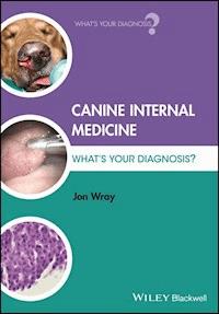
An 11 year-old male-neutered Border terrier (Figure 1.1) weighing 10.4 kg is presented with a history of lethargy, anorexia, vomiting and progressive dullness resulting in a reluctance to walk. His owners report that he has had previous episodes of self-limiting vomiting lasting 2–3 days of which he has had four episodes over the previous 6 months. They have noticed both his water consumption and urination increased dramatically over the previous 2 weeks. No haircoat or skin abnormalities have been noted but he has lost some weight over this time. His appetite up until 48 hours ago has been excellent. He has vomited, accompanied by abdominal effort, 8 times in the last 24 hours and the vomitus has been bile-stained fluid.
Figure 1.1 The patient at presentation.
Physical examination demonstrates a very depressed dog in overweight body condition (body condition score 4/5) who is reluctant to walk. The eyes are sunken and normal skin turgor is lost. Mucous membranes are very tacky to the touch but are of a normal colour with a capillary refill time of 2.5 s. Heart rate is 144/min, respiratory rate is 32/min and rectal temperature is 38.3 °C. Peripheral pulse quality is slightly poor. Auscultation of the heart and respiratory tract is normal and abdominal palpation generates reproducible cranial abdominal discomfort but no palpable structural abnormalities. The urinary bladder is moderately full and after palpation the dog spontaneously voids urine without apparent difficulty, and some is collected for analysis.
A blood sample is also taken and the following results were obtained:
Haematology
Parameter
Value
Units
Range
RBC
6.89
× 10
12
/l
(5.5–8.5)
Haemoglobin
14.4
g/dl
(12.0–18.0)
Haematocrit
0.49
l/l
(0.37–0.55)
Mean cell volume
71.3
fl
(60–77)
Mean cell haemoglobin cconcentration
30
g/dl
(30.0–38.0)
Mean cell haemoglobin
20.9
pg
(19.5–25.5)
Total white cell count
19.26
× 10
9
/l
(6.0–15.0)
Neutrophils
16.37
× 10
9
/l
(3.0–11.5)
Lymphocytes
0.96
× 10
9
/l
(1.0–4.8)
Monocytes
1.96
× 10
9
/l
(0.2–1.4)
Eosinophils
0
× 10
9
/l
(0.1–1.2)
Basophils
0
× 10
9
/l
(0.0–0.1)
Platelets
223
× 10
9
/l
(200–500)
Biochemistry
Parameter
Value
Units
Range
Total protein
78
g/l
(54–77)
Albumin
39
g/l
(25–40)
Globulin
39
g/l
(23–45)
Urea
13.5
mmol/l
(2.5–7.4)
Creatinine
132
μmol/l
(40–145)
Potassium
3.6
mmol/l
(3.4–5.6)
Sodium
143
mmol/l
(139–154)
Chloride
118
mmol/l
(105–122)
Calcium
2.2
mmol/l
(2.1–2.8)
Inorganic phosphate
0.9
mmol/l
(0.60–1.40)
Glucose
34
mmol/l
(3.3–5.8)
ALT
385
IU/l
(13–88)
AST
96
IU/l
(13–60)
ALKP
2130
IU/l
(14–105)
GGT
32
IU/l
(0–10)
Bilirubin
3
μmol/l
(0–16)
Cholesterol
10.2
mmol/l
(3.8–7.0)
Triglyceride
2.4
mmol/l
(0.56–1.14)
Creatine kinase
168
IU/l
(0–190)
Urinalysis
Parameter
Value
Units
Range
Appearance
Clear, straw
Chemistry
Specific gravity
1.032
pH
8.1
Protein
Trace
Nitrite
–
Blood / Hb
–
Glucose
++++
Ketones
++++
Bilirubin
–
Urobilinogen
–
Cytology
Red cells
2
/hpf
(0–2)
White cells
1
/hpf
(0–2)
Epithelial cells
0
/hpf
(0–5)
Casts
–
Crystals
–
Bacteria
–
Other
Questions
How would you interpret these laboratory results in the context of the history and clinical examination findings, and what is this dog's clinical diagnosis?
How might this have arisen and what further information might be gained to plan appropriate treatment?
What are this dog's immediate fluid and other medical therapy needs and what problems may be anticipated/pre-empted during the initial stages of treatment?
Answers
How would you interpret these laboratory results in the context of the history and clinical examination findings, and what is this dog's clinical diagnosis?
The haematology shows a mild neutrophilia, a monocytosis and concurrent lymphopenia and eosinopenia, all components of a stress leukogram. Inflammation may also account for neutrophilia and monocytosis but would not be expected to cause the concurrent lymphopenia and eosinopenia.
The biochemistry demonstrates:
A marked hyperglycaemia which, with concurrent glucosuria, is consistent with diabetes mellitus.
A rise in urea without a concomitant rise in creatinine, which suggests that a pre-renal azotaemia may be present and indeed the urine SG of 1.032 (i.e. > 1.030) is supportive of this.
Total protein is marginally elevated, though albumin and globulin are just within the upper end of their respective reference intervals. With the pre-renal azotaemia, haematocrit that is in the upper end of the reference interval and clinical examination findings, this is most likely due to dehydration.
A moderate rise in ALT and AST together with a more marked raise in ALKP and GGT is seen. This is likely to represent hepatopathy with a cholestatic component or response to endogenous or exogenous glucocorticoids.
The Similarity in Biochemistry between Diabetic and Cushingoid Patients
It is important to remember that such changes in hepatocellular and cholestatic markers are common in untreated diabetic patients due to the effects of hepatic lipidosis induced by insulin deficiency. Such biochemical findings are very similar to those seen in patients with hyperadrenocorticism (in which hepatic glycogen is stored in excess, not lipid) and it is common in the author's experience that veterinary surgeons commonly have suspicion aroused of concurrent hyperadrenocorticism by such findings, rather than considering them an expected effect of untreated diabetes mellitus. Improvement in these with serial biochemical monitoring after institution of insulin therapy is the key differentiator.
A hyperlipidaemia comprising rises in both cholesterol and triglyceride is present. This may be present in diabetes mellitus, pancreatitis, hypothyroidism, hyperadrenocorticism, protein-losing nephropathy and cholestasis (though more usually cholesterol only is elevated in these last two).
Urinalysis demonstrates glucosuria and ketonuria.
Remember that urine test strips detect acetoacetate and acetone but not β-hydroxybutyrate.
The patient has been demonstrating polyuria, polydipsia, weight loss and until very recently a healthy appetite. In conjunction with the hyperglycaemia and glucosuria a diagnosis of diabetes mellitus can be confidently made. It is unnecessary to perform a fructosamine level to confirm this diagnosis although many clinicians advocate assessment of a fructosamine level at diagnosis for future comparison with subsequent fructosamine levels.


