74,99 €
Mehr erfahren.
- Herausgeber: Thieme
- Kategorie: Fachliteratur
- Sprache: Englisch
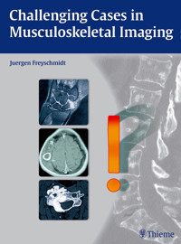
This book demonstrates how even difficult cases can be diagnosed by taking a systematic approach to image interpretation. Drawing upon decades of experience, Dr. Freyschmidt guides readers from case to case while solving the core problems that arise in making a diagnosis. He shows how initially challenging cases can be turned into cases that only seemed difficult at the outset. Step-by-step approach: Systematic case presentations: prior history and clinical questions, radiologic findings, pathoanatomic classification, synopsis and discussion, final diagnosis, and comments Arranged by anatomic region: skull, spine, pelvis, shoulder girdle and thoracic cage, upper and lower limbs More than 1,400 high-quality images from the author’s case files Over 150 difficult and challenging cases from skeletal radiology Answers questions such as: What are the requirements of a good diagnosis? How is a good diagnosis defined? Which imaging procedure will yield the desired information most quickly and accurately? Describes imaging modalities and gives recommendations on selecting a particular modality Your instructor in book form: a systematic, case-by-case approach to making a diagnosis!
Das E-Book können Sie in Legimi-Apps oder einer beliebigen App lesen, die das folgende Format unterstützen:
Seitenzahl: 719
Veröffentlichungsjahr: 2015
Ähnliche


Challenging Cases in Musculoskeletal Imaging
Juergen Freyschmidt, MDProfessorReference Center for OsteoradiologyZentralklinikum Bremen MitteUniversity Teaching Hospital of the University of GöttingenBremen, Germany
1438 illustrations
ThiemeStuttgart · New York · Delhi · Rio de Janeiro
Library of Congress Cataloging-in-Publication Data
Freyschmidt, J. (Jürgen), author. [Schwierige Diagnosen in der Skelettradiologie. English]Challenging cases in musculoskeletal imaging/Juergen Freyschmidt; translator, Terry C. Telger. p.; cm.This book is an authorized translation of the German edition published and copyrighted 2013 by Georg Thieme Verlag, Stuttgart. Title of the German edition: Schwierige Diagnosen in der Skelettradiologie.Includes bibliographical references and index.ISBN 978-3-13-176401-0 (hardback: alk. paper) – ISBN 978-3-13-176411-9 (eISBN) I. Title. [DNLM: 1. Bone Diseases–diagnosis–Case Reports. 2. Diagnostic Imaging– Case Reports. 3. Fractures, Bone–diagnosis–Case Reports. WE 225] RC925.7 616.7'07548–dc23
2014048126
This book is an authorized translation of the German edition published and copyrighted 2013 by Georg Thieme Verlag, Stuttgart. Title of the German edition: Schwierige Diagnosen in der Skelettradiologie
Translator: Terry C. Telger, Fort Worth, TX, USA
Illustrator: Roland Geyer Weilerswist, Germany
© 2015 Georg Thieme Verlag KG
Thieme Publishers StuttgartRüdigerstrasse 14, 70469 Stuttgart, Germany+49 [0]711 8931 421, [email protected]
Thieme Publishers New York333 Seventh Avenue, New York, NY 10001 USA+1 800 782 3488, [email protected]
Thieme Publishers DelhiA-12, Second Floor, Sector-2, Noida-201301Uttar Pradesh, India+91 120 45 566 00, [email protected]
Thieme Publishers Rio, Thieme Publicações Ltda.Argentina Building 16th floor, Ala A, 228 Praia do BotafogoRio de Janeiro 22250-040 Brazil+55 21 3736-3631
Cover design: Thieme Publishing GroupTypesetting by DiTech Process Solutions Pvt. Ltd., India
Printed in China by Everbest Printing Ltd
ISBN 9783131764010
Also available as an e-book:eISBN 9783131764119
Important note: Medicine is an ever-changing science undergoing continual development. Research and clinical experience are continually expanding our knowledge, in particular our knowledge of proper treatment and drug therapy. Insofar as this book mentions any dosage or application, readers may rest assured that the authors, editors, and publishers have made every effort to ensure that such references are in accordance with the state of knowledge at the time of production of the book.
Nevertheless, this does not involve, imply, or express any guarantee or responsibility on the part of the publishers in respect to any dosage instructions and forms of applications stated in the book. Every user is requested to examine carefully the manufacturers’ leaflets accompanying each drug and to check, if necessary in consultation with a physician or specialist, whether the dosage schedules mentioned therein or the contra-indications stated by the manufacturers differ from the statements made in the present book. Such examination is particularly important with drugs that are either rarely used or have been newly released on the market. Every dosage schedule or every form of application used is entirely at the user’s own risk and responsibility. The authors and publishers request every user to report to the publishers any discrepancies or inaccuracies noticed. If errors in this work are found after publication, errata will be posted at www.thieme.com on the product description page.
Some of the product names, patents, and registered designs referred to in this book are in fact registered trademarks or proprietary names even though specific reference to this fact is not always made in the text. Therefore, the appearance of a name without designation as proprietary is not to be construed as a representation by the publisher that it is in the public domain.
This book, including all parts thereof, is legally protected by copyright. Any use, exploitation, or commercialization outside the narrow limits set by copyright legislation without the publisher’s consent is illegal and liable to prosecution. This applies in particular to photostat reproduction, copying, mimeographing or duplication of any kind, translating, preparation of microfilms, and electronic data processing and storage.
Contents
Preface
Acknowledgment
Abbreviations
1 From Symptom to Diagnosis
1.1 Selecting the Correct Imaging Modality
1.2 Image Interpretation
2 Skull
2.1 Sclerotic Changes
2.2 Osteolytic Lesions and Lesions with Mixed Features
3 Spine
3.1 Mono- and Bisegmental Changes
3.2 Oligo- and Multisegmental Changes
3.3 Diseases of Spinal Entheses and Joints
3.4 Sacrum
4 Pelvis
4.1 Sclerotic Changes
4.2 Osteolytic Changes and Changes Associated with Decreased Bone Density
4.3 Unusual Fractures
4.4 Bone Lesions with a Predominantly Extraosseous Component
4.5 Soft-Tissue Mineralization
4.6 Hip Region
5 Shoulder Girdle and Thoracic Cage
5.1 Clavicle
5.2 Scapula
5.3 Ribs
5.4 Sternum
5.5 Anterior Chest Wall as a Whole
6 Upper Limb
6.1 Upper Arm
6.2 Forearm
6.3 Hands (Bone and Joint Diseases)
7 Lower Limb
7.1 Equivocal MRI Findings
7.2 Predominantly Osteolytic Changes
7.3 Predominantly Osteosclerotic Changes
7.4 Changes with Mixed Features
7.5 Extraosseous Lesions
Bibliography
Index
Preface
Long years of experience in the daily practice of radiology and observations in the training of radiologists have shown that factual knowledge alone is not always enough to make a clinically useful diagnosis. Often more can be gained by taking a systematic approach to image analysis and piecing together available facts. Based on my observations in daily conferences and refresher courses, I have repeatedly found that even less experienced readers with relatively little factual knowledge could solve difficult or challenging cases simply by taking a systematic approach to image analysis and then interpreting their findings in the light of clinical data. By contrast, “old hands” at radiology can diagnose most cases at a glance owing to their extensive experience, but this “shooting from the hip” will often miss the mark when it comes to more challenging diagnoses. Thus, having made several blunders in the past, I have made it a practice to recheck a seemingly obvious diagnosis for its logical plausibility and correctness.
In writing this book, I hope to show how even challenging cases can be diagnosed by following a systematic approach to image interpretation, and how many cases that initially presented as difficult can be turned into ones that only “seemed” difficult.
Acknowledgment to Prof. Helmut Ostertag, MD
Every abnormal finding in diagnostic imaging is based on an underlying structural pathoanatomic change that must be identi-fied before an accurate diagnosis can be made.
For many years I have collaborated with the Hannover pathologist Prof. Helmut Ostertag, MD, whose comprehensive general and specialized knowledge in osteopathology has unveiled many of the secrets of musculoskeletal diseases, helping me to understand and interpret even complex diseases and disorders.
I thank him sincerely for all the time that he sacrificed in reviewing the manuscript for this book. As a dilettante in macro-scopy and histology, I needed his help to make sure that the pathoanatomic details of the cases in this book conformed to reality.
Abbreviations List
ADC
apparent diffusion coefficient
ARA
American Rheumatism Association
ARCO
Association for Research of Circulation Osseus
ASAS
Ankolysing Spondylitis Assessment Study
BPOP
bizarre parosteal osteochondromatous proliferation
CNO
chronic nonbacterial osteitis or osteomyelitis
CPPD
calcium pyrophosphate dehydrate deposition disease
CRMO
chronic recurrent multifocal osteomyelitis
CRP
C-reactive protein
CSF
cerebrospinal fluid
CT
computed tomography
CUP
cancer of unknown primary
DISH
diffuse idiopathic skeletal hyperostosis
DSA
digital subtraction angiography
EPI
echo planar imaging
FDG
fluorodeoxyglucose
FLAIR
fluid attenuated inversion recovery
FS
fat suppression
HRCT
high-resolution computed tomography
MPR
multiplanar reconstruction
MRA
magnetic resonance angiography
MRI
magnetic resonance imaging
NSAID
nonsteroidal anti-inflammatory drug
PAO
pustular arthro-osteitis
PDW
proton density weighted
PEO
pustular enthesio-osteitis
PET
positron emission tomography
PPP
pustulosis palmoplantaris
PVNS
pigmented villonodular synovitis
RAP
regional acceleratory phenomenon
SAPHO
synovitis, acne, pustulosis, hyperostosis, osteitis (syndrome)
SCCH
sternocostoclavicular hyperostosis
SPAIR
spectral presatuation inversion recovery
SPECT
single-photon emission computed tomography
SPIR
spectral attenuated inversion recovery
STIR
short tau inversion recovery
STT
scapho-trapezio-trapezoid
SUV
standard uptake value
TIRM
turbo-inversion recovery magnitude
TSE
turbo spin echo
WHO
World Health Organization
1From Symptom to Diagnosis
Orthopedics and trauma surgery, rheumatology, the vast field of oncology and, to a degree, even biopsy-based pathology must rely heavily on skeletal imaging. Radiologists must be well versed in these areas to ensure that their services will continue to be relevant. What, then, do these specialties require in terms of a good diagnosis? And how should a “good diagnosis” be defined?
First and foremost, a radiologic diagnosis must be useful. It must alter the treatment strategy for a patient. On the other hand, it does not necessarily have to be definitive; it does not have to be the final diagnosis like that supplied by histology, clinical chemistry, molecular biology, or the clinical course. Above all it must be logical and well founded and it must reflect due diligence. That is the only justifiable aspect of a radiologic diagnosis—not its absolute correctness, which may draw even highly specialized experts into a dispute that no judge could resolve.
Every radiologist must provide two essential services:
Selecting the correct imaging modality
Image interpretation
1.1Selecting the Correct Imaging Modality
The following imaging modalities should be available and selectively utilized at centers where skeletal examinations are performed:
Plain radiography
CT (computed tomography)
Bone scintigraphy including SPECT (single-photon emission computed tomography)
MRI (magnetic resonance imaging)
PET (positron emission tomography) and PET-CT fusion
Ultrasound
The patients who are seen in the everyday practice of skeletal radiology fall into one of three scenarios:
A patient undergoes initial imaging for the investigation of a clinical problem. The patient has had no previous imaging studies, and ideally the radiologist is challenged to tailor the imaging study to the specific problem, using his or her skills and experience.
A patient is referred for the further investigation of findings noted in one or more previous imaging studies. In this situation the radiologist must select the study that can supply the fastest and most accurate, clinically useful diagnosis.
A patient with an established diagnosis presents for follow-up imaging.
1.1.1Available Imaging Studies
What, then, are the most suitable and rewarding imaging studies for a particular problem? Which of the procedures listed above will yield the desired information most rapidly and accurately while causing the least amount of patient compromise?
To answer these questions, it is helpful to review briefly the capabilities and limitations of the imaging modalities listed above.
Plain Radiography
Plain radiography creates an image based on differences in the X-ray absorption characteristics of different tissues. Thus, bone tissue or any bony or calcified structures are portrayed in high contrast while most soft tissues are imaged in low contrast, unless contrast medium is used. The main disadvantage of plain radiography in skeletal studies is the presence of summation effects, since radiographs give only a superimposed, two-dimensional view of three-dimensional body structures. Thus, from a modern-day perspective (and based on current requirements), plain radiography is unsuitable for detecting focal lesions (1.5–2 cm in diameter) in body regions that are sensitive to projection effects such as the thoracic and lumbar spine, sacrum, and skull base. On the other hand, there are systemic bone diseases with more or less fine structural changes (e.g., osteopathies such as osteoporosis, hyperparathyroidism, osteomalacia, Paget disease) in which summation effects are diagnostically useful and may detect structural changes that would often be missed in sectional imaging procedures.
Plain radiography is inherently unsuitable for the evaluation of soft-tissue structures such as bone marrow, muscle, tendons, ligaments, cartilage, etc. A major advantage of skeletal plain radiography, which has been in use for over a century, is that valid pattern-recognition algorithms and engrams (cognitive links) have been established that permit the fast and accurate detection of abnormalities. This capability is useful, for example, in the analysis of osteolytic areas by Lodwick grading and for recognizing ossification patterns in a tumor matrix, such as the “ground-glass” pattern for woven bone (see ▶ Case 139) and the “popcorn” pattern for cartilaginous matrix (see ▶ Case 108).
Computed Tomography
CT basically works like plain radiography by converting tissue X-ray absorption values into an image. The advantage of CT is that it can generate sectional, nonsuperimposed images of body structures with a contrast resolution many times higher than that of conventional radiographs. As a sectional modality, it is particularly useful for providing nonsuperimposed views of the axial skeleton and of complex processes in the appendicular skeleton. It can also provide limited views of soft-tissue structures like muscle, fat, and hematomas. Scanning after the administration of intravenous contrast medium can significantly expand the ability of CT to define soft-tissue structures, especially when the proper window setting is used. CT densitometry permits the accurate identification of fat, fluid, and other media based on their attenuation values. Modern multidetector-row scanners can produce three-dimensional images that are essential for the accurate localization of structures and lesions. From a physical standpoint, the indications for CT are the same as those for plain radiographs, aside from its sectional imaging capabilities. Also, owing to the establishment of pattern-recognition algorithms and engrams with high diagnostic specificity, CT, which is slightly less than 40 years old, has features in common with plain radiography in skeletal examinations and offers numerous advantages over the “younger” modalities of MRI and PET-CT.
Bone Scintigraphy
Bone scintigraphy with Tc-99m (technetium-99m methylene diphosphonate) is an imaging technique based on radioactive tracer uptake in bones and adjacent soft tissues. The amount of tracer uptake is dependent on local bone metabolism, local perfusion, and regional tracer affinity, so that planar “bone scans” with a gamma camera supply information on global, regional, and focal bone metabolism. This is helpful in determining, for example, whether a systemic process with increased bone turnover, such as hyperparathyroidism (see ▶ Case 15 and ▶ Fig. 6.30g in ▶ Case 111), is present as opposed to a focal or a multifocal process representing a bone metastasis. Bone scans can reliably determine whether a sclerotic lesion is active or not, which may be particularly helpful in oncologic evaluations (see ▶ Case 145 and ▶ Case 149). Bone scintigraphy, then, is a functional imaging study. Today there is no radiologic technique that can supply such precise information on bone metabolism with “one look” and with a relatively simple protocol. In patients thought to have smaller and less active lesions, another sectional imaging technique, SPECT, is of great value in many cases owing to its ability to provide nonsuperimposed three-dimensional views.
Unfortunately, bone scintigraphy is not widely practiced today despite the fact that it is widely available and supplies functional information not obtainable with CT or MRI. In recent years we have been able to define more than 20 tracer distribution patterns which are highly specific for certain skeletal diseases and refute the common argument against bone scans, namely that “they are not specific enough.1 The radiation safety aspects of bone scintigraphy will not be explored here, but suffice it to say that radiation exposure is relatively low and such risk is greatly outweighed by the benefits of the study for sick patients.
Magnetic Resonance Imaging
MRI, in which the relaxation times and proton content of body tissues are measured, is a sectional imaging modality that has fundamental physical differences from the X-ray absorption modalities. It is the method of first choice for imaging soft-tissue structures (bone marrow, muscle, tendons, fascia, neurovascular bundles, cartilage, synovial membrane, intra-articular fluid) and currently has a relatively broad role in skeletal investigations. Yet the initial euphoria that this modality could handle all skeletal imaging tasks without X-rays has been tempered by the results of clinical imaging studies. Meanwhile, sound and appropriate indications for MRI have been crystalizing, especially in the diagnosis of articular, bone-marrow and muscular diseases and in the staging of skeletal tumors. It should be noted that bony structures (especially healthy cortical bone) appear as signal voids on MR images and thus can be identified only indirectly; this may lead to problems of differential diagnosis. The same applies to processes that are associated with more or less subtle ossifications or calcifications, which are clearly demonstrable by CT.
The information content of MRI can be increased by dynamic contrast-enhanced imaging (e.g., to assess the perfusion of a lesion) and by diffusion-weighted imaging. But these techniques cannot in themselves provide reliable information on general and local bone metabolism in the strict sense, and unlike bone scintigraphy they cannot answer the question of whether a sclerotic lesion, for example, is active. Unlike plain radiographs, CT scans, and bone scans, MRI still does not have well-established pattern recognition algorithms and engrams (e.g., Lodwick grades, ground-glass appearance, systemic sclerosing diseases), apart from joint trauma, which could guide the radiologist quickly and confidently to a correct diagnosis. Moreover, there is still no good, comprehensive catalog of normal variants like that available for the more conventional modalities.
Note
A very important rule for MRI protocols: If scanning is performed with contrast medium, the sequences before and after contrast administration should be identical (e.g., T1 weighting before and after contrast injection). Otherwise, possible enhancement cannot be evaluated. Unfortunately this technical principle is often violated in clinical practice. For example, a T1-weighted (T1w) sequence is performed before contrast administration but a fat-suppressed T1w sequence or water-sensitive sequence is performed afterward.
Positron Emission Tomography (and PET-CT)
PET, like bone scintigraphy, is a functional imaging modality. Today, over 90% of all PET scans employ the glucose analog 18F-FDG (18F-2-fluoro-2-deoxyglucose), tagged with the fluorine radioisotope 18F. The method is based on the imaging of a circumscribed change in specific cellular functions, such as the intensity of glucose metabolism as determined by 18F-FDG PET. Increased glucose metabolism is a relatively nonspecific indicator, however, which is seen in inflammatory processes as well as neoplasms and tumorlike lesions. The standard uptake value (SUV) provides a semiquantitative indicator of tumor metabolism (see below).
PET can be combined with simultaneous CT imaging in two interconnected scanners to produce a system known as PET-CT. This integrated system can generate a PET-CT fusion image without having to change the patient position, respiratory position, slice thickness, etc. Increasingly, PET-CT is becoming an established tool in oncologic musculoskeletal studies, as in the diagnosis of lymphoma and plasmacytoma2, 3 and skeletal metastases. In a recent meta-analysis of 23 studies, however, Liu et al4 found that MRI was superior to 18F-FDG PET and bone scintigraphy for detecting skeletal metastases in breast cancer patients (pooled sensitivity on a per-patient basis: 97.1% for MRI, 83.3% for PET, and 87.0% for bone scintigraphy; pooled sensitivity on a per-lesion basis: 97% for MRI, 94.5% for PET, and 88.1% for bone scintigraphy).
As for the diagnosis of primary bone tumors,5, 6 to date there have been only scattered publications, based largely on case reports, which do not provide conclusive information (see below). But given the generally low incidence of bone tumors, it is still too early to expect definitive answers.
FDG-PET can be very useful for monitoring bone tumors during treatment, since by its very nature it is accurate in assessing tumor viability. The SUV for FDG provides a semiquantitative measure of metabolic tumor activity. Because tumor metabolism is usually heterogeneous, the SUV in a region of interest (ROI) within the tumor provides an excellent index for evaluating tumor metabolism. Hawkins et al7 investigated the maximum SUV before and after neoadjuvant chemotherapy for the Ewing sarcoma family of tumors. Patients with an SUV of less than 2 after therapy tended to have an excellent response (10% or less viable tumor) with a 4-year disease-free survival. Hawkins et al8 found similar results for osteosarcoma.
Gaston et al9 obtained somewhat different results in their recent study: the change in maximum SUV between baseline examination and post-treatment scanning was not significantly associated with histologic response for either osteosarcoma or Ewing sarcoma. The metabolic tumor volume and the percentage of injected 18F-FDG dose were different in the osteosarcoma and Ewing sarcoma response subgroups. A 50% reduction in metabolic tumor volume was found to be significantly associated with a good histologic response in osteosarcoma but not in Ewing sarcoma. When the cut-off value for Ewing sarcoma was increased to a 90% reduction in metabolic tumor volume, a good correlation was found with histologic response.
These essentially positive results of FDG-PET scans cannot be applied directly to the problem of benign/malignant differentiation (see above). Aoki et al5 found no significant difference between the SUVs of osteosarcoma and giant cell tumor or between fibrous dysplasia and chondrosarcoma; this is not surprising (see above). Feldman et al,6 on the other hand, studied 29 cartilage tumors (11 enchondromas, 6 osteochondromas, 11 chondrosarcomas) and found that 18FDG-PET, using a maximum SUV cut-off of 2.0, could discriminate between malignant and benign lesions with a sensitivity of 90.9%, a specificity of 100%, and an accuracy of 96.6%. On closer analysis, the patients in their study comprised a very heterogeneous group. Bredella et al10 evaluated FDG-PET in differentiating benign from malignant compression fractures (33 patients with 43 fractures). In a total of 14 malignant and 29 benign compression fractures, five cases with a benign fracture were falsely classified as malignant (false-positive). Three of those patients had undergone prior treatment with bone-marrow stimulating agents. There were two false-negative results. Sensitivity, specificity, and accuracy were in the range of approximately 70 to 90%. In our view these results are not too significant when we consider that the patients participating in the study had multiple fractures that most likely had the same cause. Moreover, only nine of the cases were histologically confirmed, and the tumor entities underlying the malignant fractures were extremely heterogeneous. The authors state that the difference between the SUV values for benign and malignant fractures was statistically significant (1.9 ± 0.97 and 3.9 ± 1.52, respectively). Shin et al11 found basically the same results in a similar population, although they performed all examinations with PET-CT and obtained measurements separately for the cortex and bone marrow.
Thus, we find no real consensus in the current literature on the efficacy of FDG-PET in diagnosing bone tumors, aside from initially positive results in the follow-up of Ewing sarcoma and osteosarcoma during treatment. This is to be expected, since this modality is relatively recent and the spectrum of primary and secondary bone tumors is very broad with respect to tumor dynamics and tumor biology. One critical note on the above follow-up studies is that none has yet compared PET with traditional modalities such as MRI, CT, scintigraphy, and plain radiography.
1.1.2Imaging Strategies
Initial Imaging Study
Given the specific characteristics of the imaging modalities described above, it is important to ask which one should be used for the initial investigation of a given clinical problem in a patient not previously imaged. See ▶ scenario 1. In ▶ Table 1.1 we have attempted to match various disease groups and entities with the imaging modality that years of personal experience has shown to provide the fastest and surest route to a correct diagnosis—one that will direct the referring colleague in selecting further clinical and laboratory tests and then planning appropriate treatment. That is the only diagnosis of any real value. A simple description of findings that does not supply a working diagnosis is of no use to the referring clinician. Nor is it enough to report “no evidence of malignancy,” especially if the clinical presentation does not raise the problem of benign/malignant differentiation, as in patients with stress-related disorders.
Fig. 1.1Diagnostic algorithm for a suspected bone tumor.15
Fig. 1.2Diagnostic algorithm for a presumed stress fracture.
Fig. 1.3Diagnostic algorithm for suspected osteomyelitis.
Note
Basic guidelines for selecting an imaging modality:
Imaging bony structures: plain radiography, CT
Imaging soft-tissue structures (muscle, tendons, cartilage, synovial membrane, “foreign tissue” in bone, etc.): ultrasound, otherwise MRI
Evaluating the activity of a lesion: whole-body bone scintigraphy
Supplemental Imaging Study
Recommendations on selecting a suitable imaging modality to supplement a previous imaging study (▶ scenario 2) are derived from the specificity of the modality in question. With some restrictions, the following rule applies:
Note
When selecting an imaging modality for the further investigation of a lesion detected by a different modality, never select a modality that is less sensitive or less specific than the previous study.
For example, if bone scintigraphy has detected small focal lesions in the spine, it would be wrong to order radiographs in two planes for further investigation of the lesions because plain radiographs usually cannot detect osteolytic or mixed osteolytic-osteoblastic lesions (e.g., metastases from breast cancer) smaller than 1.5–2 cm due to their low sensitivity. Since it is reasonable to expect that scintigraphic abnormalities are true bone metastases rather than metastases confined to bone marrow—that is, metastases that have already interacted with the bone—further investigation by CT (not MRI) would be indicated owing to the higher specificity of CT for an osseous process.
A more detailed look at ▶ scenario 2:
If plain radiographs show an indeterminate lesion that appears to involve bony structures, CT should be ordered next to provide a nonsuperimposed view in the “bone mode.” We do not recommend changing the physical mode in this situation (e.g., from measuring absorption differences to measuring proton content and relaxation times) because this would address entirely different questions (see above). But if radiographs suggest that the lesion originates from soft tissues (e.g., tumor involvement of the medullary cavity), MRI is recommended (see ▶ Case 8, ▶ Case 9, ▶ Case 10, ▶ Case 12, ▶ Case 14, ▶ Case 16, ▶ Case 30, ▶ Case 34, ▶ Case 45, ▶ Case 48, ▶ Case 49, ▶ Case 58, ▶ Case 94, ▶ Fig. 6.21 in ▶ Case 107, ▶ Case 118, ▶ Case 119, ▶ Fig. 7.12 in ▶ Case 135, ▶ Case 139, ▶ Case 142, and ▶ Case 145).
If MRI was performed first and could not answer questions relating specifically to bone structure or ossifications, it should be supplemented with a modality that can directly image the bone: plain radiography or CT. CT should be chosen whenever nonsuperimposed views are required. As a sectional modality, it provides image slices that can “unmask” a detail of interest (see ▶ Case 7, ▶ Case 17, ▶ Case 19, ▶ Case 20, ▶ Case 22, ▶ Case 29, ▶ Case 30, ▶ Case 36, ▶ Case 39, ▶ Case 60, ▶ Case 63, ▶ Case 64, ▶ Case 74, ▶ Case 101, ▶ Case 104, ▶ Case 107, ▶ Case 113, ▶ Case 127, ▶ Case 129, ▶ Case 131, ▶ Case 136, ▶ Case 145, ▶ Case 146, and ▶ Case 149).
If the initial study was PET or PET-CT (the latter may be nondiagnostic due to less-than-optimal image quality of the CT component), dedicated spiral CT is recommended for the further investigation of a bony process (see ▶ Fig. 4.50 in ▶ Case 77). If the goal is to investigate a soft-tissue lesion revealed by PET-CT, MRI will provide an effective adjunct. With a negative PET scan (screening for metastases, suspected plasmacytoma), the recommended course of action is clinical follow-up, depending on the patient’s symptoms.
If the goal is to determine the activity of a lesion detected by other methods (especially important in oncologic radiology), the next recommended study is bone scintigraphy. For example, a sclerotic lesion that is negative on bone scans (osteoma, old fibrous dysplasia, old lipoma, old infarction, healed reactive inflammatory process, etc.) is more likely to be old and harmless than a lesion that shows uptake (e.g., sclerosing metastasis, bone-forming tumor, etc.). When uptake is found, bone scintigraphy can advance the differential diagnosis by determining whether the lesion is solitary (e.g., fibrous dysplasia, which is often multifocal, versus lipoma, which is usually solitary; metastasis, which is usually multiple, versus a bone-forming tumor like osteoid osteoma, which is almost always solitary; see also ▶ Case 12, ▶ Case 31, ▶ Case 32, ▶ Case 47, ▶ Case 50, ▶ Case 73, ▶ Case 74, ▶ Case 78, ▶ Case 102, ▶ Case 107, and ▶ Case 133).
If the initial study was bone scintigraphy showing one or more sites of focal uptake, the next study should be one that directly images the bone, depending on the site(s) of involvement. The options, are plain radiography and CT (see ▶ Case 20), since scintigraphy demonstrates the activity of a bony process. In many cases, however, the distribution pattern on scintigraphy will in itself suggest the correct diagnosis (see ▶ Fig. 4.22 in ▶ Case 62, ▶ Fig. 5.8h, i in ▶ Case 82, ▶ Case 83, ▶ Fig. 5.13 in ▶ Case 84, and ▶ Case 103).
If initial CT scans showed one or more lesions originating from soft tissue, the recommended supplemental study is MRI. Of course, this modality is also used to investigate a bone tumor detected by CT or by supplemental CT after plain radiography, as it can detect tumor spread into adjacent soft tissues and supply valuable additional information (see ▶ Case 45, ▶ Case 54, ▶ Case 57, ▶ Case 70, ▶ Case 86, ▶ Case 106, ▶ Case 111, and ▶ Fig. 7.12 in ▶ Case 135).
Finally a word about costs: the use of one or more necessary imaging modalities can be very expensive. But such costs must be weighed against the benefits for properly selected patients. A frequent benefit from a financial standpoint is that a proper imaging work-up can eliminate many unnecessary invasive procedures (e.g., open biopsy) and prevent diagnostic errors. Moreover, when we consider the relatively low incidence of nontraumatic skeletal lesions, which form the major portion of our topic, the costs are almost negligible when compared with other costs incurred in the health care industry (approximately 0.1% or less).
Follow-up Imaging
Whenever possible, the follow-up imaging of patients with an established diagnosis (▶ scenario 3) should employ the same modality and should follow a consistent protocol. This is essential to allow a meaningful comparison of images.
1.2Image Interpretation
Before any remarkable image details are interpreted, they must be described. This does not require a lengthy narrative but may be done concisely in a “key-word” format. The description of findings should nevertheless be exacting and, above all, should avoid potentially misleading language. For example, a lucent area on a radiograph or CT scan should be described as such and should not be called a “cystic” or “cystlike” osteolysis, as this could bias further conclusions that may be drawn in the light of other findings. Another example is “bone marrow edema,” a term that is frequently overused in MRI reporting (see below).
Note
Image analysis requires a detailed knowledge of the normal radiologic anatomy of the musculoskeletal system and its principal variants.
1.2.1Pathoanatomic Background
When a finding is classified as definitely abnormal, the first essential question should address its possible pathoanatomic background, because each of the known basic entities (see below) is associated with a more or less specific pathoanatomic change. For example, a tumor or inflammatory process is associated with destruction, necrosis with fragmentation, and a reparative process with new bone formation. An effort should also be made to determine whether a finding may be only an epiphenomenon (e.g., hemorrhage, callus) that accompanies an underlying process. In assigning findings to a pathoanatomic substrate, it is always important to consider the capabilities for tissue discrimination (fat, fluid, etc.) that are available with the modality being used. Unfortunately, this capability is often forgotten in the case of CT (see ▶ Case 58).
In the process of image interpretation, it is dangerous to use terms that describe either a disease entity in the strict sense or a sign with a variegated background. This principle will be explained more fully based on the two examples mentioned above:
Example: Rounded Lucency
In many cases a rounded lucency seen on a radiograph or CT scan is described as a “cyst” or a “cystic or cystlike lucency,” even though it is more often caused by a solid destructive lesion. This wording may steer case deliberations still further in the wrong direction, implying that the finding may be innocent. The term “cyst” is reserved exclusively for a fluid-filled cavity such as a juvenile bone cyst, a subchondral synovial cyst, or an aneurysmal bone cyst. The fluid content should first be established by CT or MRI, except with a classic juvenile bone cyst, which is identified by its typical location, “fallen fragment sign,” and ultrashort history.
Example: Ill-defined Hyperintensity
An area of increased signal intensity with ill-defined margins seen in water-sensitive MRI sequences is often described simply as “edema.” This is frequently offered as a diagnosis when no other suggestive findings are seen. Edema in bone marrow or in structurally altered cortical bone is an extravascular interstitial fluid collection that may accompany, for instance, a traumatic, inflammatory, degenerative or even a neoplastic process. “Edema” is a term that runs the gamut from benign to malignant disorders. Simply put, the resulting MRI signal is based partly on an increased proton density in the imaged region, regardless of the cause, which is usually but not always edema (see below).
Based on the MR image alone, it cannot be stated with certainty whether “bone marrow edema” actually represents pure edema, meaning an extravascular interstitial fluid collection. Taking degenerative changes in the knee joint as an example, “bone marrow edema” may result from fat necrosis, fibrosis, or trabecular bone changes such as a bone bruise, as Zanetti et al12 have shown. A pathologic process need only have enough protons to cause increased signal intensity in water-sensitive sequences. This applies to traumatic, inflammatory, degenerative, and neoplastic processes, the latter also showing an increased intracellular water content (see ▶ Case 145, ▶ Case 146, and ▶ Case 149). Even contrast administration cannot positively distinguish, for example, between tumor tissue and edema. It is clearer and more accurate, therefore, to report an edema equivalent13, 14 or an “edema-like hyperintensity,” or simply to report the presence of a proton-rich area.
1.2.2Lesion Localization
The second question raised by an abnormal finding is its location, which is considered in terms of functional anatomy. Is the finding located in a stress-exposed area (e.g., entheses), a critical area for perfusion deficits (e.g., epiphyses, toes or fingers), or a region where hematopoiesis occurs (e.g., a vertebral body)? These are key questions that aid in the classification of findings.
1.2.3Epicenter of a Lesion
The third question relates to the epicenter of a lesion or other finding. Most neoplastic processes exhibit a concentric pattern of tumor growth. Thus, for example, if the center of a lesion is located in the cortex, it is reasonable to assume that the process originated in the cortex or periosteum (cortical osteoid osteoma, periosteal osteosarcoma, stress fracture, etc.). If there is an enthesis in the affected area, it should also be considered that stress at the insertion site may have led to cortical destruction or new bone formation (see ▶ Case 103 and ▶ Case 104).
1.2.4Solitary, Bilateral, Multiple or Disseminated Lesions
The fourth question is whether a lesion is solitary, bilateral, multiple, or disseminated. It should also be asked whether the finding initially imaged and detected is only the “tip of an iceberg.” Whole-body imaging techniques are available for answering this question. If it is believed that focal lesions (e.g., metastases) have already interacted with bone, the screening test of first choice is bone scintigraphy. Other options are whole-body MRI or, if a plasmacytoma is suspected, whole-body CT. A solitary osteolytic lesion in younger patients is more likely to be a primary bone tumor or tumorlike lesion. Multiple foci would be more consistent with fibrous dysplasia, Langerhans cell histiocytosis, sarcoidosis, angiomatosis, or a rheumatic enthesis (e.g., in a patient with psoriasis or ankylosing spondylitis). Disseminated lesions would be more consistent with, say, a generalized Langerhans cell histiocytosis, whether as single-organ or multiorgan disease. A generalized systemic process usually suggests a congenital disorder. In older patients, a solid osteolytic focus is most likely a solitary plasmacytoma or metastasis, while multiple or disseminated lesions are suspicious for a CUP syndrome (cancer from unknown primary) or systemic plasmacytoma.
These are just a few examples; the great variety of other potential causes of osteolytic, osteosclerotic, and mixed changes cannot be explored within the limited scope of this book. Imaging of the contralateral side is a tactic that may be rewarding in both children and adults who present with unusual, difficult-to-classify findings that are not located on the midline. If the same or similar findings are noted on the opposite side, this would always suggest a syndromic or dysplastic cause.
Note
When considering etiologic possibilities, always consider the prevalence of a given disease in addition to biological data and location. For practical reasons, always consider the more common entities first and the rarer ones last.
1.2.5Assigning a Lesion to a Basic Entity
The fifth question seeks to determine to which of the seven basic entities, or major nosologic groups a finding should be assigned. The seven basic entities listed below provide the basis for lesion classification:
Normal variant (possibly symptomatic) or malformation
Trauma (acute or chronic)
Inflammation
Tumor and tumorlike lesions
Perfusion disorder with hyper- or hypoperfusion and necrosis
Regression (degeneration)
Systemic disease (metabolic, disease of the reticulohistiocytic system, storage disease, neoplasia)
The answer to the fifth and final question is obtained by systematically answering the first four questions and logically tying them together in a synoptic review.
In closing, we offer this useful rule to follow in making a diagnosis: be wary of Ockham’s razor. This is a principle of economy in logic and problem-solving that is still employed today in scientific theory and methodology. Ockham’s razor states that when faced with a choice among several possible explanations for the same phenomenon, one should always favor the explanation that requires the fewest hypotheses and thus represents the “simplest” theory. This principle encourages the scientist to recognize just one adequate explanation for any given phenomenon.
If we were to apply this principle consistently, it would mean the end of modern, rational differential diagnosis in medicine. For example, if the radiologic sign of osteosclerosis were simplified to a single cause (the “simplest” theory) such as an osteosclerotic metastasis, then the many other possible causes (e.g., a reactive process due to nonspecific osteitis, a reparative process due to bacterial osteomyelitis or trauma, a neoplastic process with an ossifying tumor matrix in osteosarcoma, or a dysplastic process in melorheostosis or osteopoikilosis) would have to be discarded. This approach would, in the author’s view, yield a wrong diagnosis in at least one-third of all nontraumatic cases in skeletal radiology. It is true that if two or three rounded osteosclerotic foci of the same density were seen on one skull radiograph, it would be helpful to start by assuming one cause (e.g., metastatic lesions in a breast cancer patient). At the same time, however, one should never simplify matters by trying to place two nonidentical imaging findings in the same box (a “reverse Ockham’s razor” approach; see ▶ Case 2).
Another misleading simplification is to make a diagnosis based on an uninformed image comparison between a current case and a case pictured in a textbook or other publication—that is, applying the diagnosis from a textbook image to a specific case without further deliberations. While it is true that predominantly visual disciplines in medicine (radiology, dermatology, histology) work by the principles of empirical pattern recognition, this does not mean that two cases with similar imaging features have the same etiology and classification. To stay with the example of osteosclerosis: both inflammation and a tumor can cause osteosclerosis, which may have the same imaging appearance in both entities.
It is only by linking the finding to other available imaging findings, to the history, and to clinical and laboratory data that we can arrive at a diagnosis that is correct and useful for the patient.
1.2.6 Summary
Note
The five key questions for image interpretation:
Pathoanatomic background (destruction, necrosis, reactive, etc.)?
Lesion localization from a functional anatomic standpoint (stress-exposed area, critical zone for perfusion, etc.)?
Epicenter of the lesion?
Solitary, bilateral, multiple, disseminated?
Assignment to one of seven basic entities:
Normal variant or malformation
Trauma (acute, chronic)
Inflammation
Tumor (including tumorlike lesions)
Perfusion disorder
Regression
Systemic disease (metabolic, disease of the reticulohistiocytic system, storage disease, neoplasia)
Image interpretation concludes with a clinical evaluation. At this time it is important to consider whether a finding that was assigned to one of the basic entities is symptomatic or whether it was detected incidentally. This applies particularly to imaging findings which ultimately could not be classified with complete certainty.
Note
The principle is this: an incidental finding is more likely to be innocent, and it may be safe to take a wait-and-see approach. But if an imaging finding is symptomatic, it is more likely to require histologic examination.
It should also be determined, in consultation with the referring physician, whether the clinical and laboratory data for symptomatic lesions fit with the radiologic findings. In the course of this discussion, the radiologist should communicate the data that formed the basis for his or her impression, and which the referring clinician does not yet know. The radiologist, being aware of the imaging findings, can ask the patient much more targeted questions than the clinician, who may know only the basic clinical presentation before requesting images. Typical examples of this are presented in ▶ Case 104, ▶ Case 152, ▶ Case 156, and ▶ Case 158. The prevalence of a disease for a given patient age and gender should always be considered in the synopsis, as mentioned earlier in connection with question 4.
The patient should not depart until the radiologist (not the technician) has critically reviewed the case from an imaging standpoint and discussed it with the patient. Patients expect a conversation in which they receive answers to their questions, without taking anything away from the office consultation with the treating physician. This is the only way that radiology can continue to assert its role as a clinical specialty, and the only way patients can know what the scope of radiology services should include.
In the chapters that follow, we will explore diagnostic approaches, tips, tricks, and pitfalls relating to difficult or seemingly difficult cases that are drawn from skeletal radiology and arranged by anatomic regions. The sample cases follow the approach to systematic image interpretation described above. The case discussions are selected out of the seven basic entities. For pragmatic reasons we have changed the sequence of the questions addressed during image interpretation by starting with an objective description of the finding, then proceeding to location, pathoanatomic background, and finally the assignment to a possible basic entity. Each sample case concludes with a synopsis that summarizes the essential aspects of the case.
This approach is similar to Sherlock Holmes’ philosophy for investigating a criminal case: “So much is observation, the rest is deduction.” We believe that a radiologist should have something of a criminologist’s passion when faced with a challenging diagnosis.
Comment on Chapters 2 to 7 In the format that is consistently followed in the case presentations, the “prior history and clinical question” is preceded by an identification of the referring party. This is the physician, or occasionally a self-referred patient, who has asked the author for a radiology consult. The author feels that providing this information helps to make the overall problem clearer. The illustrations do not have detailed captions, as they are described fully in the text. “Unwieldy” terms are abbreviated. On the first occurrence of an abbreviation or acronym, the full term is spelled out in parentheses; each subsequent occurrence is not expanded. Some of the more common abbreviations and acronyms are explained in the List of Abbreviations.
2Skull
2.1Sclerotic Changes
Case 1 (▶ Fig. 2.1)
Fig. 2.1a–e▶ Case 1: Too much bone: normal variant or pathologic?
Case description
Referring physician: radiologist.
Prior history and clinical question: A 24-year-old woman with syndromic epilepsy underwent cranial MRI that showed generalized thickening of the cranial vault. A CT examination was then ordered for further investigation of the bony changes. The experienced radiologist made the correct interpretation (below), but she sought consultation due to the unusually pronounced changes.
Radiologic Findings
The principal finding on MRI (▶ Fig. 2.1a) is a large, left-sided arachnoid cyst. The CT scout view (▶ Fig. 2.1b) shows cloudy areas of increased attenuation that are most conspicuous in the frontal and high parietal areas. Axial CT scans (▶ Fig. 2.1c–e) show variable, generalized thickening of the inner table. The outer table appears normal and the diploë is intact.
Location
The principal changes are confined to the inner table.
Pathoanatomic Background of the Findings
The sectional images identify the findings as primary bone changes and not as meningeal hyperostosis. Contrast-enhanced MR images (not pictured here) did not show meningeal enhancement.
Assignment to a Possible Basic Entity
Normal variant or malformation?Yes. This case represents an incidental finding in an MRI examination for syndromic epilepsy. The patient had no complaints (e.g., bone pain) that might be referable to the bone findings. The “harmonious” thickening and increased density of the inner table are consistent with a long-standing process.
Trauma?No. At least the patient was not known to have had any serious falls, so it would be unrealistic to suggest patchy meningeal calcifications secondary to recurrent subdural bleeds (hemorrhagic pachymeningitis; ▶ Fig. 2.2, see following section ▶ Synopsis and Discussion). This pathogenesis would also imply a complete “obliteration” of the subdural space, probably resulting in impaired drainage. But the MRI shows no evidence of this.
Fig. 2.2a–f Differential diagnosis of patchy ossification.
Inflammation? The history is negative for an inflammatory process. Moreover, it is difficult to imagine a generalized osteitis confined to the inner table and not involving the diploë.
Tumor?No. One might consider a plaque-like meningioma growing in and on the bone, but contrast-enhanced MRI shows no evidence of a meningeal process (compare with ▶ Case 2 and ▶ Case 3). Fibrous dysplasia (see also ▶ Case 4 and ▶ Case 7) is unlikely to cause hyperostosis limited to the inner table; it would almost always involve both the inner and outer tables with expansion of the intervening space.
Synopsis and Discussion
The correct diagnosis is diffuse hyperostosis cranialis interna (diffuse calvarial hyperostosis), which is not an uncommon incidental finding, especially in pre- and postmenopausal women, and can be classified as a normal variant. The hyperostotic changes are especially pronounced in both the cranial vault and the phalanges in Morgagni syndrome, which occurs in postmenopausal women and is combined with obesity and hirsutism. The 24-year-old patient had undergone premature menopause, apparently related to her complex syndromic epilepsy. The pathogenesis of the hyperostosis is not fully understood but is definitely the result of an altered hormonal status.
▶ Fig. 2.2 illustrates an old, ossified subdural hematoma for comparison. When questioned closely, this 47-year-old woman recalled that she had suffered a head injury many years ago. The plain radiographs (▶ Fig. 2.2a, b) show an area of increased density in the right frontoparietal region, which appears on CT (▶ Fig. 2.2c, d) as an ossified mass abutting the inner table but not fused with it. T1-weighted MRI shows no enhancement after contrast administration (▶ Fig. 2.2e, f). The ossification is very mature and consists of cancellous bone that apparently contains a medullary cavity with fatty tissue. This explains the high signal intensity in T1-weighted images.
Final Diagnosis
Diffuse hyperostosis cranialis interna relating to precocious menopause.
Comments
Even pronounced skeletal changes can still be classified as a normal variant when they are asymptomatic and are encountered with some frequency.
Case 2 (▶ Fig. 2.3)
Fig. 2.3a–h Too much bone: normal variant or pathologic?
Case description
Referring physician: oncologist.


