129,99 €
Mehr erfahren.
- Herausgeber: Thieme
- Kategorie: Fachliteratur
- Sprache: Englisch
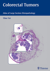
Large-section histopathology widens your perspectives... Correct diagnosis and staging are essential in determining the appropriate therapy of colorectal carcinoma, one of the most common malignancies in America and Europe. As medical science continues to develop rapidly, histopathology remains an essential part of diagnosis in most malignant diseases. In this era of interdisciplinary medicine, the role of pathology has expanded to provide images that easily correlate with endoscopic, radiological, or operative findings. In this remarkable atlas, Tibor Tot presents colon pathology in large histological sections, with cross-sections of entire tumors in their anatomic environments and their circumferential surgical margins. These unique images form a guide to diagnosis, tumor typing and staging according to TNM criteria. They help to assess the completeness of a surgical excision and to understand the heterogeneity of colorectal carcinomas. Features: Cases illustrated in two-page spreads with clinical information, conventional histopathology, and large-section histology images enlarged to almost a full page. Pathology seen in the context of surrounding tissues The margins of malignant tumors visible in their entirety Schematic guides to interpretation of the large-section images Emphasis on diagnostic advantages of using large section technique Technical guidelines for obtaining large-section histopathology specimens This atlas is the result of seven years of studying almost 2,000 cases of colorectal carcinoma and other intestinal lesions and is highly recommended for pathologists, radiologists, surgeons, and oncologists alike.
Das E-Book können Sie in Legimi-Apps oder einer beliebigen App lesen, die das folgende Format unterstützen:
Seitenzahl: 128
Veröffentlichungsjahr: 2005
Ähnliche


Library of Congress Cataloging-in-Publication Data
Tot, Tibor. Histopathology of colorectal tumors / Tibor Tot. p.; cm. Includes bibliographical references and index. ISBN 3-13-140591-0 (alk. paper) — ISBN 1-58890-384-2 (alk. paper) 1. Colon (Anatomy)—Cancer—Histopathology—Case studies. 2. Rectum—Cancer-Histopathology—Case studies. [DNLM: 1. Colorectal Neoplasms—pathology—Case Reports. WI 529 T717h 2005] I. Title. RC280.C6T68 2005 616.99'434707—dc22
2005009276
© 2005 Georg Thieme Verlag, Rüdigerstrasse 14, 70469 Stuttgart, Germanyhttp://www.thieme.de Thieme New York, 333 Seventh Avenue, New York, NY 10001 USAhttp://www.thieme.com
Typesetting by primustype R. Hurler GmbH, NotzingenPrinted in Germany by Grammlich, Pliezhausen
ISBN 3-13-140591-0 (GTV) ISBN 1-58890-384-2 (TNY)
1 2 3 4 5
Important note: Medicine is an ever-changing science undergoing continual development. Research and clinical experience are continually expanding our knowledge, in particular our knowledge of proper treatment and drug therapy. Insofar as this book mentions any dosage or application, readers may rest assured that the authors, editors, and publishers have made every effort to ensure that such references are in accordance with the state of knowledge at the time of production of the book.
Nevertheless, this does not involve, imply, or express any guarantee or responsibility on the part of the publishers in respect to any dosage instructions and forms of applications stated in the book. Every user is requested to examine carefully the manufacturers' leaflets accompanying each drug and to check, if necessary in consultation with a physician or specialist, whether the dosage schedules mentioned therein or the contraindications stated by the manufacturers differ from the statements made in the present book. Such examination is particularly important with drugs that are either rarely used or have been newly released on the market. Every dosage schedule or every form of application used is entirely at the user's own risk and responsibility. The authors and publishers request every user to report to the publishers any discrepancies or inaccuracies noticed. If errors in this work are found after publication, errata will be posted at www.thieme.com on the product description page.
Some of the product names, patents, and registered designs referred to in this book are in fact registered trademarks or proprietary names even though specific reference to this fact is not always made in the text. Therefore, the appearance of a name without designation as proprietary is not to be construed as a representation by the publisher that it is in the public domain.
This book, including all parts thereof, is legally protected by copyright. Any use, exploitation, or commercialization outside the narrow limits set by copyright legislation, without the publisher's consent, is illegal and liable to prosecution. This applies in particular to photostat reproduction, copying, mimeographing, preparation of microfilms, and electronic data processing and storage.
Contents
Introduction
1 Adenomas
2 Early Colorectal Cancer
3 Advanced Colorectal Cancer
4 Other Intestinal Neoplasms
5 Non-neoplastic Lesions
6 Technical Considerations
References
Index
Acknowledgement
To produce a high-quality large histological section is a complex and difficult task. The most important factor in this applied art is the skillful and ambitious laboratory technician. I wish to thank all my co-workers involved in producing large histological sections for the pleasure of looking at their artistic masterpieces and to dedicate this book to Britta Elborg, Britt-Marie Ericsson, Marie Hög-ström, Evelyn Karlsson, Birgitta Norén, to Lena Arvidsson, Irene Bergman, Kajsa Engfeldt, Birgitta Lind, Ingegerd Lindquist, Anita Paulsson and their colleagues. I hope that the reader will share my appreciation.
Introduction
Intestinal and, especially, colorectal diseases represent one of the most common medical problems of humankind, with colorectal carcinoma being one of the most frequently occurring malignancies in Europe and America (Parkin et al. 1992). General practitioners, gastroenterologists, oncologists, radiologists, surgeons, and nurses treat thousands of patients with intestinal diseases every day all over the world. The correct diagnosis is essential for administering the proper therapy. In spite of the rapid developments that have taken place in many fields of modern medical science, histopathology remains essential in making or confirming a diagnosis in a substantial proportion of intestinal diseases.
The attention of the medical community has gradually shifted from morphology towards molecules and genes, which has led to significant progress in many fields of modern medicine, pathology included. However, this shift also risks replacing knowledge about diseases with knowledge about numbers, expressed in a language made up of hardly understandable abbreviations. In addition, in the minds of many medical students and medical doctors, digital images generated by modern radiology have replaced the „gold standard“ of autopsy findings. As a result, knowledge derived from advanced technology is gradually replacing knowledge that has been diligently collected over a period of hundreds of years. The danger of this process is that not only will information be lost, but, more importantly, it will be replaced with something less accurate. Physicians who regularly correlate their endoscopic and radiological images with pathoanatomical and histopathological findings may avoid this mistake. Unfortunately, in this era of unacceptably low autopsy rates, such a physician has less and less opportunity to do so.
Although it is hardly possible to offer equally valuable alternatives to information obtained from autopsy findings, pathologists must try to help clinicians in correlating clinical, radiological and endoscopic findings with morphology. Today, there are many different options that can be explored, such as clinical pathology conferences, telepathology and publishing macroscopic and histologic photographs digitally or on the World Wide Web.
In addition to the clinical picture, the macroscopic appearance of the colorectal mucosa and the nature of the lesions on it, as seen with an endoscope, represent the basis for clinical diagnosis. Endoscopists regularly take biopsies of the mucosa and/or endoscopically evident lesions for histopathological analysis in order to confirm the clinical diagnosis or to detect alterations not visible endoscopically. However, the histologic details cannot be directly compared with the endoscopic appearance of the lesions. For this reason, the pathology report most often provides descriptive information, which the endoscopist cannot directly connect to the image seen in vivo. Large histologic sections, by preserving the contiguity of the tissue in a representative transection of the bowel lesion, bridge the gap between the endoscopic and the histologic appearance. Projected as an overhead, the large section not only resembles the endoscopic findings but at the same time also magnifies them, allowing the clinician to analyze the relief and the details of the lesions on the gross and subgross levels. Placing the same large histologic section under the microscope, the pathologist can demonstrate the histologic and cellular details behind the subgross findings. This makes large histologic sections an ideal approach to educating and training residents as well as specialists performing gastrointestinal endoscopy.
The advantages of using large histologic sections are not exclusively educational. This technique has been routinely used in our laboratory for every operative breast specimen since 1984, and convincing evidence about its suitability in routine diagnostic procedures has been collected. While the resolution of the cellular details are on the level of those obtained with the conventional small block technique, large histologic sections reveal much more useful information than derived from small section, since they usually include a representative transection of the entire tumor together with its environment and the circumferential surgical margins. This technique has proven to be more reliable in documenting the size of the tumor and the extent and distribution of the disease in the breast. It is also superior in demonstrating the multifocal nature of the tumor and intertumoral heterogeneity, compared to the traditional techniques of tissue sampling (Jackson et al. 1994; Tot et al. 2000). In fact, some prognostically important parameters of certain types of breast carcinoma can only be reliably assessed using large histologic sections (Tot 2003).
Surgery remains fundamental in treating patients with gastrointestinal tumors, and it is the best option for cure in patients with rectal cancer, provided that the procedure is radical. During the last two decades, a modified surgical intervention, called total mesorectal resection, has been introduced as a procedure of choice, since it has been shown to lower the rate of local recurrences from 30–40% to less than 10% (Heald and Ryall 1986; Leong 2003; Cecil et al. 2004). The procedure is based on removal of the tumor together with the mesorectal fat. Assessment of the radicality of surgical intervention has always been an important task of the pathologist. In the technique of total mesorectal resection, it has become increasingly important since 2 mm or more of free tissue in the circumferential margin may assure local control of the disease (Nagtegaal et al. 2002). By integrating the gross and subgross appearances with the details of the histologic examination, the technique of large-section histology is an ideal tool for assessing the circumferential margin in total mesorectal resection specimens. It may also influence the content of the histopathology report, which varies considerably from institution to institution (Wei et al. 2004).
With the support of our gastrointestinal surgeons, in 1997, it was decided to include large-section histology in the routine histologic work-up of total mesorectal resection specimens. Based on our experience regarding the advantages of including this technique in the pathology work-up and in demonstrating and interpreting the findings in clinical conferences, the use of large-section histology was soon widened to every operated case of colorectal neoplasm and to some non-neoplastic intestinal lesions. During the last 7 years, almost 2000 cases of colorectal carcinoma and other intestinal lesions have been documented on large histologic sections, a collection that represents the basis for the present atlas.
Proper staging of colorectal carcinomas is essential for treatment and prognosis. The most widely used staging systems are the Dukes' classification (Dukes 1940) and the TNM classification (AJCC cancer staging handbook. TNM classification of malignant tumors, 6th edn. 2002). Despite the numerous attempts to modify the Dukes system, it has remained simple and easy to apply, although somewhat limited in its ability to stratify patients for proper therapy. The TNM classification system, by contrast, offers a more sensitive prognostic categorization due to the larger number of combinations of parameters for describing both the primary tumor and the presence or absence of metastasis in the examined lymph nodes or distant organs. However, new editions of the TNM system have become increasingly complicated and difficult to reproducibly apply. Large histologic sections, by showing the deepest level of invasion of the primary tumor together with the peritumoral tissue, which may contain isolated tumor foci and several lymph nodes, can help in proper tumor classification and staging. This technique brings the so-called pTNM (pathological TNM classification), based on histologic parameters, much closer to the clinical TNM classification. Thus, in this atlas, the term pTNM is not used.
Recent advances in modern radiology have resulted in more detailed imaging of the colorectum (Koh et al 2004). So-called virtual endoscopy may revolutionize gastroenterology as it offers a non-invasive method as an alternative to endoscopy. The technique is based on computer-tomography-produced images of horizontal transections of the bowel wall at different levels. As this approach is identical to that offered by the histopathologic technique of large sections, described in this atlas, it opens the way to new, previously unexplored perspectives in radiopathological correlation of intestinal lesions. Testing the accuracy of this new radiological method is obligatory, and large sections represent the ideal basis for correlation.
Although large histologic sections provide an advantageous alternative to the conventional small block technique, it is infrequently used in routine pathology laboratories. Many pathologists have criticized the technique of large histologic sections as being too expensive, unacceptably prolonging the technical procedure, causing problems in archiving of the slides, and adding nothing more than obtained with the traditional method of tissue sampling. However, our surgeons, gastroenterologists, oncologists, radiologists, pathologists, residents, medical students, and technicians, including those who have been more or less regularly involved in producing and interpreting large histologic sections and correlating them with their own clinical findings, as well as those who have only sporadically seen such sections, are enthusiastic. The usual feedback I get from them is that the information obtained from viewing large histologic slides has positively influenced their everyday practice. Several of them suggested that I share some of the large-section images of colorectal tumors with a wider professional audience. Thus, this atlas is as much a result of their support as of my own efforts.
1 Adenomas
Case 1.1 Villous Adenoma of the Rectum
Patient data: 63-year-old woman presenting with rectal bleeding. Endoscopically, a large, broad-based, soft and exophytic tumor was seen in her rectum, diagnosed on preoperative biopsy as adenoma.
Surgical treatment: Rectosigmoidal resection, no preoperative irradiation.
Specimen: 35-cm-long rectosigmoideum with an 8×6-cm soft exophytic tumor, 3 cm from the distal margin.
Histopathologic diagnosis: Villous adenoma of the rectum with moderate dysplasia. No signs of severe dysplasia or invasion.
Follow-up: 81 months, without signs of disease recurrence.
Villous adenoma of the rectum is often a large non-pedunculated tumor, covering the rectal mucosa over an area of several centimeters. Endoscopic biopsies usually reach only the superficial part of the tumor, the delicate long villous structures covered by dysplastic epithelium. The large histologic section (Fig. 1.1) also demonstrates the intact lamina muscularis mucosae: no signs of invasion are present. It is easy to compare the structures of the normal bowel wall (indicated by the red arrow in the schematic image, Fig. 1.1d) to the structures of the adenoma (the left side of the image, green arrows in Fig. 1.1d). Adenomas exhibit a dysplastic epithelium. The transition from normal to dysplastic epithelium is illustrated in Figure 1.1a (corresponding to the area marked with a red rectangle in Fig. 1.1d). Figure 1.1b represents a microscopic image of the delicate villous structures of the adenoma (corresponding to the blue rectangle in Fig. 1.1d). Figure 1.1c is a magnified detail of Figure 1.1b, illustrating the moderately dysplastic epithelium of this adenoma exhibiting elongated, stratified and crowded cell nuclei and a reduced number of goblet cells.
Fig. 1.1a
Fig. 1.1b
Fig. 1.1c
Practical points
Villous adenomas are usually non-pedunculated, soft tumors covering a large area of the surface of the bowel wall. Large histologic sections are needed to visualize the entire lesion in one transection.The large histologic section is an ideal tool for assessing and demonstrating invasive tumor foci in such a large lesion, as these can be easily missed during the macroscopic examination and sampling of standard small blocks.Fig. 1.1 Large-section histology image of a villous adenoma of the rectum.
Fig. 1.1d Schematic guide to the morphologic details in the large section in Fig. 1.1.
Case 1.2 Villous Adenoma of the Rectum
Patient data: 84-year-old woman with intermittent rectal bleeding and a long history of recurrent adenomas in her rectum.
Surgical treatment: Rectosigmoidal resection, no preoperative irradiation.
Specimen: 33-cm-long rectosigmoideum with a 9-cm segment containing a circumferential exophytic tumor, 6 cm from the distal margin.
Histopathologic diagnosis: Villous adenoma of the rectum with low-grade, partly moderate dysplasia. No signs of invasion.
Follow-up: 6 months, without signs of disease recurrence.
The large section in Figure 1.2 demonstrates another case of villous adenoma, which covers the entire circumference of the inner surface of the rectum. No signs of invasion are seen. The architecture of villous adenomas is complex, as they consist of primary finger-like processes extending outward from the bowel wall and containing fibromuscular stroma (indicated with blue arrows in Fig. 1.2c). Secondary finger-like processes, consisting of loose fibrous stroma with delicate vasculature and a surface covered by dysplastic epithelium, cover these structures. Figure 1.2a, b


