64,99 €
Mehr erfahren.
- Herausgeber: Thieme
- Kategorie: Fachliteratur
- Sprache: Englisch
A widely popular option for vision correction, contact lenses are also useful for the treatment of many serious optical disorders. Like any therapeutic tool, however, they have both positive and negative effects, and a variety of complications can result from their use. Diagnosing and treating these complications and injuries - which sometimes mimic eye diseases - can be complex. Drawing on more than 25 years of knowledge and experience, Contact Lens Complications is the first text to provide a practical, comprehensive overview of the eye injuries caused by wearing contact lenses. This book is filled with hundreds of outstanding, full-color illustrations of problems associated with contact lenses, including eyelid lesions, eye tears, hygiene complications, and lens accidents. You will learn how to confidently manage these problems and effectively distinguish them from common ophthalmic diseases. Additionally, the book covers: treatment guidelines; patient examination and counseling; types of contact lenses available today; common lens fitting and wearing problems; and much more! More than 450 exquisite color photographs and illustrations to facilitate quick injury/disease diagnosis Meticulous, step-by-step techniques for eye examination Easy-to-understand terminology, even to those new to contact lens patient care Clear and concise disease prevention guidelines Practical patient management tips Contact Lens Complications gives medical professionals factual, clear, current information on the injuries and complications caused by wearing contact lenses. Essential for anyone involved in vision care, this authoritative and eminently practical work is certain to become a key resource in the field.
Das E-Book können Sie in Legimi-Apps oder einer beliebigen App lesen, die das folgende Format unterstützen:
Seitenzahl: 301
Veröffentlichungsjahr: 2003
Ähnliche


Library of Congress-in-Publication Data is available from the publisher
This book is an authorized translation of the Germanedition published and copyrighted 2002 by GeorgThieme Verlag, Stuttgart, Germany. Title of the Germanedition: Kontaktlinsenkomplikationen, Ätiologie,Pathogenese, Prophylaxe, Therapie.
Translated byDonald L. MacKeen, M.S., Ph.D., Bethesda MD, USAand Ethan Taub, M.D., Zurich, Switzerland.
© 2003 Georg Thieme Verlag,Rüdigerstrasse 14, 70469 Stuttgart, Germanyhttp://www.thieme.deThieme New York, 333 Seventh Avenue,New York, NY 10001 USAhttp://www.thieme.com
Typesetting by Druckhaus Götz, 71636 LudwigsburgPrinted in Germany by Appl, Wemding
ISBN 3-13-127791-2 (GTV)ISBN 1-58890-132-7 (TNY) 1 2 3 4 5
Important note: Medicine is an ever-changing science undergoing continual development. Research and clinical experience are continually expanding our knowledge, in particular our knowledge of proper treatment and drug therapy. Insofar as this book mentions any dosage or application, readers may rest assured that the authors, editors, and publishers have made every effort to ensure that such references are in accordance with the state of knowledge at the time of production of the book.
Nevertheless, this does not involve, imply, or express any guarantee or responsibility on the part of the publishers in respect to any dosage instructions and forms of applications stated in the book. Every user is requested to examine carefully the manufacturers’ leaflets accompanying each drug and to check, if necessary in consultation with a physician or specialist, whether the dosage schedules mentioned therein or the contraindications stated by the manufacturers differ from the statements made in the present book. Such examination is particularly important with drugs that are either rarely used or have been newly released on the market. Every dosage schedule or every form of application used is entirely at the user's own risk and responsibility. The authors and publishers request every user to report to the publishers any discrepancies or inaccuracies noticed. Some of the product names, patents, and registered designs referred to in this book are in fact registered trademarks or proprietary names even though specific reference to this fact is not always made in the text. Therefore, the appearance of a name without designation as proprietary is not to be construed as a representation by the publisher that it is in the publics domain.
This book, including all parts thereof, is legally protected by copyright. Any use, exploitation, or commercialization outside the narrow limits set by copyright legislation, without the publisher's consent, is illegal and liable to prosecution. This applies in particular to photostat reproduction, copying, mimeographing, preparation of microfilms, and electronic data processing and storage.
Foreword
Contact lenses are currently used electively by 80 to 100 million patients worldwide to correct refractive errors of vision. With so many patients at increasing risk for device-related ocular complications, Dr. Roth's excellent treatise, Contact Lens Complications, is a timely and important work. Long familiar and available to European readers in German, the current translation and publication of an English edition is a most welcome development for all practitioners and academics who practice both the scientific and clinical art of contactology throughout the world. The timing of publication of this superb monograph is particularly fortunate because it coincides with the introduction into clinical practice of a new generation of contact lenses: hyper oxygen-transmissible contact lenses recently approved for up to 30-night extended wear by the Food and Drug Administration, USA. Thus, it is more important than ever for contactology practitioners to be familiar with contact-lens–induced eye problems.
Outstanding features of the text are brevity without sacrificing comprehensiveness or clarity, and excellent color photographs. These features will ensure instant popularity of the English edition among both a generation of optometric students and ophthalmology residents. Indeed, it appears that an old classic has been reborn for the world community at large.
H. Dwight Cavanagh, M.D., Ph.D., F.A.C.S.Professor and Vice-ChairmanThe Dr. W. Maxwell Thomas Chair in OphthalmologyAssociate Dean for Clinical Servicesand Medical DirectorZale Lipshy University Hospital, TX, USA
Preface
Contact lenses are unquestionably a useful aid in the treatment of refractive problems and of many diseases of the eye. They can be used to compensate for optical errors or as an ocular bandage, protective covering, or drug delivery system. They improve the wearer's appearance and expand the field of vision; they perform the function of a destroyed cornea or cover an iris that has been damaged by trauma or disease. They facilitate many sporting activities and indeed open certain professions to persons with impaired vision who would not otherwise be able to pursue them.
Yet contact lenses also have disadvantages. They exert chronic pressure on the anterior surface of the eye, disturb the convection of tears, and impair immune responses in the eye through chronic foreign-body irritation, thus potentially causing complications in the anterior ocular segment. Finally, contact lenses can mask certain types of primary ocular illness, which, in the setting of contact lens wear, may be difficult to diagnose.
Like any effective medical treatment, contact lenses have not only a therapeutic effect, but also undesirable side effects. A wide variety of contact lens complications have been reported in recent years, most of which require treatment by an ophthalmologist. Contact lens complications are sometimes easily mistaken for other ocular diseases, and vice versa; the diagnostic evaluation of contact lens complications thus requires considerable expertise on the ophthalmologist's part.
A cursory glance at this book reveals that the ocular complications of contact lenses are numerous and varied, and may create the impression that the risk of wearing them is incalculably high. Yet this is certainly not the case. The multiplicity of complications in no way diminishes the therapeutic value of contact lenses, as the actual frequency of complications, according to many well-performed, international, longitudinal studies, is well below 1%—a rate that is hardly matched by many other forms of routine treatment in ophthalmology. Safety is assured by proper lens care and handling on the part of the patient, and by frequent and thorough follow-up examinations on the part of the ophthalmologist.
This book is meant to be useful to everyone involved in contactology, including resident physicians and ophthalmologists who have just started fitting contact lenses. We have avoided extensive discussions of basic science that can be found in other texts and have tried to use terminology that will be transparent even to persons with little experience in contactology. Examining techniques and findings are presented in such a way that they could be duplicated by any ophthalmologist, whether in an academic institution or in private practice.
In writing this book, the author has had to face certain inevitable difficulties. This is the first comprehensive publication in ophthalmology addressing the more important complications of contact lenses, and the causes and interrelationships of many of the findings discussed here are still imperfectly understood. Furthermore, contact lenses have not been in use long enough to allow a complete assessment of their long-term complications. This will have to await future publications by other investigators.
This book is the fruit of more than 25 years of research and teaching in applied contact optics in the University Ophthalmology Department and Army Hospital in Ulm, Germany, at the Institute for Contact Optics in Ulm, and at the Center for Sight, Georgetown University Medical Center, Washington, DC.
I would like to thank my teachers in contactology, Drs R Marquardt in Ulm, W Erich in Homburg/Saar, the late H Kemmetmueller in Vienna, the late P Halberg in New York, M Lemp and D MacKeen in Washington, and the late Jonathan Kersley in London. I would also like to thank the numerous others who taught me through their published works, and, finally, the staff of Georg Thieme Verlag for their invaluable assistance in producing this book.
Washington DC–Ulm/Danube, Spring 2003
Hans-Walter Roth
Contents
1 Problems Caused by Contact Lenses
Overview
Materials and Manufacture
Fitting Technique
Contraindications
Hygiene
Loss of Contact Lenses
Ocular Trauma
Maximum Wearing Time
Patients with Systemic Diseases
Patients at High Risk of Complications
Psychological Factors
Summary: The Risk of Injury
2 Patient Counseling and Examination
Patient Counseling
History Taking
Examination
General
Lid
Conjunctiva
Cornea
Inner Eye
Intraocular Pressure
Refractive Error and Visual Acuity
Binocular Vision
Inspection of Contact Lenses
Inspection of Contact Lens Cases
Documentation of Findings
Follow-up
3 Pathologic Findings
Lid
Anatomy and Physiology
Increased Lid Tension and Blepharospasm
Ptosis
Lid Swelling and Edema
Infectious Blepharitis, Squamous Blepharitis, Ulceration
Lid Injury
Conjunctiva
Anatomy and Physiology
Tarsal Conjunctiva
Follicular Swelling, Papillary Hypertrophy, Giant Papillary Conjunctivitis
Bulbar Conjunctiva
Conjunctival Edema and Acute Chemosis
Subconjunctival Hemorrhage
Focal Conjunctival Hyperemia
Sclera
Anatomy and Physiology
Pathology
Vasodilatation and Inflammation
Injury and Hemorrhage
Cornea
Anatomy and Physiology
Epithelial Changes
Edema and Staining
Stippling
Streaks and Spiral Traces
Indentations
Cracks
Spiders
Mosaics
Nummules and Pseudoinfiltrates
Corneal Erosions
Bizarre Epithelial Defects, Branched Defects, Pseudoherpetic Defects
Bubble Formation and Pseudokeratitis Bullosa
Stromal Changes
Edema
Tight Lens Syndrome
Toxic Keratopathy
Mixed Solution Syndrome
Corneal Deprivation Syndrome
Post-Heat Syndrome
Vascularization
Infection
Clouding and Scars
Changes of Descemet's Membrane and the Endothelium
Changes of Descemet's Membrane
Endothelial Changes
Topographic Changes
Contour Changes
Pachymetric Changes
Inner Eye
Iritis, Uveitis, Panophthalmitis
Cataract
Intraocular Hypertension, Glaucoma
Retinal Diseases and Detachment
4 Visual Impairment
Visual Impairment
Causes of Visual Impairment
Changes in Refraction
Faulty Lens Handling and Insertion, Lens Loss
Permanent Visual Impairment
Primary Ocular Diseases Masked by Contact Lenses
Material Defects and Defective Lenses
Intermittent Visual Impairment
Fitting Errors
Orthokeratological Side Effects
Spectacle-Blur Phenomenon and Corneal Distortion Syndrome
Wetting Problems
Glare
Causes of Glare
Diagnostic Evaluation of Visual Impairment and Glare
5 Causes of Contact Lens Damage
Handling, Hygiene, Wearing Times
The Patient's Hands
The Lens Case
Wearing Times
Fitting Errors
6 Alterations of Contact Lenses
Wetting Problems
Material Defects
Discoloration and Fading
Deposits
Components of Tear Fluid
Jelly Bumps
7 Primary Fitting and Wearing Problems
Systemic Disease
Febrile Illnesses, Immune Compromise, Medication Use
Allergy
Primary Ocular Disease
Lid Diseases
Conjunctival Diseases
Corneal Diseases
Tear Deficiency, Disorders of Lacrimation, DryEye
Pathophysiology
Clinical Course
Clinical Testing
Other Eye Diseases
8 Eye Injuries in Contact Lens Wearers
Types of Ocular Trauma
Trauma Involving Foreign Bodies
Perforated Globe
Ocular Contusion
Chemical Injury
Consequences for Contact Lens Wearers
9 Frequency of Contact Lens Complications
Analysis of Contact Lens Complications
10 Treatment of Contact Lens Complications
General Aspects
General Treatment Procedures
Special Treatment Procedures
Lid Diseases
Blepharospasm
Ptosis
Lid Edema
Squamous Blepharitis
Lid Trauma
Conjunctival Diseases
Giant Papillary Conjunctivitis
Conjunctival Chemosis
Conjunctival Hemorrhage
Conjunctival Trauma
Nonspecific Conjunctival Hyperemia
Bacterial Conjunctivitis
Viral Conjunctivitis
Fungal Conjunctivitis
Scleral Diseases
Corneal Diseases
Corneal Edema
Corneal Erosion
Bullous Keratopathy
Tight Lens Syndrome
Toxic Keratopathy
Corneal Deprivation Syndrome
Post-Heat Syndrome
Keratitis, Corneal Infiltrates, Corneal Ulcers
Corneal Vascularization
Corneal Scars
Corneal Deformation
Folds in Descemet's Membrane, Endothelial Changes
Intraocular Findings
Glaucoma
Dry Eye, Tear Deficiency Syndrome
Accidents, Mechanical Trauma, Chemical Trauma
References
Index to Text
Index to Illustrations
1 Problems Caused by Contact Lenses
Overview
Materials and Manufacture
Fitting Technique
Contraindications
Hygiene
Loss of Contact Lenses
Ocular Trauma
Maximum Wearing Time
Patients with Systemic Diseases
Patients at High Risk of Complications
Psychological Factors
Summary: The Risk of Injury
Overview
A contact lens is a rigid optical device that maintains close, prolonged contact with the living tissue of the eye. Contact lenses can be worn for many years without difficulty, but only if they are made of a well-chosen material, correctly shaped, and properly fitted. Complications can be prevented by precise fitting and regular follow-up by an ophthalmologist experienced in the recognition and management of the associated pathological conditions. The universally acknowledged importance of contact lenses in contemporary ophthalmology does not obviate the need for careful weighing of indications and contraindications (Tables 1 and 2), advantages and disadvantages (Tables 3 and 4), and risks and benefits, as with any other form of medical treatment.
Contact lenses are a valuable aid to vision in the presence of severe refractive errors, and an irreplaceable component of treatment for certain chronic diseases of the eye. Yet their use is not without risk. Like any other form of medical treatment, they have both desired and undesired effects. Some wearers of contact lenses will develop ocular complications, ranging from harmless irritation of the lids or conjunctiva to sight-threatening corneal ulceration. In a few reported cases, eyes have even been lost through the wearing of contact lenses. Fortunately, these disasters are extremely rare, but they serve to remind us of the dangers against which we must be on guard.
Table
1
Indications for contact lenses
Cosmetic reasons
Myopia
Hypermetropia
Regular or irregular astigmatism
Anisometropia, aniseikonia
Corneal scars
Aphakia
Post-keratoplasty
Occupation or athletic activities
Iris lesion, pupillary dysfunction
Bandage lens
Drug-delivery vehicle
Particular diseases of the anterior segment
Table
2
Contraindications of contact lenses
Chronic irritation of the lids, conjunctiva, or cornea
Malformation of the anterior segment
Absence of lacrimation
Inability to comply with lens hygiene regimen
Difficulty in handling lenses
Long-term use of ocular medications
Table
3
Advantages of contact lenses over spectacles
Near invisibility
Improvement of vision in high ametropia
Improvement of vision in irregular astigmatism
Equal image size for patients with different refractive errors in the two eyes
Containment effect in keratoconus and progressive myopia
Bandage effect in chronic ocular diseases
Medication vehicle for long-term delivery
Table
4
Disadvantages of contact lenses compared with spectacles
Need for conscientious lens care
Limited wearing time
Greater risk of injury
Easier to lose, harder to find
Risk of infection
Risk of allergic reaction
Greater difficulty in handling
More frequent ophthalmological follow-up necessary
Shorter life of lenses
Limited durability
Metabolic and mechanical disturbance of the eye
Possible psychological disturbance
Need for thorough patient education
Materials and Manufacture
Contact lenses are apt to cause complications because they pose a constant challenge to the normal functioning and immune defenses of the anterior segment of the eye. Their adverse mechanical effects may include deformation of the corneal curvature and thinning of the corneal stroma due to chronic pressure upon it. By disturbing corneal metabolism, they may induce swelling of the corneal stroma and alter the structure and cellular composition of the endothelium. They also raise the temperature of the lids, conjunctiva, and cornea by a few degrees Celsius, and thereby promote inflammation.
The manufacture of contact lenses thus demands not only high mechanical precision in the shaping of the lens, but also a lens material that is transparent, chemically pure, durable, nontoxic, and nonallergenic. These requirements are not easy to meet.
A further difficulty is that nearly all errors in fitting, choice of lens material, or disinfecting technique do not become manifest when the patient begins to wear the lens, but only later, through the pathological changes they produce in the eye. Thus, a careful prefitting history and examination, close supervision of the wearer, and regular follow-up are essential for the prevention of later problems.
There are innumerable reports of contact lens complications due to improper fitting, careless follow-up, lack of adherence to the guidelines for lens hygiene, and wearing the lenses for longer than the recommended time. Such complications are the principal difference between spectacles and contact lenses. Spectacles make prolonged contact only with the skin and thus cause, at worst, skin irritation, while contact lenses make prolonged contact with sensitive, metabolically active epithelial cells bathed in a bodily fluid (tears), and can easily damage them. The risk of complications is immediately clear when contact lenses are properly regarded as a type of synthetic medical implant, comparable to other devices such as artificial hips, synthetic vascular grafts, silicone breast implants, and artificial heart valves.
Many problems result from the interaction of synthetic materials with tissue, the most common of which is giant papillary conjunctivitis (GPC). A perfectly nonallergenic, hapten-free material for contact lenses has yet to be developed.
Fitting Technique
The chronic pressure exerted by a contact lens on the anterior surface of the eye can be kept to a minimum by the exact fitting of a lens with an aspheric inner surface, but will nevertheless, in most cases, cause topographical changes of the cornea after prolonged wearing. The continual changes in corneal thickness and contour over the course of a day, and over the years, cannot easily be summarized by a mathematical formula. There will never be an ideal contact lens that always lies exactly parallel to the corneal surface at every point, exerting a constant and spatially unvarying pressure on it, yet also—as the physiologists demand—gliding freely over it with every blink. Optimal fitting thus represents a compromise between an even distribution of pressure and a minimal disturbance of corneal metabolism. Poor fitting may lead to corneal damage, such as irregular astigmatism or Type C keratoconus.
Contraindications
Contact lenses are contraindicated by all acute and chronic processes affecting the anterior segment of the eye. For example, squamous blepharitis, chronic conjunctivitis, recurring keratitis, hordeola, and chalazia may all recur or worsen acutely when contact lenses are worn. Growths on the corneal or conjunctival surface, such as pinguecula or pterygia, contraindicate the wearing of certain kinds of lenses. Many conditions affecting the endothelium are also contraindications; patients with endothelial dystrophy should not wear contact lenses for cosmesis. Nor should contact lenses be worn in the presence of a filtering bleb after the surgical creation of a fistula.
Patients who must regularly use eye drops for the treatment of glaucoma or other disorders are only rarely suitable candidates for contact lenses. Lens wearing alters the pharmacokinetics of instilled agents and may necessitate a change of the dosage.
Contact lenses must sit on a cushion of tear fluid to be able to glide freely over the corneal surface. Soft lenses need a small additional amount of tear fluid to stay elastic. Therefore, each contact lens, depending on its type, composition, and mode of wearing, requires up to 1 ml of tear fluid daily, a quantity equaling the entire daily production of tears in a healthy eye. Disorders of lacrimation thus predispose to contact lens complications. Artificial tears may compensate temporarily for diminished lacrimation but must not be used as a permanent measure, because, by eliminating the reflex drive to lacrimation, they diminish it further. The preservatives found in moisturizing eye drops may also damage the eye with prolonged use.
Hygiene
All contact lenses, whether conventional, disposable, or for extended wear, should be regularly removed for cleaning and disinfection. Among the many different disinfecting and cleaning solutions now available, some may irritate or injure eyes that are sensitive to particular components of their formulation. Allergic reactions are seen much less frequently with the newer types of solution, but there is still a danger of severe toxic keratopathy, particularly when incompatible solutions are mixed.
Loss of Contact Lenses
Contact lenses are easier to lose than spectacles, and much harder to find. A surefire way to lose contact lenses is to wear them while swimming, diving, or surfing. Sudden jerking of the head, as when it is struck in a boxing match or brawl, can also easily dislodge contact lenses. Such events sometimes have medicolegal consequences.
Sports involving sudden, massive bodily movements, such as horseback riding or sky diving, necessitate lenses of larger than usual diameter, but such lenses cannot be worn as long as ordinary lenses. Thus, persons engaging in such sports should also have a pair of ordinary lenses for normal wear.
Ocular Trauma
All contact lenses cause a certain amount of epithelial microtrauma during normal wear. The corneal and conjunctival surfaces may be injured when the lens is inserted or removed, and may even be cut if the lens is defective. In such cases, lens wearing is discontinued and the wound generally heals within a few days, though the potential for infection or ulceration exists.
Maximum Wearing Time
No type of contact lens can be worn indefinitely, and only a few types can be worn during sleep. The recommended maximum wearing time depends on the age of the lens and the amount of surface deposition. Changing disposable lenses in a weekly rhythm is a safe practice only if new lenses are used every week. Perhaps surprisingly, soft contact lenses developed for extended wear cause the most complications; when they are worn without interruption, the rate of complications rises exponentially with the duration of wear. Corneal ulcers are more than 30 times as common when contact lenses are worn for extended periods than when they are worn for no more than 18 hours a day.
Patients with Systemic Diseases
Most of the reported severe complications of contact lenses involve infection with Pseudomonas or Acanth-amoeba infection. Corneal ulceration is usually caused by poor hygiene and excessively prolonged wear.
Diabetic patients are more likely to develop fungal infections, while immunocompromised patients, such as those with leukemia or AIDS and patients undergoing antineoplastic chemotherapy, are more likely to develop bacterial ulcers. The high risk of ocular infection in these groups of patients contraindicates the wearing of contact lenses.
Because many orally or parenterally administered medications, and their metabolic products, are secreted into the lacrimal fluid, the use of such medications contraindicates the wearing of hydrophilic or water-absorbing contact lenses. For example, in a patient taking oral calcium supplements, soft contact lenses of high water content may be irreversibly damaged within a few hours by deposition of insoluble calcium salts and proteins on their surface.
Many systemic diseases, including rheumatic diseases, thyroid and renal dysfunction, and other diseases treated with hormone supplementation, can alter the secretion of tears and thereby interfere with the wearing of contact lenses. The same is true of oral contraceptive use. The impairment of lacrimation may be asymptomatic until the patient begins wearing contact lenses.
Patients at High Risk of Complications
Contact lens fitting is not just a matter of measuring the refractive error and the corneal radius and diameter, calculating the desired lens parameters, and choosing the lenses accordingly. Rather, it requires a thorough knowledge of the anatomy and physiology of the living eye. For example, there is no textbook method for fitting contact lenses in an aphakic neonate. In such difficult cases, the ophthalmologist and contact lens team may need to call on many years of accumulated expertise.
Patients with keratoconus are also at high risk of complications. If the lens that is fitted is too flat, acute decompensation of the cone may occur practically overnight, leading to perforation and the need for immediate keratoplasty. An uneven distribution of pressure from the edge of the lens on the peripheral zone of the cornea may injure the epithelium. In patients with Type B keratoconus, the severe reduction of tear flow usually makes wearing lenses very difficult.
Older aphakic patients, too, are prone to complications of various types. Frequent lens loss and difficulties in handling limit the potential for successful contact lens wear in these patients. Lens fitting is especially difficult in this group after perforating injury or invasive ocular surgery. The resultant irregular astigmatism tends to make contact lenses unstable. Successful fitting requires both longstanding expertise and a sound biophysical understanding of the effect of lens pressure and the consequent corneal molding on a lacerated or scarred cornea.
Psychological Factors
Some patients are psychologically ill equipped to wear contact lenses. It is often advisable to recommend against wearing contact lenses for psychological reasons, even if the patient explicitly desires to wear them. The contact lens specialist should be able to recognize the potential for self-destructive and suicidal behavior, particularly in young, female patients with relationship difficulties, and should be ready to refer such patients for psychiatric help if necessary. A problem of another kind occurred in one of our patients, who complained of severe pain and visual disturbances while wearing contact lenses, although the ocular findings were normal. It turned out that her mother had forced her to wear contact lenses against her wishes. The ocular symptoms resolved when, on the doctor's orders, she stopped wearing the lenses.
Summary: The Risk of Injury
As we have seen, any contact lens may cause complications, potentially as severe as the loss of an eye. The most important preventive measures are a preliminary assessment and repeated follow-up by an ophthalmologist, and fitting by an experienced lens fitter. The patient must be made aware of the need to consult the ophthalmologist immediately for any problem that may arise. Only an explicit instruction to this effect can counteract the false impression of safety created by mass marketing, disposable lenses, internet advertising, and discount selling practices. As many years of clinical research have shown, contact lenses are not an optical product to be obtained by mail order or in the bargain basement. Only an experienced fitter can realize their potential as a valuable form of treatment for visual impairment and other ocular disorders.
2 Patient Counseling and Examination
Patient Counseling
History Taking
Examination
General
Lid
Conjunctiva
Cornea
Inner Eye
Intraocular Pressure
Refractive Error and Visual Acuity
Binocular Vision
Inspection of Contact Lenses
Inspection of Contact Lens Cases
Documentation of Findings
Follow-up
Patient Counseling
All patients need thorough counseling and instruction before they choose the type of contact lenses they will wear and as they begin to wear them. The major issues to be addressed are listed in Table 5. The choice of contact lens is not simply a matter of finding the type that gives the best cosmetic result, as patients may at first assume, but requires consideration of patient-specific risks and other factors, particularly the patient's living and working environment. Despite recent advances in soft-lens technology, hard lenses are still better tolerated over the long term, though hydrophilic lenses may be better suited to the patient's lifestyle and desired manner of wearing. For patients who plan to wear contact lenses only occasionally, soft lenses are preferable; those who plan to wear contact lenses instead of spectacles over the long term are better served with hard lenses. Patients should be informed, however, that switching from hard contact lenses back to spectacles may be difficult. Long-term wearing of hard contact lenses can cause irregular astigmatism, leading to diminished visual acuity and increased glare; this is the so-called spectacle blur phenomenon. It often takes several months after the cessation of contact lens wear for these symptoms to resolve so that vision with spectacles is as good as before.
Lens care products are to be regarded as medications, and due attention should be paid to their storage, stability, and expiration dates. Every contact lens wearer should be adequately instructed about their use, effects, and potential complications. The proper choice of lens care system depends not only on the type of lens, but also on the type and rate of deposition on the lens surface, which varies from patient to patient. Those for whom lens care is difficult, such as patients with arthritis, should use only one cleaning agent, if possible. Those with particularly sensitive eyes should use hydrogen peroxide. Careful consideration should be given to the possible use of an additional cleaning solution, or of enzyme tablets.
The ever-decreasing cost of contact lenses and the introduction of disposable lenses have misled many wearers to imagine that contact lens optics is a routine matter, and that their purchase and use are a retail business like any other. Yet the fitting of contact lenses actually demands the highest expertise of the ophthalmologist and optician. Additionally, their physical specification and manufacture are an intricate technical process, at whose end the lenses can be safely handed over to the patient only when all of the problems discussed above have been resolved, unnecessary risks avoided, and possible contraindications ruled out. Finally, the prescriber of contact lenses must be certain that the patient can care for them properly and knows the problems that may arise with improper use.
Table
5
Patient education
General reasons for wearing contact lenses
Pros and cons of contact lenses vs. spectacles
Problems in switching between contact lenses and spectacles
Problems in the workplace
Limited wearing time
Lens handling, insertion, removal
Lens cleaning
Lens disinfection
Frequency of replacement of short-term lenses
Contact lenses and use of medication
Contact lenses in patients with chronic illnesses
Follow-up examinations by the fitter or ophthalmologist
What to do in an emergency
History Taking
The patient's past medical history and review of systems may indicate the presence of an acute or chronic illness that would increase the risk of wearing contact lenses. Such illnesses, a history of major trauma, and the chronic use of medications of certain types may contraindicate wear. The use of oral contraceptives, pregnancy, and breastfeeding are also limiting factors. The patient should be asked about prior ocular injuries, infections, other diseases, and the prior use of eye drops.
If the data are available, the physician should also note any changes over time of the patient's spectacle prescription, which may indicate a progression of the refractive error. Finally, the patient should be asked whether contact lenses were previously prescribed or worn, with what result, and why a previous fitting was unsuccessful, if this was the case.
Examination
General
The tasks of the ophthalmologist include ocular examination before contact lens fitting, monitoring for potential problems in the initial adaptation phase, and regular follow-up thereafter. The routine ophthalmological examination of the contact lens wearer is the same as that of any other patient; its most important elements are listed in Table 6. Yet a few special considerations deserve mention in this section.
Most problems due to contact lenses develop slowly and become manifest only after a variable interval. It is thus desirable to document the findings regularly with any of the numerous photographic and video systems now available for clinical use, with which the data may be digitally stored and statistically processed.
The improved safety of modern synthetic materials and lens care products has greatly lessened the risks associated with contact lenses, but wearers must nonetheless be followed up regularly for the prevention of ocular damage, and particularly of slow, cumulative damage over the long term.
Table
6
Initial visit or annual follow-up examination of the contact lens patient
General medical history, ocular history
Lids
Conjunctiva
Cornea
Sclera
Anterior chamber
Pupillary mobility
Crystalline lens
Vitreous body
Fundus
Visual fields
Refractive correction (objective, subjective)
Visual acuity
Binocular vision
Corneal diameter
Corneal thickness
Corneal topography
Tear flow
Intraocular pressure (in all patients over age 40 and those with progressive myopia, suspected glaucoma, or a positive family history)
Ocular examination before, during, and after contact lens fitting thus serves not only for the immediate detection of acute complications, but also for the early detection of incipient long-term complications, which may affect either the anatomy or the physiology of the eye. We therefore recommend adherence to the classical sequence of the routine ophthalmological examination: visual acuity, motility, lids, conjunctiva, cornea, anterior and posterior chambers, retina, and intraocular pressure.
Not only the eyes, but also the contact lenses should be examined. Any abnormalities of lens position, surface depositions, staining, defects, and changes in parameters should be noted. Microbiological tests may reveal poor lens hygiene and an elevated risk of infection. One should also try to determine whether the patient has a proper understanding of lens care. Dirty hands or a dirty lens case indicate that this is not so.
Lid
Inadequate opening or closure of the eyelids, excessive blinking, or blepharospasm will be immediately visible to the examiner without special equipment. Ordinary daylight is best for examination of the lids. A check for symmetry readily reveals any monocular abnormality, such as ptosis.
The normal interpalpebral space is almond-shaped and of equal height on the two sides. The lids snugly overlie the globe and point directly upward (lower lid) and downward (upper lid). The nasal canthus makes a sharp corner, while the temporal canthus is rounded. The lacrimal puncta are fully immersed in the lacrimal menisci.
The skin of the eyelids is very thin and elastic, and care must be taken to avoid injuring it when spreading the lids apart to insert contact lenses, particularly in infants and toddlers. Children's lids are soft and smooth, those of elderly persons baggy and wrinkled.
Pathological changes of the lids are often the first sign of an adverse reaction to the lens itself, or to lens disinfectants or cleaners. The skin between the lashes should be examined carefully with a slit lamp under high magnification to detect early reddening and scaling. These findings mandate discontinuation of the contact lenses, as they are predictive of toxic keratopathy, which does not appear till much later. Particular vigilance is required in patients with a history of squamous blepharitis, which predisposes to such problems and is thus a relative contraindication to the wearing of contact lenses.
Conjunctiva
The conjunctiva, an elastic, highly vascularized tissue lying in an exposed position between the globe and the lids, responds to all forms of irritation from within or without by vasodilatation, which is visible as conjunctival hyperemia (injection). This most common abnormal finding associated with contact lens problems often provides the key to their detection and differential diagnosis. Its location, extent, and type depend on the cause of irritation of the anterior segment, which may be mechanical, toxic, or metabolic, and may arise from the lens itself or from lens care products. The tarsal conjunctiva may display massive (follicular) swelling of the papillae in what is known as cobblestone conjunctivitis or giant papillary conjunctivitis (GPC).
Zones of conjunctival necrosis are occasionally seen. Inspection of the conjunctiva of the upper and lower lids is thus mandatory not only before fitting, but also at every follow-up examination.
Pathological changes of the bulbar conjunctiva range from simple injection to complex isolated or diffuse changes.
The form and distribution of any perilimbal injection in the area covered by a soft lens are important clues to the differential diagnosis, as are superficial and deep conjunctival vasodilatation and neovascularization. A circular impression from the lens on the conjunctiva is an early sign of tight lens syndrome (TLS) and should be carefully inspected and documented, as should any microerosions or hemorrhages under the surface of the lens. Fluorescein or rose bengal staining is helpful.
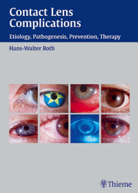
Fig. 1 Hard lens (PMMA), myopia, worn without problems for 20 years. Normal findings.
Fig. 2 Soft hydrophilic lens (hydroxymethyl methacrylate: HEMA), myopia, worn without problems for 10 years. Normal findings.
Fig. 3 Hard lens (fluorosilicone carbonate), high myopia, worn for 6 years without problems. Normal findings.
Fig. 4 Hard lens, hyperopia +7.5 diopter (D), worn without problems for 8 years. Normal findings.
Fig. 5 Absence of eye irritation, myopia, hard oxygen-permeable (RGP) CAB lenses worn for 8 years. Normal findings.
Fig. 6 Absence of eye irritation, keratoconus, spheric equivalent –16.25 D, CAB lens worn without problems for 13 years. Normal findings.
Fig. 7 Myopia, gel lens of high water content, worn without problems for 10 years. Normal findings.
Fig. 8 Myopia –3.75 D, disposable lenses worn without problems for 5 years. Normal findings.
Fig. 9 Absence of eye irritation, irregular astigmatism after photorefractive keratectomy (PRK). Piggyback system (hard lens on soft lens) worn for 3 years without complication.
Fig. 10 Aphakia, irregular astigmatism, previous iritis, complication cataract operation with secondary glaucoma. Piggyback system worn for 2 years without complication.
Cornea
The cornea is inspected in the usual manner, with a slit lamp, before and after removal of the contact lens. First, the position and mobility of the lens are examined under low magnification. Tear lenses are more easily examined after instillation of a fluorescein solution of suitable type. Fluorescein sodium, which is water-soluble, must not be used with soft gel lenses, as it will bind to them and cause permanent staining. High-molecular-weight fluorescein preparations are now available for use with gel lenses, but their fluorescence is much weaker than that of fluorescein sodium.
Rose bengal 2% is another useful dye for examination of the conjunctival and corneal epithelium after lens removal. Staining of epithelial cells may indicate a dry-eye condition associated with lens wearing. Rose bengal can bind to any type of synthetic material and thus is not routinely instilled when the contact lens is in place. Both fluorescein and rose bengal must be completely rinsed from the eye with sterile saline, and the lens should only be reinserted after a suitable interval, because even minute quantities of dye remaining in the conjunctival sac can stain it irreversibly.
The location and extent of neovascularization at the limbus indicate whether a metabolic, mechanical, or toxic disturbance is present. These vessels are best seen with slit-lamp illumination at 10–20 × magnification under red-free light.


