209,99 €
Mehr erfahren.
- Herausgeber: Thieme
- Kategorie: Fachliteratur
- Sprache: Englisch
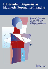
Organized by findings to reflect how radiologists really work, this abundantly illustrated book offers more than 2,000 magnetic resonance images depicting commonly seen congenital and acquired disorders, as well as many rare and unusual cases. Along with the radiographic findings, you will enjoy brief tabular summaries of essential demographic, pathologic, and clinical features of each disease. The book is divided into anatomical sections, including: the brain; head and neck; spine; musculoskeletal system; chest; abdomen; and pelvis. All diseases and findings are cross-referenced, providing quick access to desired information. Special features: Chapters arranged by anatomic location instead of by disease - mirroring the approach you apply in daily practice Hundreds of tables listing pathological features to assist in the diagnostic process Detailed descriptions allow you to differentiate between diseases and conditions that have similar appearances More than 2,000 state-of-the-art images, along with detailed diagrams and charts, give helpful examples of actual findings Extensive cross-referencing of information leads you to further resources Here is the quintessential guide to magnetic resonance imaging that radiologists and other physicians need to enhance their diagnostic skills. Residents and fellows will use it as an invaluable board preparation tool. Keep this practical text close at hand.
Das E-Book können Sie in Legimi-Apps oder einer beliebigen App lesen, die das folgende Format unterstützen:
Seitenzahl: 1319
Veröffentlichungsjahr: 2002
Ähnliche


Library of Congress Cataloging-in-Publication Data is available from the publisher.
Contributors of illustrations
Kevin G.J. Ibach, M.D., F.R.C.P.C.
Lecturer
University of Toronto
University Health Network
Toronto General Hospital
Toronto, Ontario, Canada
Uresh Patel, M.D.
Radiologist
Ide Group, PC
Rochester, NY, USA
Brian K. Tan, M.D.
Chief Resident
Department of Radiology
University of Rochester Medical Center
Rochester, NY, USA
© 2002 Georg Thieme Verlag,
Rüdigerstraße 14, D-70469 Stuttgart, Germany
Thieme New York, 333 Seventh Avenue,
New York, N.Y. 10001 USA
Cover design by Renate Stockinger, Stuttgart
Drawings by Christiane and Dr. Michael von Solodkoff,
Neckargmünd
Typesetting by primustype Robert Hurler GmbH
D-73274 Notzingen, Germany
Printed in Germany by Druckhaus Götz, Ludwigsburg
ISBN 3-13-108121-X (GTV)
ISBN 1-58890-085-1 (TNY) 1 2 3 4 5
Any reference to or mention of manufacturers or specific brand names should not be interpreted as an endorsement of advertisement for any company of product.
Some of the product names, patents, and registered designs referred to in this book are in fact registered trademarks or proprietary names even though specific reference to this fact is not always made in the text. Therefore, the appearance of a name without designation as proprietary is not to be construed as a representation by the publisher that it is in the public domain.
This book, including all parts thereof, is legally protected by copyright. Any use, exploitation, or commercialization outside the narrow limits set by copyright legislation, without the publisher's consent, is illegal and liable to prosecution. This applies in particular to photostat reproduction, copying, mimeographing or duplication of any kind, translating, preparation of microfilms, and electronic data processing and storage.
Important Note: Medicine is an ever-changing science undergoing continual development. Research and clinical experience are continually expanding our knowledge, in particular our knowledge of proper treatment and drug therapy. Insofar as this book mentions any dosage or application, readers may rest assured that the authors, editors, and publishers have made every effort to ensure that such references are inaccordance with the state of knowledge at the time of production of the book.
Nevertheless this does not involve, imply, or express any guarantee or responsibility on the part of the publisher sinrespect of any dosage instructions and forms of application stated in the book. Every user is requested to examine carefully the manufacturer's leaflets accompanying each drug and to check, if necessary in consultation with a physician or specialist, whether the dosage schedules mentioned there in or the contraindications stated by the manufacturers differ from the statements made in the present book. Such examination is particularly important with drugs that are either rarely used or have been newly released on the market. Every dosage schedule or every form of application used is entirely at the user's own risk and responsibility. The authors and publishers request every user to report to the publishers any discrepancies or inaccuracies noticed.
Foreword
Since 1985, when I had the pleasure and honor of publishing the first edition of the textbook “Differential Diagnosis in Conventional Radiology” with Dr. Francis A. Burgener, it has become customary to use a similar approach in writing radiological textbooks, i.e. to structure the books on the basis of radiographic findings instead of disease entities. When the book first appeared, the approach was well received by radiology residents preparing for their specialist examinations. Even non-radiologist physicians, occasionally called upon to interpret radiographic findings, used the text as it does not require extensive reading to find the essential diagnostic criteria.
In 1996 we published another textbook using a similar approach, namely “Differential Diagnosis in Computed Tomography,” which also proved successful. Even then, we had plans to continue the series by writing another volume on differential diagnosis in magnetic resonance imaging. To my sincere dismay, I was unable to invest enough time and energy to finish my part of the planned MRI textbook in time. So it was a great relief both to myself and Dr. Burgener that he found three talented and proficient younger radiologists, Dr. Stephen P. Meyers, M.D. Ph.D and Raymond K. Tan, M.D. of the University of Rochester Medical Center, Rochester, N.Y., and Wolfgang Zaunbauer, M.D. of Kantonsspital, St. Gallen, Switzerland to complete the book on differential diagnosis in MRI without undue delay.
MRI is still developing rapidly and new imaging sequences are published almost daily. New diagnostic signs continue to be discovered in great numbers. With this overflow of new information, selecting the essential information for a single-volume textbook has become a very demanding task. It goes without saying that a differential diagnostic textbook about MRI will be substantially larger than one on CT. The authors are to be congratulated for their success in fitting a significant amount of essential radiological information, hundreds of differential diagnostic tables, and approximately 2000 state-of-the-art drawings, charts, and MR images into a textbook of almost 700 pages.
The structure and layout of Differential Diagnosis in Magnetic Resonance Imaging is essentially similar to the previous two differential diagnostic textbooks, published by Thieme, of which Dr. Burgener is the first author. It is divided into anatomical sections, including the brain, head and neck, spine, musculoskeletal system, chest, abdomen, and pelvis. The book covers the most common diseases and a multitude of rare and unusual conditions, providing quick access to differential diagnostic information.
I am convinced that this guide to differential diagnosis in MRI will receive the same enthusiastic acceptance as the previous books in this series. It will be helpful to radiologists and other physicians involved in interpreting MR images. Residents and fellows will find it an invaluable board preparation tool.
Martti Kormano, M.D.
Professor and Chairman
Department of Diagnostic Radiology
University of Turku
Turku, Finland
Preface
All good things must come to an end. After having coauthored with Dr.Martti Kormano the textbooks “Differential Diagnosis in Conventional Radiology” and “Differential Diagnosis in Computed Tomography,” which were translated into five languages and produced several spin-off books, Dr.Kormano had to back out at the last minute from this project because of too many other professional commitments as chairman of a radiology department and many national and international societies, as clinician, researcher, and teacher, and last but not least as berry farmer. It was impossible to replace Dr. Kormano with his extensive knowledge in the entire field of diagnostic radiology by a single person. I was, however, fortunate enough to find three colleagues and friends of mine to write the part originally assigned to Dr. Kormano and I believe they performed an outstanding job.
MRI has gained worldwide acceptance and, in addition to many new indications, has replaced other diagnostic imaging techniques. MRI is no longer the exclusive domain of the radiologist, but is also practiced and/or interpreted by a large number of clinicians and surgeons. With each examination, one is confronted with a number of MRI findings that require interpretation in order to arrive at a general diagnostic impression and a reasonable differential diagnosis. To assist the physician in attaining this goal, our book is based upon MRI findings. This offers a contrast to most other radiology textbooks, which are disease oriented. Since many diseases present with MRI in a variety of manifestations, some overlap in the text is unavoidable. To minimize repetition, the differential diagnosis is presented in tabular form wherever feasible. The tables list not only the various diseases that may present with MRI in a specific way, but also include the characteristically associated MRI findings and pertinent clinical data in a succinct form. Illustrations and drawings are enclosed to visually demonstrate the MRI findings under discussion.
This book is intended for radiologists and physicians with some expertise in diagnostic imaging who wish to strengthen their diagnostic acumen in MRI. It is a compre-hensive outline of MRI findings and we expect it to be particularly useful to radiology residents who are preparing for their specialist examinations. Any physician involved in the interpretation of MRI studies should find this book helpful in direct proportion to his or her curiosity. It is my hope that this new textbook will be as well received by medical students, interns, residents, radiologists, and physicians—all being involved in the interpretation of MRI examinations—as my previous texts “Differential Diagnosis in Conventional Radiology” and “Differential Diagnosis in Computed Tomography,” written with Dr. Kormano, which were based on the same concept.
Francis A. Burgener, M.D.
Acknowledgements
It is impossible to thank individually all those who helped to prepare this textbook. We wish to acknowledge the Thieme staff, in particular Dr. Clifford Bergman and Mr. Gert A. Krüger.
We wish also to express our gratitude to many radiologists whose cooperation helped make this illustrative collection of MRI cases available. We are indebted to Drs. Barry R. Armandi Jr., Ja-Kwei Chang, Gary M. Hollenberg, Deborah J. Klein, Valeh Levy, Amit L. Mehta, Bradley R. Peters, Rodolfo Queiroz, Stuart J. Rubin, Gwy Suk Seo, John G. Strang, Eric P. Weinberg, Per-Lennart Westesson, and Ian J. Wilson, all current or former members of the University of Rochester Radiology Department, and to Drs. Masoom Haider and Naeem Merchant of the University of Toronto, who all supplied cases from their personal files. Valuable assistance in preparation and execution of the manuscript was given by Drs. Mark J. Adams, Patrick J. Fultz, and Johnny U.V. Monu, all of the University of Rochester Radiology Department, and by Dr. Claude Jean-Pierre of the Radiology Department of the University of Oslo, Norway.
We wish to express our thanks to Therese Burgener for preparing a large number of drawings and to Catherine Tan, M.D. for typing and proofreading a portion of the manuscript. Iona Mackey's work in preparing the references is also greatly appreciated. Last, but not least, we are most grateful to Alyce Norder who orchestrated the execution of this project. Not only did she do a superb job in typing, editing, and proofreading the manuscript, but she was also in charge of the transatlantic coordination of this undertaking. Without Alyce there would be no manuscript yet.
Finally, we appreciate the support of our families. They have generously given of their precious family time for the preparation of this book.
Table of Contents
Introduction
Section IBrain
1A Brain
1B Cerebral Vasculature with Magnetic Resonance Angiography
2A Ventricles and Cisterns
2B Lesions Involving the Meninges (Dura/Leptomeninges) and Skull
Section IIHead and Neck
3 Skull Base and Temporal Bone
4 Orbit and Eye
5 Paranasal Sinuses and Nasal Cavity
6 Upper Neck
7 Lower Neck, Larynx, and Hypopharynx
Section IIISpine
8 Spine and Spinal Cord
Section IVMusculoskeletal System
9 Soft-Tissue Disease
10 Joint Disease
10A Shoulder
10B Elbow
10C Wrist and Hand
10D Hip
10E Knee
10F Ankle and Foot
10G Temporomandibular Joint
11 Generalized Bone and Bone Marrow Disease
12 Localized Bone and Bone Marrow Disease
Section VChest
13 Lungs
14 Pleura, Diaphragm, Chest Wall, and Breast
15 Heart and Mediastinum
Section VIAbdomen and Pelvis
16 Liver
17 Biliary Tract
18 Spleen
19 Pancreas
20 Abdominal Wall
21 Gastrointestinal Tract
22 Peritoneum and Mesentery
23 Retroperitoneum
24 Kidneys
25 Adrenal Glands
26 Pelvis
References
Index
Abbreviations
ABC
aneurysmal bone cyst
ACTH
adrenocorticotropic hormone
ADEM
acute disseminated encephalomyelitis
AFP
alpha-fetoprotein
AIDS
acquired immune deficiency syndrome
ALL
acute lymphoblastic leukemia
AML
acute myeloblastic leukemia
ANCA
antineutrophil cytoplasmotic autoantibodies
ANT
anterior
AP
anteroposterior
APUD
amme precursor uptake and decarboxylation
APVR
anomalous pulmonary venous return
ARDS
acute respiratory distress syndrome
ATN
acute tubular necrosis
AV
arteriovenous
AVE
arteriovenous fistula
AVM
arteriovenous malformation
AVN
avascular necrosis
Bx
biopsy
+C
after gadolinium contrast enhancement
Ca
calcium
CAD
coronary artery disease
CAM
cystic adenomatoid malformation
CBD
common bile duct
CHD
common hepatic duct
CHE
congestive heart failure
CLL
chronic lymphatic leukemia
CMV
cytomegalovirus
CNS
central nervous system
COPD
chronic obstructive pulmonary disease
CPA
cerebellopontine angle
CPPD
calcium pyrophosphate dihydrate deposition disease
CRMD
chronic recurrent multifocal osteomyelitis
CSE
cerebrospinal fluid
CSI
chemical shift imaging
CT
computed tomography
2D
two-dimensional
3D
three-dimensional
DD
differential diagnosis
DDH
developmental dysplasia of the hip
DES
diethylstilbestrol
3DFT
three-dimensional Fourier transform
Die
disseminated intravascular coagulation
DISH
diffuse idiopathic skeletal hyperostosis
DISI
dorsal intercalated segmental instability
EAC
external auditory canal
ECA
external carotid artery
EG
eosinonhilic granuloma
ERCP
endoscopic retrograde cholangiopancreatography
E
female
EAST
Fourier-acquired steady state
EISP
fast imaging with steady-state precession
ELAIR
fluid attenuated inversion recovery
ELASH
fast low-angle shot
ENH
follicular nodular hyperplasia
ES
fat-suppressed (saturation)
ESE
fast spin echo
ESPGR
fast spoiled gradient-recalled echo
GB
gallbladder
Gd
gadolinium chelates
GD-contrast
gadolinium chelate contrast
Gd-DTPA
gadolinium diethylene-triamine-pentaacetic acid
GE
gastroesophageal
GI
gastrointestinal
GRASS
gradient-refocused acquisition in the steady state
GRE
gradient-refocused echo pulse sequence
GTD
gestational trophoblastic disease
GU
genitourinary
Hb
hemoglobin
HD
Hodgkin disease
HIV
human immunodeficiency virus
HRCT
high-resolution CT
Hx
history
IAC
internal auditory canal
ICA
internal carotid artery
IM
intramuscular
IMA
inferior mesenteric artery
IMV
inferior mesenteric vein
IR
inversion recovery
IS
ileosacral, internal standard
IV
intravenous
IVC
inferior vena cava
IVP
intravenous pyelogram
L
left
LA
left atrium
LCH
Langerhans cell histiocytosis
LDH
lactate dehydrogenase
LE
lupus erythematosus
LL
lower lobes
LLL
left lower lobe
LLQ
left lower quadrant
LUL
left upper lobe
LUO
left upper quadrant
LV
left ventricle
M
male
MAI
Mycobacterium avium intracellulare
MCK
multicystic kidney
MCP
metacarpophalangeal
MEA
multiple endocrine adenomas
MEN
multiple endocrine neoplasia
MFH
malignant fibrous histiocytoma
MIP
maximum intensity projection
ML
middle lobe
MPGR
multiplanar gradient recall
MPS
mucopolysaccharidosis
MR
magnetic resonance
MRA
magnetic resonance angiography
MRC
magnetic resonance cholangiography
MRCP
magnetic resonance cholangiopancreatography
MRl
magnetic resonance imaging
MRS
magnetic resonance spectroscopy
MTP
metatarsophalangeal
NHL
non-Hodgkin lymphoma
NUC
nuclear medicine
PA
posteroanterior
PAPVR
partial anomalous pulmonary venous return
PATH
pathology
PAVM
pulmonary arteriovenous malformation
PCKD
polycystic kidney disease
PCP
Pneumocystis carinii pneumonia
PD
proton density weighted imaging (long TR/short TE sequences)
PDA
patent ductus arteriosus
PDWI
proton density weighted imaging
PE
pulmonary embolism
PET
positron emission tomography
PHPV
persistent hyperplastic primary vitreous
PIP
proximal interphalangeal
PNET
primitive neuroectodermal tumor
PO
per oral
PSA
prostate specific antigen
PTHC
percutaneous transhepatic cholangiography
PVNS
pigmented villonodular synovitis
RA
rheumatoid arthritis
RA
right atrium
RBC
red blood cell
RDS
respiratory distress syndrome
RES
reticuloendothelial system
RE
radio frequency
RLL
right lower lobe
RLQ
right lower quadrant
RML
right middle lobe
RSD
reflex sympathetic dystrophy
RUL
right upper lobe
RV
right ventricle
SE
spin echo
SI
sacroiliac
SLAC
scapholunate advanced collapse
SLE
systemic lupus erythematosus
SMA
superior mesenteric artery
SMV
superior mesenteric vein
SR
surface-rendering
SR
saturation recovery
STIR
short tau (Tl) inversion recovery
SVC
superior vena cava
Tl
spin-lattice or longitudinal relaxation time
T2
spin-spin or transverse relaxation time
T2*
effective spin-spin relaxation time
T1WI
Tl-weighted imaging
T2WI
T2-weighted imaging
TAPVR
total anomalous pulmonary venous return
TB
tuberculosis
TE
echo time
TEC
triangular fibrocartilage
TFCC
triangular fibrocartilage complex
TI
inversion time
TNM
tumor-node-metastasis
TOE
time-of-flight
TR
repetition time
TURP
transurethral resection of prostate
UGI
upper gastrointestinal series
UPJ
ureteropelvic junction
US
ultrasound
VIP
vasoactive intestinal peptides
VISI
volar intercalated segmental instability
WBC
white blood cells
WDHA
watery diarrhea, hypokalemia, achlorhydria
WDHH
watery diarrhea, hypokalemia, hypochlorhydria
Introduction
Magnetic Resonance Imaging (MRI) is a method which can provide in vivo anatomic images of portions of the human body with high soft-tissue contrast resolution. The magnetic resonance (MR) images can be obtained in multiple planes, i.e. sagittal, axial, coronal, or various oblique combinations. The “signal” used to generate an MR image comes from hydrogen nuclei (protons) within a human body. In essence, MRI is basically a hydrogen scan.
The hydrogen nucleus has a net charge of +1 and spins at a frequency that is dependent on the ambient magnetic field and its particular physical characteristic known as its gyromagnetic ratio. The spinning charge of each hydrogen nucleus gives off a tiny magnetic field perpendicular to the axis of spin, thus acting like a tiny bar magnet. Outside of the bore of a magnet, the net magnetic properties (magnetic moment) of a person will be zero because the spinning hydrogen nuclei will be oriented randomly resulting in an overall cancellation of the sum total of tiny magnetic fields. Once placed into a high field strength magnet, spinning hydrogen nuclei within the human body become aligned or magnetized along the magnetic field of the magnet. This net magnetization of hydrogen nuclei is oriented in a low energy alignment (ground state) that is parallel to the magnetic field of the magnet. The hydrogen nuclei spin (precess) at a frequency proportional to their specific gyromagnetic ratio and the magnetic field, in a relationship known as the Larmor equation. The precessional frequency of hydrogen nuclei at 1.5T (tesla) is 64 MHz.
To generate an MR signal, energy is transferred to the hydrogen nuclei within the magnet by using a radio frequency (RF) pulse at the Larmor frequency. The Larmor frequency is dependent on the field strength of the magnetic device and the gyromagnetic ratio–which is specific for the element or molecule of interest. For MRI, that element is the hydrogen nucleus. The hydrogen nuclei absorb this energy and move out of their ground-state alignment. When the RF pulse is turned off, the energy absorbed by the hydrogen nuclei is emitted at the same frequency. This emitted energy, or MR signal, can be detected by the receiver coils (which act like antennae) in the magnet, and used to produce an MR image. Soft-tissue contrast results from: 1) the densities of protons (hydrogen nuclei) within different tissues; 2) the different rates at which the protons in various tissues realign themselves with the magnetic field of the magnet (also referred to as T1 relaxation, longitudinal or spin-lattice relaxation); 3) rates of signal decay or dephasing (also referred to as T2 relaxation, transverse or spin-spin relaxation). Using these biophysical properties of different normal and abnormal tissues allows MRI to have greater soft-tissue contrast than computed tomography (CT).
The main components of a typical MRI scanner include: 1) a large-bore magnet with high field strength (0.3 to 1.5T); 2) RF coils within the magnet which can transmit and receive properly tuned RF pulses, as well as set spatially-dependent magnetic fields (gradients) that allow localization of specific regions of anatomic interest; 3) a computer that operates the device and processes the RF signal data received from the patient to form an anatomic image. To generate an MR image, a person is placed onto a table that can be specifically located within the bore of the magnet. Once in the magnet, the operator selects programs that include the RF pulse sequences necessary to generate images with the desired contrast parameters based on the proton densities, T1 and T2 values of the various tissues. The data received from the subject or patient is processed by the computer using computer algorithms (2D or 3D Fourier transformation). The images are displayed on the monitor console and transferred to film or other computers. Many systems store the image data on digital tape or optical discs for easy retrieval.
Not all patients can have MRI examinations. Intracranial aneursym clips, cardiac pacemakers, and metallic foreign bodies in the eyes are absolute contraindications for MRI. In addition, the presence of surgical clips, metallic rods, wires, and other orthopedic hardware can produce artifacts obscuring visualization of the anatomic structures in the region of interest.
Section I Brain
Introduction
Major advantages of MRI include excellent soft-tissue contrast resolution, multiplanar imaging capabilities, dynamic rapid data acqusition, and various available contrast agents. MRI has proven to be a powerful imaging modality in the evaluation of: 1) congenital anomalies of the brain; 2) disorders of histogenesis; 3) neoplasms of the central nervous system; cranial nerves, pituitary gland, meninges, and skull base; 4) traumatic lesions; 5) intracranial hemorrhage; 6) ischemia and infarction; 7) infectious and noninfectious diseases; 8) metabolic disorders; and 9) dysmyelinating and demyelinating diseases. MR data can also be used to generate images of arteries and veins (MR angiography [MRA]) in displays similar to conventional angiography. Another option with clinical MRI scanners is the acquisition of spectral data to characterize the biochemical properties of selected regions of interest in the brain (MR spectroscopy [MRS]).
The appearance of brain tissue depends on the MRI pulse sequence used as well as the age of the patient imaged. Myelination of the brain begins in the fifth fetal month and progresses rapidly during the first 2 years of life. The degree of myelination affects the appearance of the brain parenchyma on MRI. In adults, the cerebral cortex has intermediate signal on T1WI, which is lower or hypointense relative to normal white matter. On T2WI, gray matter has intermediate signal, which is higher in signal (hyperintense) relative to white matter. For infants less than 6 months of age, this pattern is reversed due to the immature myelination of brain tissue. Maturation or myelination of the brain tissue as seen on T1WI versus T2WI occurs at different rates. The myelination proceeds in a predictable and characteristic pattern with regard to locations and timing. These changes on T1WI become most evident during the first 6 months, whereas the changes are most apparent on T2WI from 6 to 18 months. At around 6 months of age, the adult MRI signal pattern of the gray and white matter begins to progressively emerge. After 18 months, the brain has a mature MRI appearance with regard to the gray and white matter signal patterns.
In addition to the commonly used standard spin-echo (SE) or fast spin-echo (FSE) sequences for evaluation of brain parenchyma, other MRI pulse sequences or imaging options are sometimes used. These include: inversion recovery (STIR, FLAIR, etc), gradient-recall-echo (GRE) imaging, magnetic transfer, diffusion/perfusion MRI, frequency selective chemical saturation, etc. Detailed discussions of these sequences and options can be found elsewhere.
Various pathologic processes can affect the T1 or T2 properties of the involved tissue or organ. For example, intraparenchymal hemorrhage can have variable appearances in the brain depending on the age of the hematoma, oxidation states of the iron in hemoglobin, hematocrit, protein concentration, clot formation and retraction, location, and size. Oxyhemoglobin in a hyperacute blood clot has ferrous iron and is diamagnetic. Oxyhemoglobin does not significantly alter the T1 and T2 values of the tissue environment other than causing possible localized edema. After a few hours during the acute phase of the hematoma, the oxyhemoglobin loses its oxygen to form deoxyhemoglobin.
Deoxyhemoglobin also has ferrous iron, although it has unpaired electrons and becomes paramagnetic. As a result, deoxyhemoglobin shortens the T2 value of the acute clot but does not significantly change the T1 value. On MRI, deoxyhemoglobin in the clot will have intermediate T1 signal and low signal on SE, FSE or GRE T2WI. Later in the early subacute phase of the hematoma, deoyxhemoglobin becomes oxidized to the ferric state, methemoglobin, which is strongly paramagnetic. Methemoglobin shortens the T1 value of hydrogen nuclei, resulting in high signal on T1WI. While the red blood cells in the clot are intact with intracellular methemoglobin, the T2 values will also be decreased resulting in low signal on T2WI. In the late subacute phase, breakdown of the membranes of the red blood cells results in extracellular methemoglobin, which now results in high signal on both T1WI and T2WI. In the chronic phase, methemoglobin becomes further oxidized and broken down by macrophages into hemosiderin, which has prominent low signal on T2WI and low to intermediate signal on T1WI.
The MRI features of subdural hematomas are variable, although the appearances can progress in patterns similar to intraparenchymal hematomas. Chronic subdural hematomas often have low to intermediate signal on T1WI and high signal on T2WI. Subarachnoid hemorrhage is often difficult to see on T1WI and T2WI, although it can sometimes be identified on long repetition time (TR)/short echo time (TE) (proton density weighted images) or FLAIR images.
Other processes that can result in zones of high signal on T1WI are fat, dermoids (intact or ruptured), teratomas, lipomas, cystic structures with high protein concentration or cholesterol, and pantopaque.
Most other pathologic processes increase the T1 and T2 relaxation coefficients of the involved tissues, resulting in decreased signal on T1WI and increased signal on T2WI relative to adjacent normal tissue. Such processes include ischemia, infarction, inflammation, infection, demyelination, dysmyelination, metabolic or toxic encephalopathy, trauma, neoplasms, gliosis, radiation injury, and encephalomalacic changes. Areas where there is breakdown of the blood-brain barrier from these disorders can also be evaluated with gadolinium(Gd-)based intravenous contrast agents. Leakage of these agents through the blood-brain barrier reduces the T1 values of the hydrogen nuclei localized to the involved regions, resulting in high signal (enhancement) on T1WI. Contrast-enhanced MR images are an important portion of most imaging examinations of the head. In addition to pathologically altered intracranial tissues, Gd-contrast enhancement can be normally seen in dura mater, veins, choroid plexus, anterior pituitary gland, pituitary infundibulum, pineal gland, and area postrema. For this book, MRI signal of the various entities will be described as low, intermediate, high, or mixed on T1WI and T2WI; presence or nonpresence of Gd-contrast-enhancement will also be noted.
Lesions or structures with low signal on T1WI and T2WI can result from calcifications, very high protein or Gd-contrast-chelate concentrations, magnetic susceptibility effects, especially from metal fragments or surgical clips, and artifacts.
Intracranial lesions are typically classified as being extra-axial or intra-axial. Extra-axial lesions arise from the skull, meninges, or tissues other than the brain parenchyma. Extra-axial lesions are characterized as being within epidural, subdural, or subarachnoid spaces or compartments. Lesions involving the meninges can further be categorized as involving the dura mater (such as with benign postoperative dural fibrosis, etc.) or involving the leptomeninges (pia and arachnoid). Abnormalities of the meninges are often best seen after intravenous administration of Gd-contrast material. Dural enhancement usually has a linear configuration, whereas pathology involving the leptomeninges appears as enhancement within the sulci and basilar cisterns. Enhancement of the leptomeninges is usually related to significant pathology such as neoplastic or inflammatory diseases.
Intra-axial lesions are located in the brain parenchyma or brainstem. Differential diagnosis of extra-axial and intraaxial mass-like lesions are presented in Tables 1A.2, 1A.3, 1A.4 and 1A.5. Intra-axial mass-like lesions are presented according to location, supratentorial versus infratentorial. Infratentorial neoplasms are more common in children and adolescents than adults. During childhood, the most common neoplasms are intra-axial tumors such as astrocytomas, medulloblastomas, ependymomas, and brainstem gliomas. In adults, metastatic lesions and hemangioblastomas are the most common intra-axial infratentorial tumors, and acoustic schwannomas and meningiomas are common extra-axial infratentorial neoplasms. Infratentorial lesions are discussed in Table 1A.4, 1A.5.
MRI is an excellent imaging modality for evaluation of the posterior cranial fossa, skull base, orbits, nasopharynx and oropharynx, and floor of mouth because of its multiplanar imaging capabitilities, high soft-tissue contrast resolution, and lack of scatter artifacts from bone that are seen with CT. The intracranial portions of the fifth, seventh, and eighth cranial nerves can be routinely seen with MRI. Occasionally, the third cranial nerve can be seen. MRI is the optimal method for imaging the location and extent of lesions at the skull base such as pituitary tumors, acoustic and trigeminal schwannomas, chordomas and chondrosarcomas at the clivus, metastatic tumors, perineural tumor spread through skull foramina, cholestrol granulomas, petrous apicitis, and inflammatory lesions of the cranial nerves.
MRI is the optimal method for evaluation of normal and abnormal brain parenchyma. Congenital and or developmental anomalies of the brain (such as semilobar holoprosencephaly, septo-optic dysplasia, schizencephaly, gray matter heterotopia, cortical dysplasias, unilateral megalencephaly, Dandy-Walker complex) can be characterized in detail by MRI.
Diseases of white matter are classified into two major groups: dysmyelinating and demyelinating diseases. Dysmyelinating diseases, also known as leukodystrophies, are a group of disorders resulting from enzyme deficiencies that cause abnormal formation and metabolism of myelin. Demyelinating diseases are a group of disorders in which myelin is degraded or destroyed after it has formed in a normal fashion.
Abnormalities involving the lateral third and fourth ventricles as well as the cerebral aqueduct are well seen with MRI because of the difference of MRI signal characteristics between the brain parenchyma and cerebrospinal fluid (CSF). CSF has prolonged T1 and T2 relaxation values, with resultant low signal on T1WI and high signal on T2WI. CSF production occurs in the choroid plexus within the ventricles. CSF circulates from the lateral ventricles through the foramen of Monro into the third ventricle. The third ventricle communicates with the fourth ventricle via the cerebral aqueduct. CSF from the fourth ventricle enters the subarachnoid space through the foramina of Luschka and Magendie. Mass lesions along the CSF pathway can result in obstructive hydrocephalus with dilatation of the ventricles proximal to the blockage.
Some asymmetry of the lateral ventricles can be seen normally. Altered morphology of the ventricles can result from various congenital anomalies (e.g., holoprosencephaly, septo-optic dysplasia, unilateral hemimegalencephaly, gray matter heterotopia, Dandy-Walker malformation), as well as from distortion from intra-axial or extra-axial mass lesions.
The sizes of sulci can vary depending on multiple variables such as age, congential malformations, vascular abnormalities (e.g., cerebral infarcts, Sturge-Weber, arteriovenous malformation [AVM]), intra-axial or extra-axial mass lesions, hydrocephalus, or inflammatory diseases. Sulci should have CSF signal within them. The presence of Gd-contrast enhancement within the sulci and basilar cisterns is usually associated with pathology such as inflammatory or neoplastic disease. Subarachnoid hemorrhage can sometimes have increased signal on proton density weighted, or FLAIR images.
MRI is a powerful imaging modality for evaluating normal and abnormal blood vessels. The appearance of blood vessels on MRI depends on various factors such as the type of MRI pulse sequence, pulsatility and range of velocities in the vessels of interest, and size, shape, and orientation of the vessels relative to the image plane. Useful anatomic information of blood vessels can be gained by using SE pulse sequences that can display patent vessels as zones of signal void (black-blood images), or GRE pulse sequences that display the moving hydrogen atomic nuclei (protons) in blood as zones of high signal (bright-blood images). The GRE pulse sequences can also be adapted to produce MR angiograms. MRA has proven to be clinically useful in the evaluation of intracranial arteries, veins, and dural venous sinuses. Pathologic processes involving intracranial blood vessels–such as aneurysms, AVM, arterial occlusions, and dural venous sinus thrombosis–can be seen with MRA.
1A Brain
Table 1A.
1
Congenital malformations of brain
Disease
MRI Findings
Comments
Holoprosencephaly
Alobar: Large monoventricle with posterior midline cyst, lack of hemisphere formation with absence of falx, corpus callosum, and septum pellucidum. Fused thalami.
Semilobar (Fig. 1A.1): Monoventricle with partial formation of interhemispheric fissure, occipital and temporal horns, partially fused thalami. Absent corpus callosum and septum pellucidum. Associated with mild craniofacial anomalies.
Lobar: Near complete formation of interhemispheric fissure and ventricles. Fused inferior portions of frontal lobes, dysgenesis of corpus callosum, absence of septum pellucidum, separate thalami, neuronal migration disorders.
Septo-optic dysplasia (de Morsier syndrome) (Fig. 1A.2): Mild form of lobar holoprosencephaly. Dysgenesis or agenesis of septum pellucidum, optic hypoplasia, squared frontal horns. Association with schizencephaly in 50%.
Holoprosencephaly: Disorders of diverticulation (4–6 weeks of gestation) characterized by absent or partial cleavage and differentiation of the embryonic cerebrum (prosenecephalon) into hemispheres and lobes.
Neuronal migration disorders:
Lissencephaly (Fig. 1A.3)
Absent or incomplete formation of gyri and sulci with shallow sylvian fissures and “figure 8” appearance of brain on axial images. Abnormally thick cortex, gray matter heterotopia with smooth gray-white matter interface.
Severe disorder of neuronal migration (7–16 weeks of gestation) with absent or incomplete formation of gyri, sulci, and sylvian fissures. Associated with severe mental retardation and seizures, early death. Other associated CNS anomalies include dysgenesis of corpus callosum, microcephaly, hypoplastic thalami, cephaloceles.
Pachygyria (nonlissencephalic cortical dysplasia) (Fig. 1A.4)
Thick gyri with shallow sulci involving all or portions of the brain. Thickened cortex with relatively smooth gray-white interface. May have areas of high T2 signal in the white matter (gliosis).
Severe disorder of neuronal migration. Clinical findings related to degree of extent of this malformation.
Gray matter heterotopia (Figs.1A.5–1A.7)
Laminar heterotopia: appears as band (s) of isointense gray matter within the cerebral white matter (Fig. 1A.5).
Nodular heterotopia: appears as nodule (s) of isointense gray matter along the ventricles (Fig. 1A.6) or within the cerebral white matter (Fig. 1A.7).
Disorder of neuronal migration (7–22 weeks of gestation) where a collection or layer of neurons is located beween the ventricles and cerebral cortex. Can have a band-like (laminar) or nodular appearance isointense to gray matter, may be unilateral or bilateral. Associated with seizures, schizencephaly.
Schizencephaly (split brain) (Figs. 1A.8, 1A.9)
Cleft in brain extending from ventricle to cortical surface, lined by heterotopic gray matter. Cleft may be narrow (closed lip, Fig. 1A.8) or wide (open lip, Fig. 1A.9).
Association with seizures, blindness, retardation, and other CNS anomalies (e.g., septo-optic dysplasia). Clinical manifestations related to severity of malformation. Ischemia or insult to portion or germinal matrix before hemisphere formation.
Unilateral hemimegalencephaly (Fig. 1A.10)
Nodular or multinodular region of gray matter heterotopia involving all or part of a cerebral hemisphere with associated enlargement of the ipsilateral lateral ventricle and hemisphere.
Neuronal migration disorder associated with hamartomatous overgrowth of the involved hemisphere.
Neural tube closure disorders
Chiari 1 malformation (Fig. 1A. 11)
Cerebellar tonsils extend more than 5 mm below the foramen magnum in adults, 6 mm in children below 10 years of age. Syringohydromyelia in 20 to 40%. Hydrocephalus in 25%. Basilar impression in 25%. Less common association: Klippel-Feil, atlanto-occipital assimilation.
Cerebellar tonsilar ectopia. Most common anomaly of CNS. Not associated with myelomeningocele.
Chiari II malformation (Arnold-Chiari) (Fig. 1A.12)
Small posterior cranial fossa with gaping foramen magnum through which there is an inferiorly positioned vermis associated with a cervicomedullary kink. Beaked dorsal margin of the tectal plate. Myelomeningoceles in nearly all patients. Hydrocephalus and syringomyelia common. Dilated lateral ventricles posteriorly (colpocephaly).
Complex anomaly involving the cerebrum, cerebellum, brainstem, spinal cord, ventricles, skull, and dura. Failure of fetal neural folds to develop properly results in altered development affecting multiple sites of the CNS.
Chiari III malformation
Features of Chiari II plus lower occipital or high cervical encephalocele.
Rare anomaly associated with high mortality.
Cephaloceles (meningoceles or meningoencephaloceles) (Fig. 1A.13)
Defect in skull through which there is either herniation of meninges and CSF (meningocele) or meninges, CSF, and brain tissue (meningoencephaloceles).
Congenital malformation involving lack of separation of neuroectoderm from surface ectoderm with resultant localized failure of bone formation. Occipital location most common in western hemisphere, frontoethmoidal location most common site in Southeast Asians. Other sites include parietal and sphenoid bones. Cephaloceles can also result from trauma or surgery.
Dysgenesis of the corpus callosum (Fig. 1A.14) (Fig. 1A.16)
Spectrum of abnormalities ranging from complete to partial absence of the corpus callosum. Widely separated and parallel orientations of frontal horns and bodies of lateral ventricles, high position of third ventricle in relation to interhemispheric fissure, colpocephaly. Associated with interhemispheric cysts, lipomas, and anomalies such as Chiari II, gray matter heterotopia, Dandy-Walker malformations, holoprosencephaly, azygous anterior cerebral artery, and cephaloceles.
Failure or incomplete formation of corpus callosum (7–18 weeks of gestation). Axons that normally cross from one hemisphere to the other are alligned parallel along the medial walls of lateral ventricles (bundles of Probst).
Dandy-Walker malformation (Fig. 1A.15)
Vermian aplasia or severe hypoplasia, communication of fourth ventricle with retrocerebellar cyst, enlarged posterior fossa, high position of tentorium and transverse venous sinuses. Hydrocephalus common. Associated with other anomalies such as dysgenesis of the corpus callosum, gray matter heterotopia, schizencephaly, holoprosencephaly, and cephaloceles.
Abnormal formation of roof of fourth ventricle with absent or near incomplete formation of Cerebellar vermis.
Dandy-Walker variant (Fig. 1A.16)
Mild vermian hypoplasia with communication of posteroinferior portion of the fourth ventricle with cisterna magna. No associated enlargement of the posterior cranial fossa.
Occasionally associated with hydrocephalus, dysgenesis of corpus callosum, gray matter heterotopia, other anomalies.
Lhermitte-Duclos disease (Fig. 1A.17)
Poorly defined zone of low T1 signal, high T2 signal with laminated appearance and localized mass effect in the cerebellum. No enhancement.
Uncommon cerebellar dysplasia with gross thickening of cerebellar folia and disorganized cellular structure.
Fig. 1A.1 Semilobar holoprosencephaly. a, b Axial T2WI of an 8-day old male shows fused frontal lobes and partial formation of interhemispheric fissure posteriorly.
Fig. 1A.2 Septo-optic dysplasia (deMorsier syndrome). a Axial T2WI shows absence of septum pellucidum and squared frontal horns of lateral ventricles.
Fig. 1A.2b Sagittal T1WI shows optic nerve hypoplasia (arrows).
Fig. 1A.3 Lissencephaly. Axial T2WI of a 2-day old male shows absent formation of gyri and sulci with shallow sylvian fissures and “figure 8” appearance of brain. Abnormally thick cortex, gray matter heterotopia with smooth gray-white matter interface.
Fig. 1A.4 Pachygyria (nonlissencephaliccortical dysplasia). a Sagittal T1WI shows thick gyri with shallow sulci involving portions of the brain.
Fig. 1A.4b Coronal T2WI shows thickened cortex with relatively smooth gray-white interface.
Fig. 1A.5a Gray matter heterotopia. Laminar heterotopia. Coronal T1WI (arrows).
Fig. 1A.5b Axial T2WI show bands of isointense heterotopic gray matter within the cerebral white matter (arrows).
Fig. 1A.6 Gray matter heterotopia. Nodular heterotopia. Axial T2WI shows nodules of isointense heterotopic gray matter along the lateral ventricles.
Fig. 1A.7 Gray matter heterotopia. Axial T2WI shows isointense heterotopic gray matter within the cerebral white matter of the left frontal lobe.
Fig. 1A.8 Schizencephaly (split brain). Coronal T1WI shows a narrow cleft (arrows) in brain (closed lip type) extending from ventricle to cortical surface lined by heterotopic gray matter.
Fig. 1A.9 Schizencephaly (split brain). Sagittal T1WI shows a wide cleft in brain (open lip type) extending from ventricle to cortical surface lined by heterotopic gray matter (arrows).
Fig. 1A.10 Unilateral hemimegalencephaly. Axial T2WI shows multinodular region of gray matter heterotopia involving an enlarged left cerebral hemisphere with associated enlargement of the left lateral ventricle.
Fig. 1A.11 Chiari I malformation. Sagittal T2WI shows the cerebellar tonsils extending 12 mm below the foramen magnum in adults, representing a Chiari 1 malformation (arrows).
Fig. 1A.12 Chiari II Malformation (Arnold-Chiari). Sagittal T1WI shows a small posterior cranial fossa with gaping foramen magnum through which there is an inferiorly positioned vermis associated with an elongated fourth ventricle. Note also dysplasia of the corpus callosum and “beaked” tectum.
Fig. 1A.13 Cephaloceles. Sagittal T1WI shows a defect in the parieto-occipital portion of the skull through which there is herniation of meninges, CSF, and brain tissue (meningoencephalocele).
Fig. 1A.14 Dysgenesis of the corpus callosum. a Sagittal T1WI shows absence of the corpus callosum.
Fig. 1A.14b Axial T2WI shows widely separated and parallel orientations of frontal horns and bodies of lateral ventricles.
Fig. 1A.15 Dandy-Walker malformation. Sagittal T1WI shows severe vermian hypoplasia, communication of fourth ventricle with retrocerebellar cyst, enlarged posterior fossa, high position of tentorium and transverse venous sinuses.
Fig. 1A.16 Dandy-Walker variant. Sagittal T1WI shows mild to moderate vermian hypoplasia with communication of posteroinferior portion of the fourth ventricle with cisterna magna. No associated enlargement of the posterior cranial fossa. Dysgenesis (partial formation) of corpus callosum is also observed.
Fig. 1A.17 Lhermitte-Duclos Disease. Axial T2WI shows a spheroid zone with heterogenous high T2 signal with laminated appearance and localized mass effect located in the left cerebellar hemisphere (arrows).
Table 1A.
2
Supratentorial intra-axial mass lesions
Disease
MRI Findings
Comments
Congenital
Gray matter heterotopia (see p. 6,Figs. 1A.5–1A.7)
Unilateral hemimegalencephaly (see p. 8,Fig. 1A.10)
Neoplastic
Astrocytoma (Figs. 1A.18–1A.21)
Low-grade astrocytoma: Focal or diffuse mass lesion usually located in white matter with low to intermediate signal on T1WI and high signal on T2WI; with or without mild Gd-contrast enhancement. Minimal associated mass effect.
Juvenile pilocytic astrocytoma: Subtype: solid/cystic focal lesion with low to intermediate signal on T1WI and high signal on T2WI; usually with prominent Gd-contrast enhancement. Lesions located in cerebellum, hypothalamus, adjacent to third or fourth ventricles, brainstem.
Cliomatosis cerebri: Infiltrative lesion with poorly defined margins with mass effect located in the white matter. Low to intermediate signal on T1WI and high signal on T2WI; usually no Gd-contrast enhancement until late in disease.
Anaplastic astrocytoma: Often irregularly marginated lesion located in white matter with low to intermediate signal on T1WI and high signal on T2WI, with or without Gd-contrast enhancement.
Low-grade astrocytoma: Often occur in children and adults (age 20–40 years). Tumors comprised of well-differentiated astrocytes. Association with neurofibromatosis type 1. Ten-year survival. May become malignant.
Juvenile pilocytic astrocytoma: Subtype: common in children, usually favorable prognosis if totally resected.
Gliomatosis cerebri: Diffusely infiltrating astrocytoma with relative preservation of underlying brain architecture. Imaging appearance may be more prognostic than histologic grade. Approximate 2-year survival.
Anaplastic astrocytoma: Intermediate between low-grade astrocytoma and glioblastoma multiforme. Approximate 2-year survival.
Glioblastoma multiorme (Fig. 1A.22)
Irregularly marginated mass lesion with necrosis or cyst. Mixed signal on T1WI and heterogeneous high signal on T2WI. Hemorrhage may be associated. Prominent heterogeneous Gd-contrast enhancement. Peripheral edema. Can cross corpus callosum.
Most common primary CNS tumor. Highly malignant neoplasms with necrosis and vascular proliferation, usually in patients above 50 years. Extent of lesion underestimated by MRI. Survival less than 1 year.
Giant cell astrocytoma–Tuberous sclerosis (Fig. 1A.23)
Circumscribed lesion located near the foramen of Monro with mixed low to intermediate or high signal on T1WI and on T2WI. Cysts and/or calcifications may be associated. Heterogenous or homogenous Gd-contrast enhancement.
Subependymal hamartoma near foramen of Monro, occurs in 15% of patients with tuberous sclerosis below 20 years of age. Slow-growing lesions that can progressively cause obstruction of CSF flow through the foramen of Monro. Long-term survival usual if resected.
Pleomorphic xanthoastrocytoma (Fig. 1A.24)
Circumscribed supratentorial lesion involving cerebral cortex and white matter. Low to intermediate signal on T1WI, intermediate to high signal on T2WI. Cyst (s) may be present. Heterogeneous Gd-contrast enhancement, with or without enhancing mural nodule associated with cyst.
Rare type of astrocytoma occurring in young adults and children, associated with seizure history.
Oligodendroglioma (Fig. 1A.25)
Circumscribed lesion with mixed low to intermediate signal on T1WI and mixed intermediate to high signal on T2WI; areas of signal void at sites of clump-like calcification; heterogenous Gd-contrast enhancement. Involves white matter and cerebral cortex. Can cause chronic erosion of inner table of calvaria.
Uncommon slow-growing gliomas with usually mixed histologic patterns (e.g., astrocytoma). Usually in adults older than 35 years of age, 85% supratentorial. If low-grade 75% 5-year survival; higher grade lesions have a worse prognosis.
Central neurocytoma (Fig. 1A.26)
Circumscribed lesion located at margin of lateral ventricle or septum pellucidum with intraventricular protrusion. Heterogeneous intermediate signal on T1WI; heterogeneous intermediate to high signal on T2WI. Calcifications and/or small cysts may be associated. Heterogeneous Gd-contrast enhancement.
Rare tumors that have neuronal differentiation. Imaging appearance similar to intraventricular oligodendrogliomas. Occur in young adults. Benign slow-growing lesions.
Ganglioglioma, ganglioneuroma, gangliocytoma (Fig. 1A.27)
Circumscribed tumor; usually supratentorial, often temporal or frontal lobes. Low to intermediate signal on T1WI, intermediate to high signal on T2WI. Cysts may be present. With or without Gd-contrast enhancement.
Ganglioglioma (contains glial and neuronal elements), ganglioneuroma (contains only ganglion cells). Uncommon tumors, below 30 years, seizure presentation, slow-growing neoplasms. Gangliocytoma (contains only neuronal elements, dysplastic brain tissue). Favorable prognosis if completely resected.
Ependymoma (Fig. 1A.28)
Circumscribed lobulated supratentorial lesion, often extraventricular. Cysts and/or calcifications may be associated. Low to intermediate signal on T1WI; intermediate to high signal on T2WI. Variable Gd-contrast enhancement.
Occurs more commonly in children than adults. One-third supratentorial, two-thirds infratentorial. 45% 5-year survival.
Hamartoma–Tuberous sclerosis (Fig. 1A.29)
Cortical: Subcortical lesion with high signal on T1WI and low signal on T2WI in neonates and infants; changes to low to intermediate signal on T1WI and high signal on T2WI in older children and adults. Calcifications in 50% in older children. Gd-contrast enhancement uncommon.
Subependymal hamartomas: Small nodules located along and projecting into the lateral ventricles. Signal on T1 Wl and T2WI similar to cortical tubers. Calcification and Gd-contrast enhancement common.
Cortical and subependymal hamartomas are non-malignant lesions associated with tuberous sclerosis. Tuberous sclerosis is an autosomal dominant disorder associated with hamartomas in multiple organs.
Hypothalamic hamartoma (Fig. 1A.30)
Sessile or pedunculated lesions at the tuber cinereum of the hypothalamus. Often intermediate signal on T1WI and T2WI similar to gray matter, occasionally slightly high signal on T2WI; usually no enhancement. Rarely contain cystic and/or fatty portions.
Usually occur in children with isosexual precocious puberty (age 0–8 years) or seizures (gelastic or partial complex) in second decade. Congenital/developmental heterotopia/hamartoma (non-neoplastic lesions).
Primitive neuroectodermal tumor (Fig. 1A.31)
Circumscribed or invasive lesions. Low to intermediate signal on T1WI; intermediate to high signal on T2WI. Variable Gd-contrast enhancement. Frequent dissemination into the leptomeninges.
Highly malignant tumors located in the cerebrum, pineal gland, and cerebellum that frequently disseminate along CSF pathways.
Dysembryoplastic neuroepithelial tumor (Fig. 1A.32)
Circumscribed lesions involving the cerebral cortex and subcortical white matter. Low signal on T1WI; high signal on T2WI. Small cysts may be associated. Usually no Gd-contrast enhancement.
Benign superficial lesions commonly located in the temporal or frontal lobes.
Lymphoma (Fig. 1A.33)
Primary CNS lymphoma: focal or infiltrating lesion located in the basal ganglia, periventricular regions, posterior fossa/brainstem. Low to intermediate signal on T1WI; intermediate to slightly high signal on T2WI. Hemorrhage/necrosis may be associated in immunocompromised patients. Usually Gd-contrast enhancement. Diffuse leptomeningeal enhancement is another pattern of intracranial lymphoma.
Primary CNS lymphoma more common than secondary, usually occurs in adults older than 40 years of age. B cell lymphoma more common than T cell lymphoma. Increasing incidence related to number of immunocompromised patients in population. MRI features of primary and secondary lymphoma of brain overlap. Intracranial lymphoma can involve the leptomeninges in secondary lymphoma more commonly than primary lymphoma.
Hemangioblastoma (Fig. 1A.94)
Circumscribed tumors usually located in the cerebellum and/or brainstem. Small Gd-contrast-enhancing nodule with or without cyst, or larger lesion with prominent heterogeneous enhancement with or without flow voids within lesion or at the periphery. Intermediate signal on T1WI; intermediate to high signal on T2WI. Occasionally lesions have evidence of recent or remote hemorrhage.
Rarely occur in cerebral hemispheres; occur in adolescents, young and middle-aged adults. Lesions are typically multiple in patients with von Hippel-Lindau disease.
Metastases (Fig. 1A.34)
Circumscribed spheroid lesions in brain that can have various intra-axial locations, often at gray-white matter junctions. Usually low to intermediate signal on T1WI; intermediate to high signal on T2WI. Hemorrhage, calcifications, cysts may be associated. Variable Gd-contrast enhancement. Often high signal on T2WI peripheral to nodular enhancing lesion representing axonal edema.
Represent approximately 33% of intracranial tumors, usually from extracranial primary neoplasm in adults older than 40 years of age. Primary tumor source in order of decreaseing frequency: lung, breast, GI, GU, melanoma.
Neurocutaneous melanosis
Extra-axial or intra-axial lesions usually less than 3cm in diameter with irregular margins in the leptomeninges or brain parenchyma/brainstem (anterior temporal lobes, cerebellum, thalami, inferior frontal lobes) with intermediate to slightly high signal on T1WI secondary to increased melanin. Gd-contrast enhancement. Vermian hypoplasia, arachnoid cysts, Dandy-Walker malformation may be associated.
Neuroectodermal dysplasia with proliferation of melanocytes in leptomeninges associated with large and/or numerous cutaneous nevi. May change into CNS melanoma.
Inflammatory
Cerebritis (Fig. 1A.35)
Poorly defined zone or focal area of low to intermediate signal on T1WI and intermediate to high signal on T2WI; minimal or no Gd-contrast enhancement. Involves cerebral cortex and white matter for bacterial and fungal infections.
Focal infection/inflammation of brain tissue from bacteria or fungi, secondary to sinusitis, meningitis, surgery, hematogenous source (cardiac and other vascular shunts), and/or immunocompromised status. Can progress to abscess formation.
Pyogenic brain abscess (Fig. 1A.36)
Circumscribed lesion with low signal on T1WI; central zone of high signal on T2WI (air-fluid level may be present) surrounded by a thin rim of low T2 signal; peripheral poorly defined zone of high signal on T2WI representing edema. Ring-like Gd-contrast enhancement that is sometimes thicker laterally than medially.
Formation of brain abscess occurs 2 weeks after cerebritis with liquefaction and necrosis centrally surrounded by a capsule and peripheral edema. Can be multiple. Complication from meningitis and/or sinusitis, septicemia, trauma, surgery, cardiac shunt.
Fungal brain abscess (Fig. 1A.37)
Vary depending on organism. Lesions occur in meninges and brain parenchyma; solid or cystic lesions with low to intermediate signal on T2WI and high signal on T2WI. Nodular or ring enhancement. Peripheral high signal in brain lesions on T2WI (edema).
Occur in immunocompromised or diabetic patients with resultant granulomas in meninges and brain parenchyma; Cryptococcus involves the basal meninges and extends along perivascular spaces into the basal ganglia. Aspergillus and Mucor spread via direct extension through paranasal sinuses or hematogenously invade blood vessels resulting in hemorrhagic lesions and/or cerebral infarcts. Coccidiomycosis usually involves the basal meninges.
Encephalitis, (Fig. 1A.38) (Fig. 1A.95)
Poorly defined zone (s) of low to intermediate signal on T1WI and intermediate to high signal on T2WI; minimal or no Gd-contrast enhancement. Involves cerebral cortex and/or white matter, minimal localized mass effect. Herpes simplex typically involves the temporal lobes/limbic system; hemorrhage may be associated. Cytomegalovirus (CMV) usually in periventricular/subependymal locations. HIV often involves periatrial white matter.
Encephalitis: infection/inflammation of brain tissue from viruses, often in immuncompromised patients (herpes simplex, CMV, HIV, progressive multifocal leukoencephalopathy) or immunocompetent (St Louis encephalitis, Eastern or Western equine encephalitis, Epstein-Barr virus).
Tuberculoma (Fig. 1A.39)
Intra-axial lesions in cerebral hemispheres and basal ganglia (adults), cerebellum (children). Low to intermediate signal on T1WI, central zone of high signal on T2WI with a thin peripheral rim of low signal, occasionally low signal on T2WI. Solid or rim Gd-contrast enhancement. Calcification may be present. Meningeal lesions: nodular or cystic zones of basilar meningeal enhancement.
Occurs in immunocompromised patients and in developing countries. Caseating intracranial granulomas via hematogenous dissemination. Lesions more common in meninges than brain.
Parasitic brain lesions
Toxoplasmosis (Fig. 1A.40)
Single or multiple solid and/or cystic lesions located in basal ganglia and/or corticomedullary junctions in cerebral hemispheres. Low to intermediate signal on T1WI; high signal on T2WI. Nodular or rim pattern of Gd-contrast enhancement, with or without peripheral high T2 signal (edema).
Most common opportunistic CNS infection in AIDS patients, caused by ingestion of food contaminated with parasites (Toxoplasma gondii).
Cysticercosis (Fig. 1A.41)
Single or multiple cystic lesions in brain or meninges.
Acute/subacute phase: Low to intermediate signal on T1WI and high signal on T2WI. Rim and possible nodular pattern of Gd-contrast enhancement, with or without peripheral high T2 signal (edema).
Chronic phase: calcified granulomas.
Caused by ingestion of ova (Taenia solium) in contaminated food (undercooked pork). Involves in order of decreasing frequency: meninges, brain parenchyma, ventricles.
Hydatid cyst
Echinococcus granulosus: Single or rarely multiple cystic lesions with low signal on T1WI and high signal on T2WI with a thin wall with low signal on T2WI; typically no Gd-contrast enhancement or peripheral edema unless superinfected. Often located in vascular territory of the middle cerebral artery.
Echinococcus multilocularis: Cystic (possibly multilocular) and/or solid lesions. Central zone of low to intermediate signal on T1WI or T2WI, surrounded by a slightly thickened rim of low signal on T2WI; Gd-contrast enhancement. Peripheral zone of high signal on T2WI (edema) and calcifications are common.
Caused by parasites: Echinococcus granulosus (South America, Middle East Australia, New Zealand) or Echinococcus multilocularis (North America, Europe, Turkey, China). CNS involvement in 2% of cases of hydatid infestation.
Radiation necrosis (Fig. 1A.42)
Focal lesion with or without associated mass effect, or poorly defined zone of low to intermediate signal on T1WI and intermediate to high signal on T2WI, with or without Gd-contrast enhancement involving tissue (gray matter and/or white matter) in field of treatment.
Usually occurs from 4–6 months to 10 years after radiation treatment. May be difficult to distinguish from neoplasm. Positron emission tomography (PET) and MR spectroscopy (MRS) might be helpful for evaluation.
Hemorrhage
Intracerebral hemorrhage (Figs.1A.43,1A.44)
The signal of the hematoma depends on its age, size, location, hematocrit, hemoglobin oxidation state, clot retraction, and extent of edema.
Hyperacute phase (4–6 hours): Hemoglobin primarily as diamagnetic oxyhemoglobin (iron Fe +2 state), intermediate signal on T1WI and slightly high signal on T2WI.
Acute phase (12–48 hours): Hemoglobin primarily as paramagnetic deoxyhemoglobin (iron, Fe +2 state), intermediate signal on T1WI and low signal on T2WI, surrounded by a peripheral zone of high T2 signal (edema).
Subacute phase (>2 days): Hemoglobin becomes oxidized to the iron Fe +3 state, methemoglobin, which is strongly paramagnetic.
When methemoglobin is initially intracellular: the hematoma has high signal on T1WI, progressing from peripheral to central, and low signal on T2WI, surrounded by a zone of high T2 signal (edema). When methemoglobin eventually becomes primarily extracellular: the hematoma has high signal on T1WI and T2WI.
Chronic phase: Hemoglobin as extracellular methemoglobin is progressively degraded to hemosiderin. The hematoma progresses from a lesion with high signal on T1WI and T2WI with a peripheral rim of low signal on T2WI (hemosiderin) to predominant hemosiderin composition and low signal on T2WI.
Can result from trauma, ruptured aneurysms or vascular malformations, coagulopathy, hypertension, adverse drug reaction, amyloid angiopathy, hemorrhagic transformation of cerebral infarct, metastases, abscesses, viral infections (herpes simplex, CMV).
Cerebral contusions (Fig. 1A.45)
The MR appearance of contusions is initially one of focal hemorrhage involving the cerebral cortex and subcortical white matter. The MR signal of the contusion depends on its age and presence of oxyhemoglobin, deoxyhemoglobin, methemoglobin, hemosiderin, etc. Contusions eventually appear as focal superficial encephalomalacic zones with high signal on T2WI, with or without small zones of low signal on T2WI from hemosiderin.
Contusions are superficial brain injuries involving the cerebral cortex and subcortical white matter that result from skull fracture and/or acceleration/deceleration trauma to the inner table of the skull. Often involve the anterior portions of the temporal and frontal lobes and inferior portions of the frontal lobes.
Metastases (Fig. 1A.46)
The MR appearance of a hemorrhagic metastatic lesion is one of an intracerebral hematoma involving a portion or all of the neoplasm. Usually associated with peripheral edema (high signal on T2WI), often multiple.
Metastatic intra-axial tumors associated with hemorrhage include bronchogenic carcinoma, renal cell carcinoma, melanoma, choriocarcinoma, and thyroid carcinoma. May be difficult to distinguish from hemorrhage related to other etiologies, such as vascular malformations and amyloid angiopathy
Vascular
Arteriovenous malformation (AVM) (Fig. 1A.47)
Lesions with irregular margins that can be located in the brain parenchyma–pia, dura, or both locations. AVMs contain multiple tortuous tubular flow voids on T1WI and T2WI secondary to patent arteries with high blood flow; as well as thrombosed vessels with variable signal, areas of hemorrhage in various phases, calcifications, and gliosis. The venous portions often show Gd-contrast enhancement. Gradient-echo (GRE) MRI shows flow-related enhancement (high signal) in patent arteries and veins of the AVM. MRA using TOF or phase contrast techniques can provide additional detailed information about the nidus, feeding arteries, and draining veins, and presence of associated aneurysms. Usually not associated with mass effect except in the case of recent hemorrhage or venous occlusion.
Supratentorial AVMs occur more frequently (80–90%) than infratentorial AVMs (10–20%). Annual risk of hemorrhage. AVMs can be sporadic, congenital, or associated with a history of trauma. Multiple AVMs can be seen in syndromes: Rendu-Osler-Weber, AVMs in brain and lungs, and mucosal capillary telangectasias; Wyburn-Mason, AVMs in brain and retina, cutaneous nevi.
Cavernous hemangioma (Fig. 1A.48)
Single or multiple multilobulated intra-axial lesions that have a peripheral rim or irregular zone of low signal on T2WI secondary to hemosiderin, surrounding a central zone of variable signal (low, intermediate, high, or mixed) on T1WI and T2WI depending on ages of hemorrhagic portions. GRE techniques useful for detecting multiple lesions.
Supratentorial cavernous angiomas occur more frequently than infratentorial lesions. Can be found in many different locations; multiple lesions >50%. Association with venous angiomas and risk of hemorrhage.
Venous angioma (Fig. 1A.49)
On postcontrast T1WI, venous angiomas are seen as a Gd-contrast-enhancing transcortical vein draining a collection of small medullary veins (caput Medusa). The draining vein can be seen as a signal void on T2WI.
Considered an anomalous venous formation typically not associated with hemorrhage. Usually an incidental finding except when associated with cavernous hemangioma.
Lipoma (Fig. 1A.50)
Lipomas have MR signal isointense to subcutaneous fat on T1WI (high signal) and on T2WI. Signal suppression occurs with frequency-selective fat suppression (FS) techniques or with a STIR method. Typically no Gd-contrast enhancement or peripheral edema. Lipomas can be nodular or curvilinear. Lipomas can occur in many locations, commonly: corpus callosum, cerebellopontine angle cistern, tectal plate.
Benign fatty lesions resulting from congenital malformation, often located in or near the midline. May contain calcifications and/or traversing blood vessels.
Neuroepithelial cyst (Fig. 1A.51)
Well-circumscribed cysts with low signal on T1WI and high signal on T2WI. Thin walls. No Gd-contrast enhancement or peripheral edema.
Cyst walls have histopathologic features similar to epithelium. Neuroepithelial cysts located in: choroid plexus, choroidal fissure, ventricles, brain parenchyma.
Porencephalic cyst (Fig. 1A.52)
Irregular relatively well-circumscribed zone with low signal on T1WI and high signal on T2WI similar to CSF, surrounded by poorly defined thin zone of high T2 signal in adjacent brain tissue. No Gd-contrast enhancement or peripheral edema.
Represent remote sites of brain injury (trauma, infarct, infection, hemorrhage) with evolution into a cystic zone with CSF. MR signal characteristics surrounded by gliosis in adjacent brain parenchyma. Gliosis (high T2 signal) allows differentiation from schizencephaly.
Demyelinating disease–Multiple sclerosis (MS), acute disseminated encephalomyelitis (ADEM) (Figs.1A.53,1A.54)
Lesions located in cerebral or cerebellar white matter, brainstem. Usually have low to intermediate signal on T1WI and high signal on T2WI. With or without Gd-contrast enhancement. Enhancement can be ring-like or nodular, usually in acute/early subacute phase of demyelination. Lesions rarely can have associated mass effect simulating neoplasms.
MS is the most common acquired demyelinating disease usually affecting women (peak age 20–40 years). Other demyelinating diseases include: ADEM–immune mediated demyelination after viral infection; toxins (exogenous from environmental exposure or ingestion–alcohol, solvents etc.–or endogenous from metabolic disorder–leukodystrophies, mitochondrial encephalopathies, etc.), radiation injury, trauma, vascular disease.
Cerebral infarct (Fig. 1A.55) (Fig. 1A.102)
MRI features of cerebral and cerebellar infarcts depend on age of infarct relative to time of examination:
<12 hours: Localized edema, usually isointense signal to normal brain on T1WI and T2WI. Diffusion weighted images can show positive findings related to decreased apparent diffusion coefficients secondary to cytotoxic edema. Absence of arterial flow void or arterial enhancement in the vascular distribution of the infarct.
12–24 hours: Intermediate signal on T1WI and high signal on T2WI. Localized edema. Signal abnormalities commonly involve the cerebral cortex and subcortical white matter and/or basal ganglia.
24 hours to 3 days: Low to intermediate signal on T1WI and high signal on T2WI. Localized edema. Hemorrhage may be associated. With or without enhancement.
4 days to 2 weeks: Low to intermediate signal on T1WI and high signal on T2WI. Edema/mass effect diminishing. Hemorrhage may be associated. With or without enhancement.
2 weeks to 2 months: Low to intermediate signal on T1WI and high signal on T2WI. Edema resolves. Hemorrhage may be associated. Enhancement may eventually decline.
>2 months: Low signal on T1WI and high signal on T2WI. Encephalomalacic changes. With or without calcification, hemosiderin.


