54,99 €
Mehr erfahren.
- Herausgeber: Thieme
- Kategorie: Fachliteratur
- Sprache: Englisch
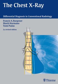
The conventional chest x-ray remains the first line in the diagnostic imaging of thoracic disease. In the new edition of this classic, you will get the essential help you need to make preliminary diagnoses of a vast range of pulmonary, cardiovascular, and other thoracic conditions. The differential diagnostic information is provided in extensive tables, organized by classes of findings, and illustrated by hundreds of brilliant radiographs and, where helpful, schematic diagrams.
Professionals value the systematic structure of the information in the Burgener series, which aids in making diagnoses confidently and cost-effectively.
Das E-Book können Sie in Legimi-Apps oder einer beliebigen App lesen, die das folgende Format unterstützen:
Seitenzahl: 283
Veröffentlichungsjahr: 2005
Ähnliche


Library of Congress Cataloging-in-Publication Data is available from the publisher.
© 2006 Georg Thieme Verlag,
Rüdigerstraße 14, D-70469 Stuttgart, Germany
http://www.thieme.de
Thieme New York, 333 Seventh Avenue,
New York, N.Y. 10001, U.S.A
http://www.thieme.de
Cover design: Martina Berge, Erbach
Typesetting by primustype Hurler GmbH,
D-73274 Notzingen
Printed in Germany by Grammlich, Pliezhausen
ISBN 3-13-107612-7 (GTV, Stuttgart)
ISBN 1-58890-446-6 (TMP, New York) 1 2 3 4 5
Important Note: Medicine is an ever-changing science undergoing continual development. Research and clinical experience are continually expanding our knowledge, in particular our knowledge of proper treatment and drug therapy. Insofar as this book mentions any dosage or application, readers may rest assured that the authors, editors and publishers have made every effort to ensure that such references are in accordance with the state of knowledge at the time of production of the book.
Nevertheless this does not involve, imply, or express any guarantee or responsibility on the part of the publishers in respect of any dosage instructions and forms of application stated in the book. Every user is requested to examine carefully the manufacturers’ leaflets accompanying each drug and to check, if necessary in consultation with a physician or specialist, whether the dosage schedules mentioned therein or the contraindications stated by the manufacturers differ from the statements made in the present book. Such examination is particularly important with drugs that are either rarely used or have been newly released on the market. Every dosage schedule or every form of application used is entirely at the user's own risk and responsibility. The authors and publishers request every user to report to the publishers any discrepancies or inaccuracies noticed.
Some of the product names, patents and registered designs referred to in this book are in fact registered trademarks or proprietary names even though specific reference to this fact is not always made in the text. Therefore, the appearance of a name without designation as proprietary is not to be construed as a representation by the publisher that it is in the public domain.
This book, including all parts thereof, is legally protected by copyright. Any use, exploitation or commercialization outside the narrow limits set by copyright legislation, without the publisher's consent, is illegal and liable to prosecution. This applies in particular to photostat reproduction, copying, mimeographing or duplication of any kind, translating, preparation of microfilms, and electronic data processing and storage.
Preface
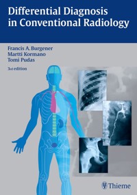
Conventional radiography remains the backbone in thoracic radiology despite the advent of newer and more exciting imaging techniques such as computed tomography, high resolution computed tomography, magnetic resonance imaging and most recently positron emission tomography. In contrast to many of these newer methods, conventional radiography is practiced not only by radiologists but also by a large number clinicians and surgeons. With each examination, one is confronted with radiologic findings that require interpretation in order to arrive at a general diagnostic impression and a reasonable differential diagnosis. To assist the film reader in attaining this goal this book is based upon radiographic finding unlike most other textbooks in radiology that are disease oriented. Since many diseases present radiographically in a varicty of manifestations some overlap in the text is unavoidable. To minimize repetition the differential diagnosis of a radiographic findings is presented in tabular form whenever feasible. Most tables do not only list the various diseases that may present radiologically in a specific pattern, but also describe in succinct form other characteristically associated radiographic findings and pertinent clinical data. Radiographic illustrations and drawings are included to demonstrate visually the radiographic features under discussion.
The transition from film to digital radiography had the greatest impact on conventional radiology since the publication of the last edition. This change however did not affect the way radiologic diagnoses are ascertained. Since the publication of the last edition in 1992 the name of a few disorders has changed (e. g., histiocytosis X to Langerhans cell histiocytosis) and a few disease are newly recognized (e. g. severe acute respiratory distress syndrome or SARS). These facts were taken into account in the new edition. The text was updated, many illustrations replaced and large numbers of new illustrations added.
A changing of the guard has also taken place. Since Dr. Martti Kormano's professional endeavors do no longer include clinical radiology, he felt no longer up to the task to update his original contributions to the text. He was however very fortunate to find in Dr. Tomi Pudas a very talented young radiologist to take over the revision of the chapters originally prepared by him.
I hope this new edition will be as well received as its predecessors in the past that produced several spin-off books and were translated into five foreign languages. The concept of an imaging pattern approach in tabular form rather than a disease oriented text was introduced in 1985 with the original edition and has since been adopted by many authors. I feel complimented by the old cliché, “imitation is the sincerest form of flattery.“
This book is meant for physicians with some experience in chest radiology who wish to strengthen their diagnostic acumen. It is a comprehensive outline of radiographic findings and it should be particularity useful to radiology residents preparing for their specialist examinations, especially since the exposure to conventional radiography during their training continuously decreased in the past in favor of newer imaging modalities. Any physician involved in the interpretation of conventional chest radiograph examinations should find this book helpful in direct proportion to his curiosity.
It is my hope that this new edition will be as well received as the previous ones by medical students, residents, radiologists and physicians involved in the interpretation of conventional chest radiographs were.
Francis A. Burgener, M.D.
Acknowledgements
It is impossible to thank individually all those who helped to prepare the third edition of this textbook. I wish to acknowledge the staff of Thieme, in particular Dr. Clifford Bergman and Mr. Gert A. Krüger.
I am deeply indebted to Dr. Gertrud Gollman, Steinach am Attersee, Austria, who translated the last edition of this text into German and suggested many alterations and corrections, which have been incorporated into this new edition.
My gratitude goes to all the radiologists whose cooperation made available illustrative cases to compliment the original collection or to replace older illustrations. I am indebted to Drs. Steven P. Meyers, Johnny U. V. Monu, and Gwy Suk Seo, all staff members of the University of Rochester Radiology Department, and to the former residents Drs. John M. Fitzgerald and Wael E. A. Saad for providing selected cases.
I wish to express also many thanks to Jeanette Griebel, Iona Mackey, and Marcella Maier for their assistance in preparing the references and to Shirley Cappiello for her general assistance. Last, but not least, I am most grateful to Alyce Norder who left the University and me after 30 years for the richness of the industry. She is the only person capable of deciphering my longhand and, as in the past, did a superb job in typing, editing, and proofreading the manuscript of the new edition of this text. Despite her heavy workload as executive assistant in her new endeavor. Alyce was kind enough to perform this task in her spare time, for which I am greatly appreciative.
Finally I appreciate the support of my wife Therese, who has generously given her precious family time for the preparation of this book.
Francis A. Burgener, M.D.
I would like to express my deepest gratitude to honorary professor Martti Kormano who invited me to carry on his work in this new edition. I continue to admire the massive work that he and Dr. Burgener originally put into the project in the early nineteen-eighties. The hundreds of hours which Dr. Kormano and I have spent together editing this edition have been a great pleasure. It was a fascinating time in my life.
I especially want to thank Drs. Kimmo Mattila and Seppo Koskinen for introducing me to musculoskeletal radiology, and for their extraordinary teaching and support. Many thanks also belong to Drs. Erkki Svedström, Risto Elo, and Peter B. Dean for encouraging me on my way in the field of radiology. The many fascinating discussions I have had with Drs. Seppo Kortelainen and Teemu Paavilainen brought me much delight, on non-radiological topics as much as on professional subjects.
I also express sincere thanks to the staff of the publishers, Thieme, especially to Dr. Clifford Bergman and Mr. Gert Krüger. Finally, much gratitude is due to Mr. Markku Livanaien for his valuable assistance with technical questions, and to Ms. Pirjo Helanko for all her help with general matters. Many other individuals helped in various ways with this project, and though I cannot name them all, I am grateful for their contributions.
Tomi Pudas, M.D.
Contents
1 Cardiac Enlargement
Martti Kormano and Tomi Pudas
2 Mediastinal or Hilar Enlargement
Martti Kormano and Tomi Pudas
3 Pleura and Diaphragm
Martti Kormano and Tomi Pudas
4 Intrathoracic Calcifications
Martti Kormano and Tomi Pudas
5 Alveolar Infiltrates and Atelectasis
Francis A. Burgener
6 Interstitial Lung Disease
Francis A. Burgener
7 Pulmonary Edema and Symmetrical Bilateral Infiltrates
Francis A. Burgener
8 Pulmonary Nodules and Mass Lesions
Francis A. Burgener
9 Pulmonary Cavitary and Cystic Lesions
Francis A. Burgener
10 Hyperlucent Lung
Martti Kormano and Tomi Pudas
References
Index
Abbreviations
ACTH
adrenocorticotropic hormone
AIDS
acquired immune deficiency syndrome
ALL
acute lymphoblastic leukemia
AML
acute myeloblastic leukemia
ANCA
antineutrophil cytoplasmotic autoantibodies
ANT
anterior
AP
anteroposterior
APVR
anomalous pulmonary venous return
ARDS
acute respiratory distress syndrome
ATN
acute tubular necrosis
AV
arteriovenous
AVF
arteriovenous fistula
AVM
arteriovenous malformation
AVN
avascular necrosis
Bx
biopsy
CAD
coronary artery disease
CAM
cystic adenomatoid malformation
CHF
congestive heart failure
CID
cytomegalic inclusion disease
CLL
chronic lymphatic leukemia
CMV
cytomegalovirus
CNS
central nervous system
COPD
chronic obstructive pulmonary disease
CT
computed tomography
DD
differential diagnosis
DIC
dissemination intravascular coagulation
DIP
desquamative interstitial pneumonitis
EG
eosinophilic granuloma
F
female
GE
gastroesophageal
GIP
giant cell interstitial pneumonitis
Hb
hemoglobin
HD
Hodgkin disease
HIV
human immunodeficiency virus
HRCT
high-resolution CT
Hx
history
IM
intramuscular
IVC
inferior vena cava
L
left
LA
left atrium
LCH
Langerhans cell histiocytosis
LE
lupus erythematosus
LIP
lymphoid interstitial pneumonitis
LL
lower lobe
LLL
left lower lobe
LLQ
left lower quadrant
LUL
left upper lobe
LUQ
left upper quadrant
LV
left ventricle
M
male
MAI
Mycobacterium avium intracellulare
MFH
malignant fibrous histiocytoma
ML
middle lobe
MPS
mucopolysaccharidosis
MR
magnetic resonance
MRI
magnetic resonance imaging
NHL
non-Hodgkin lymphoma
NUC
nuclear medicine
PA
posteroanterior
PAPVR
partial anomalous pulmonary venous return
PATH
pathology
PAVM
pulmonary arteriovenous malformation
PCP
Pneumocystis carinii pneumonia
PDA
patent ductus arteriosus
PE
pulmonary embolism
PET
positron emission tomography
PNET
primitive neuroectodermal tumor
PO
per oral
RA
rheumatoid arthritis
RBC
red blood cell
RDS
respiratory distress syndrome
RES
reticuloendothelial system
RLL
right lower lobe
RUL
right upper lobe
RV
right ventricle
SARS
severe acute respiratory distress syndrome
SLE
systemic lupus erythematosus
TAPVR
total anomalous pulmonary venous return
TB
tuberculosis
TNM
tumor-node-metastasis
UIP
usual interstitial pneumonitis
US
ultrasound
1 Cardiac Enlargement
Most of the diseases involving the heart will cause either generalized or localized enlargement of the heart or great vessels. A small heart is usually constitutional (asthenia, senility, wasting) and only rarely reflects a disease (Fig. 1.1). Exceptions to this are adrenal insufficiency, constrictive pericarditis, dehydration, oran asthmatic paroxysm with emphysema, where a relatively small heart may be considered to reflect the effects of the disease process itself.
The size of the heart can be estimated in several ways. A simple approach is the cardiothoracic index: the ratio between the total transverse diameter of the cardiac shadow and the internal diameter of the chest (Fig. 1.1a). After the age of 5 years, this ratio normally varies, between 0.4 and 0.5. In smaller children and neonates, the ratio may be as high as 0.6, and during the second month after birth even 0.65.
The cardiac volume can be estimated with a fair degree of accuracy by measuring the relative volume of the heart according to the following equation:
Fig. 1.1a Small heart in an asthenic woman, age 20. The method of measuring the cardiothoracic index is shown.
Fig. 1.1b, c
Fig. 1.2 Nomogram for the determination of body surface area of adults (adapted from Documenta Geigy, Scientific Tables, Basel). Join patients weight and weight with a line. In the mid column you find the body surface.
Fig. 1.3 Great change in the appearance of heart size is produced by the Valsalva effect. The two exposures were taken a few minutes apart.
Fig. 1.4 Typical manifestations of the enlargement of individual cardiac chambers
Evaluation of a suspected congenital heart disease is a difficult task and often it is impossible to name a specific diagnosis without ultrasound examination, catheterization or angiography. A dynamic MRI examination is also diagnostic. When analyzing plain films, the following morphological features should be evaluated in each case:
1 cardiac enlargement – which chambers are enlarged?
2 the size of the aorta- small, wide, narrow, dilated?
3 the vascular pedicle – narrow or wide?
4 right-sided aortic arch?
5 a notch in the aortic arch, resulting in a number 3 appearance?
6 pulmonary vascularity: are the main trunks of the pulmonary artery wide or unusually narrow; is there pulmonary oligemia or hyperemia?
7 the pulmonary artery segment in the left cardiac shadow: large and convex or small and concave.
8 rib notching (Fig. 1.39).
The following clinical information is important in evaluating suspected congenital heart disease:
1 is the patient cyanotic or not?
2 Is there right- or left-sided dominance in electrocardiography?
3 Are there typical murmurs?
By using the radiographic and clinical information, it is possible to classify congenital heart diseases of childhood into various groups as presented in Table 1.1 or often to predict the correct diagnosis, unless multiple defects exist.
In a neonate, only two congenital heart diseases can be diagnosed with great certainty because of their characteristic appearance in chest roentgenogram, namely, total anomalous pulmonary venous return (snowman heart) (Fig. 1.47) and pulmonary stenosis with atrial septal defect (prominent main pulmonary trunk with oligemic lungs). In other cases further clinical information is very helpful in narrowing down the differential diagnostic possibilities. Nowadays ultrasound and Doppler ultrasound examinations have largely replaced plain radiographic analysis of heart.
Based on the time of appearance of cardiac failure and cyanosis, the patients can be divided into the following groups:
1 Cardiac failure manifests early (at birth or during the first week) but there is no cyanosis: hypoplasia of the left heart; atresia of the aortic (and possibly mitral) valve; coarctation of aorta.
2 Cardiac failure develops later: ventricular septal defect; patent ductus arteriosus; truncus arteriosus; total anomalous pulmonary venous return (TAPVR).
3 Cyanosis develops at birth or within a week: transposition of great vessels; hypoplastic right heart; Ebstein's anomaly; tricuspid atresia; obstructive type of TAPVR.
4 Cyanosis develops later during childhood: tetralogy of Fallot; pulmonary stenosis with atrial septal defect.
Table 1.3 gives the approximate relative frequency of the congenital heart diseases. In Table 1.5, the diseases are grouped according to their dominant, most common or earliest findings. The differential diagnostic groupings in Table 1.2 may be helpful in narrowing the differential diagnosis in congenital heart diseases in children.
In the adult patient with suspected congenital heart disease, the following features are of particular importance: the position of the diaphragm (high or low); degree of inspiration; calcifications; Kerley B-lines; the appearance of the branches of the pulmonary artery; possible arteriovenous malformations in the lung parenchym anomalous veins across the lung field.
Evaluation of Blood Flow and Blood Pressure in Lungs
When blood circulation through the lungs is increased, both arteries and veins are full of blood. This occurs in left-to-right shunting (Table 1.1) and to a milder degree in hyperkinetic conditions (hypervolemia, anemia, polycytemia, pregnancy, hyperthyroidism). A clear oligemia may be a result of a right-to-left shunt or narrow or obliterated pulmonary arterial channels (narrow pulmonary artery, thromboembolic disease or emphysema). Pulmonary blood flow can increase two to three times before the arterial pressure increases. When the precapillary pressure increases, the peripheral branches of the pulmonary arteries appear narrow, but the central trunks are wide.
An increase in the pulmonary venous pressure causes characteristic changes in the distribution of blood in the lungs. On upright films of an adult untreated patient, such changes relatively accurately reflect the pulmonary venous and left atrial pressure as diagrammatically presented in Fig. 1.5. These changes, together with the less sensitive changes in the size and configuration of the heart, are the cornerstone of the radiographic diagnosis of congestive heart failure. The findings in the most common conditions involving left ventricular strain are presented in Table 1.4. Although the radiologic evaluation of the pulmonary venous pressure is relatively accurate, the evaluation of venous pressure of the right side is difficult on roentgenograms and is better assessed from the filling of the veins of the patient's neck. Occasionally a prominent azygos vein may be seen (Fig. 1.6a). In conditions such as acute myocardial infarction, even the central venous pressure readings do not reliably reflect the patient's hemodynamic condition.
Pericardial effusion mimics generalized cardiomegaly. Although it sometimes may cause a characteristic (bottle-like) configuration of the heart shadow, most of the cases are difficult to diagnose on plain films, and as much as 200 ml of pericardial fluid usually goes undetected in the primary reading. Ultrasound examination CT or MRI are far more sensitive than plain films in detecting pericardial effusion.
Fig. 1.5 Manifestations of increased pulmonary venous (PV) and arterial (PA) pressure.
Table 1.1 Pulmonary Vasculature in Congenital Heart Disease
Pulmonary vasculature
Cyanosis
Diseases
Increased
Cyanotic
Mixed lesions with major malformations:
Complete transposition
Truncus arteriosus (Type I)
Single ventricle
Common atrioventricular canal
Total anomalous pulmonary venous return
Acyanotic
Left to right shunts:
Atrial septal defect
Ventricular septal defect
Patent ductus arteriosus
Aorticopulmonary defect
Partial anomalous pulmonary venous return
Normal
Acyanotic
Coarctation of aorta
Aortic stenosis
Subaortic stenosis
Pulmonary arterial stenosis
Congenital mitral stenosis
Endocardial fibroelastosis
Decreased
Cyanotic
Tetralogy of Fallot
Trilogy of Fallot
Truncus (pseudotruncus) arteriosus
Tricuspid atresia
Ebstein's anomaly
Pulmonary stenosis with ASD or transposition
Table 1.2 Some General Differential Diagnostic Features of Congenital Heart Disease
Unusually small ascending aorta and aortic arch
Atrial septal defect Ventricular septal defect Uncorrected transposition of great vessels
Enlarged ascending aorta and aortic arch
Patent ductus arteriosus Valvular congenital aortic stenosis
Coarctation of aorta Truncus arteriosus
Concave pulmonary artery segmented in posteroanterior view
Tetralogy of Fallot
Tricuspid atresia
Ebstein's anomaly
Pseudotruncus arteriosus
Uncorrected transposition of great vessels
Prominent pulmonary artery segment (a moderate prominence is normal under age 20 or in pregnancy)
Patent ductus arteriosus
Atrial septal defect
Ventricular septal defect
Eisenmenger's complex
Valvular pulmonary stenosis (poststenotic dilatation)
Cor pulmonale
Enlarged left atrium in a child
Patent ductus arteriosus
Ventricular septal defect
Congenital mitral stenosis
Table 1.3 Relative Frequency of Various Entities Among Congenital Heart Diseases
(Significant congenital heart diseases occur in approximately 1% of babies)
Common
Ventricular septal defect
20–25%
Patent ductus arteriosus
12–15%
Tetralogy of Fallot
11–15%
Pulmonary stenosis without ventricular septal defect
10–15%
Atrial septal defect
7–14%
Transposition of great vessels
5–9%
Coarctation of aorta
5–9%
Congenital aortic stenosis
3–6%
Rare
Tricuspid atresia
1.2–3%
Truncus arteriosus
1–3%
Single ventricle
2–3%
Corrected transposition
1.2–3%
Common atrioventricular canal
2%
Total anomalous venous return
2%
Aortic atresia
2%
Pulmonary atresia
1–;1.7%
Endocardial fibroelastosis
1%
Ebstein's anomaly
1%
The rest are very rare (less than 1%).
Table 1.4 Radiologic Findings in Common Conditions which Cause Left Ventricular Strain
Pulmonary Veins
Size of the Left Ventricle
Normal
Enlarged
Normal
No heart disease
Aortic valve stenosis
Arterial hypertension
Athlete's heart
Aortic regurgitation
Myocardial damage
Venous congestion
Acute myocardial infarction
Mitral stenosis
Hypervolemia
Constrictive pericarditis
Congestive heart failure
Mitral regurgitation
Both arteries and veins distended
Atrial septal defect
Patent ductus arteriosus
Ventricular septal defect
Arteriovenous malformation
Possible enlargement of the left ventricle depends on the magnitude of shunting.
Table 1.5 Predominantly Left Ventricular or Generalized Cardiac Enlargement
Disease
Radiographic Findings
Comments
Athlete's heart (no disease)
Generalized cardiomegaly with left ventricular prominence, no pulmonary vascular changes.
Associated with bradycardia (large stroke volume); secondary to excessive training.
Arteriosclerotic heart disease (myocardial ischemia)
Chest roentgenogram is usually unrevealing. Variable degrees of generalized or left ventricular enlargement may occur.
Calcification of the coronary arteries indicates coronary atherosclerosis.
Acute myocardial infarction (Figs. 1.6–1.8)
Chest roentgenogram is often unrevealing. Some cardiac dilatation, with variable degrees of pulmonary venous congestion, occurs frequently. Acute pulmonary edema may ensue.
Myocardial aneurysm may develop early in the infarcted muscle or later in the scar. It is seen as a bulge or an unusual prominence in the left ventricular border, which occasionally calcifies.
Postmyocardial infarction syndrome (Dressler's syndrome)
Rapid increase in the heart size due to pericardial effusion, always accompanied by either pleural effusion and/or pulmonary infiltrates in the left base or bilaterally.
Can occur a few days or up to two months following an acute infarction.
Left-sided or bilateral pleural effusion is the most common finding (80%) and may occur alone. Pericardial effusion is present in 70% and pulmonary infiltrates in 60% of cases. Response to steroid therapy is striking.
Congestive myocardial failure
Left-sided failure (Fig. 1.9)
Cardiac enlargement, related to the severity of failure and the precongestive heart size. The increase of pulmonary venous pressure can be evaluated as presented in Fig. 1.5). Pleural effusions (bilateral or right-sided).
Pulmonary signs of congestive failure without significant cardiac enlargement. Pericardial calcification is seen in 50%. Often the cardiac configuration resembles that of mitral stenosis.
Unilateral left-sided effusion is likely due to causes other than congestive failure.
Right-sided failure (Fig. 1.10)
Dilatation of the right ventricle and atrium. Widening of the superior vena cava and azygos vein.
Liver enlargement.
If right heart failure develops following left heart failure, pulmonary venous congestion from left-sided failure diminishes.
Chronic arterial hypertension (hypertensive heart)
Left ventricular hypertrophy may produce no radiographic changes or some rounding of the left margin. Left ventricular dilatation causes typical signs of left ventricular enlargement. The aortic knob enlarges concomitantly. Congestive failure may supervene.
Hypertension is most often essential but may be associated with renal or renovascular disease, coarctation of the aorta, adrenal diseases with adrenal hyperfunction, collagen diseases (lupus erythematosus, polyarteritis nodosa), or hyperthyroidism.
Aortic valve insufficiency (Aortic regurgitation) (Fig. 1.11)
Dilated left ventricle, concave left cardiac border, some dilatation of the ascending aorta and sometimes calcification of the aortic annulus or even the valves. Congestive failure may supervene in advanced cases. Radiographic findings may be subtle although findings in ultrasonography may be obvious.
Insufficiency of the aortic valve can be secondary to:
1 primary damage of the valve (e.g., rheumatoid endocarditis, bacterial endocarditis, congenital valvular deformity, lues, ankylosing spondylitis and Reiter's disease, mucopolysaccharidosis, spontaneous or traumatic rupture of the valve, or degenerative phenomena);
2 diseases of the aortic wall or annulus fibrosus (e.g., cystic medial necrosis, Marfan's syndrome, dissecting aneurysm, lues, arterial hypertension); or
3 congenital malformation of the aortic root (e.g., aneurysm of the sinus of Valsalva, congenital coronary fistula, or high ventricular septal defect with prolapse of the noncoronary cusp).
Congenital aortic insufficiency is usually due to a bicuspid valve.
Acute aortic insufficiency occurs in bacterial endocarditis, rupture of the valve or dissecting aneurysm.
Combined aortic valve insufficiency and stenosis (Fig. 1.12)
Dilatation of the left ventricle and ascending aorta is more prominent and left atrium may enlarge. May mimic pure aortic stenosis.
Rheumatoid heart disease usually causes a combination of aortic valve stenosis and insufficiency.
Subvalvular aortic stenosis (idiopathic hypertrophic subaortic stenosis)
Enlarged left ventricle, often enlarged left atrium. Poststenotic aortic dilatation is absent.
Narrowing of the left ventricular outflow tract by muscular hypertrophy. Essentially ultrasonographic diagnosis.
Supravalvular aortic stenosis
The heart may be enlarged or normal, the aorta is normal or small.
Congenital fibrous ring or thickening above the sinuses of Valsalva. Frequently associated with idiopathic hypercalcemia of infancy.
Aortic stenosis
See Table 1.9
High-output heart
Cardiac enlargement, dilatation of the main pulmonary artery, prominent pulmonary vasculature (both arteries and veins).
Associated conditions:
Pregnancy or athletic (physiological).
Severe anemia including sickle-cell anemia, leukemia, or primary polycythemia.
Beriberi (vitamin B deficiency). Hypervolemia (fluid overload). Extrapulmonary arteriovenous fistula.
Mild forms occur in extreme obesity, thyrotoxicosis, and pyrexia.
Advanced Paget's disease is a rare cause of high-output heart.
Myocardiopathy (Figs. 1.13 – 1.14)
Diffuse enlargement of the heart, predominantly of the left ventricle. The aorta appears small as compared with the size of the heart. Pulmonary venous congestion may develop. Moderate left atrial enlargement and fullness of the main pulmonary artery occurs. Superimposed pericardial effusion may further enlarge the cardiac shadow.
The many causes of myocardiopathy may be classified into five groups:
1 Idiopathic, unassociated with extracardiac disease: non-specific myocardosis; endocardial fibroelastosis (neonate or child); postpartum cardiopathy.
2 Infectious: Coxsackie B and other viral infections; Chagas' disease (South American trypanosomiasis); rheumatic, septic, diphtheric, toxoplasmic.
3 Infiltrative: amyloidosis (especially primary), generalized glycogen storage disease (Pompe's disease, infant); leukemia.
4 Endocrine: hypothyroidism; acromegaly; Cushing's disease; thyrotoxicosis.
5 Miscellaneous causes: ischemic; uremia; collagen diseases; nutritional deficiency (beriberi, alcoholism, potassium or magnesium depletion); toxicity (drugs, chemicals, cobalt); sarcoidosis; neuromuscular dystrophy.
A combination of cardiac enlargement and normal pulmonary vasculature is usually due to one of the following four entities:
1 myocardiopathy; 2 aortic stenosis; 3 aortic coarctation, or 4 athlete's heart.
Mitral insufficiency Coarctation of aorta Patent ductus arteriosus Ventricular septal defect
Left ventricular enlargement is a feature in these conditions as well as in a number of rare congenital heart diseases.
Since other characteristics are more useful for differential diagnosis they will be discussed elsewhere.
Pericardial defect (Fig. 1.15)
1 Displacement of the heart to left without displacement of trachea.
2 Unusual configuration of the left heart border (bulging aortic, pulmonary arterial and left ventricular segments).
3 Bulging through a partial defect may mimic left ventricular aneurysm, but shows no paradoxal pulsations.
Either total (in 3/4) or partial absence of pericardium, usually on the left side, which usually is symptomless and discovered incidentally. Displacement of the heart may simulate left ventricular enlargement. A deep notch between the aorta and pulmonary artery is characteristic.
Acute glomerulonephritis
Generalized cardiomegaly
Pleural effusions
Pulmonary interstitial or alveolar edema
Cardiac dilatation occurs in over 50% of acute in children cases in children. The mechanism is unknown.
The radiographic changes subside together with healing of the underlying disease.
Pericardial effusion (Figs. 1.16–1.17)
Enlarged cardiac shadow mimics generalized cardiomegaly.
Large effusions tend to obliterate normal cardiac markings.
There is less posterior displacement of the barium-filled esophagus in the lateral view than could be expected from the apparent heart size.
Rapid changes occur in the cardiac silhouette in consecutive films, in the absence of pulmonary vascular enlargement.
Postural alterations change cardiac contours. The pulmonary vasculature is decreased and the superior vena cava may be prominent if tamponade of the right atrial inflow occurs.
Common causes of pericardial effusion are: Pericarditis (especially Coxsackie virus, but also in other infections)
Congestive heart failure
Collagen diseases (LED)
Cardiac surgery or trauma
Renal failure
Postmyocardial infarction syndrome
Tumor invasion (from lung or mediastinal lymphoma) or metastases (from lung, breast, or melanoma)
Radiation therapy
Still's disease
Transposition of great vessels (Fig. 1.18)
“Egg-on-side” cardiomegaly that appears during the first week after birth together with congestive heart failure.
Concave pulmonary artery segment, although vascularity in the lungs is increased unless transposition is associated with pulmonary stenosis, the latter occurs in 15%.
Narrow vascular pedicle in frontal projection (aorta and pulmonary artery are superimposed), seen in 50%.
A rare anomaly, but the most frequent form of congenital heart disease associated with cyanosis present since birth. Due to lack of spiralization of the spiral septum, the aorta remains anterior and originates from the right ventricle. The pulmonary artery is behind the aorta and originates from the left ventricle. Pulmonary and systemic circulation are connected through a defect in the atrial and/or ventricular septum or through a patent ductus arteriosus.
Tricuspid atresia
See Table 1.7
Anomalous left coronary artery
Radiographic changes are identical to other myocardiopathies such as endocardial fibroelastosis (see under myocardiopathy in this Table, above).
Left coronary artery originating from the pulmonary artery is the most common significant anomaly of the coronary circulation.
Fig. 1.6a, b Acute myocardial infarction. a dilatation of the heart, pulmonary edema, and congestion of the azygos veins (arrowhead). b Lateral view reveals pleural effusion in the pleural fissures.
Fig. 1.7a, b Left ventricular aneurysm after myocardial infarction. A bulge in the cardiac contour is seen in both projections (arrows). Pulmonary vasculature indicates mild venous congestion.
Fig. 1.8a, b A large left ventricular aneurysm bulges posteriorly, a complication of myocardial infarction. A double density is seen in the AP projection.
Fig. 1.9a, b Chronic left heart failure. There is left ventricular enlargement, pulmonary venous congestion and bilateral pleural Effusion, more on the right side.
Fig. 1.10a, b Congestive left heart failure developing into both left- and right-sided failure. a Left ventricular enlargement and pulmonary venous congestion. b Enlargement of the heart, including the right side, widening of the mediastinum (superior vena cava, and severe pulmonary venous congestion). The azygos vein is also congested (arrow).
Fig. 1.11 Arteriosclerotic aortic valve insufficiency with mild left ventricular enlargement, no congestive heart failure, and a normal-looking aorta.
Fig. 1.12a, b Combined aortic valve stenosis and insufficiency of rheumatic origin. Extensive enlargement of the left ventricle and dilated ascending aorta. Aortic valve calcification is demonstrated (arrows)
Fig. 1.13 Uremic myocardiopathy. Left ventricular enlargement without congestive failure. The aorta is relatively small.
Fig. 1.14 Diabetic cardiomyopathy in a child aged 10. The heart is large, and left ventricles is very prominent. Pulmonary vasculature is normal.
Fig. 1.15 Pericardial defect, age 5. The heart (not mediastinum) is displaced to the left. The aortic, pulmonary, and left ventricular segments are sharply bulging, a characteristic pattern.
Fig. 1.16a, b Pericardial effusion. The cardiac shadow is large, but the esophagus in the lateral view is not proportionally displaced posteriorly.
Fig. 1.17a, b Still's disease. Fig. 1.17a was taken one month before than Fig. 1.17b. Massive increase in heart size is due to pericardiac effusion which also caused emerging heart tamponade and pulmonary constriction.
Fig. 1.18 Transposition of great vessels. “Egg-on-side” cardiomegaly and a concave pulmonary segment associated with increased vascularity of lungs. Narrow vascular pedicle.
Table 1.6 Predominantly Left Atrial Enlargement
Disease
Radiographic Findings
Comments
Mitral stenosis (acquired or congenital) (Figs. 1.19 and 1.20)
1. Left atrial enlargement
2. Possible calcification of the mitral valve, not to be confused with the heavy calcification of the mitral anulus
3. Pulmonary venous congestion
4. Prominent main pulmonary artery segment, enlarged hilar vessels
5. A small aortic knob
6. Right ventricular enlargement, normal-sized left ventricle
7. Dilatation of the central pulmonary arteries and narrowing of peripheral arteries (pulmonary arterial hypertension)
8. Pulmonary parenchymal changes due to hemosiderosis (granular opacities) (Fig. 5.47, p. 117), multiple up to 8 mm ossifications, or pulmonary fibrosis (pulmonary infarctions)
Obstruction of flow from the left atrium into the left ventricle during diastole, resulting in increased pressure and enlargement of the left atrium. The increased pressure is transmitted to the pulmonary veins and eventually to the pulmonary arteries and the right heart. The usual cause is a rheumatic valvular lesion. Congenital mitral stenosis can be identical to the rheumatic one (short chordae ten-dineae, fibrotic valves and fused commissures). A rare congenital form of mitral stenosis is the parachute deformity (all chordae tendineae originate from a single papillary muscle). The latter is associated with other anomalies. In the early phase, congenital mitral stenosis and a left-to-right shunt may appear similar but in mitral stenosis the vascular shadows are hazier due to venous and lymphatic congestion. Enlarged confluence of right pulmonary veins may mimic left atrial enlargement or tumor.
Mitral insufficiency (mitral regurgitation) (Figs. 1.21–1.24)
1 Enlarged, sometimes enormous left atrium
2 Enlarged left ventricle
3 Small or normal aortic knob
4 Normal pulmonary vasculature, sometimes venous congestion and prominent pulmonary artery are present.
Most commonly caused by rheumatic valvulitis, but may also be caused by functional dilatation of the mitral ring secondary to other cardiac diseases (congestive heart failure, acute rheumatic fever, aortic valve disease, coarctation of aorta), or from papillary muscle dysfunction


