18,49 €
Mehr erfahren.
- Herausgeber: The Crowood Press
- Kategorie: Lebensstil
- Sprache: Englisch
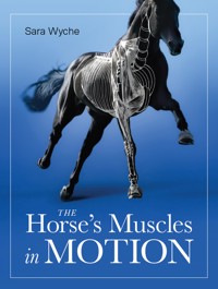
New in paperback for 2022, this book aims to show how – and why- the horses muscles work and explains how this knowledge can be put to good use in all aspects of horse care and riding. With careful anatomical drawings supported by explanatory text, Sara Wyche 'dissects' the horse's musculoskeletal system and describes how the various muscles work together with bones, joints, ligaments and nerves to produce movement. Throughout, there are valuable insights into how man's use of the horse can adversely affect this movement, how good riding practice can help to avoid problems, and why the horse is sometimes unable to meet the strenuous demands that are so often placed upon him. Riders, trainers, saddle-fitters – all who have an interest in the way the horse moves – will find this book to be a fascinating exploration of the horse's musculoskeletal system. More than this, it is an important guide to understanding exactly what it is they ask of the horse and, therefore, an aid to improving the horse's performance.
Das E-Book können Sie in Legimi-Apps oder einer beliebigen App lesen, die das folgende Format unterstützen:
Seitenzahl: 122
Veröffentlichungsjahr: 2022
Ähnliche


First published in 2002by The Crowood Press LtdRamsbury, MarlboroughWiltshire SN8 2HR
www.crowood.com
Paperback edition 2022
This e-book first published in 2022
© Sara Wyche 2002
All rights reserved. This e-book is copyright material and must not be copied, reproduced, transferred, distributed, leased, licensed or publicly performed or used in any way except as specifically permitted in writing by the publishers, as allowed under the terms and conditions under which it was purchased or as strictly permitted by applicable copyright law. Any unauthorised distribution or use of this text may be a direct infringement of the author’s and publisher’s rights, and those responsible may be liable in law accordingly.
British Library Cataloguing-in-Publication Data
A catalogue record for this book is available from the British Library.
ISBN 978 0 7198 4087 6
Dedication
To Jim.
Acknowledgements
It goes without saying that any book on the subject of anatomy owes its inspiration to the living model: in this case, the horse. However the author is particularly indebted to the inspiration provided by certain special horses and, inseparably, their dedicated owners. Horses such as Mia, Estoril, Lucy and Tom, My Liege and Girlie, ponies such as Fly, and the occasional donkey, who, together with their owners – N. Kerslake, M. Brownell, R. Tyrrell. J. Beasley, M. Phillips – are among the many who overturned poor prognoses by questioning the opinions of mainstream veterinary medicine.
The work in this book complements the many discussions of joint mechanics and muscle function that were begun over the stable door or over the telephone. It is gratefully acknowledged that such a project would not have been possible without this dialogue.
Thanks are also due to Beth Shaw, with whom the author is developing the Sumchi tool (www.sumchi.com), an important new treatment modality for soft tissue injuries.
A special word of thanks is due to Mrs Jean Hammond, principal of Tollers Design Centre, who condensed the history of painting movement into a tea break and who, in the world’s shortest art lesson, managed to sort out the white from the black.
Line-drawings by Sara Wyche
Cover design by Sergey Tsvetkov
Contents
Author’s Note
Introduction
1 Introducing Anatomy
2 The Frame: Bones, Joints and Ligaments
3 The Driving Forces: Muscles
4 The Wiring: Nerves
Postscript
Index
Author’s Note
The author is not a trained artist and hopes that any faults in technique will be forgiven. Occasionally it has been necessary to elongate structures and widen apertures in order to reveal elements that would normally be hidden. Any small degree of ‘artistic licence’ will be noted on the appropriate page.
It should be said that under no circumstances does the book claim to make any new discoveries in the science of biomechanics. The anatomy of the horse (for example, skeletal conformation, ligament placement, origin and insertion of muscles) is well documented: this book simply presents anatomical fact from a new perspective. Therefore, although the full orchestration of movement has yet to be analysed, the standard anatomical tunes are freely available to all. In this case, they have been taken from the following textbooks:
Stashak, Ted S., Adams’ Lameness in Horses, 4th edition, Lea & Feibiger, Philadelphia, 1976.
Nickel, Schummer, Seiferle, Lehrbuch der Anatomie der Haustiere, 4th edition, Verlag Paul Parey, Berlin and Hamburg, 1977.
If the reader should find discrepancies (for example, sites of muscle insertion) between this and other anatomy books, it is hoped that these inconsistencies will be negligible and not affect the picture as a whole.
Introduction
‘Have you ever noticed the fact that children always ask “why”, but adults invariably tell them “how”?’ This observation was made by a dressage rider who had problems finding a satisfactory saddle. Consequently he took several saddles apart to discover how they were made, then, by asking the question why, set out to challenge the use of traditional materials and methods of construction. The result was a uniquely flexible saddle-tree that eliminated many of the pitfalls associated with conventional design.
When riders have problems with their horse’s movements, they cannot, of course, physically dismember the horse. However, as long as the horse is sound, perhaps only the most inquisitive rider wants to know how. It’s not until the horse goes lame that we all want to know why.
The examination of horses for lameness has always occupied a large part of the equine veterinary practice. It has done in the past and will probably do so well into the future. With the sophisticated techniques available in specialist clinics, it is now possible to diagnose diseased tissue, often with pinpoint accuracy. But whereas the discovery of acute trauma (for example, bone fracture or tendon rupture) immediately explains how the horse went lame, the diagnosis of chronic wear and tear (for example, degenerative spinal or joint disease) often fails to explain why.
There are many factors that contribute to degenerative processes in the horse’s musculoskeletal system, but there can be no doubt that some are linked to the way in which the horse is ridden. Although nobody rides with the intention of constricting the horse’s joints, compressing the ligaments, or compromising the muscles, the fact remains that chronic unsoundness can develop only when the horse’s body is, in some way, repeatedly misdirected. Since it’s not likely that the horse inflicts such unsoundness on himself (at least, not without good reason), we have to consider the possibility that degeneration is unwittingly caused by a rider’s lack of awareness of simple relationships in the horse’s anatomy.
The musculoskeletal system can be divided into two types of tissue: rigid and elastic. The rider directs the horse’s movements through the elastic tissues (muscles, tendons and ligaments), which in turn direct the rigid tissues (bones) to move the body from place to place. If the system is to remain intact, the energy released into it must be distributed correctly. The source of this energy is in the horse’s muscles.
Ideally, the rigid and elastic tissues should be equal partners in the business of producing movement. They combine their efforts in a sort of musculoskeletal ‘co-operative’, which protects everyone’s investment of energy and pays dividends in the conservation of fuel. Nevertheless, muscles have the ultimate power. If they abuse it, or abdicate their responsibility, then unhealthy cracks develop in the musculoskeletal organization, and these threaten the whole fabric of movement.
Of course horses run on the forehand, of course they hollow their backs, of course they trot along with their noses stuck out in front of them and their hocks insufficiently engaged under their bodies. It may be considered unaesthetic, but these movements are well within the horse’s natural repertoire. They are not inherently harmful. Movements become damaging to the horse only when they are either unfairly dominated by the weight of a rider, interrupted by the rider’s aids, or forced by the rider against the natural conformation of the joints. If then, as riders, we choose to disregard, and even deliberately disconnect, certain coupling mechanisms essential to the survival of the horse’s musculoskeletal system, we do so to its detriment.
To describe the movement of muscles using the printed page obviously has its limitations, especially when computer programs now do this with greater realism. Nevertheless, pen and paper have the advantage of fixing an image in the mind’s eye. In this book, the purpose of line-drawings is to freeze movements at certain moments in time in order to high-light the effect of muscles on vulnerable parts of the horse’s skeleton.
However, regardless of the means, it is only the hallmark of good teaching if eventually the teacher becomes redundant. It is hoped that by the time the reader reaches the final pages of this book, he will be able to go out to the stable, look at the living horse, and understand the parameters of movement – without recourse to a textbook or indeed a laptop.
If, in addition, the book furnishes students and therapists with an accessible compendium of anatomy, that will be a bonus. At the very least, this book looks at the role of the horse’s muscles in movement and, for the adult – and the child – in every horseperson, attempts to answer how they work, why they work, and why sometimes (despite our best efforts) they don’t.
1Introducing Anatomy
The assessment of conformation is an essential part of breeding and training horses. The aim is to evaluate an individual’s potential by taking stock of his strengths and weaknesses. In the first instance, judgement is based on the appearance of the horse’s exterior – for example, the visible circumference of bone and the angles of the joints. This may seem akin to judging a book by its cover, but it is a practical application of anatomy and it undoubtedly results in winners.
Bones and joints are justifiably regarded as the foundation of a horse’s performance, not least because their size and shape determines an animal’s capacity to carry muscle. Nevertheless, this preoccupation with the horse’s skeleton also permeates the conventional way of looking at lameness. Many lameness investigations confine themselves to the evidence of radiographic and ultrasonic images: in other words they give diagnostic priority to diseases that affect the immediate vicinity of ligaments, bones and joints.
Whether it is the result of the high profile of physiotherapists in sport, the growing popularity of alternative medicine, or the widespread use of scintigraphy in equine clinics, horse owners are now moving away from a traditionally regimented interpretation of lameness. There is a collective and spontaneous will in the horse world (which once could envisage only joint disease and tendon breakdown as a cause of lameness) to adopt a more holistic approach to the horse’s locomotor system. Suddenly, at the top of everyone’s agenda are muscles.
For example, if ever it could be said that riders, like the average car user, had very little notion of ‘what goes on under the bonnet’, that cannot be said today. Education in the relevance of anatomy for a new generation of saddle-fitters, and the continued popularity of dressage riding, are two factors responsible for the upsurge of interest in the role of the horse’s muscles.
However, there are more than one hundred and fifty muscles in the horse’s body, and it is not surprising that different professional groups emphasize those muscles that are most relevant to their areas of expertise. Unfortunately, this process has led to some muscles being given an importance that is not entirely deserved.
If horse owners should ever feel intimidated by anatomical one-upmanship (on the part of saddlers, trainers or even vets), they may rest assured that there are many more muscles that contribute to the horse’s movement or his saddle fit than the much quoted ‘trapezius’ or ‘longissimus’.
THE LANGUAGE OF THE BODY
All horse people are familiar with the term ‘body language’. Whether or not we subscribe to current trends in natural horsemanship, body language is an essential part of communication in any branch of equestrianism. It comprises the sum total of conscious and subconscious gestures that enable humans and horses to participate in a ‘conversation’. However, if it’s the body that speaks the language then it’s the muscles that supply the words.
Students of equine or veterinary science will probably agree that learning the anatomy of muscles is like jumping a cross-country course. The track is lengthy, often uphill, with a variety of imposing obstacles. For example, some muscles have names the size of tree-trunks and are therefore not for the faint-hearted. Some muscles form tricky combinations and cannot be taken individually. And some muscles are fixed to more than one point: understanding how these work depends on which route you take.
However, if there are hurdles in learning anatomy they are not very different from those encountered in learning a language. For example, in language we have to know how to arrange words correctly before we can grasp the meaning of a sentence. Similarly, we have to learn where to place muscles correctly in relation to the skeleton before we can understand what they mean in movement.
We could even think of the muscles as letters of an alphabet. Individually, muscles have no meaning. It is only when they work as a group that they produce a single movement – the equivalent of a word. When one muscle group links up with another group, they produce a pattern of movements – the equivalent of a phrase or sentence. Just as words can be combined to make sense, or even nonsense, so there are combinations of movements that make sense, and combinations (usually enforced) that make no sense at all.
If movement is language, then anatomy holds the key to its grammar. Knowing the position of muscles in relation to the skeleton is part of understanding that grammar. For any additional explanations, we can always consult a dictionary. Unfortunately, the dictionary of anatomy is written in Greek and Latin, which is a pity since clues to the skeletal whereabouts of muscles are often concealed in their names.
On the face of it, the lengthy and rather venerable names of muscles make for a fairly indigestible diet of words. It is true that ‘biceps’ and ‘triceps’ have found their way into the vernacular, ‘pecs’ are body-building jargon, and ‘cruciates’ are what footballers regularly sacrifice on Saturday afternoons. But ‘common digital extensor’ is barely common parlance, brachiocephalicus’ sounds like a dinosaur, and ‘teres major’ could be the key signature of a symphony. When it comes to ‘vastus intermedius’, ‘latissimus dorsi’, or ‘rectus capitus’, one might be forgiven for thinking these were heroes in a Roman epic!
The reason for the apparent inaccessibility of anatomy labels is that their origins lie in the history of medicine. In centuries gone by, without the provision of an internationally available computer database, each newly discovered muscle, tendon, nerve and ligament had to be described in a way that was intelligible the world over. Furthermore the names had to include connotations of shape, function or situation (and preferably all three), yet, at the same time, not be liable to misinterpretation. This meant that a common terminology was necessary, and this was automatically derived from the scholarly, classical languages of Greek and Latin.
Science still coins words from these languages, but because the words describe new technology, they melt unobtrusively into modern English.
A PLACE FOR EVERYTHING ...
Leornardo da Vinci did it. So did George Stubbs. In order to portray humans and horses in the most life-like way, they undertook to study anatomy. This did not mean going down to the local library; it meant collecting corpses, and taking them apart. Working from cadavers, these artists taught themselves the principles of anatomy by peeling back the layers of flesh and systematically drawing what they saw.


