45,99 €
Mehr erfahren.
- Herausgeber: John Wiley & Sons
- Kategorie: Fachliteratur
- Serie: At a Glance
- Sprache: Englisch
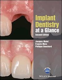
The second edition of Implant Dentistry at a Glance, in the highly popular at a Glance series, provides an accessible, thoroughly revised and updated comprehensive introduction that covers all the essential sub-topics that comprise implant dentistry.
- Features an easy-to-use double-page spread, with text and corresponding images
- Expanded and updated throughout, with 13 new chapters and coverage of many advances
- Includes access to a companion website with self-assessment questions and illustrative case studies
Sie lesen das E-Book in den Legimi-Apps auf:
Seitenzahl: 457
Veröffentlichungsjahr: 2018
Ähnliche


Table of Contents
Cover
Title page
Copyright
Dedication
Preface
Acknowledgments
About the companion website
Chapter 1: Quality of life associated withimplant‐supported prostheses: An introduction to implant dentistry
Oral health quality of life
Dental implants and oral health
Should missing teeth be replaced?
Does implant dentistry improve the patient’s quality of life?
Is implant dentistry a cost‐effective option?
Chapter 2: The basics: Osseointegration
Implant neck
Implant body
Implant loading
Chapter 3: The basics: The peri‐implant mucosa
Soft tissue interface dimensions
Soft tissue seal
Soft tissue components
Soft tissue healing
Chapter 4: The basics: Surgical anatomy of the mandible
Anterior area
Posterior area
Chapter 5: The basics: Surgical anatomy of the maxilla
Anterior area
Posterior area
Chapter 6: The basics: Bone shape and quality
Bone shape
Bone quality
Clinical examination
Chapter 7: Implant macrostructure: Shapes and dimensions
Implant length
Implant diameter
Implant shape
Thread design of implants
Cylindrical versus tapered dental implants
Chapter 8: Implant macrostructure: Short implants
Definition
Survival and success rates
Limitations
Clinical recommendations
Chapter 9: Implant macrostructure: Special implants
Orthodontic mini‐implants
Mini dental implants
Chapter 10: Implant macrostructure: Implant/abutment connection
Abutment connection
External connection
Internal connection
Load transmission
Abutment screw loosening
Interface location
Bacterial colonisation
Platform switching
Chapter 11: Implant microstructure: Implant surfaces
Surface topography
Surface configurations of some commercially available implants
Surface chemistry
Chapter 12: Choice of implant system: General considerations
Scientific background
Dental implant engineering
Cost‐effectiveness
Commercial efficiency
The dental implant ‘network’
Chapter 13: Choice of implant system: Clinical considerations
Aesthetics
Timing of implant placement
Bone dimension
Bone density
Periodontally compromised patients
Chapter 14: Success, failure, complications and survival
Dental implants: The best treatment option?
What is a success?
Failure
Survival
Complication
Chapter 15: The implant team
Who is the head of the team?
Chapter 16: Patient evaluation: Medical evaluation form and laboratory tests
Chapter c17: Patient evaluation: Surgery and the patient at risk
Absolute contraindications
Relative contraindications
Patients at risk during the surgical procedure
Chapter 18: Patient evaluation: The patient at risk for dental implant failure
Age
Smoking
History of treated periodontitis
Number of teeth
Ectodermal dysplasia
AIDS/HIV
Diabetes/hyperglycaemia
Bone diseases
Radiotherapy
Miscellaneous
Chapter 19: Patient evaluation: Local risk factors
Implant stability
Bone density
Interproximal space
Infected sites
Soft tissue thickness
Keratinised soft tissue
Surgical procedure
Chapter 20: Patient evaluation: Dental history
Compliance
Oral hygiene
Bruxism
History of tooth loss
Dental inflammatory or infectious processes
Periodontal history
Chapter 21: Patient evaluation: Dental implants in periodontally compromised patients
Treated periodontitis subjects
Untreated periodontitis subjects
Chapter 22: Patient evaluation: Aesthetic parameters
Compliance and the patient’s demand
Smile line
Biotype and soft tissue thickness
Tissue modification after tooth extraction
Immediate implant
Three‐dimensional positioning of implant
Aesthetic limitations in implant therapy
Chapter 23: Patient evaluation: Surgical parameters
Surgical accessibility
Aesthetic complexity
Alveolar mucosa
Alveolar process dimensions
Dimensions of the edentulous area
Adjacent teeth
Chapter 24: Patient evaluation: Surgical template
Characteristics
Technical procedures
Limitations
Chapter 25: Patient evaluation: Imaging techniques
Imaging techniques
Preoperative examination
Radiographic monitoring of implants
Chapter 26: Patient records
Informed consent
Dental quote
Traceability
Medical record
Chapter 27: The pretreatment phase
First appointment
Prosthetic evaluation
Surgical evaluation
Decision‐making process
Treatment plan
Chapter 28: Treatment planning: Peri‐implant environment analysis
Dimensions of the edentulous area
Biomechanics
Chapter 29: Treatment planning: The provisional phase
Timing
Role of the temporary prosthetic restoration
General specifications of temporary prosthetic restorations
Removable solutions
Tooth‐supported solutions
Transitional implants
Implant‐supported solutions (immediate function)
Chapter 30: Treatment planning: Immediate, early and delayed loading
Rationale
Background
Clinical situations
Recommendations
Indications for immediate/early loading of implants
Chapter 31: Treatment planning: Single‐tooth replacement
Advantages/disadvantages of tooth‐supported FPDs
Indications
Contraindications
Single‐tooth implant in the anterior area
Provisional fixed restoration and soft tissue modelling
Single‐tooth implant in the posterior area
Prosthetic considerations
Chapter 32: Treatment planning: Implant‐supported fixed partial denture
Rationale
Advantages
Disadvantages
Indications
Implant distribution
Splinting of implants
Cantilevers
Implant/natural tooth connection
Screw‐retained or cemented restoration
Complications
Chapter 33: Treatment planning: Fully edentulous patients
Surgical specificities
Number and position of dental implants
Prosthetic specificities
Removable options
Fixed options
Chapter 34: Treatment planning: Edentulous mandible
Removable options
Fixed options
Success/survival rates
Chapter 35: Treatment planning: Edentulous maxilla
Removable options
Fixed options
Success/survival rates
Chapter 36: Treatment planning: Aesthetic zone
Three‐dimensional implant positioning
Timing of implant placement
Bone augmentation procedures
Soft tissue augmentation
Provisional restoration and soft tissue remodelling
Chapter 37: Dental implants in orthodontic patients
Dental implants for patients with tooth agenesis
Dental implants used as ‘absolute’ orthodontic anchorage
Chapter 38: Surgical environment and instrumentation
Surgical team
Operating room
Operating suite
Preparation of the patient
Preparation of the surgical team (see Appendix C)
Preparation of the surgical table
Basic instrumentarium (see Appendix B)
Chapter 39: Surgical techniques: Socket preservation
Rationale
Products and devices
Technical procedures
Indications
Complications
Chapter 40: Surgical techniques: The standard protocol
Rationale
Products and devices
Technical procedure
Healing duration
Indications
Contraindications
Complications
Chapter 41: Surgical techniques: Implants placed in postextraction sites
Definitions
Outcomes
Rationale
Products and devices
Technical procedures
Indications
Aesthetics
Contraindications
Complications
Chapter 42: Surgical techniques: Computer‐guided surgery
Rationale
Definitions
Products and devices
Accuracy
Technical procedures
Computer‐guided versus computer‐navigated surgery
Indications
Limitations
Chapter 43: CAD/CAM and implant prosthodontics: Background
General process
Advantages and indications
Limitations and contraindications
Note
Chapter 44: CAD/CAM and implant prosthodontics: Technical procedure
Scanning
Modelling
Milling
Note
Chapter 45: Bone augmentation: One‐stage/simultaneous approach versus two‐stage/staged approach
Optimal timing for bone augmentation
Optimal timing for implant placement
Risk of complication with bone augmentation procedures
One‐stage/simultaneous approach
Two‐stage/staged approach
Implant survival, marginal bone loss and implant complications
Chapter 46: Bone augmentation: Guided bone regeneration – product and devices
Non‐resorbable membranes
Resorbable membranes
Chapter 47: Bone augmentation: Guided bone regeneration – technical procedures
Rationale
Technical procedures
Indications
Contraindications
Complications
Chapter 48: Bone augmentation: Graft materials
Autografts (grafts from a donor who is the also the recipient)
Allografts (graft from a donor of the same species as the recipient)
Xenografts (graft from a donor of a different species to the recipient)
Alloplast (synthetic material)
Growth factors and platelet‐rich plasma
Chapter 49: Bone augmentation: Block bone grafts
Rationale
Results
Technical procedures
Advantages
Disadvantages
Indications
Complications
Chapter 50: Bone augmentation: Split osteotomy (split ridge technique)
Rationale
Products and devices
Technical procedure
Indications and advantages
Contraindication and limitations
Complications
Chapter 51: Bone augmentation: Sinus floor elevation – lateral approach
Rationale
Products and devices
Technical procedures
Indications
Contraindications
Complications
Chapter 52: Bone augmentation: Sinus floor elevation – transalveolar approach
Rationale
Advantages and disadvantages of the transalveolar technique
Products and devices
Technical procedures
Indication
Contraindications
Complications
Chapter 53: Bone augmentation: Alveolar distraction osteogenesis
Rationale
Products and devices
Technical procedures
Indications
Contraindications
Complications
Chapter 54: Soft tissue integration
Soft tissue integration evaluation
Influence of dental implant materials
Influence of surgical techniques
Influence of abutment connection
Chapter 55: Soft tissue augmentation
Rationale
Indications
Technical procedures
Timing for soft tissue augmentation
Chapter 56: Prescriptions in standard procedure
Anxiolytic premedication
Local antiseptics
Systemic antibiotics
Analgesics
Non‐steroidal anti‐inflammatory drugs (NSAIDs)
Steroids
Chapter 57: Postoperative management
Standard procedure
Advanced procedures
Chin bone harvesting
Ramus harvesting
Sinus procedures
Chapter 58: Surgical complications: Local complications
Perioperative complications
Postoperative complications
Chapter 59: Surgical complications: Rare and regional complications
Ecchymosis and haematoma
Neurosensory dysfunctions
Rare complications
Chapter 60: Life‐threatening surgical complications
Haemorrhages
Foreign body ingestion/aspiration
Chapter 61: Peri‐implant diseases: Diagnosis
Diagnostic parameters
Peri‐implant mucositis
Peri‐implantitis
Chapter 62: Peri‐implant diseases: Treatment
Non‐surgical procedures
Surgical procedures
Conclusion
Chapter 63: Dental implant maintenance
Rationale for plaque elimination around dental implants
Individual plaque control
Professional plaque control
Appendix A: Glossary
Appendix B: Basic surgical table and instrumentation
Appendix C: Preparation of the Members of the Sterile Team
Appendix D: Medical history form
Appendix E: Consent form for dental implant surgery
Appendix F: Postoperative patient records: stage 1
Appendix G: Postoperative patient records: stage 2
Appendix H: Postoperative instructions
Appendix I: Treatment planning: fully edentulous patient
Appendix J: Overdenture supported by two implants: surgical procedure
Appendix K: Overdenture supported by two implants: prosthetic procedure
Appendix L: Fixed prosthesis (mandible) supported by four implants
Appendix M: Fixed prosthesis (maxilla) supported by four implants
Appendix N: Overview of the digitalimplant dentistry
Appendix O: The double scanning method
Appendix O: The double scanning method
Appendix Q: Guided bone regeneration
References and further reading
Preface
Chapter 1
Chapter 2
Further reading
Chapter 3
Chapter 4
Chapter 5
Chapter 6
Further reading
Chapter 7
Further reading
Chapter 8
Chapter 9
Chapter 10
Chapter 11
Further reading
Chapter 12
Chapter 13
Chapter 14
Further reading
Chapter 17
Further reading
Chapter 18
Further reading
Chapter 19
Chapter 20
Chapter 21
Chapter 22
Chapter 24
Further reading
Chapter 25
Chapter 27
Further reading
Chapter 28
Further reading
Chapter 29
Chapter 30
Chapter 31
Chapter 32
Chapter 33
Chapter 34
Chapter 35
Chapter 36
Chapter 37
Chapter 38
Chapter 39
Further reading
Chapter 40
Further reading
Chapter 41
Further reading
Chapter 42
Further reading
Chapter 43
Chapter 44
Chapter 45
Chapter 46
Chapter 47
Chapter 48
Chapter 49
Chapter 50
Chapter 51
Chapter 52
Chapter 53
Chapter 54
Chapter 55
Chapter 56
Chapter 57
Chapter 58
Chapter 59
Chapter 60
Chapter 61
Chapter 62
Further reading
Chapter 63
Further reading
Index
End User License Agreement
List of Tables
Chapter 7: Implant macrostructure: Shapes and dimensions
Table 7.1 Commercially available dental implants
Table 7.2 Implant length and diameter: indications compared to standard implants
Chapter 10: Implant macrostructure: Implant/abutment connection
Table 10.1 Some commercially available implant connection designs
Chapter 13: Choice of implant system: Clinical considerations
Table 13.1 Main characteristics of five well‐documented implant systems
Chapter 16: Patient evaluation: Medical evaluation form and laboratory tests
Table 16.1 International normalised ratios (INR) for specific conditions
Chapter 23: Patient evaluation: Surgical parameters
Table 23.1 Surgical options according to the risk factors
Table 23.2 Surgical complexity associated with anatomical deformities
Chapter 25: Patient evaluation: Imaging techniques
Table 25.1 Characteristics of different imaging techniques
Table 25.2 Recommendations for radiographic preoperative examination
Chapter 28: Treatment planning: Peri‐implant environment analysis
Table 28.1 Minimal buccolingual bone volume
Chapter 30: Treatment planning: Immediate, early and delayed loading
Table 30.1 Loading definitions
Table 30.2 Different loading protocols: level of scientific evidence (edentulous patient)
Chapter 32: Treatment planning: Implant‐supported fixed partial denture
Table 32.1 Survival rate of prosthetic fixed partial dentures
Table 32.2 Advantages and disadvantages of screw‐retained and cemented fixed partial dentures
Chapter 36: Treatment planning: Aesthetic zone
Table 36.1 Guidelines in the aesthetic zone
Chapter 42: Surgical techniques: Computer‐guided surgery
Table 42.1 Deviation (error) between computer‐assisted planning and actual surgery
Chapter 45: Bone augmentation: One‐stage/simultaneous approach versus two‐stage/staged approach
Table 45.1 Risk of complication with bone augmentation procedures
Chapter 46: Bone augmentation: Guided bone regeneration – product and devices
Table 46.1 Impact of the resorbability of the membrane on GBR outcomes and procedures
Table 46.2 Biological characteristics of collagen membranes
Table 46.3 Characteristics of commercially available collagen membranes
Table 46.4 Characteristics of commercially available synthetic membranes
Chapter 49: Bone augmentation: Block bone grafts
Table 49.1 Harvesting sites of autogenous bone block grafts
Chapter 54: Soft tissue integration
Table 54.1 Recommendations for soft tissue integration
Chapter 55: Soft tissue augmentation
Table 55.1 Guidelines for soft tissue augmentation procedures
List of Illustrations
Appendix Q: Guided bone regeneration
Figure Q.1 Time difference between the staged and the combined approach
Chapter 2: The basics: Osseointegration
Figure 2.1 Healing phases of ‘non‐cutting’ dental implants placed in Labrador dogs (Berglundh
et al
., 2003). (a, b)
Four days of healing
. The fibrin clot has been replaced by granulation tissue. (c)
One week
. Woven bone formation. (d, e)
Four weeks
. The newly formed bone includes woven bone combined with lamellar bone. In the pitch regions, the bone remodelling appears to be intense (e). (f)
Twelve weeks
. Mature bone (lamellar bone and marrow) is in close contact with the implant and covers most of the surface.
Chapter 3: The basics: The peri‐implant mucosa
Figure 3.1 (a,b) Clinical appearance of the peri‐implant mucosa. Red circles indicate the implant‐supported prosthesis
Figure 3.2 Histological differences between tooth and dental implant. AB, alveolar bone; BE, barrier epithelium; BII, bone/implant interface; C, cementum; CT, connective tissue; CTF, connective tissue fibres; GE, gingival epithelium; JE, junctional epithelium; P, periosteum; PIB, peri‐implant bone; PIE, peri‐implant epithelium; PL, periodontal ligament
Figure 3.3 The biological width around dental implants
Chapter 4: The basics: Surgical anatomy of the mandible
Figure 4.1 Mandible: mental foramen. Two anatomical variations of the inferior alveolar nerve. (a) Anterior extension: incisive canal. (b) Anterior loop. 1. Inferior alveolar nerve; 2. mental nerve; 3. incisive canal; 4. anterior loop of the inferior alveolar nerve.
Figure 4.2 Mandible: horizontal section/occlusal view. 1. Mandibular foramen; 2. mandibular canal (inferior alveolar nerve); 3. mental foramen; 4. lingual nerve; 5. incisive canal.
Figure 4.3 Mandible: posterior vertical section. 1. Lingual cortex concavity: submandibular fossa; 2. mandibular canal (inferior alveolar nerve); 3. lingual foramen; 4. mental spines: (a) genioglossus, (b) geniohyoid.
Figure 4.4 Mandible: lingual view. 1. Mandibular foramen; 2. lingual nerve.
Chapter 5: The basics: Surgical anatomy of the maxilla
Figure 5.1 Maxilla: palatal view. 1. Incisive foramen; 2. greater palatine foramen; 3. descending palatine artery; 4. greater palatine nerve; 5. nasopalatine nerves
Figure 5.2 Maxilla: front view.
Right side: intra‐bony structures
: 1. nasal cavity; 2. infraorbital artery and nerve; 2a. anterior superior alveolar arteries and nerves; 2b. middle superior alveolar arteries and nerves.
Left side: soft tissue structures
: 2c. infraorbital artery and nerve branches; 3. infraorbital foramen; 4. facial artery and superior labial artery; 5. facial nerve
Figure 5.3 Maxilla: horizontal section. 1. Lateral pterygoid plate; 2. maxillary sinus; 3. inferior nasal meatus; 4. nasal septum
Figure 5.4 Maxilla: lateral view. 1. Maxillary sinus; 2. maxillary tuberosity; 3. lateral pterygoid plate; 4. palatine bone (pyramidal process); 5. anterior nasal spine; 6. alveolar antral artery; 7. posterior superior alveolar artery and nerve; 8. infraorbital artery branch
Chapter 6: The basics: Bone shape and quality
Figure 6.1 Classification of the host bone. (A–E) Bone shape. (Group 1 to Group 4) Bone quality: 1. cortical bone; 2. dense cortico‐cancellous bone; 3. sparse cortico‐cancellous bone; 4. thin cortical and very sparse medullar bone
Figure 6.2 Bone volume resorption and interocclusal relationship. (a) The axis of the dental implant and the natural axis of the tooth are similar (blue arrow) when the postextractional bone resorption is moderate. (b) After advanced vertical and horizontal bone resorption, the axis of the implant (red arrow) does not allow an adequate interocclusal relationship
Figure 6.3 (a, b) Clinical examination shows a thin edentulous alveolar ridge with horizontal and vertical bone resorption. (c) The clinical conditions are confirmed by tomography
Chapter 7: Implant macrostructure: Shapes and dimensions
Figure 7.1 Implant dimensions. L, length; D, diameter; P, platform
Figure 7.2 Selection of implant diameter depending on the location (tooth dimension)
Figure 7.3 Wide implants (teeth 36 and 37, diameter 5 mm, length 8.5 mm)
Figure 7.4 Narrow implant (length 13 mm, diameter 3.3 mm)
Figure 7.5 Currently available implant thread patterns. (a) V threads; (b) square threads; (c) buttress threads; (d) reverse buttress threads; (e) spiral threads. Adapted from Abuhussein
et al
., 2010. Reproduced with permission of John Wiley & Sons
Chapter 8: Implant macrostructure: Short implants
Figure 8.1 Single tooth (# 17) restoration supported by a short implant (7 mm length, 6 mm diameter). (a) Dental implant before placement; (b) clinical view after 2 years of loading; (c) corresponding X‐ray (2 years).
Figure 8.2 Fixed partial denture supported by two short implants (7 mm length, 4 mm diameter) in positions 35 and 36 and one wide implant (8.5 mm length, 5 mm diameter) in position 37. (a) clinical view; (b) radiographic control.
Figure 8.3 Patient at risk for sinus lift procedure. Fixed partial denture supported by three dental implants. Standard implants are used for 25 and 27 replacement. One short implant (7 mm length, 4 mm diameter) in position 26 avoids bone augmentation procedure. Radiographic control after 5 years of loading.
Figure 8.4 Short implant use in aesthetic areas. (a) Buccal bone concavity (arrow) does not allow the use of standard implant; (b) standard implant simulation showing the protrusion of the apex out of the cortical plate; (c) optimal 3D short implant position; (d) one‐year follow‐up.
Chapter 9: Implant macrostructure: Special implants
Figure 9.1 A classical orthodontic mini‐implant (OMI) design. 1: Head; 2: gingival portion; 3: body. Note the hole of the head’s groove that allows orthodontic wire insertion
Figure 9.2 A classical mini dental implant (MDI) design (denture stabilisation). 1: Body; 2: implant‐abutment connection; 3: abutment; 4: prosthetic component. Note the abutment design matching with the prosthetic component (attachment) included in the overdenture
Figure 9.3 Orthodontic mini‐implant (OMI) used as distal anchorage to increase the interdental distance. (a) Preoperative view – note the tilting of the second molar; (b) clinical view at six months; (c) detail of the orthodontic appliance
Figure 9.4 Mini dental implants
(
MDIs) used to support a provisional fixed restoration. (a) Preoperative view – treatment plan includes extraction of teeth 43, 33 and 34; (b) preoperative X‐ray; (c) immediate provisional bridge (34–43) supported by three MDIs after teeth extractions; (d) immediate X‐ray control; (e) clinical view at six weeks – note the location of MDIs between the future implant sites (black circles)
Chapter 10: Implant macrostructure: Implant/abutment connection
Figure 10.1 Three types of implant/abutment connection. Coronal part of the implant
Figure 10.2 Three types of implant/abutment connection. Schematic abutment connected
Figure 10.3 Interface location (arrows) for submerge‐designed implants (1, subcrestal; 2, crestal) and transmucosal‐designed implants (A, sulcular; B, supragingival)
Figure 10.4 The standard implant/abutment interface
Figure 10.5 The platform switching concept
Chapter 11: Implant microstructure: Implant surfaces
Figure 11.1 Scanning electron microscopy images showing the surface morphology of some commercially available implants. (a) Machined surface; (b) Osseotite™ surface (data from www.biomet3i.com.br/implantenanotite_pg04.asp); (c) SLA™ surface; (d) TiUnite™ surface; (e) HA‐coated surface; (f) OsseoSpeed™ surface. Source: Jarmar
et al
.,
2008
. Reproduced with permission of John Wiley & Sons.
Chapter 12: Choice of implant system: General considerations
Figure 12.1 Schematic illustration of the different parameters involved in the choice of an implant system (general considerations only)
Chapter 13: Choice of implant system: Clinical considerations
Figure 13.1 Choice of implant collar according to aesthetic requirements. (a) This implant collar is designed to be placed at the bone level; (b) this implant collar is smooth and is not indicated in aesthetic zones
Figure 13.2 Choice of implant design according to primary stability requirements. (a) This implant body is cylindrical and is advisable in standard situations when the bone is completely healed; (b) this implant body is tapered and is advisable to improve primary stability in low‐density bone and/or non‐completely healed bone
Figure 13.3 Type of dental implant adapted to bone dimensions. (a) Standard implant; (b) very short implant; (c) narrow implant. Standard and very short dental implants are also available with larger diameters
Figure 13.4 (a) Narrow‐diameter implant used to avoid bone augmentation procedure at a mandible narrow ridge, allowing placement of a 3.3 mm diameter implant (right) instead of a standard implant with a bone dehiscence (left/red arrow). (b) Clinical view (five‐year follow‐up) of the four‐units fixed partial dentures, including standard dental implants (4.0 mm diameter) to replace teeth # 34 and 37, and a narrow dental implant to replace tooth # 36. (c) radiographic control 5 years after dental implant placement.
Chapter 14: Success, failure, complications and survival
Figure 14.1 Factors that may influence implant longevity
Figure 14.2 Estimated implant loss after five years of function according to the type of prosthesis. FPDs, fixed partial denturesData sources: 1. Lang
et al
., 2004; 2. Berglundh
et al
., 2002.
Figure 14.3 Ten‐year survival estimate of tooth‐supported fixed partial dentures (FPDs), implant‐supported FPDs, tooth‐implant FPDs and implant‐supported single crowns (SCs)
Figure 14.4 Cumulative five‐year biological and mechanical complications
Figure 14.5 Implant‐supported single crowns: cumulative five‐year biological complicationsData source: Pjetursson, 2008.
Chapter 15: The implant team
Figure 15.1 The basic team
Figure 15.2 The extended team
Chapter 16: Patient evaluation: Medical evaluation form and laboratory tests
Figure 16.1 A basic physical examination that can be performed in the dental practice. (a) Body Mass Index: scales and a measuring rod should be used to calculate the patient’s BMI. (b) High blood pressure: the blood pressure should be measured with a sphygmomanometer and stethoscope. The patient must be in a sitting position and at rest. (c) Diabetic patients: the surgeon may use a blood glucose meter to measure the glucose level before a surgical procedure.
Chapter c17: Patient evaluation: Surgery and the patient at risk
Figure 17.1 The patient at risk during the surgical procedure: the risk of bleeding. (a) Extraction socket in an anticoagulated patient. (b) A collagen sponge is inserted in the socket. (c) Clinical view of the collagen sponge in situ. (d) The soft tissues are tightly sutured over the resorbable material. Note the immediate cessation of bleeding
Chapter 18: Patient evaluation: The patient at risk for dental implant failure
Figure 18.1 Patient at risk for implant failure: combination of smoking and periodontitis. (a) Clinical view of a periodontitis patient four years after implant placement. The patient is a heavy smoker and has not received a periodontal treatment. (b) The panoramic radiograph indicates bone loss around teeth and implants (yellow arrows)
Figure 18.2 Complete edentulism in a 9‐year‐old boy with ectodermal dysplasia. (a) Clinical view of the maxilla. (b) Clinical view of the mandible. (c) Three‐dimensional reconstruction of the mandible. Note the thin edge of the alveolar ridge that may preclude the placement of temporary dental implants. (d) Panoramic radiography.
Figure 18.3 Complete edentulism of the mandible in a 3‐year‐old boy with ectodermal dysplasia. This young boy, born in 2000, was implanted in 2003. The clinical view was taken in 2007. The lower denture has been stabilised by two implants placed between the mental foramina.
Chapter 19: Patient evaluation: Local risk factors
Figure 19.1 (a) Loss of papilla (arrow) due to insufficient distance between the implant shoulder (22) and the proximal tooth (21). Note the inflammatory process of the soft tissues. (b) Note the proximity between tooth 21 and the dental implant on the X‐ray corresponding with the clinical view
Figure 19.2 Apical peri‐implantitis. (a) The dental implant has been inserted in a completely healed site. The tooth had been extracted for endodontic reasons. Note the presence of a fistulous tract two months after implantation. (b) A flap is raised. Note the apical localisation of the bone defect
Figure 19.3 Mucosal recession due to a thin tissue biotype
Figure 19.4 The minimally invasive flapless procedure. (a) The alveolar mucosa is punched out over the implant site. (b) The tissue punch is removed to expose the alveolar ridge. (c) After drilling, a direction indicator is placed in the implant bed. (d) The dental implant is inserted. (e) Clinical view of the implant platform. (f) A prosthetic abutment is placed at the end of the surgery (one‐stage procedure)
Chapter 20: Patient evaluation: Dental history
Figure 20.1 Extraction of the left cuspid after successive endodontic surgeries. (a) Successive surgeries have failed to treat the endodontic lesion. (b) Clinical view of multiple bone defects when extracting the canine. (c) The canine has been extracted and endodontic surgery has been performed on the lateral. Note the complete destruction of the buccal cortical plate at the canine site
Figure 20.2 Traumatic injury of the anterior teeth. (a) The maxillary incisors have been knocked out following a trauma. Clinical view of the alveolar ridge one year after the trauma. The horizontal bone resorption beneath the soft tissue is clinically visible. (b) The thin alveolar ridge is confirmed during surgery
Figure 20.3 Bone and soft tissue collapse following extraction of teeth 21 and 22 for periodontal reasons (severe periodontitis)
Figure 20.4 Posterior vertical bone resorption due to a removable denture worn for 20 years
Chapter 21: Patient evaluation: Dental implants in periodontally compromised patients
Figure 21.1 Dental implant therapy in a well‐maintained periodontitis patient. (a) Panoramic radiography, seven years after implant placement. (b) Clinical view at the time of the radiography
Figure 21.2 Full arch fixed restoration in a periodontitis patient. Note that without bone augmentation procedures, the aesthetic outcome is compromised by the restoration, which cannot compensate for bone resorption. In addition, plaque control may be difficult due to the reduction of the vestibule (see tooth 21)
Figure 21.3 Treatment plan for dental implant therapy according to the periodontal status of the patient
Chapter 22: Patient evaluation: Aesthetic parameters
Figure 22.1 Patient with a high lip line (‘gummy’ smile)
Figure 22.2 The determinants of aesthetics. 1. Symmetry; 2. gingival line; 3. crown shape and proportions; 4. interproximal papilla
Figure 22.3 Thin scalloped biotype: high aesthetic risk
Figure 22.4 Three‐dimensional ideal positioning of implant in the aesthetic zone
Figure 22.5 Parameters to be considered to obtain a papilla filling after implant placement. PDL, periodontal ligament
Figure 22.6 Adjacent implants 21–22. Red circles indicate the implant‐supported prosthesis
Chapter 23: Patient evaluation: Surgical parameters
Figure 23.1 A minimum of 40–45 mm of mouth opening is required for surgical and prosthetic accessibility
Figure 23.2 A drill extension may be necessary to avoid contact between the handpiece and the adjacent teeth, without altering the drilling direction
Figure 23.3 Buccal bone concavity. (a) A meticulous palpation of the alveolar process identifies a buccal bone concavity (white arrow). (b) The CT scan corroborates the clinical examination. In order to respect the prosthetic axis (red arrow), a short implant (8.5 mm) is selected. (c) The apical fenestration will require a guided bone regeneration (GBR) procedure
Figure 23.4 Management of the keratinised mucosa. The keratinised mucosa is located between the mucogingival junctions (white lines). The incision (black line) preserves sufficient keratinised tissue around the implant to allow a favourable environment after healing
Figure 23.5 Prosthetic management of a reduced interdental space due to a mesial migration of 27. The implant is not placed in the middle of the original interdental space (red arrow), but slightly more mesial, in the middle of the residual interdental space (green arrow). The size of the crown is reduced (premolar design)
Chapter 24: Patient evaluation: Surgical template
Figure 24.1 Radiographic template: titanium guide sleeves allow visualisation of implant direction and indicate surgical placement
Figure 24.2 Surgical template with a denture design. Note the exact positioning of the implants
Figure 24.3 Radiographic/surgical template. (a) Artificial teeth are covered with barium sulfate. The drilling holes are filled with a radiopaque cement for radiographic examination. The cement is removed before the surgical procedure (white arrows). (b) CT scan (cross‐sectional images): visualisation of tooth shape (green arrow) and identification of future implant position and direction (red arrow). (c) Drilling with a surgical guide
Figure 24.4 Dental‐borne surgical guide. (a) Surgical template stabilised on the proximal incisal edges. (b) A wide hole allows modification of drilling only in the palatal direction
Chapter 25: Patient evaluation: Imaging techniques
Figure 25.1 Conventional tomography image
Figure 25.2 Computed tomography (CT) scan image
Figure 25.3 Cone beam computed tomography (CBCT) image
Figure 25.4 The periapical parallel technique: the detector (film, X‐ray sensor) is positioned parallel to the long axes of the implants, and the central X‐ray is directed perpendicular to both the film and the implant
Figure 25.5 Proper parallel technique: good position of the film and correct orientation of the X‐ray. Note the accurate appearance of the threads of the implant
Figure 25.6 Incorrect technique: wrong position of the film and/or non‐perpendicular orientation of the X‐ray
Chapter 26: Patient records
Figure 26.1 Patient records
Figure 26.2 Surgical planning form
Chapter 27: The pretreatment phase
Figure 27.1 The decision‐making process in dental implant therapy
Figure 27.2 The pretreatment phase
Figure 27.3 The treatment plan
Chapter 28: Treatment planning: Peri‐implant environment analysis
Figure 28.1 Minimal interdental space to place a standard‐diameter dental implant
Figure 28.2 Minimal distance between the centres of two standard‐diameter dental implants
Figure 28.3 Optimal orientation of the implant: the implant axis should emerge in the central fossa and in the direction of the opposing supporting cusps
Figure 28.4 Three implants placed in a tripod alignment to minimise stress and torque distribution
Chapter 29: Treatment planning: The provisional phase
Figure 29.1 Single‐tooth replacement: removable denture. (a) Extraction of tooth 11. (b) Temporisation with a removable denture. (c) Buccal view after insertion of the provisional denture. Note the lack of artificial gingiva to avoid any buccal compression
Figure 29.2 Multiple‐teeth replacement: removable denture. (a) Modified vacuum‐formed clear resin tray (flexible, 0.5 mm thick) replacing the four maxillary incisors during the osseointegration phase. (b) The buccal portion is removed for aesthetics and commercially available artificial teeth are attached to the tray
Figure 29.3 Multiple‐teeth replacement: fixed provisional restoration. (a) Extraction of teeth 11 and 21. (b) Temporisation with a resin‐bonded cast metal bridge cemented to the abutment teeth without any tooth preparation (buccal view). (c) Palatal view.
Figure 29.4 Single‐tooth replacement: fixed provisional restoration. (a) Tooth‐supported provisional bridge on tooth 11, replacing tooth 21 (cantilever). (b) Clinical view without the provisional restoration
Chapter 30: Treatment planning: Immediate, early and delayed loading
Figure 30.1 Evolution of dental implant stability in time, after surgical placement. 1. Favourable conditions for immediate loading: good primary stability and rapid osteogenesis; 2. unfavourable conditions for immediate loading: insufficient primary stability and slow osteogenesis
Figure 30.2 Immediate loading (edentulous mandible). (a) Four implants are inserted between the mental foramina. An impression is taken at the end of surgery. (b) The fixed restoration (10 teeth and no cantilevers) is delivered within 24 hours. (c) Radiographic control on the day of prosthesis insertion
Figure 30.3 Immediate restoration (tooth 14). (a) After insertion of the implant, a temporary prosthetic abutment is placed. (b) A temporary crown is elaborated chairside and cemented without occlusal contact. (c) Radiographic control
Chapter 31: Treatment planning: Single‐tooth replacement
Figure 31.1 Replacement of two deciduous incisors retained at the mandible in a 35‐year‐old adult patient. (a) Preoperative radiograph. (b) Orthodontic treatment aiming to make optimal space for a single dental implant. (c) Clinical view one year after loading. (d) Corresponding radiograph of the tapered narrow implant (diameter 3.25 mm)
Figure 31.2 Replacement of a maxillary premolar (no. 25) with a standard dental implant (diameter 4.0 mm). (a) Clinical view five years after loading. (b) Corresponding radiograph
Chapter 32: Treatment planning: Implant‐supported fixed partial denture
Figure 32.1 (a–d) Anterior area recommendations. The number of implants is reduced to avoid adjacent implants and optimise aesthetics
Figure 32.2 Anterior area alternatives. (a) More implants are necessary when the occlusal load is high and/or when the bone volume is reduced. The distance between adjacent implants (red arrow) should be more than 3 mm. (b) Cantilever replacement of a lateral incisor is an alternative to avoid bone augmentation procedures. Excursive tooth contact must be avoided on the cantilever
Figure 32.3 Posterior area recommendations. The number of implants depends on biomechanical parameters. (a, c) Short span bridges are preferred for lower financial cost and to avoid implants that are too close. (b) Each molar is replaced by an implant
Figure 32.4 Posterior area alternative. Mesial cantilever can avoid bone augmentation procedures. A minimum of two adjacent implants is required
Chapter 33: Treatment planning: Fully edentulous patients
Figure 33.1 Modification of jawbone dimensions in the fully edentulous patient
Figure 33.2 Modification of the orofacial support in the fully edentulous patient
Figure 33.3 Anatomical areas at the maxilla and at the mandible where dental implants can normally be placed in the native bone (shaded areas)
Figure 33.4 Bone resorption in edentulous patients
Figure 33.5 The simplest option: overdenture supported by two dental implants
Figure 33.6 A sophisticated option: implant‐supported fixed partial denture replacing the entire arch
Chapter 34: Treatment planning: Edentulous mandible
Figure 34.1 Removable options: overdentures with attachment systems. (a) Two dental implants. Ball attachment system. (b) Four dental implants. Bar attachment system. Distal bar extensions are possible
Figure 34.2 Fixed options: screw‐retained denture designs. (a) Four dental implants. The two distal implants are tilted (see Appendix L). (b) Six dental implants. (c) Eight dental implants
Figure 34.3 Fixed options: screw‐retained or cemented bridge designs. (a) Six dental implants. The second premolar may have a molar shape. (b) Eight dental implants
Chapter 35: Treatment planning: Edentulous maxilla
Figure 35.1 Removable options: overdentures with bar attachment systems. (a) Four parallel dental implants. The denture base has full palatal coverage. Distal bar extensions are possible. (b) Six parallel dental implants. The palatal area of the denture is relieved
Figure 35.2 Fixed options: screw‐retained denture designs. (a) Four dental implants. The two distal implants are tilted (see Appendix M). (b) Six dental implants. (c) Eight dental implants
Figure 35.3 Fixed options: screw‐retained or cemented bridge designs. (a) Six dental implants. The second premolar may have a molar shape. (b) Eight dental implants. (c) Ten dental implants
Chapter 36: Treatment planning: Aesthetic zone
Figure 36.1 Anterior single‐tooth replacement. Preoperative clinical view
Figure 36.2 Postoperative clinical view (one year)
Figure 36.3 The emergence profile on the provisional restoration is gradually modified. Note the shape of the peri‐implant mucosa at the end of this process. The provisional restoration is positioned on the initial cast and the emergence profile is transferred to the laboratory
Chapter 37: Dental implants in orthodontic patients
Figure 37.1 Disharmony after passive migration of adjacent teeth surrounding an implant. (a) Clinical view of the final dental implant restoration (no. 11) in a 20‐year‐old patient. (b) Ten years following implant loading an aesthetic impact due to passive eruption of the teeth is observed. (c) Corresponding radiograph
Figure 37.2 Agenesis of teeth 12 and 22. (a) Preoperative clinical view. (b) A horizontal bone deficiency is observed after flap elevation. (c) A block bone graft is adapted to compensate for the alveolar deficiency. (d) Clinical view three years after loading
Figure 37.3 Dental implants used as orthodontic anchorage. (a) A temporary crown is placed on the osseointegrated implant. The orthodontic appliance is connected to the crown. (b) Straightening and mesial translation of tooth 37. Note the distal bone apposition (red arrows)
Chapter 38: Surgical environment and instrumentation
Figure 38.1 A dental practice setting is not adapted for dental implant surgery
Figure 38.2 Key equipment for the operating room: operating table or dental chair; dental cart; surgical aspirator; over‐the‐patient stainless steel rolling table adjustable in height to set up instruments; stainless steel rolling dressing cart to set up surgical motors; X‐ray viewer; stainless steel bucket on castors; two stools (if the surgeon works seated); containers for disposables.
Figure 38.3 Key aspects of the operating suite. (a) Preparation room for the patient; (b) nurses’ room, in the storage room of the operating suite; (c) dental X‐ray generator; (d) recovery room. Note the monitoring devices and the window where the nurse can provide attention to the patient
Chapter 39: Surgical techniques: Socket preservation
Figure 39.1 The Bio‐Col socket‐preservation technique. (a) Soft tissue recession on 22 (hopeless tooth). (b) Intraoral radiography showing an endodontic lesion and root perforation. (c) Stabilisation of a connective tissue graft with mattress sutures. (d) Three months healing, before implant surgery. (e) Three‐month CT scan showing bone preservation. (f) Implant bed preparation. (g) Five‐year clinical result. (h) Intraoral radiography (five years)
Figure 39.2 Combined surgical protocol (flap elevation). (a) Intraoral radiography showing the complex periodontal lesions in the upper lateral jaw. (b) A flap elevation is combined with teeth extraction. An autologous particulated bone graft associated with xenograft (Bio‐Oss® material) filled the fresh alveolar socket extraction. (c) The vestibular mucoperiosteal flap is raised to cover the extraction area. (d) Eight months later, radiography to evaluate the gain of osseous volume. (e) CT scan control at eight months; note the bone gain. (f) The dental implants are placed in the regenerated edentulous area. The bone maturation is partial and Bio‐Oss® granules are visible at the top of the alveolar crest (second‐stage procedure)
Figure 39.3 Decision making in socket‐preservation procedures
Chapter 40: Surgical techniques: The standard protocol
Figure 40.1 Standard protocol: surgical technique. (a) Mid‐crestal incision in the keratinised tissue. (b) Mucoperiosteal flap elevation. (c) Alveolar ridge preparation to obtain a flat surface (if necessary) followed by perforation of the cortical bone at the exact implant location. (d) Drilling (2 mm diameter) to the appropriate depth and direction. (e) The depth gauge is inserted in the implant bed to check the drilling depth. (f) The direction indicators are placed in the sites to verify the parallelism and direction of the implants. (g) Drilling is continued to the desired final diameter (pilot drills may be used). (h) Before implant placement, direction indicators confirm the final implant bed
Figure 40.2 Dental implant installation (speed: 25 rpm; torque: 35 Ncm)
Figure 40.3 One‐stage procedure. (a) Healing abutment tightened with light finger force. (b) Flap adaptation around healing abutment (mattress sutures)
Figure 40.4 Two‐stage procedure. (a) Cover screw tightened with light finger force. (b) Flap carefully closed over cover screw (mattress sutures)
Chapter 41: Surgical techniques: Implants placed in postextraction sites
Figure 41.1 Relationship between healing events and time of implant placement
Figure 41.2 Immediate implant placement in anterior site at the maxilla. (a) Drilling palatal to the axis of the socket. Note the gap with buccal native bone. (b) A bone substitute is placed between the implant and the native bone
Figure 41.3 Immediate implant placement in molar site at the mandible. The inter‐radicular septum is used to drill the implant bed and to ensure primary stability
Figure 41.4 Immediate implant placement in molar site at the maxilla (one‐stage procedure). (a) Preoperative X‐ray of a hopeless first molar (pulp floor perforation). (b) Perioperative view of the extraction: root separation to preserve the inter‐radicular septum. (c) Clinical view of the healing abutment after completion of the surgical procedure. (d) Postoperative X‐ray four months after surgery
Chapter 42: Surgical techniques: Computer‐guided surgery
Figure 42.1 Edentulous maxilla: computer‐guided surgery and immediate loading protocol. Radiopaque markers (white spots) are inserted in the denture, which serves as a radiographic template
Figure 42.2 During radiographic examination, the template is stabilised with an occlusal index to avoid incorrect positioning
Figure 42.3 Implant positions and stabilising pins are planned virtually. The data are transferred to the implant company that will fabricate the template
Figure 42.4 End of the surgery. Six implants are placed. Note that the surgical template is stabilised on the maxilla with three pins
Figure 42.5 An immediate provisional implant‐supported fixed partial denture, fabricated from the surgical template
Figure 42.6 The provisional implant‐supported fixed partial denture is screwed on the implants at the end of the surgery
Figure 42.7 Radiographic control
Chapter 43: CAD/CAM and implant prosthodontics: Background
Figure 43.1 The three basic steps of the CAD/CAM process. PEEK, polyether ether ketone; PMMA, polymethyl methacrylate
Figure 43.2 The three types of solid block used for the CAD/CAM process. Co‐Cr, cobalt chromium; PEEK, polyether ether ketone; PMMA, polymethyl methacrylate
Figure 43.3 Comparison between intraoral and laboratory scanning
Chapter 44: CAD/CAM and implant prosthodontics: Technical procedure
Figure 44.1 Single‐implant scanning: intraoral abutments. (a) Clinical view; (b) digital view
Figure 44.2 Multiple implant scanning: scanning abutments fixed on the model
Figure 44.3 Tooth positioning at the maxilla. (a) Digital view of the emergence of the dental implants; (b) virtual positioning of the tooth onto the dental implants
Figure 44.4 Implant‐retained bar at the mandible. (a, b) Preset design; (c) cast metal framework
Chapter 45: Bone augmentation: One‐stage/simultaneous approach versus two‐stage/staged approach
Figure 45.1 The simultaneous approach. (a) Tooth extraction; (b) implant insertion; (c) bone grafting (Bio‐Oss®) – the final prosthetic abutment is screwed; (d) clinical view after suturing; (e) four‐year outcome
Figure 45.2 One‐stage/simultaneous approach (maxilla)
Figure 45.3 The staged approach. (a) Tooth 21 must be extracted for periodontal reasons; (b) extraction and socket preservation (Bio‐Oss®); (c) bone substitute covered by connective tissue graft; (d) four‐month clinical view; (e) implant placement; (f) two‐year outcome
Figure 45.4 Two‐stage/staged approach (maxilla)
Chapter 46: Bone augmentation: Guided bone regeneration – product and devices
Figure 46.1 Configuration of membranes used for bone augmentation procedures. (a) Titanium‐reinforced e‐PTFE membrane (Gore‐Tex®). (b) Titanium‐reinforced high‐density PTFE membrane (Cytoplast®). (c) Polyglycolic acid membrane (Resolut Adapt LT®). (d) Collagen porcine membrane (Biogide®). (e) Polylactic acid membrane (GUIDOR® bioresorbable matrix barrier.
Chapter 47: Bone augmentation: Guided bone regeneration – technical procedures
Figure 47.1 Staged approach without bone grafting
Figure 47.2 Staged approach with bone grafting
Figure 47.3 Combined approach with bone grafting
Figure 47.4 Combined approach with bone grafting. Clinical case. (a) Initial clinical view; (b) clinical view six weeks after extraction; (c) implant; (d) graft material (BioOss®); (e) collagen membrane (Osseoguard®); (f) soft tissue closure; (g) final result (two years)
Chapter 48: Bone augmentation: Graft materials
Figure 48.1 Autogenous bone. (a, b) Bone is harvested at the donor site with a trephine. (c, d) Bone is ground in a bone mill
Figure 48.2 Autogenous bone plus xenograft. (a–c) Bone is harvested with a trephine mill system. (d, e) The retrieved bone is collected and may be blended with a bone substitute
Figure 48.3 Xenograft. (a) Implant placement with two important bone dehiscences requiring horizontal bone augmentation. (b) The bone substitute is soaked with blood. (c) The xenograft covers the defects. (d) A collagen barrier membrane is secured with mattress sutures over the bone material. (e) Periosteal fenestration is performed to achieve soft tissue closure
Chapter 49: Bone augmentation: Block bone grafts
Figure 49.1 Intraoral autogenous bone block grafts: anatomical areas (shaded) at the mandible where cortical bone can be harvested
Figure 49.2 Bone block graft harvested from the chin. (a) Donor site after harvesting; (b) adaptation and fixation of the blocks in the anterior area of the maxilla
Figure 49.3 Horizontal bone augmentation. (a) Preparation of the recipient site; (b) bone block graft harvested from the ramus; (c) graft fixation with a titanium screw; (d) a bone graft substitute (Bio‐Oss®) is placed to fill the spaces and a resorbable membrane is adapted on the site; (e) soft tissue covering and sutures without any tension; (f) second‐stage surgery four months later and implant placement
Chapter 50: Bone augmentation: Split osteotomy (split ridge technique)
Figure 50.1 Split ridge technique for single dental implant placement at the maxilla. (a) The dental implant cannot be placed due to the knife‐edge alveolar bone morphology. (b) Lamellar cortical splitting is initiated by using a burr. (c) A set of chisels of increasing width is used to split the alveolar ridge. (d) The gap created by splitting is left empty. (e) Transverse expansion allows for immediate dental implant installation
Figure 50.2 Set of chisels for split osteotomy
Figure 50.3 Split osteotomy at the mandible. (a) Lamellar cortical splitting with a 3 mm chisel. (b) The gap is progressively created by the use of different chisels. (c) The dental implants are placed and the gap is filled with a bone substitute. (d, e) A resorbable membrane is trimmed and adapted over the implants before closure. (f) Three months later, the uncovering procedure can be performed. The regenerated tissue has filled the gap. (g) The prosthetic phase can begin one month after the uncovering procedure
Chapter 51: Bone augmentation: Sinus floor elevation – lateral approach
Figure 51.1 Lateral approach, two‐stage technique. (a) A mucoperiosteal flap is reflected to expose the buccal wall of bone. (b) The osteotomy is prepared with a piezo tip (or round burr). (c) The osteotomy is complete. The osseous window is reflected medially and superiorly using a spoon elevator. (d) The sinus membrane is elevated with blunt sinus curettes. At the end of this stage, the Valsalva manoeuvre indicates that the sinus membrane is intact. (e) The compartment is filled with the grafting material through the window. (f) The bone graft material is extended to the osteotomy border and packed into the osteotomy site. A resorbable membrane is trimmed and adapted to the lateral window (at least 3 mm beyond the osteotomy lines). (g) The window is completely covered with the resorbable membrane. Care is taken not to leave graft material particles outside the sinus cavity. (h) Soft tissues are closed with mattress sutures and the graft material is used as a scaffold for new bone formation. (i) The dental implants are inserted about four months after sinus elevation
Figure 51.2 Surgical complication: perforation of the sinus membrane. (a) Clinical view of the perforation, which occurred during membrane elevation. The membrane contours around the perforation are in the operator’s field of view. (b) A collagen membrane is used to obliterate the perforation
Figure 51.3 Identification of the alveolar antral artery. (a) The artery adheres to the sinus membrane. (b) The artery is carefully separated by dissection from the outer portion of the membrane with a sinus curette, before elevation. Care is taken to avoid any artery injury during filling with the graft material
Chapter 52: Bone augmentation: Sinus floor elevation – transalveolar approach
Figure 52.1 Transalveolar approach. (a) Depending on bone quality, a minimum residual bone height of 4–6 mm is necessary to ensure primary stability of the implant. (b) The alveolar bone is drilled (diameter 2 mm) to within 2 mm of the sinus floor. Care is taken not to drill the sinus floor. (c) The sinus floor is carefully fractured with the tip of the first osteotome (diameter >2 mm). (d, e) Osteotomes of wider diameters are used to compress and expand the bone opening. (f) The sinus is filled with a bone substitute through the canal created by the osteotomes. (g) Prior to implant placement, the bone graft material is packed with the tip of the wider‐diameter osteotome into the osteotomy site. (h, i) The implants are inserted immediately. The prosthetic phase can begin about four months after sinus elevation
Figure 52.2 (a) Replacement of tooth # 26. Preoperative radiograph showing a 4 mm bone height that does not allow dental implant placement. (b) The osteotomy is gradually performed with tapered osteotomes with different diameters. The figure shows a 3.9 mm diameter osteotome, which corresponds to the placement of a 4.1 mm implant. (c) The integrity of the sinus membrane can be checked through the implant bed using the Valsalva’s maneuver. (d) Graft material is placed in the osteotomy and pushed with the osteotome within the sinus cavity. (e) An 8 mm long dental implant is selected and placed into the osteotomy. (f) Postoperative radiograph. Courtesy of Dr Eric Maujean, Rothschild hospital, AP‐HP, Paris France
Chapter 53: Bone augmentation: Alveolar distraction osteogenesis
Figure 53.1 The three components of a miniature intraoral distraction device
Figure 53.2 Alveolar distraction technique at the mandible. (a) Osteotomy is performed. (b) The stabilising plate is fixed to the jawbone and the transport plate is secured to the transport fragment. (c) The activation key is slightly turned to ensure the mobility of the transport fragment. (d) The distractor is activated with the key by the patient or a family member. (e) The transport segment is slowly mobilised in a coronal direction. (f) A consolidation period in static mode allows calcification of the regeneration chamber. (g) The distractor is removed. (h) The implants are inserted at the same time. (i) The flap is secured around the implant neck. A two‐stage procedure can be also performed
Figure 53.3 Traditional time schedule of the distraction protocol
Chapter 54: Soft tissue integration
Figure 54.1 Transmucosal implant. Note the lack of inflammation of the soft tissues surrounding the dental implant neck
Figure 54.2 Bone‐level implant. (a) Clinical view (tooth # 11). (b) Clinical view after crown removal showing the non inflammatory aspect of the soft tissues after epithelial downgrowth
Figure 54.3 Clinical view of two transmucosal dental implants after abutment placement. Note the excellent soft tissue integration
Figure 54.4 Zyrconium abutment
Chapter 55: Soft tissue augmentation
Figure 55.1 Connective tissue graft. Thin biotype. (a) The deciduous tooth must be replaced by a dental implant. Note the thin biotype. (b) A connective tissue graft is performed during implant surgery, to thicken the buccal mucosa. (c) Clinical view after six years
Figure 55.2 Soft tissue ridge augmentation. (a) Horizontal and vertical defect. A three‐unit bridge supported by two dental implants (teeth 12 and 14) is planned. (b) A connective tissue graft increases the soft tissue volume in the area of ridge defect to improve the shape of the pontic (tooth 13). (c) Clinical view after five years showing the aesthetic improvement due to the soft tissue augmentation
Figure 55.3 Peri‐implant soft tissue recession coverage. (a) Peri‐implant soft tissue recession. (b) Connective tissue graft before suturing. (c) Sutures (tunnel technique). (d) Clinical view after one year showing aesthetic improvement
Chapter 56: Prescriptions in standard procedure
Figure 56.1 Medications that can be prescribed after dental implant placement according to patient condition. NSAIDs, non‐steroidal anti‐inflammatory drugs
Chapter 57: Postoperative management
Figure 57.1 Patients are not allowed to wear a removable denture for 10 days after the surgical procedure
Figure 57.2 An ice pack is applied immediately after surgery
Figure 57.3 Advanced surgical procedures: specific postoperative management. (a) Chin bone harvesting. (b) Elastic tapes are used to provide pressure to the chin
Chapter 58: Surgical complications: Local complications
Figure 58.1 Bone dehiscence management. (a) Bone dehiscence (white arrow) and bone fenestration (black arrow) at the end of implant drilling. (b) A graft material (BioOss®) is placed after the implants. (c) A guided bone regeneration membrane (Osseoguard®) is stabilised with resorbable sutures. (d) Tension‐free flap closure
Figure 58.2 Implant placed a few millimetres into the sinus cavity (red arrow). Radiography at five years. In this case, no clinical signs were reported
Figure 58.3 Wound dehiscence. (a) Clinical view one week after surgery. Note the wound opening with exposure of underlying tissues. (b) Clinical view three weeks after surgery and after two weeks of topical application of chlorhexidine. Note second‐ intention healing
Chapter 59: Surgical complications: Rare and regional complications
Figure 59.1 Haematoma with skin discoloration
Figure 59.2 Clinical management of inferior alveolar nerve injury. (a) Implant 34 is too close to the mental foramen, leading to paraesthesia. (b) The implant is removed. (c) A shorter implant is placed three months later. At this stage, paraesthesia has decreased but persists. (d) Five years later, the paraesthesia persists (area outlined in blue on the skin)
Figure 59.3 Displacement of an implant into the posterior region of the maxillary sinus cavity (red arrow)
Chapter 60: Life‐threatening surgical complications
Figure 60.1 Monitoring and emergency devices
Figure 60.2 The Heimlich manoeuvre
Chapter 61: Peri‐implant diseases: Diagnosis
Figure 61.1 Radiographic interpretation of bone loss around dental implants
Figure 61.2 Peri‐implant mucositis. (a) Clinical view (implant no. 12). Note the redness of the marginal mucosa, which reflects the inflammatory process. (b) Radiographic control. Note the absence of marginal bone loss
Figure 61.3 Peri‐implantitis on a dental implant replacing tooth # 46. (a) Postoperative radiograph 12 months after loading. Note the lack of marginal bone loss. (b) Clinical view after two years. Peri‐implantitis has occurred, which is diagnosed by a thorough clinical examination. This examination includes peri‐implant probing and radiographic control. Note the suppuration after probing (arrow). (c) Corresponding radiograph showing a typical saucerisation of the bone surrounding the dental implant neck
Chapter 62: Peri‐implant diseases: Treatment
Figure 62.1 Localised peri‐implantitis: surgical procedure. (a) Preoperative view. (b) Preoperative radiography. (c) Perioperative view. Note the bone defect morphology, which is characteristic of peri‐implant defects. (d) One‐year postoperative view. (e) One‐year postoperative radiography
Chapter 63: Dental implant maintenance
Figure 63.1 Materials for individual plaque control around dental implants. (a) Powered toothbrush; (b) soft sulcular toothbrush; (c) interproximal brush; (d) special floss
Figure 63.2 Materials for professional plaque control around dental implants. (a) Plastic periodontal probe for peri‐implant probing; (b) graphite tip scaler for sonic scalers; (c) graphite implant scalers; (d) plastic tip scaler for sonic scalers
Guide
Cover
Table of Contents
Preface
Begin Reading
Pages
viii
ix
x
2
3
5
4
6
7
9
8
11
10
12
13
15
14
16
18
19
20
22
23
24
27
26
28
31
30
32
33
35
34
36
38
39
40
43
42
45
44
47
46
48
51
50
52
54
55
56
58
59
60
61
62
63
67
66
68
69
70
71
72
74
75
76
79
78
81
80
83
82
84
87
86
88
91
90
92
95
94
96
99
98
101
100
103
102
104
105
107
106
108
111
110
113
112
114
117
116
118
119
120
123
122
124
126
127
128
129
130
132
133
134
137
136
138
140
141
142
145
144
146
147
148
150
151
152
154
155
156
158
159
160
163
162
164
165
166
168
169
170
172
173
174
175
177
176
178
180
181
182
183
184
185
186
188
189
190
192
193
194
195
196
197
198
199
200
201
202
203
204
205
206
207
208
209
210
211
212
213
214
215
216
217
218
219
231
232
233
234
235
236


