64,99 €
Mehr erfahren.
- Herausgeber: Thieme Medical Publishers
- Kategorie: Fachliteratur
- Sprache: Englisch
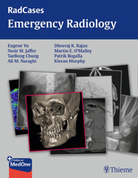
All the key Radiology cases for your rounds, rotations, and exams
Emergency Radiology presents the challenging cases that are most likely to be encountered by physicians and radiologists in the emergency room. It helps radiologists and emergency physicians correctly interpret images and thus quickly make initial diagnoses on both common and less common disorders. This book can also be used as an effective review guide for those studying for the radiology board exams.
Key Features:
- Examples of critical cases that must be accurately diagnosed to avert potential disaster in daily practice and on exams
- Clearly labeled, high-quality images help you quickly absorb key findings
RadCases contains cases selected to simulate everything that you'll see on your rounds, rotations, and exams. RadCases also helps you identify the correct differential diagnosis for each case—including the most critical.
RadCases covers:
- Cardiac Imaging · Interventional Radiology · Musculoskeletal Radiology · Neuro Imaging · Thoracic Imaging · Pediatric Imaging · Gastrointestinal Imaging · Breast Imaging · Nuclear Medicine · Ultrasound Imaging · Head and Neck Imaging · Genitourinary Imaging
Each RadCases title features 100 carefully selected, must-know cases documented with clear, high-quality radiographs. The organization provides maximum ease of use for self-assessment. Each case begins with the clinical presentation on the right-hand page; simply turn the page for imaging findings, differential diagnoses, the definitive diagnosis, essential facts, and more.
This RadCases book comes with a code providing access to additional online cases: 100 from this book plus 150 more cases.
Learn your cases, diagnose with confidence, and pass your exams. RadCases.
This print book includes complimentary access to a digital copy on https://medone.thieme.com.
Publisher's Note: Products purchased from Third Party sellers are not guaranteed by the publisher for quality, authenticity, or access to any online entitlements included with the product.
Das E-Book können Sie in Legimi-Apps oder einer beliebigen App lesen, die das folgende Format unterstützen:
Veröffentlichungsjahr: 2015
Ähnliche


To access the additional media content available with this e-book via Thieme MedOne, please use the code and follow the instructions provided at the back of the e-book.
RadCases Emergency Radiology
Edited by
Eugene Yu, MD, FRCPC
Associate Professor of Radiology and OtolaryngologyHead and Neck SurgeryPrincess Margaret Cancer CentreUniversity of TorontoToronto, Ontario, Canada
Nasir M. Jaffer, MD, FRCPC
Professor of RadiologyUniversity Health NetworkMount Sinai Hospital and Women's College HospitalDepartment of Medical ImagingFaculty of MedicineUniversity of TorontoToronto, Ontario, Canada
TaeBong Chung, MD, FRCPC
University Health NetworkMount Sinai Hospital and Women's College HospitalDepartment of Medical ImagingFaculty of MedicineUniversity of TorontoToronto, Ontario, Canada
Ali M. Naraghi, MD
University Health NetworkMount Sinai Hospital and Women's College HospitalDepartment of Medical ImagingFaculty of MedicineUniversity of TorontoToronto, Ontario, Canada
Dheeraj K. Rajan, MD, FRCPC, FSIR
Head of Vascular and Interventional RadiologyUniversity Health NetworkMount Sinai Hospital and Women's College HospitalDepartment of Medical ImagingFaculty of MedicineUniversity of TorontoToronto, Ontario, Canada
Martin E. O'Malley, MD, FRCPC
University Health NetworkMount Sinai Hospital and Women's College HospitalDepartment of Medical ImagingFaculty of MedicineUniversity of TorontoToronto, Ontario, Canada
Patrik Rogalla, MD
Professor of RadiologyJoint Department of Medical ImagingUniversity Health NetworkMount Sinai Hospital and Women's College HospitalToronto General HospitalUniversity of TorontoToronto, Ontario, Canada
Kieran Murphy, MB, FRCPC, FSIR
University Health NetworkMount Sinai Hospital and Women's College HospitalDepartment of Medical ImagingFaculty of MedicineUniversity of TorontoToronto, Ontario, Canada
Series Editors
Jonathan Lorenz, MD
Associate Professor of RadiologyDepartment of RadiologyThe University of ChicagoChicago, Illinois
Hector Ferral, MD
Senior Clinical EducatorNorthShore University HealthSystemEvanston, Illinois
ThiemeNew York • Stuttgart • Delhi • Rio de Janeiro
Executive Editor: William LamsbackManaging Editor: J. Owen Zurhellen IVEditorial Assistant: Heather AllenSenior Vice President, Editorial and Electronic Product Development: Cornelia SchulzeProduction Editor: Teresa Exley, Maryland CompositionInternational Production Director: Andreas SchabertInternational Marketing Director: Fiona HendersonDirector of Sales, North America: Mike RosemanInternational Sales Director: Louisa TurrellSenior Vice President and Chief Operating Officer: Sarah VanderbiltPresident: Brian D. ScanlanCompositor: MPS Limited
Library of Congress Cataloging-in-Publication Data
RadCases emergency radiology/edited by Eugene Yu, Nasir M. Jaffer, TaeBong Chung, Ali M. Naraghi, Dheeraj K. Rajan, Martin E. O'Malley, Patrik Rogalla, Kieran Murphy, Jonathan Lorenz, Hector Ferral. p.; cm.—(RadCases)Emergency radiologyISBN 978-1-60406-832-0—ISBN 978-1-60406-323-3 (eISBN) I. Yu, Eugene, editor. II. Title: Emergency radiology. III. Series: Rad-Cases.[DNLM: 1. Radiology--Case Reports. 2. Emergencies--Case Reports. WN 100]RC78616.07’57--dc23 2014038289
Copyright © 2015 by Thieme Medical Publishers, Inc.Thieme Publishers New York333 Seventh AvenueNew York, NY 10001 USA1-800-782-3488, [email protected]
Thieme Publishers StuttgartRüdigerstrasse 14 70469 Stuttgart, Germany+49 [0]711 8931 421, [email protected]
Thieme Publishers DelhiA-12, Second Floor, Sector -2, NOIDA -201301Uttar Pradesh, India+91 120 45 566 00, [email protected]
Thieme Publishers Rio, Thieme Publicações Ltda.Argentina Building 16th floor, Ala A, 228 Praia do BotafogoRio de Janeiro 22250-040 Brazil+55 21 3736-3631
Printed in China by Everbest Printing Ltd.
ISBN 978-1-60406-832-0
Also available as an e-book:eISBN 978-1-60406-833-7
Important note: Medicine is an ever-changing science undergoing continual development. Research and clinical experience are continually expanding our knowledge, in particular our knowledge of proper treatment and drug therapy. Insofar as this book mentions any dosage or application, readers may rest assured that the authors, editors, and publishers have made every effort to ensure that such references are in accordance with the state of knowledge at the time of production of the book.
Nevertheless, this does not involve, imply, or express any guarantee or responsibility on the part of the publishers in respect to any dosage instructions and forms of applications stated in the book. Every user is requested to examine carefully the manufacturers’ leaflets accompanying each drug and to check, if necessary in consultation with a physician or specialist, whether the dosage schedules mentioned therein or the contraindications stated by the manufacturers differ from the statements made in the present book. Such examination is particularly important with drugs that are either rarely used or have been newly released on the market. Every dosage schedule or every form of application used is entirely at the user's own risk and responsibility. The authors and publishers request every user to report to the publishers any discrepancies or inaccuracies noticed. If errors in this work are found after publication, errata will be posted at www.thieme.com on the product description page.
Some of the product names, patents, and registered designs referred to in this book are in fact registered trademarks or proprietary names even though specific reference to this fact is not always made in the text. Therefore, the appearance of a name without designation as proprietary is not to be construed as a representation by the publisher that it is in the public domain.
This book, including all parts thereof, is legally protected by copyright. Any use, exploitation, or commercialization outside the narrow limits set by copyright legislation without the publisher's consent is illegal and liable to prosecution. This applies in particular to photostat reproduction, copying, mimeographing or duplication of any kind, translating, preparation of microfilms, and electronic data processing and storage.
We dedicate this book to our families for their continued support and encouragement.
RadCases Series Preface
The ability to assimilate detailed information across the entire spectrum of radiology is the Holy Grail sought by those preparing for the American Board of Radiology examination. As enthusiastic partners in the Thieme RadCases Series who formerly took the examination, we understand the exhaustion and frustration shared by residents and the families of residents engaged in this quest. It has been our observation that despite ongoing efforts to improve Web-based interactive databases, residents still find themselves searching for material they can review while preparing for the radiology board examinations and remain frustrated by the fact that only a few printed guidebooks are available, which are limited in both format and image quality. Perhaps their greatest source of frustration is the inability to easily locate groups of cases across all subspecialties of radiology that are organized and tailored for their immediate study needs. Imagine being able to immediately access groups of high-quality cases to arrange study sessions, quickly extract and master information, and prepare for theme-based radiology conferences. Our goal in creating the RadCases Series was to combine the popularity and portability of printed books with the adaptability, exceptional quality, and interactive features of an electronic case-based format.
The intent of the printed book is to encourage repeated priming in the use of critical information by providing a portable group of exceptional core cases that the resident can master. The best way to determine the format for these cases was to ask residents from around the country to weigh in. Overwhelmingly, the residents said that they would prefer a concise, point-by-point presentation of the Essential Facts of each case in an easy-to-read, bulleted format. This approach is easy on exhausted eyes and provides a quick review of Pearls and Pitfalls as information is absorbed during repeated study sessions. We worked hard to choose cases that could be presented well in this format, recognizing the limitations inherent in reproducing high-quality images in print. Unlike the authors of other case-based radiology review books, we removed the guesswork by providing clear annotations and descriptions for all images. In our opinion, there is nothing worse than being unable to locate a subtle finding on a poorly reproduced image even after one knows the final diagnosis.
The electronic cases expand on the printed book and provide a comprehensive review of the entire subspecialty. Thousands of cases are strategically designed to increase the resident's knowledge by providing exposure to additional case examples—from basic to advanced—and by exploring “Aunt Minnie's,” unusual diagnoses, and variability within a single diagnosis. The search engine gives the resident a fighting chance to find the Holy Grail by creating individualized, daily study lists that are not limited by factors such as radiology subsection. For example, tailor today's study list to cases involving tuberculosis and include cases in every subspecialty and every system of the body. Or study only thoracic cases, including those with links to cardiology, nuclear medicine, and pediatrics. Or study only musculoskeletal cases. The choice is yours.
As enthusiastic partners in this project, we started small and, with the encouragement, talent, and guidance of Tim Hiscock at Thieme, we have continued to raise the bar in our effort to assist residents in tackling the daunting task of assimilating massive amounts of information. We are passionate about continuing this journey, hoping to expand the cases in our electronic series, adapt cases based on direct feedback from residents, and increase the features intended for board review and self-assessment. As the American Board of Radiology converts its certifying examinations to an electronic format, our series will be the one best suited to meet the needs of the next generation of overworked and exhausted residents in radiology.
Jonathan L orenz, MDHector Ferral, MDChicago, IL
Preface
Emergency Radiology is the front line of medical imaging; it is where we are most relevant, most engaged, and most alive as a diagnostic medical specialty. It is best practiced face-to-face with the radiologist in the emergency room, or one door away. It's where we demonstrate our impact and where we get the most personal reward for our skills and art. It's where a resident or staff develops friendship and clinical trust based on face-to-face service. It's everything teleradiology is not. Teleradiology may perhaps be the most cost-effective model with the leanest staffing and the optimal utilization of subspecialist skills, but the interpersonal disconnection from the point of care will cost us our specialty. Radiology is not mere pattern recognition, because that, in isolation of clinical context, can never achieve the level of clinical relevance that our referring physician (or our patient) wants. Further, our absence from the emergency room creates a vacuum that will be readily filled by other services.
This book is written for the young trainee radiologist who will be in the trenches on call, and the radiologist who is taking general call and wants to refresh their knowledge. Remember that to err is human, and we all have and will. Listen to the little voice in your head when it tells you to look at something again. Be bothered by things, pick up the phone and call the emergency department or walk out that door and ask the staff or resident what's going on and explain your conundrum. They will recognize that you are showing commitment and good conscience. Thank you for using our book and its accompanying electronic version; may these serve you well.
Kieran Murphy, MB, FRCPC, FSIR
Acknowledgments
We are grateful for the hard work and efforts of the many medical students, residents, fellows, and colleagues who gave their time and expertise in helping write this book. Without their support, this book would not have been possible. A special thanks to the medical students from the Royal College of Surgeons in Ireland (RCSI), Dublin, who spent the summer of 2012 working on this project with us, including Rimpy Cheema, Keri Delaney, Alexandra Haw, Ramia Jameel, Hilary Kerr, Ravneet Mann, Caitlin Moran, Kevin Moran, Sameer Patel, Priya Sapra, and Noorin Walji.
Table of Contents
RadCases Series Preface
Preface
Acknowlegments
Case 1
Case 2
Case 3
Case 4
Case 5
Case 6
Case 7
Case 8
Case 9
Case 10
Case 11
Case 12
Case 13
Case 14
Case 15
Case 16
Case 17
Case 18
Case 19
Case 20
Case 21
Case 22
Case 23
Case 24
Case 25
Case 26
Case 27
Case 28
Case 29
Case 30
Case 31
Case 32
Case 33
Case 34
Case 35
Case 36
Case 37
Case 38
Case 39
Case 40
Case 41
Case 42
Case 43
Case 44
Case 45
Case 46
Case 47
Case 48
Case 49
Case 50
Case 51
Case 52
Case 53
Case 54
Case 55
Case 56
Case 57
Case 58
Case 59
Case 60
Case 61
Case 62
Case 63
Case 64
Case 65
Case 66
Case 67
Case 68
Case 69
Case 70
Case 71
Case 72
Case 73
Case 74
Case 75
Case 76
Case 77
Case 78
Case 79
Case 80
Case 81
Case 82
Case 83
Case 84
Case 85
Case 86
Case 87
Case 88
Case 89
Case 90
Case 91
Case 92
Case 93
Case 94
Case 95
Case 96
Case 97
Case 98
Case 99
Case 100
Further Readings
Index
Case 1
Clinical Presentation
A 54-year-old woman with sudden onset of severe headaches and decreased level of consciousness.
Further Work-up
Imaging Findings
(A, B) Non–contrast-enhanced computed tomography (NECT) shows the hyperdensity in the subarachnoid spaces in the supratentorial subarachnoid cisterns extending laterally to the sylvian fissures (arrows). There is mild dilatation of the temporal horns of the lateral ventricles (asterisk), a sign of early hydrocephalus. (C, D) Coronal computed tomography angiography (CTA) maximum intensity projection (MIP) reformats show a superiorly oriented anterior communicating artery (ACoA) aneurysm (arrow), better characterized on the three-dimensional (3D) reformatted image.
Differential Diagnosis
• Aneurysmal subarachnoid hemorrhage (SAH): The presence of SAH in the basal cisterns extending laterally to the sylvian fissure, especially if it is associated with early hydrocephalus, must raise the suspicion for a ruptured aneurysm. CTA should be performed immediately for the initial assessment and treatment planning.
• Nonaneurysmal SAH: Fifteen percent to 20% of patients with nontraumatic SAH will have no visible cause on CTA or digital subtraction angiography (DSA). Around 50% of these cases will have a classic bleeding pattern with a more benign clinical course, the so-called perimesencephalic SAH.
• “Pseudo” SAH: Patients with diffuse brain edema may show increased density of the basal cisterns because of crowding of the meninges and engorgement of the venous structures.
Essential Facts
• SAH represents 6% of all strokes, with an incidence of <9/100 per year.
• Eighty-five percent of nontraumatic SAH hemorrhages are secondary to a ruptured aneurysm.
• The most common locations for ruptured aneurysm are the ACoA (29.0%), posterior communicating artery (19.6%), basilar artery (14.7%), and middle cerebral artery (11.8%).
• Size and location are the main factors used to predict the risk of rupture.
• Risk factors include female gender, age, personal or family history of SAH, smoking, hypertension, and alcohol abuse.
• Treatment options include surgical clipping and endovascular coiling. The latter has better outcomes for aneurysms suitable for both strategies.
• Prognosis is poor with 35% mortality and 45 to 64% risk of dependency.
• Risk of a new aneurysmal SAH in survivors is 15 times higher than it is in the general population.
Other Imaging Findings
• Computed tomography perfusion (CTP): Increased mean transit time (MTT) with normal cerebral blood volume (CBV) may depict areas at risk for delayed brain ischemia before the onset of vasospasm.
• Magnetic resonance/magnetic resonance angiography (MR/MRA):
• Fluid-attenuated inversion recovery (FLAIR): increased signal of the cerebrospinal fluid (CSF) spaces due to blood
• MRA: Time of flight (TOF) or contrast-enhanced magnetic resonance angiography (CE-MRA) are also good options for detecting the aneurysm, although less sensitive for smaller lesions.
Pearls &Pitfalls
CTA has a high sensitivity and specificity for detecting aneurysms.
Imaging work-up must prove the necessary details for treatment decision (location, size, neck, at-risk branches).
3D reformatted images may help in differentiating a vessel loop from a small aneurysm.
Thrombosed aneurysms and nonsaccular aneurysms (i.e., blood blister, dissecting) may have a false-negative CTA.
Case 2
Clinical Presentation
A 27-year-old man with congenital heart disease on Coumadin (Bristol-Myers Squibb, New York, NY) presents with new headache and aphasia.
Imaging Findings
(A, C) Unenhanced axial computed tomography (CT) shows a large area of low axial T1-weighted, gadolinium-enhanced image attenuation in the left temporal lobe. There is an irregular nodular rim enhancement along the area of slight hyperattenuation. (B) Axial T2-weighted margins of the lesion. (D) Diffusion weighted study and image shows the presence of vasogenic edema and an apparent diffusion coefficient (ADC) image (not shown) demonstrates the lesion to be irregularly shaped lesion with a low signal rim restricted.
Differential Diagnosis
• Cerebral abscess: This is a focal pyogenic infection of the brain. The causes are bacterial, fungal, or parasitic. Imaging findings vary with the stage of abscess formation. Seen as a ring-enhancing lesion with central high signal (restricted diffusion) on diffusion weighted imaging (DWI). T2 imaging may show a hypointense rim with surrounding edema.
• Primary tumor: Radiographically can mimic an abscess. Has a thick nodular enhancing wall with surrounding edema. Neoplasms typically show low signal on DWI. More often a gradual onset of symptoms. Lack of elevated white cell count and fever.
• Demyelination: Tumefactive demyelination can present as a mass lesion with irregular enhancement. Peripheral ring enhancement often incomplete. Look for other characteristic lesions of multiple sclerosis such as corpus callosum involvement, foci in the posterior fossa, and “Dawson fingers” representing perivenular demyelination.
Essential Facts
• Can affect any age group
• Males are affected twice more than females.
• Headache is the most common symptom; others include seizure and decreased level of consciousness.
• Peak incidence is in the third and fourth decades.
• Common causes include hematogenous seeding from a distant infective source, mastoiditis, meningitis, and penetrating injury.
• Surgical drainage with or without excision is the primary therapeutic approach.
Other Imaging Findings
• CT: low-density mass with edema and mass effect, peripheral enhancement
• Magnetic resonance imaging:
• T1WI: hypointense mass
• T2WI: hypointense abscess rim with surrounding edema; centrally high signal
• TI with contrast: enhancing rim; irregular margins
• DWI: increased signal intensity, low ADC
Pearls &Pitfalls
It is important to investigate local causes such as sinusitis, otitis media, and mastoiditis.
In patients who are successfully treated, the T2 hypointense abscess rim resolves before enhancement.
Assess for communication with the ventricular system—increased risk of ventriculitis and hydrocephalus.
Clinical history showing acute onset with associated malaise, fever, and elevated white cell count are helpful clinical features.
Case 3
Clinical Presentation
A 26-year-old woman involved in a road traffic accident and presents with neck pain and an acute right Horner syndrome.
Imaging Findings
(A) Axial contrast-enhanced computed tomography (CT) scan demonstrates a medial bulge arising from the right internal carotid artery (arrow). (B, C) Coronal maximum intensity projection (MIP) image and three-dimensional (3D) computed tomography angiography (CTA) reconstruction demonstrate two medial areas of focal outpouching arising from the right internal carotid artery (ICA) (arrows).
Differential Diagnosis
• Traumatic pseudoaneurysms: These “false” aneurysms appear as a focal outpouching from the wall of the affected artery. A pseudoaneurysm is a focal hematoma that is not contained by the normal components of the vessel wall.
• Fibromuscular dysplasia (FMD): FMD lesions appear as a characteristic beading of the carotid arteries. In the internal carotid arteries, FMD usually affects the extracranial portion at the C1–C2 levels.
• Mycotic pseudoaneurysm: Mycotic aneurysms are more likely to be saccular, eccentric, or multilobulated. There may also be accompanying inflammation around the vessel. A clinical history of sepsis or the presence of a septic source is contributory to the diagnosis.
Essential Facts
• Usually caused by blunt or penetrating trauma that disrupts the arterial wall and allows blood to leak into the surrounding tissue
• Traumatic pseudoaneurysms occur due to penetrating and nonpenetrating injuries and are responsible for less than 1% of all aneurysms.
• Pseudoaneurysms can also arise secondary to deep neck space infection, atherosclerosis, collagen vascular disorders, radiation therapy, and tumor invasion.
• The risk of hemorrhage in posttraumatic pseudoaneurysms is <20% with a peak incidence of rupture at 2 to 3 weeks after injury resulting in 30 to 50% mortality.
• Horner syndrome indicates an associated disruption of the sympathetic nerves, which travel with the carotid artery.
Other Imaging Findings
• CT:
• “Blister-like” bulge in the vessel wall on CTA
• Magnetic resonance imaging (MRI):
• Focal outpouching from the vessel wall
• May see a thrombosed surrounding “wall” that contains the pseudoaneurysm
• Wall may enhance
• Gradient-recalled echo (GRE) imaging will show intense low signal corresponding to clot within the pseudoaneurysm.
Pearls &Pitfalls
Delayed onset of cranial nerve palsy may be a sign of pseudoaneurysm formation following neck trauma.
Evaluate the brain for evidence of stroke and multiple emboli.
Case 4
Clinical Presentation
A 52-year-old woman with right-sided headache, right-sided ptosis diplopia, and fever. Blood cultures positive for Staphylococcus aureus. Elevated leukocyte count in cerebrospinal fluid (CSF).
Imaging Findings
(A, B) Axial T2 image with fat saturation and coronal T1 postgadolinium with fat saturation show expansion of the right cavernous sinus (arrows in A and B). The right lateral rectus muscle is also slightly T2 hyperintense, consistent with extraocular muscle edema. Filling defects are seen within the right cavernous sinus (arrowheads in B). There is total opacification of the bordering right sphenoid sinus with T1 hypo- and T2 heterogeneous signals as well as peripheral enhancement consistent with sinusitis. The left sphenoid sinus and some ethmoidal air cells also show mucosal inflammatory changes (asterisk in A). (C) There is expansion of the right superior ophthalmic vein as well as a filling defect within this vein (dashed arrow).
Differential Diagnosis
• Cavernous sinus thrombosis: Complication of midface infection (including sinusitis, orbital or skin infection, tonsillitis) or trauma. Most cases are secondary to bacterial infection (most commonly S. aureus). Presents with expansion and convexity of the cavernous sinus/filling defects within the cavernous sinus. Dilatation/filling defects within the superior ophthalmic vein may be present.
• Cavernous sinus neoplasm: Most frequently meningiomas but other lesions such as metastases, lymphoma, leukemia, hemangioma, or nerve sheath tumors may occur in this location.
• Cavernous sinus inflammation: Either secondary to systemic disease (such as Wegener granulomatosis or sarcoidosis) or idiopathic (Tolosa-Hunt syndrome). Look for the presence of sinonasal or lung involvement in Wegener granulomatosis. In addition to typical chest findings in sarcoidosis, intracranial and/or orbital involvement must be sought. Any part of the orbits can be involved in sarcoidosis; however, lacrimal gland involvement is more frequent.
Essential Facts
• The cavernous sinuses receive blood from multiple valveless veins. Infection from anatomical structures with venous drainage to the cavernous sinus (e.g., the sphenoid sinus) is the major risk factor for cavernous sinus thrombophlebitis.
• Patients will typically present with proptosis, cranial nerve palsy, and headache.
Other Imaging Findings
• CT:
• On postcontrast CT, the normal cavernous sinus has a flat or concave lateral margin. The internal carotid artery normally cannot be differentiated from the normal, opacified, cavernous venous sinus.
• With thrombosis, the lateral margin of the cavernous sinus becomes convex. The ICA can be identified from the thrombosed, nonopacified, cavernous sinus.
• CTA may allow for identification of ICA involvement (stenosis, occlusion, or pseudoaneurysm formation).
• Look for sinusitis or signs of midface or orbital infection.
• MRI:
• Must use high-resolution MRI to assess cavernous sinuses including 1- to 3-mm thick sections through the orbits and cavernous sinuses in the axial and coronal planes
• T1: May show T1 hyperintense subacute venous thrombosis in cavernous sinus and/or superior ophthalmic vein.
• T2 with fat saturation or short T1 inversion recovery (STIR): Variable but often hyperintense clot signal. May show extraocular muscle edema and intraorbital fat stranding.
• T1 postcontrast with fat saturation: may show filling defects within the cavernous sinus/superior ophthalmic vein
• Magnetic resonance venography: May show other sites of dural venous sinus thrombosis
• Diffusion weighted imaging (DWI): may be useful in identifying thrombosis and/or abscess formation
Pearls &Pitfalls
Beware of cavernous sinus thrombosis in the setting of midface, orbital, or sinonasal infection especially in patients presenting with cranial nerve palsy and/or proptosis.
Perform appropriate high-resolution MRI of the orbits and cavernous sinuses before excluding the possibility of this diagnosis.
Search for secondary ICA and intracranial complications.
Patients who have diabetes and are immunocompromised are at risk of developing invasive fungal infection.
Case 5
Clinical Presentation
A 44-year-old woman with a history of previously treated breast carcinoma presents with new left L2–L3 numbness and back pain.
Imaging Findings
(A) Sagittal T1-weighted image (T1WI) magnetic resonance (MR) shows diffuse hypointensity throughout the vertebral column and posterior elements due to metastatic disease. Epidural extension causes cord compression at T12 (arrow). (B) Sagittal T2-weighted image (T2WI) MR shows heterogeneous hypointensity in the vertebral column and posterior elements with moderate collapse of T12 and L3 and mild collapse of L4. Again, cord compression is seen at T12. (C) Axial T2-weighted image (T2WI) MR shows heterogeneous hypointensity within the body and posterior elements of T12. Epidural extension of tumor causes cord compression (arrow).
Differential Diagnosis
• Vertebral column metastases from breast carcinoma: Generalized vertebral hypointensity on T1WI and T2WI are in keeping with diffuse sclerotic metastases; may diffusely enhance with gadolinium. May cause pathologic fracture with epidural retropulsion.
• Multiple myeloma: Most often occurs in spine, skull, ribs, and pelvis. Multiple well-circumscribed lytic lesions on radiography. Multifocal, diffuse, or heterogeneous T1 hypointensity. Bone scintigraphy typically negative.
• Lymphoma: Bone destruction with soft tissue extension seen on radiography. Most lesions are homogeneous and hypointense on T1 and enhance uniformly with gadolinium.
Essential Facts
• Vertebral column metastases occur in 60% of patients with breast cancer. Epidural spinal cord compression occurs in 4% of patients with breast cancer.
• Myelopathy can occur in the form of limb weakness, numbness, paresthesia, and alterations in bladder and bowel function.
• Once spinal cord compression is diagnosed, steroids should be administered immediately along with emergency radiation therapy and/or surgical decompression.
Other Imaging Findings
• Plain spine radiographs: require 50 to 70% bone destruction for detection
• Anteroposterior (AP): “absent pedicle sign”
• Lateral: destroyed posterior cortical line on lateral view
• Look for areas of lucency or sclerosis.
• Computed tomography (CT): Location proportional to red marrows (lumbar [L] > thoracic [T] > cervical [C] spine). Posterior vertebral body involved in almost all cases/paraspinous/epidural soft tissue mass. May see lytic and/or sclerotic foci of disease.
• Nuclear medicine: Technetium-99m (Tc-99m) single-photon emission computed tomography (SPECT) bone scan has high sensitivity for vertebral metastases. Diffuse disseminated disease can give rise to a “super scan.”
Pearls &Pitfalls
MR scan should cover entire spinal axis when metastasis is suspected.
Persistence of low T1 marrow signal after radiation therapy may be either residual active tumor or fibrosis.
Gadolinium administration is used to assess for leptomeningeal metastases.
Use of gadolinium will also cause enhancement of vertebral lesions; this can make these lesion less conspicuous as they may become isointense with normal vertebral yellow marrow. Consider obtaining postcontrast T1 sequences with fat saturation to avoid this potential pitfall. Alternatively, precontrast T1 images would also help in this regard.
Case 6
Clinical Presentation
An 86-year-old woman presents with fever, neck pain, and confusion. The patient was treated recently for sinusitis.
Imaging Findings
Sagittal T1 (A), T1 postgadolinium contrast (B), and T2 (C) images of the cervical spine show irregularity along the vertebral body endplates bordering the C6–C7 disk. There is abnormal enhancement along the endplate margins and there is also abnormal enhancing soft tissue protruding anteriorly into the prevertebral space (solid arrow). Posteriorly, there is also thick enhancing tissue running up and down along the anterior epidural space (dashed arrows). Note the mass effect on the cervical cord that demonstrates high parenchymal signal on the T2-weighted image.
Differential Diagnosis
• Diskitis: This is a bacterial infection of an intervertebral disk and adjacent vertebral bodies. Characteristic imaging features include disk height loss, endplate erosion, and irregularity. Marrow edema within the vertebral bodies is evident. The disk and endplates often enhance and show a bright T2 signal. Complications include the presence of epidural and prevertebral phlegmon (as seen in this case) as well as frank abscess formation.
• Degenerative disk disease: This includes spondylosis deformans (degenerative changes to the annulus fibrosus and ring apophysis with the formation of osteophytes) and intervertebral osteochondrosis (degenerative changes to the nucleus pulposus and adjacent vertebral endplates). Results in characteristic progression of Modic-type changes (I, II, III) in the vertebral endplates. Imaging features include the presence of signal change in the disk (can be T2 bright or low depending on the stage of disease), endplate sclerosis and irregularity, disk herniation, and rim osteophyte formation.
• Neuropathic spine: Arises in patients with decreased proprioception and pain perception (patients with diabetes and spinal cord trauma). Prone to repetitive stress on the spine, leading to injury to the disks and facets. Most commonly in the lumbar spine. Features include disk height loss, marked vertebral and endplate sclerosis, osteophyte formation and deformity, fracture, and pseudoarthrosis.
Essential Facts
• Most commonly secondary to hematogenous seeding from a bacteremia such as upper respiratory tract or genitourinary tract infections
• Diskitis is most common in the lumbar spine.
• Staphylococcus aureus is the most common invading pathogen; Salmonella is common in patients with sickle cell anemia.
• Risk factors: intravenous (IV) drug users, immunocompromised individuals, recent invasive (spine) procedures (surgery, diskography, myelography)
• Diskitis can also arise from the contiguous spread of infection from adjacent areas of inflammation, such as diverticulitis and appendicitis.
Other Imaging Findings
• Conventional radiography: Can be normal early in disease. Endplate erosions, disk height loss, paraspinal opacity (representing possible paraspinal edema, abscess).
• Computed tomography (CT): Similar features as with plain film imaging. Loss of disk height and endplate erosion. Also may see disk and vertebral body enhancement. Will better depict paraspinal abscess.
• Magnetic resonance imaging (MRI): Edema within the vertebral bodies and disk will manifest at low T1 and bright T2 signal. Disks and vertebrae will often enhance with gadolinium. Fat saturation or short T1 inversion recovery (STIR) imaging will best show the presence of edema and fluid.
• Nuclear medicine imaging: Three-phase technetium 99m (Tc-99m) diphosphonate bone scan, gallium scan, and white blood cell scan will show areas of elevated uptake.
Pearls &Pitfalls
Plane film radiography can be falsely negative if completed too early; it may be 2 to 6 weeks before findings are evident.
MRI is the imaging modality of choice.
Earliest imaging sign is the loss of disk space height.
Always assess for paravertebral and epidural extension of inflammation. The presence of an abscess may necessitate surgical drainage.
Tuberculous spinal infection is characterized by multilevel involvement, paraspinal abscess formation, vertebral collapse, and kyphotic deformity.
Case 7
Clinical Presentation
A 29-year-old woman, 3 months postpartum, with a left frontal headache. Patient was started on oral contraceptive pill 2 months postpartum.
Imaging Findings
(A) Sagittal T1-weighted image (WI) with contrast demonstrates a filling defect in the transverse sinus with surrounding dural enhancement (“empty delta” sign, white arrow). (B) Axial T2WI demonstrates loss of the normal flow void in the left transverse sinus (white arrows). (C) Axial T1WI with contrast demonstrates a large filling defect in a distended left transverse sinus (white arrows). (D) Frontal three-dimensional maximum intensity projection from a magnetic resonance (MR) venogram demonstrates absence of flow-related enhancement in the left transverse sinus (white arrow) as compared with the normal right transverse sinus (white arrowhead).
Differential Diagnosis
• Dural venous sinus thrombosis: This is caused by occlusion of the dural venous sinus. Associated cortical vein thrombosis is not uncommon. Thrombosis of the venous sinus has a varied appearance on both computed tomography (CT) and magnetic resonance imaging (MRI) depending on the age of the clot. CT and MR venography are considered the mainstay for diagnosis and typical imaging features include a filling defect in the affected sinus on venography and absence of the expected flow void on MRI.
• Arachnoid granulation: Arachnoid granulations are aggregates of tissue that are contiguous with the subarachnoid space and are the passageway through which cerebrospinal fluid (CSF) circulates. These structures protrude into the lumen of the dural venous sinus, creating ovoid filling defects, which can mimic a dural venous thrombus. They do not enhance and the main distinguishing characteristic is that they follow CSF density and signal on all CT and MR sequences, respectively.
• Dural venous sinus aplasia/hypoplasia: This congenital anatomic variant is most commonly associated with the transverse sinus. A hypoplastic sinus will appear small but smoothly patent on CT and MR with an associated small ipsilateral bony groove in the calvarium, best seen on CT. Angiography will demonstrate an absent sinus in the setting of aplasia, which can mimic dural sinus thrombosis.
Essential Facts
• Dural sinus thrombosis is most commonly seen in the setting of dehydration (particularly in infants), trauma, malignancy, coagulopathy, infection, and with certain medications (such as oral contraceptives and chemotherapy).
• There is a varied clinical presentation of headaches, visual disturbances, seizures, and focal neurologic deficits, which can develop over the course of a few hours to several weeks.
• The superior sagittal and transverse sinuses are most commonly involved.
• In the setting of renal insufficiency or other contraindications to intravenous (IV) contrast, a time-of-flight MR angiography can be performed, which utilizes a “flow-related enhancement” phenomenon technique to determine vascular patency.
• Treatment is usually with anticoagulation; endovascular recanalization is reserved for more severe cases.
Other Imaging Findings
• Common CT and MRI features:
• Filling defect on contrast-enhanced imaging
• The appearance of clot varies with age.
• Empty delta sign—filling defect in the venous sinus surrounded by enhancing dura
• Numerous enlarged collateral veins adjacent to the thrombosed sinus
• Additional CT features:
• Acute thrombus can appear hyperdense on noncontrast imaging.
• Additional MR features:
• T2: loss of the normal flow void
Pearls &Pitfalls
Consider dural sinus thrombosis in the setting of hemorrhagic or atypical cortical or subcortical infarcts in a distribution that does not correspond to any major arterial territory.


