119,99 €
Mehr erfahren.
- Herausgeber: Thieme
- Kategorie: Fachliteratur
- Sprache: Englisch
A comprehensive guide providing essential information for successful surgery
Winner of the First Prize in ENT at the 2008 BMA (British Medical Association) Medical Book Competition
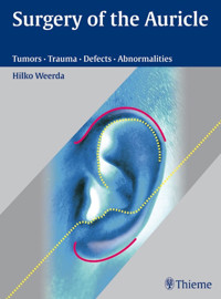
This book is a comprehensive guide to the delicate and complex reconstructive procedures for the external ear. Featuring concise descriptions, step-by-step instructions, and numerous before and after photos, this book provides surgeons with the essential knowledge that successful surgery in this difficult field demands.
The text opens with an overview of the anatomy and anthropometry of the external ear, aesthetic principles of auricular reconstruction, and the basic principles of plastic surgery. Separate sections of the book provide in-depth discussion of the techniques for managing tumors, trauma and non-inflammatory processes, auricular defects, and abnormalities.
Features:
- More than 1,300 illustrations and photographs that aid comprehension of auricular problems and surgical steps
- Detailed discussion of classification of auricular defects and abnormalities
- Coverage of the radiologic examination of malformations of the petrous temporal bone
An incomparable reference for all surgeons specializing in treating defects and disorders of the external ear, this volume succeeds beautifully in capturing the myriad creative, scientific, and technical facets of auricular reconstruction.
Das E-Book können Sie in Legimi-Apps oder einer beliebigen App lesen, die das folgende Format unterstützen:
Seitenzahl: 689
Veröffentlichungsjahr: 2007
Ähnliche


Dedicated in gratitudeto my wife Barbara
Library of Congress Cataloging-in-Publication Data is available from the publisher.
This book is an authorized and revised translation of the German edition published and copyrighted 2004 by Georg Thieme Verlag, Stuttgart, Germany. Title of the German edition: Chirurgie der Ohrmuschel
Translator: Grahame Larkin, MD, Wimborne, UK
Illustrator: Katharina Schumacher, Munich, Germany
© 2007 Georg Thieme Verlag,Rüdigerstrasse 14, 70469 Stuttgart, Germanyhttp://www.thieme.deThieme New York, 333 Seventh Avenue,New York, NY 10001, USAhttp://www.thieme.com
Typesetting by Primustype Hurler, NotzingenPrinted in Germany by Druckhaus Götz, Ludwigsburg
ISBN 978–3-13–139411-8 (TPS, Rest of World)ISBN 978–1-58890-359–4 (TPN, The Americas) 1 2 3 4 5 6
Important note: Medicine is an ever-changing science undergoing continual development. Research and clinical experience are continually expanding our knowledge, in particular our knowledge of proper treatment and drug therapy. Insofar as this book mentions any dosage or application, readers may rest assured that the authors, editors, and publishers have made every effort to ensure that such references are in accordance with the state of knowledge at the time of production of the book.
Nevertheless, this does not involve, imply, or express any guarantee or responsibility on the part of the publishers in respect to any dosage instructions and forms of applications stated in the book. Every user is requested to examine carefully the manufacturers’ leaflets accompanying each drug and to check, if necessary in consultation with a physician or specialist, whether the dosage schedules mentioned therein or the contraindications stated by the manufacturers differ from the statements made in the present book. Such examination is particularly important with drugs that are either rarely used or have been newly released on the market. Every dosage schedule or every form of application used is entirely at the user's own risk and responsibility. The authors and publishers request every user to report to the publishers any discrepancies or inaccuracies noticed. If errors in this work are found after publication, errata will be posted at www.thieme.com on the product description page.
Some of the product names, patents, and registered designs referred to in this book are in fact registered trademarks or proprietary names even though specific reference to this fact is not always made in the text. Therefore, the appearance of a name without designation as proprietary is not to be construed as a representation by the publisher that it is in the public domain.
This book, including all parts thereof, is legally protected by copyright. Any use, exploitation, or commercialization outside the narrow limits set by copyright legislation, without the publisher's consent, is illegal and liable to prosecution. This applies in particular to photostat reproduction, copying, mimeographing, preparation of microfilms, and electronic data processing and storage.
Forewords
Surgical creation of the outer ear with autogenous tissues is a unique blend of art and science. The result will be influenced by the surgeon's adherence to the basic principles of plastic surgery, but his or her sculpture and design skills will ultimately determine the final outcome.
Over the years numerous surgeons have visited me in the operating room, but only a handful have blossomed into auricular artists. Dr Hilko Weerda is one of these gifted few.
I have been honored not only to watch Dr Weerda perform ear surgery, but also to be the recipient of a number of his wonderful self-made Christmas card paintings over the years. During my visit to Lübeck, I was most impressed with his artistic flair and the lovely paintings that he had created both for his home and office.
Both the art and the surgical world are privileged to have Dr Weerda's presence, for the impact he has made on both is outstanding.
His book on ear reconstruction will certainly be another wonderful contribution of artistic and scientific endeavor.
Professor Burton Brent, MD
El Camino Hospital,
Woodside, CA, USA
My acquaintance with Professor Hilko Weerda of the Lübeck University School of Medicine in Germany began in May 1995 at the 11th International Congress of Plastic, Reconstructive, and Aesthetic Surgery (IPRAS) in Yokohama, Japan. Professor Weerda's concern for the treatment of auricular and middle ear defects led to the organization of the 3rd International Symposium on Auricular and Middle Ear Malformation, Ear Defects and Their Reconstructions in Lübeck, Germany, September 1997. At this meeting, Professor Weerda was able to invite prominent surgeons from throughout the world to present lectures and perform demonstration surgeries: this was indeed one of the most educational and scientific meetings concerned with problems related to the auricle to take place in the twentieth century.
Auricular reconstruction is still considered to be one of the most difficult reconstructive surgeries for a plastic reconstructive surgeon to perform. This is due to the fact that anatomical and morphological knowledge is required prior to being able to reconstruct an auricle in the proper anatomical location with all the detailed morphological features of the auricle. In addition, there is the problem of insufficient skin surface area to cover the fabricated three-dimensional costal cartilage framework (3-D frame), and its associated complications like low hairline and craniofacial skeletal defects. The Nagata method described in this textbook by Professor Weerda is a two-stage method for total auricular reconstruction, where anatomical and morphological problems have been solved; and with Professor Weerda's expertise, the problem of middle ear malformation (auditory function) is in the midst of being solved for the attainment of the ultimate goal, the reconstruction of a functional auricle of normal appearance.
It is an honor for me to write a foreword for Professor Weerda's textbook. I would like to recommend this textbook to all surgeons involved in, and/or considering a clinical practice in, reconstructive surgery for auricular and middle ear defects.
Professor Satoru Nagata, MD, PhD
Department of Reconstructive Plastic Surgery
AKIBA-Hospital, Saitama, Japan
Preface
When I began my work as a physician at the Hospital for Oral and Maxillofacial Surgery in Erlangen in 1965, the thalidomide disaster was just emerging and we had to make external prostheses for a great many children with auricular malformations. Because I had studied sculpture at an art academy before attending medical school, I was called upon to fabricate the prostheses. The relatively poor psychological rehabilitation of these children moved me deeply and prompted me to seek more satisfactory methods of reconstruction.
I have maintained a keen interest in this area during more than 30 years’ practice as a plastic and reconstructive surgeon, working first at the Department of Otolaryngology at Freiburg University Hospital under Fritz Zöllner and Chlodwig Beck and later at the Department of Otolaryngology of Lübeck Medical University.
Particularly in Lübeck, I was able to pursue my work in this field along with a number of colleagues and doctoral candidates, some of whom have kindly contributed their results to this volume.
I also wish to acknowledge all the specialists who enriched this book with their contributions.
I extend special thanks to our photographer, Mrs Ellen Liegmann, who was responsible for years of difficult image documentation and effectively illustrated essential diseases and operations. I also thank Mrs Bettina Villmann, who worked tirelessly to put my partially handwritten notes into an intelligible form.
I am grateful to Thieme Publishing and its dedicated staff, particularly Ms. Elisabeth Kurz, Ms. Stefanie Langner, and Mr. Stephan Konnry. I appreciate the high quality of the illustrations prepared from my sketches by Mrs K. Schumacher and her patience in implementing my suggested changes.
For didactic reasons, the majority of conditions and procedures have been illustrated for the right ear.
I hope that this book will encourage my colleagues to explore auricular reconstruction and to advance both the theory and practice of this challenging field.
Hilko Weerda
Contributors
Franz Xaver Brunner, MDProfessor and DirectorDepartment of OtorhinolaryngologyAugsburg Central HospitalAugsburg, Germany
Guenter Burg, MDProfessorDepartment of DermatologyUniversity Hospital of ZürichZürich, Switzerland
Stefan Gottschalk, MDInstitute of NeuroradiologyUniversity Hospital Schleswig-HolsteinCampus LübeckLübeck, Germany
Ingo GreinerGreiner Epithesen GmbHKiel, Germany
Andreas Haisch, MDDepartment of OtorhinolaryngologyCharité—University Medicine BerlinCampus Benjamin FranklinBerlin, Germany
Albert A. Hartmann, MDProfessorDermatology, AllergologyAachen, Germany
Andreas Hasse, MDSchlosspark-KlinikDepartment of Oral-Maxillofacial SurgeryBerlin, Germany
Dirk Hoehmann, MDAssociate ProfessorPrivate Practice ClinicOtorhinolaryngology, Plastic Surgery, AllergologyNuremberg, Germany
Robert A. Jahrsdoerfer, MDProfessor of Clinical Otolaryngology—Head and Neck SurgeryUniversity of Virginia Medical CenterDepartment of Otolaryngology—Head and Neck SurgeryDivision of Otolgy, NeurotologyCharlottesville, VA, USA
R. Katzbach, MDDepartment of OtorhinolaryngologyUniversity Hospital Schleswig-HolsteinCampus LübeckLübeck, Germany
Jeffrey Hung N. Kim, MDAssistant ProfessorGeorgetown University Medical CenterDepartment of Otolaryngology—Head and Neck SurgeryWashington, DC, USA
Susanne Klaiber, MDDepartment of OtorhinolaryngologyUniversity Hospital Schleswig-HolsteinCampus LübeckLübeck, Germany
Michael Landthaler, MDProfessorClinic and Polyclinic for DermatologyUniversity of RegensburgRegensburg, Germany
Dirk Petersen, MDProfessorInstitute of NeuroradiologyUniversity Hospital Schleswig-HolsteinCampus LübeckLübeck, Germany
Rainer Schoenweiler, MDProfessorDepartment of OtorhinolaryngologyDivision of PhoniatricsUniversity Hospital Schleswig-HolsteinCampus LübeckLübeck, Germany
Ralf Siegert, MDProfessorDepartment of OtorhinolaryngologyProsper HospitalRecklinghausen, Germany
Hilke Sommer, MDDepartment of OtorhinolaryngologyKlinikum OldenburgOldenburg, Germany
Hilko Weerda, MD, DMDProfessor and Former HeadDepartment of OtorhinolaryngologyUniversity Hospital Schleswig-HolsteinCampus LübeckLübeck, Germany
Monika Wimmershoff, MDDermatologist and VenereologistPrivate PracticeUlm, Germany
Table of Contents
1 Basic Principles
1.1 Anatomy of the External Ear(H. Weerda)
1.2 Anthropometry of the Auricle(R. Siegert)
1.2.1 Introduction
1.2.2 Variables Relative to Age and Height
1.2.3 Summary
1.3 Aesthetic Principles of Auricular Reconstruction(H. Weerda)
1.4 Basic Principles of Plastic Surgery(H. Weerda)
1.4.1 Instruments and Auxiliary Equipment
1.4.2 Wound Management, Care of Small Defects, and Scar Revision
1.4.3 Composite Grafts
1.4.4 Graft and Implant Terminology
2 Tumors of the External EarH. Weerda, A.A. Hartmann, F.X. Brunner, G. Burg, and D. Hoehmann
2.1 Introduction
2.2 Basal Cell Carcinoma and Squamous Cell Carcinoma
2.2.1 Basal Cell Carcinoma
2.2.2 Keratoacanthoma
2.2.3 Spinal Cell Carcinoma
2.3 Malignant Melanoma
Histomorphology
Therapy
2.4 Keloid
2.5 Neurofibroma
2.6 Gout Tophi
2.7 Chondrodermatitis Nodularis Chronica Helicis
3 Trauma and Non-inflammatory ProcessesH. Weerda
3.1 Acute Trauma of the Auricle
3.1.1 Chemical Burns
3.1.2 Thermal Injuries
First-degree Burns
Second-degree (Partial-thickness) Burns
Third-degree (Full-thickness) Burns
Frostbite
3.1.3 Acute Otoseroma and Otohematoma
3.2 Chronic Otohematoma
3.3 Auricular Injuries
3.3.1 Bite Injuries
3.3.2 Piercing
3.3.3 Tears and Avulsions of the Auricle
Classification
First-degree Auricular Injuries
Second-degree Auricular Injuries
Third-degree Auricular Injuries
Historical Review
Epidemiology of Auricular Trauma
Theoretical Principles
Cartilage Survival Time
Emergency Care of Auricular Avulsions
3.3.4 Replantation of the Auricle
Preoperative Care of the Amputated Part
Suture Technique
Perioperative Measures
Replantation Techniques
Classic Replantation
Pocket Techniques
Distant Banking
Banking Beneath Retroauricular Mastoid Skin
Mladick's Technique
Flap Coverage
Other Techniques of Replanting Auricular Parts
Baudet's Technique
Arfai's Technique
Microvascular Ear Replantation
Concluding Summary and Assessment of Replantation Techniques
4 Classification and Surgery of Auricular DefectsH. Weerda
4.1 History
4.2 Classification
4.2.1 Central Defects: Recommended Defect Coverage
Conchal Defects
Defects of the Antihelix and Combined Central Defects
4.2.2 Peripheral Defects
Helix Reconstruction with Auricular Reduction
Wedge Excision
Gersuny's Technique
Nonrecommended Methods of Defect Reconstruction
Helix Reconstruction without Auricular Reduction
4.2.3 Partial Reconstruction of the Auricle
Upper Third Auricular Defects
Reconstruction with Auricular Reduction
Reconstruction without Auricular Reduction
Recommended Reconstruction
Methods of Reconstruction Described in the Literature
Middle Third Auricular Defects
Reconstruction with Auricular Reduction
Reconstruction without Auricular Reduction: Recommended Methods of Reconstruction
Lower Third Auricular Defects
4.2.4 Reconstruction of the Earlobe
Traumatic Earlobe Cleft
Reconstruction without Preservation of the Earring Perforation
Reconstruction with Preservation of the Earring Perforation
Defects of the Earlobe
Loss of the Earlobe
Hypertrophic Scar Formation on the Earlobe
Earlobe Keloids
4.2.5 Posterior Defects
Postauricular Defects
Retroauricular Defects
Combined Post- and Retroauricular Defects
4.2.6 Subtotal Defects
4.2.7 Reconstruction After Total Auricular Loss
Reconstruction with Local Skin
Reconstruction Using an Approach via an Incision in the Hair-bearing Scalp
Reconstruction with a Fan Flap
4.2.8 Upper (and Lower) Preauricular Defects
Small defects
Reconstruction of the Tragus
Larger Preauricular Defects
Preauricular Defects with Partial Loss of the Auricle
4.2.9 Reconstruction of Defects of the Auricular Region after Partial or Total Amputation
Free Skin Graft
Deltopectoral Flaps
Myocutaneous Pectoralis Major Muscle Island Flap After Petrosectomy
Myocutaneous Latissimus Dorsi Muscle Island Flap
Myocutaneous Scapula and Parascapula Flaps
Transplants Anastomosed Using Micro-vascular Technique
4.2.10 Auricular Burns
5 Abnormalities
5.1 Epidemiology(H. Weerda)
5.2 Embryology and Classification of Auricular Malformations(H. Weerda)
5.2.1 Development of Malformations
Deformities Involving Individual Mesenchymal Hillocks
Combined Deformities Involving Several Hillocks
5.3 Classification and Surgery of Auricular Overgrowth(H. Weerda)
5.3.1 Auricular Appendages
Melotia and Polyotia
Treatment
5.3.2 Fistulae and Sinuses of the Ear
Treatment
5.4 Classification and Surgery of Auricular Dysplasias(H. Weerda)
5.4.1 Grade I Dysplasias
Prominent Ear
Standard Measurements
Historical Review
Rotation of the Ear without Reconstruction of the Antihelical Fold
Antihelix Reconstruction
Modern Techniques of Antihelix Reconstruction
Current Methods
Approaches
Dressing Technique for Autoplasty
Operative Techniques
Suture Technique (Mustardé’s Technique)
Anterior Scoring Technique (Congchet's Technique)
Stenström's Technique
Incision-Suture Technique (Converse's Technique)
Conchal Setback without Incision
Conchal Setback with Cartilage Excision
Earlobe Setback
Synopsis of Otoplasty
Special Forms of Otoplasty
Weerda's Technique
Conservative, Noninvasive Otoplasty Techniques
Results and Complications of Otoplasty Procedures
Early Complications
Late Complications
Hypertrophic Scars and Keloids
Deformities of the Auricle
Macrotia
Wedge Excisions and Reduction of the Earlobe
Gersuny's Technique
Cryptotia
Cleft Auricle (Coloboma)
Scaphoid Ear
Stahl's Ear
Satyr Ear
Minor Deformities
Excision of a Very Pronounced Tubercle
Tragus Deformities
Antitragus Deformities
Lobular deformities
Adherent Earlobe
Macrolobule
Cleft Earlobes
Microlobule
Aplasia of the Earlobe
Cup-ear deformities
Classification
Type I Cup-ear Deformity
Type II Cup-ear Deformities
Type IIA Cup-ear Deformity
Type IIB Cup-ear Deformity
Operative Techniques—Posterior Approach
Less Popular Operative Techniques—Reconstruction with Additional Skin Flaps
Type III Cup-ear Deformities
Summary
5.4.2 Grade II Dysplasias
Type III Cup-ear Deformities (and Mini-ear with a Deficit in the Superior Region)
Weerda's Technique of the Reconstruction of Dystopic Type III Cup-ear Deformity
Reconstruction of the Upper Mini-ear
Brent's Reconstruction of the Mini-ear or Severe Cup-ear Deformities
Nagata's Reconstruction of Severe Cup-ear Deformities
Mini-ear (and Type III Cup-ear Deformity)
Auricular Reconstruction for Grade II Microtia with Dystopia of the Auricular Remnant and Presence of the Auditory Canal(P. Katzbach, S. Klaiber)
5.4.3 Grade III dysplasias (Microtias)
Historical Review
Supportive Frameworks
Modern Surgery for Grade III Microtia
Unilateral Grade III Microtia
Construction of the Auricular Templates
Determining the Position of the New Auricle
Harvesting Costal Cartilage
Brent's Method of Reconstruction of the Auricle for Grade III Dysplasia
Fine-tailoring After Operations for Dysplasias
Corrective Measures for a Low Hairline-Epilation
Dressing the Ear
Complications
Bilateral Dysplasia
Anotia
5.4.4 Classification of Stenosis of the Auditory Canal and Atresia
Malformations of the Auditory Canal
5.4.5 Skin Expansion in Preparation for Auricular Construction or Reconstruction(R. Siegert)
Introduction
Basic Principles
Clinical Use
Objective and Expansion Protocols
Complications and Their Prevention
Conclusion
6 Diagnostics and Auxiliary Therapy
6.1 Radiological Examination of Malformations of the Petrous Temporal Bone(S. Gottschalk and D. Petersen)
6.1.1 Introduction
6.1.2 High-Resolution CT (HRCT)
6.1.3 CT Scores for Assessing Malformations of the Petrous Bone
6.1.4 Radiological Analysis—Normal and Pathological Anatomy
External Auditory Canal
Mastoid Process
Tympanic Cavity
Auditory Ossicles
Labyrinthine Windows
Facial Nerve
Vessels
Inner Ear
Summary
6.2 Treatment of the Malformed Middle Ear—Techniques and Results(R. Jahrsdoerfer and J.H.N. Kim)
6.2.1 Introduction
6.2.2 Incidence
6.2.3 Clinical Evaluation
Hearing Assessment
Grading System to Predict Surgical Outcome: Jahrsdoerfer Score
6.2.4 Minor Malformations
Vascular Anomalies of the Middle Ear
6.2.5 Major Malformations—Congenital Aural Atresia
Cholesteatoma and Aural Stenosis
Surgical Treatment
Positioning of the Patient
Surgical Approach
Anesthesia Gases and Middle Ear Pressure
Alternative Methods of Ossicular Chain Reconstruction
Facial Nerve Injury
Transposition of the Facial Nerve
6.2.5 Results
6.2.6 Risks and Complications
Hearing Loss
Meatal Stenosis
Canal Stenosis
6.3 Surgery of the Middle Ear for Grade III Microtia in the Presence of Congenital Aural Atresia(R. Siegert)
6.3.1 Introduction
6.3.2 Middle Ear Surgery Combined with Reconstruction of the Auricle: Method
Preoperative Preparation
Surgery
Postoperative Care
6.4 Provision of Hearing Aids for Congenital Aural Atresia(H. Sommer and R. Schoenweiler)
6.4.1 Bone-Conduction Hearing Aids
Principle
Bone-Conduction Receiver, Bone-Conduction Spectacles
Bone-Anchored Hearing Aid (BAHA)
Audient Bone Conductor
Air Conduction Hearing Aids
6.4.2 Strategies for Provision of Hearing Aids
Unilateral Atresia of the Auditory Canal: Early Provision of a Hearing Aid
Bilateral Atresia of the Auditory Canal: Immediate Provision of a Hearing Aid
6.5 Anchorage of Bone-Anchored Hearing Aids and Epitheses(S. Klaiber and H. Weerda)
6.5.1 Bone-Anchored Hearing Aid
Why Titanium?
Demands for the Long-term Stability of Implants
Type and Timing of Functional Loading
Indications for a Bone-Anchored Hearing Aid
Exclusion Criteria
Preoperative Diagnostics
Available Hearing Aids
BAHA Classic 300
BAHA Compact
BAHA Cordelle II
BAHA Divino
Surgical Procedure
Implantation Site
One-stage Approach
Complications
Intraoperative Complications
Postoperative Complications
Skin Reactions
Implant Loss
Postoperative Care
Two-stage Operation
One-stage Operation
Long-term Care
Acceptance/Quality of Life
Implants for Bone-Anchored Epitheses
6.6 Auricular Prostheses
6.6.1 The Development of Auricular Prostheses(H. Weerda and I. Greiner)
6.6.2 Provision of Prostheses for Defects of the Auricle(I. Greiner and H. Weerda)
Introduction
Aims
Advantages and Disadvantages of Modern Epitheses
Anchorage of Epitheses
Temporary Provision of an Epithesis
Manufacture of Epitheses
6.7 Maxillofacial Management of Hemifacial Microsomia(A. Hasse)
6.7.1 Clinical Findings
6.7.2 Age for Treatment
6.7.3 Forms of Treatment
Transplantation
Sagittal Split Osteotomies/Sandwich Plasties
Genioplasty
Distraction Osteogenesis
Course of Treatment
6.8 Methods of Epilation(M.B. Wimmershoff and M. Landthaler)
6.8.1 Electroepilation
6.8.2 Chemical Depilators
6.8.3 Light-based Epilation
Mode of Action
Adverse Reactions
Treatment Results
6.8.4 Summary
6.9 Tissue Engineering of Human Cartilage(A. Haisch)
6.9.1 Introduction
6.9.2 Cartilage Morphology
6.9.3 Carrier Material
6.9.4 Review and Prospectives
6.10 Psychosocial Aspects of Severe Microtia(R. Siegert)
6.10.1 Introduction
6.10.2 Perceived Burden of Suffering Due to the Disorder
6.10.3 Motivation for Auricular Reconstruction
6.10.4 Retrospective Assessment of the Operative Result
6.10.5 Personality Traits Before and After Surgery
6.10.6 Assessment
Reactions of Parents
Reactions of Children
7 Epilogue
8 References
Appendix
Appendix 1: Examination Form For External Ear Patients
Appendix 2: Suture Materials
Appendix 3: Self-help Groups
1Basic Principles
1.1 Anatomy of the External Ear
H. Weerda
The external ear (auris externa) comprises the auricle (Fig. 1.1a, b) and the auditory canal.
The anterior surface of the ear is referred to as the anteroauricular (lateral, anterior auricular) surface, the posterior surface of the ear as the postauricular (medial, cranial, posterior auricular) surface (Rogers 1974).
The auricle consists of a skin envelope about 0.8–1.2 mm thick, which is firmly attached to the perichondrium (Fig. 1.2). The posterior surface bears an additional layer of fat between the skin and perichondrium, which, unlike the anterior surface, allows good mobility of the skin (1.2–3.0 mm) on the posterior surface (Smahel and Converse 1980).
The framework of the auricle (Fig. 1.3a, b) consists of a convoluted elastic cartilage, 1.0–3.0 mm thick.
Anterior auricular surface (Fig. 1.1a). The anterior relief of the auricle is characterized by its typical convolutions: the helix in the marginal region, the shell-like concha in the middle of the ear merging into the antihelix, which divides into an upper and lower crus. Between these lies the triangular fossa. The antihelix blends inferiorly into the anti-tragus, and between the tragus and antitragus lies the intertragal notch. For evolutionary reasons, there may be a small duplication above the tragus, which is referred to as the tubercle of His. Darwin's tubercle can be found in the superior portion of the helix, towards the scapha. The concha gives rise to the cartilaginous part of the external auditory canal.
Fig. 1.1 External ear and adjacent structures (after Weerda 1994 d). a Anterior auricular surface. b Posterior auricular surface.
Fig. 1.2 Histological structure of the auricle.
Fig. 1.3 Anterior view (a) and posterior view (b) of the elastic auricular cartilage (after Feneis 1982; Weerda 1985 a; Quatela and Cheney 1995).
Fig. 1.4 Arterial supply of the anterior (a) and posterior (b) auricular surface (after Weerda 1985 a).
The cartilaginous auditory canal extends into the bony part of the auditory canal, is about 3.5 cm long in all, and ends with the tympanic membrane. Note its proximity to the parotid gland anteriorly, as well as to the facial nerve, which courses in a lateral direction after emerging from the stylomastoid foramen and divides within the parotid (Davis 1987; Weerda 1994 d; see Figs. 1.1a; 1.15).
Posterior auricular surface. The posterior auricular surface (see Fig. 1.1b) is characterized by the eminences of the scapha, the triangular fossa, and the concha, between which are found the antihelical sulcus and fossa (see Fig. 1.1b).
Auricular cartilage. The cartilaginous relief of the anterior (see Fig. 1.3a) and the posterior surfaces (see Fig. 1.3b) corresponds to the structure of the anterior and posterior auricular surfaces. The earlobe lacks any elastic cartilage.
Vascular supply. Knowledge of the vascular supply of the external ear and the surrounding area is essential for auricular reconstruction (Rauber-Kopsch 1987; Park et al. 1992). The supply of the ear is based on two arteries: the superficial temporal and the posterior auricular (Fig. 1.4a, b). The anterior surface is supplied by numerous perforating vessels; the branches of the superficial temporal artery are most variable. The arteries have a diameter of between 0.4 and 0.7 mm, the veins between 0.3 and 2.0 mm.
Fig. 1.5 Sensory supply of the anterior auricular surface (a) and the posterior auricular surface (b; Quatela and Cheney 1995).
Fig. 1.6 Muscles and ligaments of the anterior (a) and posterior (b) auricular surface (after Davis 1987).
Sensory supply. The sensory supply of the anterior surface is via the auriculotemporal and great auricular nerves (Fig. 1.5a), the posterior surface by the great auricular nerve and the mastoid branch of the lesser occipital nerve (Fig. 1.5b; Quatela and Cheney 1995).
Muscles and ligaments. Although the muscles and ligaments play only a minor role for the human ear, the position of the large muscles, particularly the posterior auricular muscle and its corresponding artery, as well as the superior auricular muscle, should be known (Fig. 1.6a, b; Davis 1987).
Lymphatic drainage system. The draining basin of the external ear lies both superficially in the preauricular region in the parotid and in the submandibular region (Fig. 1.7a, lymph node groups 1, 2, 5, and 6) as well as in the postauricular and mastoid region (Fig. 1.7a, lymph node groups 3 and 4).
In the peripheral region, drainage is into the superficial lymph node groups and the deep cervical lymph node groups 7–11 (Fig. 1.7a, b).
Position. The position of the external ear plays an extremely important role in auricular reconstruction. The determination of its position, axis, etc. is discussed in the section on anthropometry (see p. 4), in the section on basic aesthetic principles (see p. 6) and in the section “Fabrication of a template” (see pp. 64, 202, 203).
Fig. 1.7 Superficial (a) and deep (b) lymph nodes (LNs) of the auricular region and the neck (after Feneis 1982; Richter and Feyerabend 1991).
Lymph node groups (see p. 3):
1 Preauricular LNs
2 Superficial parotid LNs
3 Infraauricular LNs
4 Occipital LN group
5 Mastoid (retroauricular) LNs
6 Deep parotid LNs
7 Intraglandular LNs
8 Superficial cervical LNs (external jugular vein)
9 Jugulodigastric LNs
10 Submandibular LNs
11 Retropharyngeal LNs
12 Superficial cervical LNs (carotid artery)
13 Lateral jugular LNs
14 Jugulomohyoideus LN
15 Supraclavicular LN group
1.2 Anthropometry of the Auricle
R. Siegert
1.2.1 Introduction
Reconstructive and corrective surgery of the external ear requires exact knowledge of the normal auricular anatomy. This includes the position of the external ear with respect to the head and the relative positions of the various structures of its relief. These data, relative to the age, height, and sex of the patient, are required for individual surgical planning.
Classic cephalometry (measurement of the dimensions of the head) gained importance from the middle to the end of the last century. Farkas in the USA concerned herself with the specific anthropometry of the ear. In her classic work, she compiled a wealth of measurements gathered manually from volunteers and patients (Farkas 1981). We conducted our own study which, with the aid of a computer, digitalized and evaluated in detail standardized photographs of over 1000 normal and malformed ears (Kaesemann 1991; Siegert et al. 1998 b). In this chapter, the clinically most important standard anthropometric values (Tables 1.1 and 1.2), and sometimes their relationship to age and height, are presented as a basis for operative planning.
1.2.2 Variables Relative to Age and Height
The following variables are relative to age and, in particular, to height. The positions of important parameters will be described below.
When reconstructing the external ears of children and adolescents who are not yet fully grown, the expected body height should first be estimated. This can be calculated from the relative height in comparison with peers and from the height of the parents. A more exact method, which is not usually necessary for surgery of the external ear, is the analysis of growth using radiographs of the carpal bones.
Horizontal position of the ear. The horizontal position of the external ear increases almost linearly with height and is therefore related to head size. The symmetry of the head should also be taken into consideration in the clinical assessment of malformations. There may be considerable differences between the left and right side in combined malformations of the ear and lower jaw, so that the position of the ear cannot be determined strictly according to standard values, but will always be a compromise between the norm and individual asymmetry. For this purpose, the position of the readily palpable mandibular joint should be taken into consideration, situated as it is immediately in front of the tragus (see Fig. 1.11, p. 7).
Length of the auricle. The length of the external ear (Fig. 1.8a–c, Table 1.3) is closely related both to height and to age. Between the 5th year of life and adulthood it increases from 53 mm by at least 10 mm. Growth of the auricle continues until the 20th year of life. By the 6th year of life, it has on average reached 85% of its final length, by the 9th year 90% and by the 15th year 95%. Smaller changes occur throughout later life, particularly soft-tissue alterations of the lower third, which has no cartilaginous support.
Table 1.
1
Reference lines
Line
Definition
Facial profile line
Line connecting glabella and most protruded point of upper lip (see Fig. 1.8a)
Pupil line
Line connecting both pupils
Table 1.
2
Landmarks
Points
Definition
Glabella
The most prominent point in the median sagittal plane between the eyebrows
Vertex
The highest point of the head in the median sagittal plane
Occipital point
The most prominent posterior point, with the head in profile, usually located in the median sagittal plane
Upper lip
Border between the red and white portions of the lip
Nasion
Landmark where the nasofrontal suture meets the median sagittal plane, projected onto the soft-tissue contour (most dorsal point of the root of the nose)
Tragion
Superior margin of the tragus. This point usually corresponds to the lowest notch between tragus and helix
Supraaurale
Highest point of the auricle
Subaurale
Lowest point of the earlobe
Postaurale
The most posterior point on the posterior helical rim
Otobasion superius
The highest point of attachment of the external ear to the scalp
Otobasion inferius
The lowest point of attachment of the external ear to the scalp
Fig. 1.8 Ear length.
a Measurement line.
b Mean values relative to body height.
c Mean values relative to age.
Fig. 1.9 Ear width.
a Measurement line.
b Mean values relative to body height.
Table 1.
3
Ear length
Height (cm)
(mm)
<120
53
120–130
54
130–140
56
140–150
57
150–160
58
160–170
60
170–180
64
> 180
66
Table 1.
4
Ear width (lateral view)
Height (cm)
(mm)
<120
32
120–130
32
130–140
33
140–150
32
150–160
33
160–170
34
170–180
34
> 180
> 35
The length of the auricle can be calculated with the aid of growth charts. Surgically constructed external ears do not demonstrate any significant increase in size, so the size of the auricle for children and adolescents should be calculated in relation to their prospective body height when fully grown.
Width of the auricle. The width of the auricle (lateral view; Fig. 1.9a, b,Table 1.4) with reference to its long axis is also related to both age and height, but here the relationship is much less pronounced. Its increase between the 5th year of life and adulthood is only about 2 mm. By the 6th year of life, the auricle has reached 95% of its final width.
1.2.3 Summary
Exact planning of the size and position of the new auricle is a prerequisite for successful corrective, constructive, and reconstructive surgery of the external ear. Furthermore, prospective growth has to be considered when dealing with children.
1.3 Aesthetic Principles of Auricular Reconstruction
H. Weerda
The external ear is paired and lies at an angle of less than 30° to the mastoid plane. Any changes or asymmetries, such as scars, differences of contour, abnormalities, differences in height, changes in size, are easily noticed (Fig. 1.10a, b). Apart from an exact reconstruction modeled on the contralateral healthy ear, skin color, skin texture, and thickness of the skin are just as important as the same size, same length, and same position (Pierce 1930; Broadbent and Mathews 1957; Gorney et al. 1971).
The following landmarks should be observed during reconstruction (Suracie 1944; Broadbent and Mathews 1957; Gorney et al. 1971; Farkas 1974; Brent 1977; Song et al. 1982; Tolleth 1978; Nagata 1994 a–c; Weerda 1985 a, 1994 a; Goedecke 1995):
• The long axis of the ear, which extends from the highest point on the superior helix to the anterior border of the earlobe, should be roughly parallel to the nasal bridge. The angle amounts to about 10–25° (Fig. 1.11, 1).
• The connection between the attachments of the anterior helix and the earlobe (otobasion) forms the extension of the posterior margin of the mandibular ramus (line PF).
• The earlobe lies roughly at the same level as the tip of the nose (Fig. 1.11, 2).
• The distance between helix attachment and the lateral orbital margin amounts approximately to the length of the ear (the distance from attachment of the ear and lateral bony orbital margin amounts to about 65–70 mm; Fig. 1.11, 4).
• The highest point of the superior helix lies approximately at the same height as the arch of the eyebrow (Fig. 1.11, 3).
• The external auditory canal lies on same level as the midpoint between the eyebrow and the tip of the nose.
• The two ears should be the same size.
• The contours of both ears should be the same.
• The protrusion of both ears should be the same (see Fig. 1.10b).
• The auricular height should be the same on each side.
• The form of the ear should not appreciably change after surgery.
• Suitable tissue should be selected for fabricating the framework and for coverage.
• The skin of both auricles should be of appropriate color, i. e., the tissue covering the auricular framework must come from the vicinity of the auricle.
Fig. 1.10 Measurements of the auricle (see Chapter 5, pp. 64, 202, 203; after Farkas 1974). a Measurements of the lateral auricle; L: length is the distance between the lowest point on the free margin of the earlobe and the highest point on the free margin of the helix; W: width is the distance between the otobasion line and the parallel line on the outermost point of the posterior helical rim; OS: otobasion superius; OI: otobasion inferius; AO: auriculoorbital distance (see Fig. 1.11).
The posterior margin of the ascending mandibular ramus and the position of the temperomandibular joint are also important for determining the position of the ear (see Fig. 1.11), provided there is only slight dysplasia of the mandibular head. The line of the otobasion lies directly on the extension of the ascending mandibular ramus, the tragus lies directly behind the mandibular head (see also p. 202).
Fig. 1.11 Anatomically aesthetic relationships of the auricle to landmarks of the face.
Ear inclination: The angle between line PF (parallel to facial profile line P) and the medial longitudinal axis (1) of the auricle is 10–25°. The otobasion line (OS–OI) lies immediately behind the posterior margin of the ascending mandibular ramus; OS: otobasion superius (helical attachment to the head); OI: otobasion inferius (attachment of the earlobe).
Lower border of the auricle:
Line 2, earlobe margin–base of nose.
Upper border of the auricle:
Line 3, upper helical margin–eyebrow height.
Distance from the lateral orbital margin to the otobasion AO (line 4):
Tragus (T): positioned behind the mandibular head.
Given that 90% of all malformations of the auricle are unilateral, the position of the normal ear can be projected onto the contralateral side (see pp. 202, 203; see Figs. 5.154 and 5.155).
1.4 Basic Principles of Plastic Surgery
H. Weerda
1.4.1 Instruments and Auxiliary Equipment
We generally use loupes with 2- to 2.5-fold magnification for the operation and for suturing. High-quality instruments are also required, including size 11, 15, and 19 scalpels (Fig. 1.12a) and a somewhat stronger needle holder for atraumatic needles (Fig. 1.12b). In addition, fine surgical forceps, e. g., Adson's forceps, and nontoothed forceps (Fig. 1.12c) are required, as well as fine, angled bipolar forceps for the coagulation of vessels, 2–3 haemostats, mucosal clamps, and assorted sharp-pointed scissors and paper scissors (Fig. 1.12d). The instrument set also includes single- and double-pronged skin hooks (Fig. 1.12e) to hold and manipulate flaps. A good alternative is the hooked forceps after Weerda (Fig. 1.13). The margins of the skin flaps should not be crushed with the forceps. A ruler and a caliper (Fig. 1.12f), sterile colored marker pens, wooden marker sticks and methylene blue, 5–0, 6–0, and 7–0 mono-filament suture material and 5–0 absorbable material, braided, and monofilament sutures (see Appendix 2 for a list of suture materials) are also required. For carving the cartilaginous framework of the ear, we use a variety of tools (Fig. 1.12g).
Fig. 1.12 a–g Instruments for surgery of the external ear (see text; K. Storz, Inc., Tuttlingen).
We also use adhesive tapes of various lengths for dressings, as well as nonaqueous ointments, usually containing petroleum jelly. Finally, we attach suction drains and mini suction drains, using the vacuum to suction off wound secretions and to improve adaptation of the skin to the wound bed (see also Weerda 1999 a).
Fig. 1.13 Weerda's hooked forceps (K. Storz, Inc., Tuttlingen).
1.4.2 Wound Management, Care of Small Defects, and Scar Revision
Knowledge of the vascular supply of the face (Fig. 1.14) as well as of the course of the facial nerve (Fig. 1.15), is essential.
Operations can be performed under local anesthesia for up to 2.5 hours. Longer operations, larger scar revisions, and time-consuming procedures are done under general anesthesia. Care should be taken not to distort the face by plaster fixation of the endotracheal tube. The face should not be covered up during operations in the region of the facial nerve. We use transparent foils to drape the face, allowing the function of the facial nerve to be monitored.
Fig. 1.14 Arterial blood supply of the face is derived from the external carotid artery and anastomotic regions.
Fig. 1.15 The facial nerve and its distribution on the face. Buccal, marginal mandibular, and cervical branches from the cervicofacial branch; zygomatic branches and temporal branches from the temporofacial branch.
Fig. 1.16 Options for harvesting two- and three-layered composite grafts with primary closure of the defects. Fat–skin graft in the region of the earlobe (for defect closure see pp. 53, 63, 166, 167).
1.4.3 Composite Grafts
Composite grafts, usually skin–cartilage transplants (two-layered composite grafts) or skin–cartilage–skin transplants (three-layered composite grafts) are normally harvested from the auricle (Fig. 1.16). They are generally used for the reconstruction of the nose, but can also be used for the auricle itself. Because the skin shrinks a little, it should be excised slightly larger than the defect and also larger than the harvested cartilage. We use templates of the defects cut from aluminum foil (the suture packaging) or from glove paper. The skin of the anterior surface is more firmly adherent to the perichondrium and cartilage than the skin on the posterior surface.
If the skin of the posterior surface is also harvested, it should be fixed to the cartilage with a few interrupted sutures because otherwise it easily becomes detached.
The wound margins of the composite graft should not be crushed with forceps, and the sutures should not be placed too close together. Dusky discoloration of the graft during the first few days is no cause for alarm, given that 20% of these transplants do not survive. It is also essential that the inset composite graft is kept as immobile as possible in a dressing for 6–7 days to prevent tearing of the small, freshly sprouting vessels.
1.4.4 Graft and Implant Terminology
• Autogenic (autologous). Donor and recipient are identical (autograft).
• Syngenic (isogenic). Donor and recipient are genetically identical (e. g., identical twins, animals of the same in-bred strain; isograft).
• Allogenic. Donor and recipient are of the same species (human–human, dog–dog; allograft).
• Xenogenic. Donor and recipient are of different species (xenograft, bovine cartilage).
• Alloplastic. Synthetic materials such as metal, plastic, ceramic (alloplasts, alloplastic implants).
2Tumors
H. Weerda, A.A. Hartmann, F.X. Brunner, G. Burg, and D. Hoehmann
2.1 Introduction
Benign and malignant lesions of the ear can derive from all types of tissue found in this region: the skin and its appendages, vessels, nerves, cartilage and, in the external auditory canal, bone. The lesions can occur primarily and solitarily; they can also initially appear in the adjacent skin and encroach on the ear. The ear can also be involved in generalized neoplastic changes, such as mycosis fungoides and Kaposi sarcoma. Tumorous alterations associated with metabolic diseases, e. g., gout tophi, or secondary to inflammatory alterations, such as relapsing polychondritis, chondrodermatitis nodularis chronica helicis, or of hamartoma-like origin, are only briefly mentioned here.
About 85% of malignant skin lesions are localized in the region of the head and neck. Over 70% are so-called light-induced (actinic or solar) lesions (Hertig 1978; Koplin and Zarem 1980; Hartmann et al. 1994; Weerda 1994 b; Lee et al. 1996).
Precancerous lesions. The common element of precancerous lesions is that they can give rise to malignant, metastatic tumors (Müller and Petres 1984). Their proportion of all skin changes in the region of the head and neck is reported to be about 20–25%. The number of precancerous lesions of the external ear is cited at 17% (Koplin et al. 1980; Müller and Petres 1984; Weerda 1994 b, 1999 a).
Here we discuss in detail only basal cell carcinoma, squamous cell carcinoma, and melanoma (see Hartmann et al. 1994).
2.2 Basal Cell Carcinoma and Squamous Cell Carcinoma
The proportion of lesions in the region of the external ear, including precancerous lesions, is considerable. In collective statistics of 6121 basal cell carcinomas of the face and neck region we found basal cell carcinomas of the external ear in 8.6% of cases, while in further collective statistics of 1796 skin carcinomas in the face and neck region there were 16.3% on the external ear. In these total statistics of 8187 malignant lesions of the face and neck region, 10.6% of the cases were malignant tumors in the region of the external ear (Müller and Petres 1984; Weerda 1994 b).
The distribution of the lesions on the external ear has been reported in only a few studies (Table 2.1). In collective statistics from 13 authors, we were able to ascertain the distribution of 1438 tumors, without however differentiating between basal cell and squamous cell carcinomas. In all, approximately 70% of the lesions were located on the anterior surface and 30% on the posterior surface of the external ear. The distribution on the external ear (Fig. 2.1) shows that the largest number (40%) are found on the helix, almost 12% on the antihelix, and 11% in the region of the concha. The remainder are distributed on the tragus, antitragus, and lobulus (see Fig. 2.1, Table 2.1; Ledermann 1965; Blake and Wilson 1974; Schiffmann 1975; Avila et al. 1977; Ceilley et al. 1979; Baillin et al. 1980; Alvares Cruz et al. 1981; Freedlander and Chung 1983; Draf 1984; Schrader et al. 1988; Lee et al. 1996; Levin et al. 1996).
2.2.1 Basal Cell Carcinoma
Synonyms: epithelioma basocellulare, basalioma.
Definition
Basal cell carcinoma (Figs. 2.2–2.4) is a tumor originating from the cells of the basal layer of the epidermis, the sebaceous glands and hair follicles. Its growth is locally destructive, so under certain circumstances its course can be fatal, although it metastasizes extremely rarely. Together with squamous cell carcinoma, it is one of the most common malignant neoplasms of the skin (Garbe 1995).
Pathogenesis
Although basal cell carcinoma sometimes arises for no obvious reason, it often appears in individuals with sun-reactive skin type I (red hair, freckles, and skin that sunburns easily) and after longer sun exposure. The effect of sun exposure is evident in patients with xeroderma pigmentosum, in whom basal cell carcinoma and spinal cell carcinoma occur with increased frequency. In certain syndromes there is also a genetic predisposition. The incidence rates have risen considerably in the last 20 years, from 53: 100 000 to 171: 100 000 inhabitants in 1992 (figures from the cancer register of Saarland, Germany). In Australia the incidence rate is over 1000: 100 000 inhabitants per year.
Almost 57% of anterior malignant tumors are found on the helix, almost 18% on the antihelix, c. 16% in the concha (see Table 2.1; after Weerda 1994 b).
Fig. 2.2 Nodular ulcerative basal cell carcinoma (Type I).
Fig. 2.3 Metatypical basal cell carcinoma (Type IV).
Fig. 2.4 Pigmented basal cell carcinoma.
Differential Diagnosis
Keratoacanthoma (see Fig. 2.7), spinal cell carcinoma (see Fig. 2.8), Bowen's disease (see Fig. 2.9).
Clinical Presentation
Because of differences in growth behavior, a distinction is made between various types of basal cell carcinoma (Table 2.2).
Type I
Nodular ulcerative solid basal cell carcinomas (see Fig. 2.2) have the macroscopic appearance of a pearly shiny nodule, often with a crater-like central depression. Sometimes cystic or ulcerative changes are seen. Pigmented basal cell carcinomas with medium-brown to dirty-brown pigmentation also belong to this group (see Fig. 2.4). Other forms are extremely rare on the ear.
Type II
The morphea-type, fibrosing, sclerodermiform basal cell carcinoma often demonstrates a yellowish, wax-like alteration, sometimes with telangiectasias.
Differential diagnosis. Circumscribed scleroderma, malignant melanoma, exophytic seborrhoeic keratosis, pigmented histiocytoma.
Table 2.
2
Differentiation of basal cell carcinomas (after Bukal et al. 1982)
Type
Differentiation
Type I
Clinical presentation: nodular ulcerative form (see Fig. 2.2)
Histological picture: solid basal cell carcinoma
Type II
Clinical presentation: morphea-like basal cell carcinoma (sclerodermiform basal cell carcinoma) Histological picture: fibrosing basal cell carcinoma
Type III
Clinical presentation: basal cell carcinoma of trunk skin
Histological picture: superficial multicentric basal cell carcinoma
Type IV
Metatypical (basosquamous) basal cell carcinoma (see Fig. 2.3; wildly growing solid basal cell carcinoma)
Basalioma Exulcerans
Synonym: rodent ulcer.
Destructive, invasive, ulcerative basal cell carcinoma, often uncontrolled, untreated basal cell carcinoma which preferentially grows deeply between the earlobe and the mastoid (Fig. 2.5). A metatypical alteration is possible.
Fig. 2.5 Basalioma exulcerans.
Fig. 2.6 Microscopically controlled tumor excision (from Weerda 1999 a).
a Tumor excision “orientated” on a cork using various sutures or needles or differently colored cannulae.
b Additional samples from the margins and tumor base for the purpose of histological (microscopically controlled) confirmation of freedom from tumor.
Differential diagnosis. Spinal cell carcinoma (see Fig. 2.8).
Surgical Treatment
Surgical excision of tumors of the external ear depends on type, size, and degree of invasion and metastatic spread. Excision should be achieved with an adequate safety margin on all sides, depending on the type of carcinoma. In the region of the face, including the region of the ear, we prefer a modified form of microscopically controlled surgery to minimize the recurrence rate (see Fig. 2.6).
The purpose of any form of tumor surgery is the radical removal of the tumor. The following method of simplified, histologically controlled tumor excision has proven reliable in our hands (Mohs 1941, 1988; Haas 1982; Weerda 1984 b, 1994 b, 1999 a):
• With increasing size and increasing malignity, a larger safety margin from the main tumor is selected.
• The exact location of the tumor is indicated to the pathologist by a marker stitch (Fig. 2.6a) and an illustration. The pathologist cuts the main tumor into slices and carefully examines both the defined excision margins and the underside of the excised lesion for freedom of tumor.
• In addition, we remove multiple, small excision samples from the margins and from the base of the tumor in a clockwise fashion (Fig. 2.6b), and these are sent separately for examination.
If primary coverage of the defect is intended, these samples are examined immediately as frozen sections. This readily allows primary reconstruction after excision of small carcinomas and nodular basal cell carcinomas with an adequate safety margin. Recurrences and all other tumors are subject to secondary reconstruction. This means that the defect is only covered after knowledge of the histopathology result and, in particular, after confirmation of clear surgical margins.
Where the tumor has penetrated the cartilage, or if there is invasion in the direction of the facial nerve, radical surgery is indicated; if necessary with dissection and resection of the nerve followed by reconstruction and, in severe cases, removal of the entire petrous bone (see pp. 102–104).
Solid nodular basal cell carcinomas (see Fig. 2.2) are excised with a surgical margin of 3 mm. Small recurrent lesions, or tumors extending more than 2 cm, the clinical margins of which are uncertain and which have histological evidence of sclerodermiform growth, are excised with a margin of 8–10 mm or more. Attention should be paid to an adequate depth of excision. If the pathologist reports marginal involvement, then histologically controlled excisions are performed in this area until the margins are free of tumor tissue (Koplin and Zarem 1980). Only then is any final coverage done.
The paramount objective is excision with adequate lateral surgical margins, depending on the type of lesion (see prognosis) (see Fig. 2.6).
Mohs’ micrographic surgical technique (MGS) (1947) is recommended for recurrent tumors, i. e., histological inspection of the lateral and deep surgical margins, with re-excision if necessary, until all cut surfaces are free of tumor. This often requires temporary coverage with some type of skin replacement. Coverage of the defect can then be undertaken as a secondary procedure after histological confirmation of complete excision.
Although basal cell carcinoma responds well to radiotherapy (total dose about 40–60 Gy), this should not be used on the ear because it will damage the underlying cartilage. The stroma-rich sclerodermiform basal cell carcinoma is less suitable for therapeutic irradiation and is associated with a recurrence rate of about 30%.
Prognosis
The prognosis of basal cell carcinoma is good, given its very low rate of metastatic spread (0.0028%) and its usually slow rate of growth (Paver et al. 1977). The vertically and destructively growing basalioma terebrans (ulcus terebrans), however, can rapidly lead to death through the destruction of vital structures. The growth pattern of many basal cell carcinomas—deeper and wider than they may at first appear, like icebergs—demands a somewhat more generous excision in width as well as in depth. Assessment of the complete excision of this flat growing, multicentric carcinoma is clinically and histologically uncertain, because the surgical margins may be free of tumor although further cancerous foci may still be present in the non-excised skin.
2.2.2 Keratoacanthoma
Definition
A rapidly growing spinocellular tumor, not infrequently located on the helix, with a tendency to spontaneous resolution within 6–12 months (Fig. 2.7).
Pathogenesis
The pathogenesis of keratoacanthoma is still uncertain. The identification of HPV type 40 virus in the tumor is patho-genetically not proof, although spontaneous involution, as is also known of verruca vulgaris, and the formation of micro-abscesses in acanthomas point in that direction.
Differential Diagnosis
Spinal cell carcinoma, giant molluscum contagiosum, nodular ulcerative basal cell carcinoma.
Therapy
If you do not want to wait for spontaneous involution, the tumour should be excised with histological confirmation of free surgical margins.
Fig. 2.7 Keratoacanthoma.
2.2.3 Spinal Cell Carcinoma
Synonyms: spinalioma, keratinizing squamous cell carcinoma, carcinoma spinocellulare.
Definition
This malignant tumor originates from the cells of the stratum spinosum (spinous or prickle-cell layer). Its formations spread via the epidermis into the dermis (Fig. 2.8; Brauer et al. 1977; Chen and Dehner 1978).
Of the malignant lesions of the external ear, spinal cell carcinomas are found about twice as frequently as basal cell carcinomas (Bauer et al. 1977; Chen and Dehner 1978; Müller and Petres 1984; Weerda 1994 b).
The degree of differentiation is described by four histopathologic grades according to the UICC (1998) (see Table 2.3).
Fig. 2.8 Spinal cell carcinoma.
Fig. 2.9 Metastatic spinal cell carcinoma of the concha.
Pathogenesis
A number of pathogenetic factors have been suggested, primarily sunlight, coal tar in road construction workers, x-rays, and arsenic (Fowler's solution, etc.). Skin damaged by sunlight with actinic keratoses often forms the basis for spinal cell carcinoma. Consequently, older individuals (50–80 years old) are most commonly affected. The tumors are preferentially found on the rim of the helix and on the anti-helix (see p. 12; see Fig. 2.1, see Table 2.1).
Clinical Presentation
The initial impression is that of a 0.5–1 cm solid, flat, skin-colored to gray papule with a firmly attached hyperkeratosis. This hard papule displays an exophytic and endophytic destructive form of growth through the cartilage or bone and gives rise to initially lymphogenic, and later hematogenic, metastatic spread. The affected lymph nodes are hard and may be adherent to each other and to the overlying skin. The rate of metastatic spread (Fig. 2.9) for spinal cell carcinomas varies considerably: between 0.1 and 4%, depending on the degree of differentiation and type of tumor.
Histomorphology
This is a squamous cell carcinoma of the epidermis; the degree of differentiation is classified after Broders (Lever and Schaumburg-Lever 1999; Table 2.3).
Therapy
The considerations are the same as in the treatment of basal cell carcinoma (see p. 14). Small spinal cell carcinomas are excised with a surgical margin of 3–6 mm. In tumors that penetrate the ear in an anterior or posterior direction, the extent of excision of healthy tissue is decided according to the rules of radical tumor surgery (Hauben et al. 1982; Weerda 1994 b, 1999 a). The rule for positive excision margins applies here, too: a histologically controlled excision is repeated until the margins are pathohistologically free (see Fig. 2.6). Chapter 4 deals in more detail with the classification of the resulting defects and defect surgery.
Surgical treatment consists of excision of the primary lesion with a margin of healthy tissue, e. g., by wedge excision and, when detected, removal of regional metastases (parotidectomy, neck dissection). Defect coverage is by primary or secondary wound closure, flap coverage, transplantation of a composite graft from the contralateral ear, split-skin graft, or full-thickness graft. The options for defect coverage using plastic surgery are manifold, and the cosmetic result usually quite acceptable. Depending on the patient's age and the prognosis of the tumor, facial pros-theses with their cosmetically and functionally good results are recommended for older patients, rather than the use of plastic reconstruction (see pp. 101–104; see Chapter 6.6, p. 270, Fig. 6.52; Schuchardt 1967; Tolsdorf and Walter 1974; Converse 1977; Kastenbauer 1977; Weerda 1978; Manktelow 1986; Kaufmann and Landes 1987; Schulz 1988; Haas 1991).
Metastases. Estimates of the incidence of regional lymph node metastasis vary widely (Weerda 1984 c). Freelander and Chung (1983) discovered that 50% of metastatic tumors were larger than 3 cm. Afzelius et al. (1980) found an incidence of metastasis of over 17% in 65 patients with an average age of 77 years. They saw a relationship to mode of invasion, depth of growth, and size (Lundt and Greensborough 1965; Hedington 1974; Byers 1983; Freelander and Chung 1983; Afzelius and Nordgreen 1984; Schrader et al. 1988). In contrast, Byers (1983) saw no correlation between size and incidence of metastasis. Lee et al. (1996) report metastatic lymph node involvement in 9.6% (14 of 147 patients). They also saw a relationship to location, with almost 80% of the metastatic tumors being found in the preauricular region and only 2% of the auricular tumors displaying metastases (see Fig. 2.9). Of periauricular tumors, Hertig (1978) finds only 3% in the preauricular region.
If metastatic spread occurs, the regional lymph nodes of the parotid, the deep cervical lymph nodes, or even both regions may be involved, independent of whether the site of the tumor is pre-, post- or retroauricular (Fig. 1.7). Apart from ultrasound, CT or MRI is used to establish the diagnosis.
Ultrasound controlled fine needle aspiration biopsy and cytologic (bioptic) examination are recommended to confirm the diagnosis when suspicious lymph nodes are discovered (Russ 1978, Lee et al. 1985; Weerda and Gehrking 2000). The status of the lymph node can be assessed with a high degree of certainty by needle biopsy under ultrasound control (Weerda and Gehrking 2000). We reject selective neck dissection without proof of metastatic lymph node involvement (Garbe 1995). If involvement is confirmed, selective, and at times even radical, neck dissection is undertaken.
Radiotherapy. We prefer excisional biopsy. Once the cartilage has been invaded, however, the prospects of cure are lower, with the 5-year cure rate being reported as below 50% (Ledermann 1965; Hansen et al. 1968). In tumors where surgical cure is no longer feasible because the invasion is too deep, or that are inoperable for other reasons, combination therapy should be considered, involving palliative surgical measures (laser surgery or cryosurgery) and subsequent irradiation, or a combination of chemotherapy and radiotherapy.
Photodynamic laser tumor therapy may be an alterative, although its value cannot yet be judged with certainty.
2.3 Malignant Melanoma
Definition
Malignant melanoma is one of the most malignant tumors of the skin and mucous membrane, displaying lymphogenic and hematogenic metastatic spread. There are few data on the frequency of malignant melanoma of the ear, but the frequency of malignant melanoma in the head and neck region is reported to be 10% in men and 9.8% in women (Weidner and Tonak, no year given). Of the total number of malignant melanomas in the head and neck region, the proportion on the ear is reported to be 12.6–18.6% (Sylven and Hamberger 1950; Fitzpatrick 1972; Conley and Pack 1974).
Pathogenesis
Genetic predisposition, UV exposure, and chronic irritation play important roles in the development of malignant melanoma (Schwartz 1980; Kaufmann et al. 1989; Garbe and Orfanos 1991; Jung 1991).
Clinical Presentation
When there is justifiable suspicion of malignant melanoma, incisional biopsy is ruled out because of the possibility of initiating metastatic spread (Blake and Wilson 1974; Altmeyer et al. 1991). However, unnecessarily large, disfiguring operations should not be performed, nor are cosmetically disturbing scars acceptable. Accordingly, if there is the slightest suspicion of malignant melanoma, the diagnosis should be clinically established or excluded before any surgical procedure.
An experienced dermatologist can differentiate a malignant melanoma clinically from other pigmented skin changes with a 95% level of certainty with the aid of history, clinical presentation, and, if necessary, light microscopy (Czarnetzki et al. 1992, 1993).
The ABCDE Rule
The so-called ABCDE rule was devised to clinically differentiate a melanoma from other skin changes of similar appearance:
In situ melanomas often appear clinically as asymmetrical, irregularly bordered macules with a diameter of more than 5 mm, indistinct marginal extensions, and irregular pigmentation, ranging from light to deep blackish-brown.
Fig. 2.10 Lentigo maligna melanoma.
Fig. 2.11 Ulcerative nodular melanoma.
Suspicious signs, which are also found in other pigmented lesions, include pruritus, signs of regression, inflammatory margins, vertical growth, and possibly loss of hair, ulceration, and bleeding.
Types of Melanoma
A distinction is made between different types of melanoma:
• Lentigo maligna melanoma (c.15–20% of all melanomas)
• Superficial spreading melanoma (c.60%)
• Nodular melanoma (c.10%)
• Acral lentiginous melanoma (c.5%)
• Amelanotic melanoma (c.5%)
• Non-classifiable melanoma (c.5%)
Lentigo maligna melanoma (Fig. 2.10). This develops from a lentigo maligna after a latency period lasting years, sometimes even decades, and is characterized by the appearance of usually darkly pigmented, nodular areas and infiltrated foci within the lentigo maligna. The clinical picture is that of the lentigo maligna.
Superficial spreading melanoma. This often has a history of only a few years. It grows firstly in a radial, then a vertical, direction and morphologically displays a large degree of variation with foci of up to 3 cm in size, polycyclic, well defined, often with tongue-shaped extensions, inhomogeneously pigmented, light to dark brown, in part interspersed with bluish, light gray, reddish inflammatory amelanotic components; during regression it has leukodermic patches and secondary nodular components.
Nodular melanoma. The primary nodular melanoma, with a history of only months to a few years, usually develops de novo, less often on the basis of a pre-existing pigmented lesion. Its appearance is nodular, exophytic tumorous in with a homogeneous dark-brown coloring. It is rarely amelanotic, and not infrequently displays erosions or ulcerations and bleeding (Fig. 2.11).
Table 2.4 presents the classification of malignant melanomas according to the TNM system (Sobin 2002).
Histomorphology
The histological diagnosis includes the Clark level of invasion (see also Table 2.4) and the Breslow vertical tumor thickness.
Invasion Level (Clark)
• Level I: intraepidermal malignant melanoma (surgically curable)
• Level II: invasion of the papillary dermis
• Level III: tumor cells have infiltrated the entire papillary dermis
• Level IV: invasion of the reticular dermis
• Level V: invasion of the subcutaneous tissue
Tumor Thickness (Breslow)
• Tumor thickness < 0.5–0.75 mm—low risk of metastatic spread
• Tumor thickness 0.75–1.5 mm—medium risk of meta-static spread
• Tumor thickness > 1.5 mm—high risk of metastatic spread
The prognostic index after Schmoeckel (in Braun-Falco et al. 1991) can be calculated from tumor thickness and tumor mitotic rate (number of mitoses/mm2). A prognostic index of 13 represents a high risk.
Patterns of Metastatic Spread
The following distinctions are made:
• Local metastases: recurrence in the surgical scar and/or satellite metastases in the immediate vicinity of the primary lesion (< 2 cm; Fig. 2.12; surgical therapy, see Figs. 4.52 to 4.55)


