Targeting Protein-Protein Interactions for Drug Discovery E-Book
133,99 €
Mehr erfahren.
- Herausgeber: Wiley-VCH
- Kategorie: Fachliteratur
- Sprache: Englisch
Up-to-date reference surveying the latest advances in the structural understanding of protein-protein interactions and developments in drug discovery and therapeutics
Targeting Protein–Protein Interactions for Drug Discovery provides a systematic and comprehensive overview of protein-protein interactions (PPIs), reviewing foundational concepts, advanced methodologies, and emerging therapeutic strategies, reflecting the multidisciplinary nature of PPI research.
This book discusses computational methods for predicting PPI structures, with a special emphasis on protein docking and deep learning-based approaches, diverse chemical scaffolds for PPI modulation, including foldamers as inhibitors of aberrant PPIs and sulfonyl-γ-AApeptides as novel modulators, and the development and application of stapled peptides as modulators of intracellular PPIs, offering enhanced stability, binding affinity, and cellular permeability.
Readers will also find information on cyclic peptides, focusing on their unique conformational stabilization and therapeutic potential across a range of diseases, small molecule inhibitors targeting BCL-family proteins, revealing their potential in cancer therapy, molecular glues as activators for PPIs, categorized into degraders, stabilizers, and inhibitors based on their biological effects, and the targeting of the APC–Asef interaction for drug discovery in colorectal cancer therapy, offering a case study of specificity and clinical relevance.
Targeting Protein–Protein Interactions for Drug Discovery explores sample topics including:
- Challenges and strategies of drug discovery targeting PPIs, including high-throughput screening and structure-based drug design
- Fluorescence resonance energy transfer (FRET) technology, a powerful tool for real-time analysis of molecular interactions in live cells
- Utility of mass spectrometry (MS) for large-scale mapping of PPI networks with high sensitivity and resolution
- Proximity ligation assays (PLA) for detecting PPIs in situ, emphasizing spatial precision and adaptability for multiplexed detection
- Application of surface plasmon resonance (SPR) for characterizing PPI specificity, affinity, and kinetics
Exploring both classical and novel approaches to PPI characterization and modulation, Targeting Protein–Protein Interactions for Drug Discovery offers a comprehensive reference for researchers aiming to unlock the therapeutic potential of PPIs along with educators and students engaged in the study of cellular mechanisms, drug discovery, and biotechnology.
Sie lesen das E-Book in den Legimi-Apps auf:
Seitenzahl: 793
Veröffentlichungsjahr: 2025
Ähnliche


Table of Contents
Cover
Table of Contents
Title Page
Copyright
Preface
1 Exploring Protein–Protein Interactions: Concepts, Methods, and Implications
1.1 General Concepts of Protein–Protein Interactions
1.2 Functional Significance of Protein–Protein Interactions
1.3 Methods for Analyzing Protein–Protein Interactions
1.4 Implications of the Basic Research on Protein–Protein Interactions
1.5 Conclusions and Perspectives
References
2 Overview of Drug Discovery Targeting PPI Systems
2.1 Introduction
2.2 Fundamentals of Protein–Protein Interactions
2.3 Challenges in Targeting PPI Systems
2.4 Approaches in Drug Discovery Targeting PPI Systems
2.5 Case Studies and Success Stories
2.6 Conclusion
References
3 Fluorescence Resonance Energy Transfer Technology and its Applications
3.1 Introduction
3.2 Mechanism of FRET
3.3 Applications of FRET
3.4 Advantages and Limitations
3.5 Recent Advances
3.6 Conclusion
Acknowledgements
References
4 Dissect Protein Interactions Using Mass Spectrometry
4.1 Introduction
4.2 Affinity Purification Coupled with Mass Spectrometry (AP‐MS)
4.3 Proximity Labeling
4.4 Cross‐linking Mass Spectrometry (XL‐MS)
4.5 Co‐fractionation Coupled with Mass Spectrometry (CF‐MS)
4.6 Thermal Proximity Co‐aggregation (TPCA)
4.7 Limited Proteolysis–Mass Spectrometry (LiP–MS)
4.8 Conclusion and Outlook
Acknowledgements
References
5 Detection of Protein–Protein Interactions In Situ via Proximity Ligation Assay
5.1 Introduction
5.2 Implementations of Proximity Ligation Assay
5.3 Applications of PLA for Detecting Protein–Protein Interactions
5.4 Conclusions and Outlooks
Acknowledgments
References
6 Application of Surface Plasmon Resonance in the Characterization of Protein–Protein Interactions
6.1 Introduction
6.2 Applications of SPR Assays in PPIs Characterization
6.3 Advantages and Limitations of SPR Application for PPIs
6.4 Future Directions
References
Note
7 Computational Methods for Protein–Protein Interactions
7.1 Introduction
7.2 Protein–Protein Docking
7.3 End‐to‐end Structure Prediction
7.4 CAPRI Experiments
7.5 Challenges and Future Directions
Acknowledgments
Author Contributions
References
8 Foldamers as Inhibitors of Aberrant Protein–Protein Interactions
8.1 Introduction
8.2 The Evolution of Hamilton's Oligopyridylamides
8.3 Limitations of a Tedious Synthetic Route
8.4 OPs as Antagonists of Neurodegeneration
8.5 OPs Inhibit HIV Infection
8.6 OPs Targeting Type II Diabetes
8.7 OPs Targeting and Reactivating Mutant Protein in Cancer
8.8 Novel Synthesis of OPs and Alzheimer's Disease
8.9 2D‐FAST
8.10 OQ Foldamers – Structure and Discovery
8.11 Synthesis of OQ Foldamers
8.12 OQs as Modulators of Type II Diabetes‐Related aPPIs
8.13 Mechanistic Insights into OQ Manipulation of aPPIs
8.14 Chemical Diversity and Structure Modulate Efficacy of OQs
8.15 Modulation of Alzheimer's Disease‐Related Aβ
8.16 OQs for the Modulation of Synucleinopathies
8.17 Epilogue
Acknowledgement
References
9 Application of Sulfonyl‐γ‐AApeptides for PPI Drug Discovery
9.1 Introduction
9.2 Application of Sulfonyl‐γ‐AApeptides
9.3 Future Directions/Conclusion
Acknowledgments
References
10 Introduction of the Application of Stapled Peptides in Protein–Protein Interactions Drug Discovery and Their Successful Examples
10.1 Introduction
10.2 Stapled Peptides: Structure Features and Benefits
10.3 Successful Applications of Stapled Peptides in Drug Discovery
10.4 Challenges and Limitations
10.5 Future Directions
Acknowledgments
References
11 Cyclic Peptides for PPI Drug Discovery
11.1 Introduction
11.2 α‐Helix Cyclic Peptides (Stapled Peptides)
11.3 β‐Hairpin Cyclic Peptides
11.4 Macrocyclic Peptides
11.5 Summary and Outlook
References
12 Small Molecule Inhibitors Targeting Protein–Protein Interactions in the BCL Protein
12.1 Introduction
12.2 Inhibitors of BCL‐2 Family Antiapoptotic Proteins
12.3 Inhibitors of β‐catenin/BCL9
12.4 Targeting BCL‐6 Small Molecule Inhibitors
12.5 BCL‐3 Inhibitors
12.6 BCL‐10 Inhibitors
12.7 Summary
References
13 Molecular Glues as Activators for PPI
13.1 Introduction
13.2 Molecular Glues as Orthosteric PPI Stabilizers/Activators
13.3 Methods for MG Discovery
13.4 Conclusions and Outlook
Contributors
References
14 Targeting APC–Asef Protein–Protein Interaction for Drug Discovery in Colorectal Cancer Therapy
14.1 Introduction
14.2 Structural Insights into APC–Asef Interaction
14.3 Current APC–Asef Inhibitors
14.4 A More Sensitive FP Method for Identifying Highly Active APC–Asef Inhibitors
14.5 Conclusions and Outlook
References
15 Computational Methods Applied to Drug Discovery of Protein–Protein Interaction Systems
15.1 Introduction
15.2 Computational Methods for PPI Prediction
15.3 Conclusions and Outlook
References
Index
End User License Agreement
List of Tables
Chapter 2
Table 2.1 Application of PPI drugs in clinical trials.
Chapter 4
Table 4.1 Principles, advantages and limitations of advanced MS‐bas...
Chapter 11
Table 11.1 The development of stapled p53‐derived peptides against...
Table 11.2 The development of SAHBs (stabilized α‐helix of BCL‐2 d...
Chapter 12
Table 12.1 Selective Bcl‐2/Bcl‐xL inhibitors.
Table 12.2 Selective Mcl‐l inhibitors.
Table 12.3 β‐catenin/BCL9 inhibitors.
Table 12.4 BCL6 inhibitors.
Chapter 13
Table 13.1 Representative small‐molecule PPI modulators approved b...
Chapter 15
Table 15.1 Currently available computational methods mentioned in ...
Table 15.2 PPI modulators mentioned in this chapter.
List of Illustrations
Chapter 1
Figure 1.1 A graphic representation of the signal transduction pro...
Figure 1.2 Simplified schematic of the gene expression processes i...
Figure 1.3 Illustration of TCR signaling (a) and BCR signaling (b)...
Chapter 2
Figure 2.1 A schematic diagram of the protein's primary to quatern...
Figure 2.2 A flowchart of FBDD. The upper panel outlines the steps...
Figure 2.3 The protein structure predicted by AF exhibits some dif...
Figure 2.4 Schematic of the Keap1‐Nrf2 Pathway and Modulators in C...
Chapter 4
Figure 4.1 Workflow of AP‐MS and proximity labeling. (a) A schemat...
Figure 4.2 Workflow of XL‐MS and CF‐MS. (a) An overview of XL‐MS w...
Figure 4.3 Workflow of TPCA and LiP‐MS. (a) A general schematic of...
Chapter 5
Figure 5.1 Principal of proximity‐induced reactions. (a) Schematic...
Figure 5.2 Schematic of in situ PLA. (a) RCA‐based in situ PLA uti...
Figure 5.3 Emerging trends in in situ PLA. (a) By strategically de...
Chapter 6
Figure 6.1 The principle of SPR technology and the sensorgram. Sou...
Figure 6.2 Segments and mutations employed in SPR assays for inter...
Figure 6.3 The typical sensorgrams for SPR‐based binning assays: t...
Figure 6.4 The typical SPR sensorgrams in PPI inhibitor and PPI st...
Chapter 7
Figure 7.1 Computational methods for protein–protein complex struc...
Chapter 8
Figure 8.1 Schematic of an oligopyridylamide interacting with the
Figure 8.2 (a) Chemical structure of ADH‐41; (b) Representative Th...
Figure 8.3 (a) Chemical structures of ADH‐19, ADH‐40, ADH‐41, ADH‐...
Figure 8.4 (a) Chemical structures of DM 1 and its derivatives; (b...
Figure 8.5 (a) Chemical structures of ADH‐1 and ADH‐6; ThT Kinetic...
Figure 8.6 (a) Synthetic scheme for the production of RD242 using ...
Figure 8.7 (a) Chemical structures of ADH‐41 and RD242; (b) Relati...
Figure 8.8 The 2‐dimensional Fragment‐Assisted Structure‐based Tec...
Figure 8.9 The relative ThT intensity of 100 μM αS after aggregati...
Figure 8.10 (a) Representative chemical structure of helical olig...
Figure 8.11 (a) Synthetic scheme of OQs bearing side chain functi...
Figure 8.12 (a) Chemical structures of OQ, peptoid, and OP pentac...
Figure 8.13 (a) Circular dichroism spectra of IAPP at various tim...
Figure 8.14 (a) Representative chemical structures of OQ analogs ...
Figure 8.15 (a) ThT kinetic aggregation assay in the absence and ...
Figure 8.16 (a) ThT aggregation assay of αS in the absence and pr...
Chapter 9
Figure 9.1 (a) Side and top views of the crystal structure of
1a
. ...
Figure 9.2 (A+B) The alpha‐helical domain of HD2 domain of BCL9. (...
Figure 9.3 (a) Structure of lead compound HC13. Structures of
2′
...
Figure 9.4 (a) Structure of lead compound XY4‐7C. (b) Structure of...
Chapter 10
Figure 10.1 Development of stapling strategies using various chem...
Chapter 11
Figure 11.1 Different cyclization modes.
Figure 11.2 Representative peptide all‐hydrocarbon stapling techn...
Figure 11.3 Other representative peptide stapling technique and t...
Figure 11.4 Representative head‐to‐tail cyclic peptides. (a) Chan...
Chapter 12
Figure 12.1 Structures of BCL‐2 family proteins. (a) composition ...
Figure 12.2 The interaction analysis of representative molecules ...
Figure 12.3 β‐Catenin/BCL9 binding sites and interaction characte...
Figure 12.4 The interaction analysis of representative molecules ...
Figure 12.5 Structural characteristics of BCL‐3 and BCL‐10. (a) d...
Chapter 13
Figure 13.1 Four different classes of small‐molecule PPI modulato...
Figure 13.2 Binding modes of two different chemical inducers of p...
Figure 13.3 Three categories of MGs based on downstream biologica...
Figure 13.4 Mechanism of action of auxin. (This figure was create...
Figure 13.5 Mechanism of action of immunomodulatory drugs (IMiDs)...
Figure 13.6 Mechanism of action of indisulam, tasisulam, and E782...
Figure 13.7 The crystal structure of DDB1 (white), CDK12 (magenta...
Figure 13.8 Mechanism of action of (
R
)‐CR8, dCeMM3, and HQ461. (T...
Figure 13.9 Molecular Glues that target CDK12‐DDB1. (This figure ...
Figure 13.10 (a) The surface model of PDE3A (magenta) and SLFN12...
Figure 13.11 Molecular Glues that target PDE3A–SLFN12. (This fig...
Figure 13.12 Mechanism of action of DNMDP, anagrelide, and naucl...
Figure 13.13 Mechanism of action of FK506, cyclosporin A, and ra...
Figure 13.14 Mechanism of action of trametinib. (This figure was...
Figure 13.15 The chemical structure of the macrocyclized scaffol...
Figure 13.16 Mechanism of action of RMC‐4998 and RMC‐6236. (This...
Figure 13.17 The basic process of rational design of MGs. (This ...
Chapter 14
Figure 14.1 (a) APC (PreARM‐ARM)/Asef (ABR‐SH3) complex structure...
Figure 14.2 (a) The structure of MAI‐203 and MAIT‐203. (b) Chemic...
Figure 14.3 Optimization of the FP tracer to improve the resoluti...
Chapter 15
Figure 15.1 The protein–protein interaction systems at different ...
Figure 15.2 Different types of PPI modulators binding to their ta...
Guide
Cover
Table of Contents
Title Page
Copyright
Preface
Begin Reading
Index
End User License Agreement
Pages
iii
iv
xiii
xiv
1
2
3
4
5
6
7
8
9
10
11
12
13
14
15
16
17
18
19
20
21
22
23
24
25
26
27
28
29
30
31
32
33
34
35
36
37
38
39
40
41
42
43
44
45
46
47
48
49
50
51
52
53
54
55
56
57
58
59
61
62
63
64
65
66
67
68
69
70
71
72
73
75
76
77
78
79
80
81
82
83
84
85
86
87
88
89
90
91
92
93
94
95
96
97
98
99
100
101
102
103
105
106
107
108
109
110
111
112
113
115
116
117
118
119
120
121
122
123
124
125
126
127
128
129
130
131
132
133
134
135
136
137
139
140
141
142
143
144
145
146
147
148
149
150
151
152
153
154
155
156
157
158
159
160
161
162
163
164
165
166
167
168
169
170
171
172
173
174
175
176
177
178
179
180
181
182
183
184
185
186
187
188
189
190
191
192
193
194
195
196
197
198
199
200
201
202
203
205
206
207
208
209
210
211
212
213
214
215
216
217
218
219
220
221
222
223
224
225
226
227
228
229
230
231
232
233
234
235
236
237
238
239
240
241
243
244
245
246
247
248
249
250
251
252
253
254
255
256
257
258
259
260
261
263
264
265
266
267
268
269
270
271
272
273
274
275
276
277
278
279
280
281
282
283
284
285
286
287
288
289
290
291
292
293
294
295
296
297
298
299
300
301
302
303
304
305
306
307
308
309
310
311
312
313
314
315
316
317
318
319
320
321
322
323
324
325
326
327
328
329
330
331
332
333
334
335
336
337
338
339
340
341
343
344
345
346
347
348
349
350
351
352
353
354
355
356
357
358
359
360
361
362
363
364
365
366
367
368
369
370
371
372
373
374
375
376
377
378
379
380
381
382
383
384
385
386
387
388
389
390
391
392
393
394
395
396
397
398
399
400
401
402
403
404
405
406
407
408
409
410
411
412
413
414
415
416
417
418
419
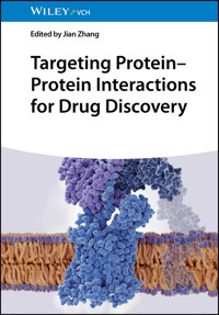
Targeting Protein–Protein Interactions for Drug Discovery
Edited by Jian Zhang
Editor
Prof. Jian ZhangShanghai Jiao Tong University227 South Chongqing RoadShanghai 200025China
Cover Design: WileyCover Image: © Juan Gaertner/Shutterstock
Library of Congress Card No.: applied for
British Library Cataloguing‐in‐Publication Data A catalogue record for this book is available from the British Library.
Bibliographic information published by the Deutsche Nationalbibliothek The Deutsche Nationalbibliothek lists this publication in the Deutsche Nationalbibliografie; detailed bibliographic data are available on the Internet at <http://dnb.d-nb.de>.
Print ISBN: 978-3-527-35360-6ePDF ISBN: 978-3-527-84580-4ePub ISBN: 978-3-527-84581-1oBook ISBN: 978-3-527-84582-8
© 2026 WILEY‐VCH GmbH, Boschstraße 12, 69469 Weinheim, Germany
All rights reserved (including those of translation into other languages, text and data mining and training of artificial technologies or similar technologies). No part of this book may be reproduced in any form – by photoprinting, microfilm, or any other means – nor transmitted or translated into a machine language without written permission from the publishers. Registered names, trademarks, etc. used in this book, even when not specifically marked as such, are not to be considered unprotected by law.
The manufacturer's authorized representative according to the EU General Product Safety Regulation is WILEY‐VCH GmbH, Boschstr. 12, 69469 Weinheim, Germany, e‐mail: [email protected].
While the publisher and the authors have used their best efforts in preparing this work, including a review of the content of the work, neither the publisher nor the authors make any representations or warranties with respect to the accuracy or completeness of the contents of this work and specifically disclaim all warranties, including without limitation any implied warranties of merchantability or fitness for a particular purpose. No warranty may be created or extended by sales representatives, written sales materials or promotional statements for this work. The fact that an organization, website, or product is referred to in this work as a citation and/or potential source of further information does not mean that the publisher and authors endorse the information or services the organization, website, or product may provide or recommendations it may make. This work is sold with the understanding that the publisher is not engaged in rendering professional services. The advice and strategies contained herein may not be suitable for your situation. You should consult with a specialist where appropriate. Further, readers should be aware that websites listed in this work may have changed or disappeared between when this work was written and when it is read. Neither the publisher nor authors shall be liable for any loss of profit or any other commercial damages, including but not limited to special, incidental, consequential, or other damages.
Preface
Protein–protein interactions (PPIs) lie at the heart of cellular processes, governing diverse biological activities such as signal transduction, gene regulation, immune responses, and enzymatic catalysis. The investigation of PPIs has not only deepened our understanding of fundamental biology but has also catalyzed breakthroughs in disease treatment and drug discovery. Dysregulation of PPIs is implicated in a wide array of diseases, including neurodegenerative disorders, infectious diseases, and cancers, underscoring the importance of elucidating their molecular mechanisms.
This book, dedicated to the exploration of PPIs, provides a comprehensive and systematic overview of the field. It encompasses foundational concepts, advanced methodologies, and emerging therapeutic strategies, reflecting the multidisciplinary nature of PPI research. Spanning 15 chapters, this volume serves as a valuable resource for researchers, educators, and students who are engaged in the study of cellular mechanisms, drug discovery, and biotechnology.
Chapter 1 introduces the essential concepts, roles, and research methodologies of PPIs, setting the stage for the in‐depth discussions that follow. Chapter 2 delves into the challenges and strategies of drug discovery targeting PPIs, including high‐throughput screening, structure‐based drug design, and computational prediction of PPI inhibitors. The subsequent chapters highlight key experimental and computational approaches to deciphering PPIs. Chapter 3 focuses on fluorescence resonance energy transfer (FRET) technology, a powerful tool for the real‐time analysis of molecular interactions in live cells, while Chapter 4 discusses the use of mass spectrometry (MS) for large‐scale mapping of PPI networks with high sensitivity and resolution. Chapter 5 explores proximity ligation assays (PLAs) for detecting PPIs in situ, emphasizing their spatial precision and adaptability for multiplexed detection. Chapter 6 reviews the application of surface plasmon resonance (SPR) for characterizing PPI specificity, affinity, and kinetics. Chapter 7 outlines computational methods for predicting PPI structures, with a special emphasis on protein docking and deep learning‐based approaches.
The discussion of therapeutic innovations continues in Chapters 8–14. Chapters 8 and 9 introduce diverse chemical scaffolds for PPI modulation, including foldamers as inhibitors of aberrant PPIs and sulfonyl‐γ‐AApeptides as novel modulators, respectively. Chapter 10 explores the development and application of stapled peptides as modulators of intracellular PPIs, offering enhanced stability, binding affinity, and cellular permeability. Chapter 11 addresses cyclic peptides, focusing on their unique conformational stabilization and therapeutic potential across a range of diseases. Chapter 12 shifts the focus to small‐molecule inhibitors targeting BCL‐family proteins, revealing their potential in cancer therapy. In Chapter 13, molecular glues are introduced as activators for PPIs, categorized into degraders, stabilizers, and inhibitors based on their biological effects. Chapter 14 highlights the targeting of the APC–Asef interaction for drug discovery in colorectal cancer therapy, offering a case study of specificity and clinical relevance.
The final Chapter 15 reflects on the future of PPI research and therapeutic discovery, discussing the application of computational methods in drug discovery, offering insights into how artificial intelligence and machine learning are shaping the discovery of PPI modulators.
Together, the chapters in this book provide a holistic perspective on PPIs, integrating basic science with translational applications. The exploration of both classical and novel approaches to PPI characterization and modulation offers a comprehensive reference for researchers aiming to unlock the therapeutic potential of PPIs. We hope this book will inspire further advancements in the field, driving the development of new technologies, therapeutics, and innovations that will ultimately benefit human health.
February 2025
Jian Zhang
1Exploring Protein–Protein Interactions: Concepts, Methods, and Implications
Mi Zhou and Renxiao Wang
Fudan University, School of Pharmacy, Department of Medicinal Chemistry, 826 Zhangheng Road, Shanghai 201203, People's Republic of China
1.1 General Concepts of Protein–Protein Interactions
Proteins are the fundamental machinery that drive the vast majority of cellular processes. These versatile biomolecules rarely perform in isolation; instead, up to 80% of proteins engage in intricate interactions with one another, forming dynamic networks that underpin the functional complexity of living organisms [1]. This chapter aims to provide a comprehensive overview of protein–protein interactions (PPIs). We will begin by elucidating the fundamental concepts of PPIs, covering their definition, structural characteristics, and pivotal roles in both physiological and pathological processes. Building upon this foundation, we delve into the diverse spectrum of current methods employed in PPI studies, showcasing both experimental and computational approaches, along with their varied applications. The chapter culminates in a discussion of the profound implications of PPI research, illuminating its potential in advancing medical understanding, revolutionizing drug discovery, and catalyzing technological innovations across various fields.
1.1.1 Definition of Protein–Protein Interactions
PPIs refer to physical contacts between two or more proteins that occur within a defined biological context. First, these interactions are intentional, not arising from random encounters, but rather precisely orchestrated by specific biomolecular forces and mechanisms. Second, they should be nongeneric, distinct from basic cellular processes like protein synthesis or degradation. Also of note, the formation and regulation of PPIs are highly organized in time and space, governed by a complex interplay of factors such as cell type, cell cycle phase, developmental stage, protein modifications, absence or presence of cofactors and binding partners, and environmental conditions [2, 3].
1.1.2 Structural Properties of Protein–Protein Interactions
The intricate architecture of proteins is critical in determining the specificity, strength, and functional outcomes of their interactions. By unraveling these structural characteristics, researchers can gain deeper insights into the molecular mechanisms underlying PPIs and develop strategies to manipulate them for therapeutic purposes.
Proteins are sophisticated macromolecules comprising an array of structural and functional units that work in concert to perform diverse biological tasks. Among these, domains and short linear motifs (SLiMs) serve as two main classes of functional modules that mediate PPIs. Domains, typically spanning 50–200 residues, are independently folding structural units within proteins that often harbor distinct biological activities. SLiMs, on the other hand, are compact, recurring functional peptides consisting of 3–10 residues, primarily found within intrinsically disordered regions (IDRs) [4]. Indeed, many PPIs can be categorized into domain–domain interactions (DDIs) or domain–motif interactions (DMIs). DDIs usually underpin the formation of stable and long‐lasting complexes, while DMIs are associated with transient and low‐affinity interactions [5].
The surface regions where the direct physical interactions between two or more proteins occur are termed interfaces. They are highly diverse in terms of size, shape, and chemical properties, determining the specific recognition and binding between proteins engaged in different biological processes. According to statistics, one‐sided size of an interface typically ranges from 200 to 2800 Å2, with the majority falling between 200 and 1200 Å2[6]. Although considered relatively flat, interfaces possess a complex topography characterized by cavities, grooves, and protruding regions. Complementary geometric features ensure that proteins bind to each other in the correct orientation and with high specificity. Moreover, interfaces encompass a variety of chemical interactions, including hydrogen bonds, hydrophobic interactions, electrostatic forces, salt bridges, and disulfide bonds, which collectively account for the specificity and stability of PPIs [7].
Within the interfaces, there exist clusters of residues known as “hot spots,” which make disproportionately large contributions to the binding affinity. Single‐point mutations of these residues to alanine may cause a substantial increase in the binding free energy (ΔΔG ≥ 2 kcal/mol), highlighting their critical roles in stabilizing the PPIs [8]. Hot spots have a distinctive amino acid composition, enriched with tryptophan, arginine, and tyrosine, due to the unique physicochemical properties of these residues like bulky side chains, the propensity to form hydrophobic surfaces, and the capacity to engage in hydrogen bonding [9]. With regard to spatial organization, hot spots cluster within tightly packed regions rather than being randomly distributed, facilitating the removal of water molecules upon binding [10]. Besides, they are surrounded by moderately conserved and energetically less important residues, which form an O‐ring to further occlude bulk solvent from the hot spots [9].
Proteins are not static, rigid structures; rather, they are dynamic entities capable of adopting multiple conformations. This inherent flexibility is critical for their diverse functions, particularly in PPIs. The traditional “lock and key” model, which implies a preexisting perfect fit between interacting proteins, fails to capture the dynamic nature of PPIs [11]. Subsequently, more precise descriptions have been put forward. The “induced fit” model suggests that upon initial contact, proteins undergo conformational changes to achieve an optimal fit with their binding partners [12]. Meanwhile, the “conformational selection” model proposes that proteins exist in an equilibrium of various conformations, with binding events selecting and stabilizing the most favorable conformation [13].
1.1.3 Diverse Types of Protein–Protein Interactions
PPIs manifest in a striking diversity of forms, each tailored to perform a specific biological role. In the following sections, we will explore several prominent types of PPIs, varying in structural properties, molecular recognition mechanisms, and functional outcomes.
1.1.3.1 Enzyme–Substrate Interactions
Enzymes are biological catalysts that bind to specific substrates through their active sites and facilitate the conversion of substrates into product molecules. Key examples that illustrate the diverse roles of enzyme–substrate interactions include: (i) protein phosphorylation and dephosphorylation, where kinases and phosphatases add and remove phosphate groups to substrates, respectively, act as molecular switches in signal transduction [14]; (ii) proteolytic cleavage, an irreversible process mediated by proteases that catalyze the hydrolysis of peptide bonds in target proteins, precisely governs protein maturation, activation, stability, and localization [15]; (iii) histone modifications, such as acetylation and methylation, are carried out by enzymes like histone acetyltransferases (HATs) and histone methyltransferases (HMTs), contributing to epigenetic regulation of gene expression [16].
1.1.3.2 Receptor–Ligand Interactions
Cells possess an intricate communication network to sense and respond to environment cues and stimuli. This network is built upon cell surface receptors that bind external signaling molecules termed ligands, initiating downstream signaling cascades within the cell and manipulating various physiological processes like growth, development, immune response, and metabolism [17]. G protein‐coupled receptors (GPCRs) constitute the largest family of these receptors, characterized by a unique architecture comprising seven transmembrane helices joined by intracellular and extracellular loops [18]. Chemokines, a class of small secreted proteins, exemplify protein ligands that bind to a specific subfamily of GPCRs called chemokine receptors. By sequentially binding to the N‐terminal region and extracellular loops of their receptors, chemokines induce receptor conformational rearrangement and activation that direct immune cell migration and positioning during inflammation [19].
1.1.3.3 Antigen–Antibody Interactions
Antibodies are glycoproteins produced by B lymphocytes to recognize and bind to foreign substances known as antigens, which can be present on the surface of invading pathogens like viruses and bacteria. This interaction launches an immune response to neutralize and eliminate pathogens, thus protecting the body from infection and disease [20]. An antibody is composed of two identical heavy chains and two identical light chains connected by disulfide bonds. Antigen recognition is achieved by the complementarity‐determining regions (CDRs) located at the N‐terminus of both heavy and light chains that precisely match antigen's epitope [21]. Beyond their pivotal role in the immune system, the exquisite specificity of antigen–antibody interactions has also been exploited for diagnostic and therapeutic applications.
1.1.3.4 Chaperone–Client Interactions
Chaperones function as protein quality control machinery, engaging with client proteins to assist in their de novo folding, subcellular translocation, and recovery from stress‐induced misfolding and aggregation. Many chaperones belong to the heat shock protein (HSP) families, including HSP60, HSP70, HSP90, and HSP100 [22]. Chaperones from different families exhibit structural and functional diversity, reflected in their distinct binding patterns to clients. Some utilize ATP binding to allosterically regulate their conformation, facilitating efficient client binding and release. Others assemble into oligomeric complexes, creating an enclosed environment for client encapsulation [23]. Structural biology studies have revealed that, in some cases, chaperones bind their clients in interconverting conformational ensembles that are locally highly dynamic. This binding behavior allows clients to undergo folding while bound to chaperones and enables chaperones to accommodate a broad range of clients with varying folding properties and limited shape complementarity [24, 25].
1.1.3.5 Scaffold Interactions
Scaffold proteins are molecular organizers that bring together two or more signaling molecules like kinases and receptors into higher‐order complexes to modulate their activities, thereby enabling the efficient spatial–temporal coordination of cellular signaling events [26]. This remarkable organizational capability of scaffolds is attributed to their unique modular architecture. Typically, they encompass discrete interaction domains capable of recognizing and binding to SLiMs (e.g. phosphotyrosine‐ and proline‐containing sequences) on target proteins, along with IDRs that confer flexibility and functional versatility [27]. In addition to simply tethering targets in proximity, scaffolds can exert allosteric regulation on the targets as well. In this way, they can handle both the linear input–output signal transmission and complicated feedback loops within distinct pathways and even promote signal integration and crosstalk among various pathways [28, 29].
1.2 Functional Significance of Protein–Protein Interactions
Far from being static or isolated events, PPIs are dynamically woven into complex networks that enable proteins to communicate, coordinate, and cooperate in carrying out the essential functions of life. Meanwhile, disruptions or aberrations in PPIs can lead to a cascade of dysfunction, contributing to the development of various diseases.
1.2.1 Cellular Signal Transduction
Signal transduction is the process by which extracellular signals are converted into cellular responses. It is organized into three distinct stages: reception, transduction, and response, each involving PPIs to ensure high fidelity of signal transmission (Figure 1.1).
Figure 1.1 A graphic representation of the signal transduction process. Extracellular signals, upon binding to their receptors, are transmitted through intracellular signaling pathways, culminating in a series of cellular responses.
(Source: Mi Zhou and Renxiao Wang.)
The reception stage commences with receptor–ligand interactions. To be exact, a variety of extracellular signaling molecules, ranging from neurotransmitters and hormones to proteins like chemokines, thrombin, and growth factors, are recognized by their corresponding receptors embedded in the membrane, such as GPCRs and receptor tyrosine kinases (RTKs) [17]. Upon ligand binding, receptors generally undergo conformational changes, activating associated proteins or triggering homodimerization and autophosphorylation to initiate downstream signaling pathways [30, 31].
During the transduction stage, the signal is propagated from the cell surface to the intracellular targets through an elaborate network of signaling cascades. These cascades are characterized by a series of posttranslational modifications, predominantly phosphorylation events driven by kinase–substrate interactions. A classic example is the mitogen‐activated protein kinase (MAPK) pathway, where a cascade of three kinases, namely MAPK kinase kinase (MAPKKK), MAPK kinase (MAPKK), and MAPK, phosphorylate and activate each other in a sequential manner [32]. A similar phosphorylation‐dependent activation mechanism is also observed in other crucial pathways, including nuclear factor kappa B (NF‐κB) pathway, phosphoinositide 3‐kinase (PI3K)/protein kinase B (AKT) pathway [33], and Janus kinase (JAK)/signal transducer and activator of transcription (STAT) pathway [34]. While some interactions serve to relay and amplify the signal, others involving phosphatases and inhibitory proteins lead to signal decay and termination [35]. Moreover, adaptor, scaffold, and docking proteins further fine‐tune signal transduction by manipulating the assembly, function, and localization of relevant proteins [36].
Finally, the signal cascades converge on the regulation of their target proteins (e.g. transcription factors, enzymes, cytoskeletal proteins, and apoptotic proteins), eliciting the desired cellular responses such as changes in gene expression, cell growth, differentiation, and apoptosis.
1.2.2 Regulation of Gene Expression
Gene expression in eukaryotes encompasses the processes of transcription, RNA processing, and translation, wherein PPIs play a critical regulatory role, contributing to the accurate transfer of genetic information from DNA to messenger RNA (mRNA) to protein.
At the onset of transcription activation, transcriptional activators bind to specific DNA sequences and then recruit coactivators to improve transcription efficiency. Some coactivators like HMTs and HATs perform histone modifications and chromatin remodeling, rendering the DNA more accessible to the transcriptional machinery [37]. Others, such as the mediator complex, bridge RNA polymerase II (Pol II) and general transcription factors onto core promoters, resulting in the assembly of a pre‐initiation complex (PIC) that directs the initiation of transcription [38] (Figure 1.2a). On the other hand, the interplay between transcriptional repressors, co‐repressors, and histone‐modifying enzymes, like histone deacetylases (HDACs), can suppress the transcription process [39].
Figure 1.2 Simplified schematic of the gene expression processes in eukaryotes, including (a) transcription, (b) RNA processing, and (c) translation.
(Source: Mi Zhou and Renxiao Wang.)
Once transcribed, the precursor mRNA (pre‐mRNA) undergoes several processing steps to become the mature mRNA. These include 5′‐end capping, splicing, and 3′‐end cleavage and polyadenylation, each orchestrated by specialized molecular machinery (Figure 1.2b). Capping involves the sequential recruitment of capping enzymes onto the Pol II, where they catalyze modifications to the 5′ end of the nascent pre‐mRNA [40]. Next, splicing is executed by a highly dynamic and massive ribonucleoprotein complex termed the spliceosome, which is assembled by ordered binding and release of small nuclear ribonucleoproteins (snRNPs) and numerous other splicing factors [41]. Finally, 3′‐end processing is performed by the cleavage and poly‐adenylation specificity factor (CPSF), a large multi‐subunit complex that utilizes endonuclease, poly(A) polymerase, and accessory proteins to cleave the pre‐mRNA and add a poly(A) tail [42].
During translation initiation, a suite of eukaryotic translation initiation factors (eIFs) mediate the assembly of the 40S and 60S ribosomal subunits at the mRNA start codon, culminating in the formation of the 80S ribosome ready for protein synthesis [43]. Elongation ensues, with eukaryotic elongation factors (eEFs) assisting the ribosome in moving along the mRNA and adding amino acids to the growing polypeptide chain [44]. At last, upon encountering a stop codon, the ribosome triggers the recruitment of eukaryotic release factors (eRFs), promoting the release of the nascent peptide and marking the termination of translation [45] (Figure 1.2c).
1.2.3 Immune Response
The immune response is built upon a foundation of intricate cell–cell communication, with PPIs serving as the key messengers. These interactions, occurring between and within immune and nonimmune cells, are indispensable for recognizing pathogens, activating signal cascades, and mounting an effective defense against invading threats.
1.2.3.1 Immune Cell Migration
Migration is crucial for immune cells to patrol for foreign antigens and traffic to inflammation or infection sites. This process often involves transmigration across epithelial barriers, relying on adhesion factors‐governed PPIs. The initial capture and rolling of immune cells along the endothelial surface are mediated by interactions between selectins (e.g. P‐selectin) on endothelial cells and their ligands (e.g. P‐selectin glycoprotein ligand‐1 [PSGL‐1]) on immune cells. Film adhesion is subsequently established through interactions between integrins (e.g. lymphocyte function‐associated antigen‐1 [LFA‐1]) on immune cells and their ligands (e.g. intercellular adhesion molecule‐1 [ICAM‐1]) on endothelial cells [46]. Finally, platelet endothelial cell adhesion molecule‐1 (PECAM‐1) molecules on both endothelial and immune cells engage in homophilic interactions, promoting transendothelial migration [47].
1.2.3.2 T‐Cell Antigen Recognition and Activation
When a foreign antigen invades the body, the T‐cell receptor (TCR) on the T‐cell surface specifically recognizes and binds to an antigen peptide presented by major histocompatibility complex (MHC) molecules on the surface of antigen‐presenting cells (APCs). In parallel, co‐receptors, such as cluster of differentiation 4 (CD4) and CD8, on the T‐cell surface also engage with the MHC molecules, reinforcing the stability of the TCR‐peptide‐MHC complex. Co‐receptor engagement brings the lymphocyte‐specific protein tyrosine kinase (Lck) into proximity with the TCR‐CD3 complex. Lck then induces the phosphorylation of CD3 molecules, initiating downstream signaling cascades that ultimately lead to T‐cell activation, proliferation, and differentiation. Additionally, PPIs between co‐stimulatory molecules, such as CD28 on T cells and CD80/86 on APCs, enhance TCR‐induced signals and prevent T‐cell anergy [48] (Figure 1.3a).
Figure 1.3 Illustration of TCR signaling (a) and BCR signaling (b) during antigen recognition.
(Source: Mi Zhou and Renxiao Wang.)
1.2.3.3 B‐Cell Antigen Recognition and Activation
Upon engagement with its cognate antigen, the B‐cell receptor (BCR) undergoes clustering on the plasma membrane. This event triggers the phosphorylation of CD79A and CD79B signaling subunits on BCR by Src family tyrosine kinases, primarily Lck/Yes‐related novel protein tyrosine kinase (Lyn). Phosphorylated CD79A and CD79B then serve as docking sites for the recruitment and activation of spleen tyrosine kinase (Syk). Once activated, Syk propagates signals to downstream signaling cascades [49, 50] (Figure 1.3b). Similarly, PPIs involving co‐receptors and co‐stimulatory molecules can augment BCR‐induced signals [51]. Ultimately, these signaling events can drive B‐cell differentiation into plasma cells, specialized in secreting antibodies that target specific antigens and neutralize or clear them from the body.
1.2.4 Protein Degradation Pathway
To maintain protein homeostasis, cells employ sophisticated quality control mechanisms that promptly identify and eliminate aberrant or unwanted proteins. Two major protein degradation systems, the ubiquitin‐proteasome system (UPS) and the autophagy‐lysosomal pathway, are essential for this vital task.
UPS selectively targets substrate proteins, tagging them with poly‐ubiquitin chains for degradation by the proteasome. The ubiquitination process is carried out by the sequential action of three key enzymes: E1 ubiquitin‐activating enzymes, E2‐ubiquitin‐conjugating enzymes, and E3 ubiquitin ligases. E1 initiates the process by activating ubiquitin, forming a thioester bond between ubiquitin's C‐terminus and its own catalytic cysteine. After that, E1 recruits E2 and transfers ubiquitin to E2's catalytic cysteine. Finally, E3 recognizes and binds to the substrate along with the E2‐ubiquitin complex, facilitating the transfer of ubiquitin from E2 onto the substrate [52]. The degradation process is executed by the 26S proteasome, a large multi‐subunit protease complex consisting of two main components: 20S core particle (CP) and 19S regulatory particles (RPs). Each component is made up of multiple distinct subunits, with dedicated chaperones assisting in stabilizing intermediate structures and ensuring the proper incorporation of individual subunits. Once assembled, the 20S CP and 19S RPs unite to form the functional 26S proteasome, poised for the degradation of poly‐ubiquitylated substrates [53].
The autophagy‐lysosomal pathway focuses on the sequestration of cargo (e.g. damaged organelles and protein aggregates) within autophagosomes, which then fuse with lysosomes for cargo degradation and recycling. This process is mainly driven by a set of autophagy‐related (ATG) proteins and their interactions. The unc‐51‐like kinase 1 (ULK1) complex initiates phagophore nucleation, followed by the recruitment of the class III phosphatidylinositol 3‐kinase (PI3K) complex that promotes autophagosome formation and elongation. Two ubiquitin‐like conjugation systems, namely the ATG12 and microtubule‐associated protein 1 light chain 3 (LC3)/GABA type A receptor‐associated protein (GABARAP) systems, further contribute to autophagosome maturation [54]. Cargo recognition and delivery are mediated by cargo receptors like sequestosome 1 (p62/SQSTM1), which interact with LC3 on the autophagosome membrane, thereby guiding the cargo for degradation within autophagolysosomal compartment [55].
1.2.5 Disease Mechanisms
PPIs are fundamental for maintaining cellular homeostasis. However, their dysregulation, whether caused by genetic anomalies or the introduction of foreign proteins, plays a significant role in the development and progression of various diseases.
1.2.5.1 Cancer
Neoplastic progression entails the acquisition of a panel of functional capabilities that enable tumor growth and metastasis, termed “hallmarks of cancer” [56]. These hallmarks are inextricably linked to dysregulated PPIs. For instance, aberrant activation of oncoproteins, such as rat sarcoma virus (RAS), induces sustained proliferative signaling by engaging downstream effectors [57]. Negative regulation of the tumor suppressor p53 by mouse double minute 2 homolog (MDM2) is one mechanism whereby cancer cells evade growth suppression [58]. Abnormal expression of anti‐apoptotic B‐cell lymphoma‐2 (BCL‐2) family proteins, through sequestering pro‐apoptotic counterparts, often contributes to cell death resistance [59]. Hypoxic tumor microenvironment stimulates the production of pro‐angiogenic factors like vascular endothelial growth factor (VEGF), which activate their receptors on endothelial cells to induce angiogenesis [60]. Various cancer cells overexpress programmed cell death ligand 1 (PD‐L1) and exploit the PD‐L1/programmed cell death protein 1 (PD‐1) pathway to escape immune destruction [61].
1.2.5.2 Neurodegenerative Diseases
Alzheimer's disease (AD) is characterized by two hallmark pathologies, amyloid‐β (Aβ) plaques and neurofibrillary tangles. The former arises from the sequential proteolytic cleavage of amyloid precursor protein (APP) by secretases, generating Aβ peptides that accumulate and deposit into amyloid plaques [62]. The latter comprises insoluble aggregates of hyperphosphorylated tau protein, a consequence of an imbalanced phosphorylation and dephosphorylation [63]. Parkinson's disease (PD) is marked by the pathological aggregation of α‐synuclein (α‐syn) into insoluble fibrillar structures called Lewy bodies. While genetic and environmental factors contribute to α‐syn aggregation, its engagements with various other proteins, including those implicated in neurodegenerative diseases (e.g. tau) and regulators of actin cytoskeleton and neurotransmission, can accelerate this process [64]. Likewise, Huntington's disease (HD) is driven by the self‐aggregation of mutant huntingtin (mHTT) protein. Abnormal PPIs between mHTT and other proteins disrupt crucial cellular processes such as axonal transport, mitochondrial fission, and gene transcription, resulting in neuronal and mitochondrial dysfunction [65].
1.2.5.3 Infectious Disease
The pathogenesis of infections caused by viruses, bacteria, fungi, or parasites hinges critically on a complex interplay between pathogen proteins and host proteins. These PPIs underpin every stage of infections, including:
(i) Invasion: Viruses gain entry into the host by utilizing their surface proteins to recognize specific host cell receptors. A notable example is the binding of the severe acute respiratory syndrome coronavirus 2 (SARS‐CoV‐2) spike (S) protein to the human angiotensin‐converting enzyme 2 (ACE2) receptor, which mediates the viral attachment and entry into the respiratory epithelial cells
[66]
. Bacteria and fungi, on the other hand, rely on surface‐exposed adhesins that interact with corresponding host cell receptors or extracellular matrix proteins, enabling their attachment and colonization [
67
,
68
].
(ii) Immune Evasion: Pathogens employ various strategies to escape or suppress host's immune defenses. Some, like human cytomegalovirus (HCMV), encode specific proteins that target and interfere with MHC molecules, disrupting antigen presentation
[69]
. Others, such as
Streptococcus pyogenes
, secrete immunoglobulin‐degrading enzymes to cleave antibodies, rendering them ineffective
[70]
.
(iii) Replication: Pathogens often hijack host cellular machinery for their replication needs. For example, expression of the human immunodeficiency virus (HIV) genome is regulated by its trans‐activator of transcription (TAT) protein, which assembles with host's positive transcription elongation factor b (P‐TEFb) onto the viral promoter, thus enhancing the production of viral transcripts
[71]
.
1.3 Methods for Analyzing Protein–Protein Interactions
The field of PPI research has undergone a transformative evolution with the advent of diverse and sophisticated methodologies, ranging from traditional biochemical techniques to high‐throughput technologies and advanced computational algorithms. Together, these complementary strategies form a powerful toolkit for researches to uncover insights into PPIs, from atomic‐level details to intricate systemic networks.
1.3.1 Experimental Methods
A diverse array of experimental methods, grounded in either biophysical, biochemical, or genetic principles, have been established to identify and characterize PPIs across various biological contexts and scales. These methods offer multifaceted capabilities, including:
1.3.1.1 Structure Determination
Elucidating the 3D structure of PPIs may provide valuable insights into the geometry and physicochemical characteristics of the binding interfaces and even shed light on potential therapeutic interventions [72]. The primary techniques currently employed for high‐resolution protein structure determination are X‐ray crystallography, nuclear magnetic resonance (NMR) spectroscopy, and cryogenic electron microscopy (cryo‐EM). X‐ray crystallography offers detailed information on atom positions and chemical bonds by analyzing X‐ray diffraction patterns generated from protein crystals [73]. As the workhorse of structural biology, it accounts for over 80% of experimental structures deposited in the Protein Data Bank (PDB), with the median resolution plateauing at ∼2 Å as of 1990 [74]. Inherent limitations of crystallography are the requirement to obtain well‐diffracting crystals and the occurrence of crystal packing artifacts. NMR spectroscopy, eliminating the crystallization process, enables structural studies of proteins in solution with the aid of structural restraints acquired from different NMR experiments. Given its strength in uncovering protein dynamics and flexibility over a range of timescales, this technique is well‐suited for investigating intrinsically disordered proteins, proteins in transient states, and weak interactions [75, 76]. Whereas drawbacks, such as the necessity of isotope labeling and size limit (typically <50 kDa), should be taken into account. Driven by technological breakthroughs over the past decade, cryo‐EM has experienced a “resolution revolution” and is gaining widespread popularity [77]. It relies on direct visualization of proteins embedded in a thin layer of vitreous ice, thus unraveling protein structures in close‐to‐physiological state with a routine resolution of 2.5–4.5 Å [78]. Compared with other techniques, it is more tolerant to sample quantity and purity. Although high‐resolution reconstruction of small‐size proteins (<100 kDa) remains challenging for cryo‐EM, novel strategies have been applied to break through the barrier [79]. In conclusion, each technique has its own pros and cons, rendering them complementary tools to decipher the intricate 3D structures of proteins and protein complexes [80, 81].
1.3.1.2 Affinity, Kinetics, and Thermodynamics Measurement
Quantitative in vitro analysis of PPIs in terms of affinity, kinetics, and thermodynamics is essential for exploring the underlying mechanisms of molecular recognition. Binding affinity, defined as the strength of the interaction between biomolecules that bind reversibly, is typically quantified through the equilibrium dissociation constant (Kd) [82]. A variety of experimental methods have been established to obtain the Kd values, including fluorescence polarization (FP), surface plasmon resonance (SPR), biolayer interferometry (BLI), isothermal titration calorimetry (ITC), differential scanning calorimetry (DSC), microscale thermophoresis (MST), fluorescence and bioluminescence resonance energy transfer (FRET and BRET), AlphaScreen, protein‐fragment complementation assay (PCA), enzyme‐linked immunosorbent assays (ELISA), and so forth [83, 84]. Of the methods mentioned earlier, SPR and BLI can also yield kinetic parameters, specifically the association rate constant (kon) and dissociation rate constant (koff) [85]. Both technology platforms enable real‐time and label‐free monitoring of binding events and have a wide range of applications, such as investigating the assembly and activation of signaling complexes [86], antibody evaluation and selection [87], and protein engineering [88]. On the other hand, calorimetric methods like ITC and DSC can furnish extra thermodynamic insight. In the case of ITC, a comprehensive thermodynamic profile, including Gibbs free energy change (ΔG), enthalpy change (ΔH), entropy change (ΔS), and heat capacity change (ΔCp), can be acquired from a single experiment [89]. Combining these data with structural information can further interpret the driving forces for the binding [90]. As a final note, prior to choosing the appropriate methods, factors such as the need for immobilization or labeling, affinity range, sample and time consumption, throughput performance, and the potential risks of false positives and false negatives should be carefully considered. Besides, integrating and validating results from multiple methods is crucial to ensure data accuracy and reliability.
1.3.1.3 Large‐Scale PPI Network Mapping
Systematic mapping of PPI network within a cell or an organism, termed the “interactome,” is an ongoing endeavor aimed at deciphering previously uncharacterized proteins, global proteome organization and function, and genotype–phenotype relationships. For nearly three decades, proteome‐wide interactomes of yeast, human, and other model organisms have been delineated and progressively expanded using diverse high‐throughput experimental techniques [91, 92]. One powerful method to identify binary PPIs is yeast two‐hybrid (Y2H) screening. In the original Y2H, PPIs are detected inside the yeast nucleus based on the functional reconstitution of a transcription factor. Thereafter alternative versions have broadened its applicability to membrane proteins, proteins with posttranslational modifications, and PPIs in mammalian cells [93]. In 2020, an ORFeome resource covering 17,408 protein‐coding genes was established, and over 150 million protein pairs were screened using Y2H, generating the largest human binary protein interactome consisting of ∼53,000 high‐quality PPIs [94]. In contrast, co‐complex mapping is typically carried out via affinity purification coupled to mass spectrometry (AP‐MS). In AP‐MS, a bait protein and its binding partners are isolated from cell lysate using bait‐specific antibody and then subjected to mass spectrometric analysis for identification of the purified components. AP‐MS excels at detecting PPIs under near physiological conditions but falls short in capturing weak or transient interactions [95]. It was recently employed in the BioPlex project for the identification of dual human interactomes that encompass 118,162 and 70,966 PPIs in 293T and HCT116 cells, respectively [92]. Despite significant experimental efforts, current human interactomes remain incomplete and present high levels of noise [96]. Methodological advances, integration of various data sources, and incorporation of context‐specific (e.g. cell‐, tissue‐, and disease‐specific) information may enhance both interactome coverage and quality, providing researchers with a more comprehensive and dynamic perspective.
1.3.2 Computational Methods
Despite remarkable efforts made by experimental techniques, they often struggle to capture transient and weak interactions or those involving intrinsically disordered proteins. Besides, they are time‐consuming and labor‐intensive in general. Computational methods offer a complementary and efficient alternative, harnessing vast datasets and predictive models to infer and analyze PPIs.
1.3.2.1 Sequence‐Based Methods
The abundance of protein sequence data in public databases has propelled the development of strategies for PPI prediction solely based on sequence information. Herein are some widely adopted approaches: (i) Ortholog‐based methods are premised on the evolutionary conservation of PPIs across species, utilizing known PPIs in one species to identify orthologous interactions (“interologs”) in another species [97]. (ii) Domain‐based methods are built on the concept that PPIs are primarily mediated by domains. By integrating experimentally determined PPI datasets with domain information sourced from databases (e.g. Pfam [98]), DDIs can be inferred and serve as the basis for predicting novel PPIs [99]. (iii) Co‐evolution analysis assumes that amino acid substitutions in one protein are often accompanied by compensatory substitutions in its interacting partner to maintain functional integrity. Multiple sequence alignments are employed to quantify the co‐evolutionary relationships between residue pairs within proteins of interact, with highly co‐evolving pairs presumed to be in physical or functional contact, suggesting a potential PPI [100]. (iv) Machine learning‐based methods have gained considerable prominence, progressing from traditional algorithms like support vector machines (SVMs) and random forests (RFs) to advanced deep learning models such as deep neural networks (DNNs) and recurrent neural networks (RNNs). These models extract sequence‐based features (e.g. amino acid composition, physicochemical properties, and evolutionary information) from large data sets, capture the underlying patterns, and make accurate predictions for novel protein pairs [101, 102]. To date, protein sequence data remain the primary source for computational PPI prediction. Ongoing improvements are driven by the growing availability of high‐quality PPI data sets, the introduction of innovative sequence encoding schemes, as well as the integration with diverse biological data such as structure information [103].
1.3.2.2 Structure‐Based Methods
Structural characterization of PPIs remains a challenge due to their inherent complexity and technological limitations. In fact, fewer than 5% of human PPIs have been structurally resolved through experimental techniques or homology modeling [104]. Molecular docking has emerged as a rapid and cost‐effective alternative to bridge this gap by predicting 3D structures of protein complexes. During the docking process, unbound protein structures or structural models are used as input, generating numerous putative binding modes through sampling. The most plausible ones are then selected by means of scoring and ranking [105]. A wide variety of sophisticated docking programs have been developed, each employing different sampling algorithms and scoring functions, including ZDOCK [106], HADDOCK [107], ClusPro [108], and RossettaDock [109]. Continuous efforts have been made to improve prediction accuracy and reliability, such as accounting for backbone and side‐chain flexibility, incorporating experimental data like interface details and distance restraints, and integrating deep learning techniques [110, 111]. Molecular dynamics (MD) simulation is another powerful computational technique widely employed to investigate the structural properties of proteins and their complexes. It applies Newtonian mechanics to study the motion of atoms and molecules within a system at nanosecond to microsecond timescales. Analysis of the output trajectories may provide atomic‐level insights into the dynamic behavior of protein molecules [112]. In the realm of PPI studies, MD simulation is utilized to assess complex stability, analyze residue flexibility, estimate interaction strengths, and explore the impact of various conditions (e.g. environmental factors, solvents, and mutations) on PPIs [113]. Popular tools for MD simulation include AMBER [114], GROMACS [115], CHARMM [116], and NAMD [117]. Future developments are focused on extending the simulation timescales, improving the accuracy of force fields, and integrating artificial intelligence techniques for enhanced prediction and analysis capabilities.
1.3.2.3 Network‐Based Methods
The identification of unmapped interactions within a PPI network can be regarded as the task of predicting missing links between nodes in a graph representation, where nodes denote proteins and links represent interactions between them. Such network‐based prediction approaches can be broadly categorized as follows [118]: (i) Similarity‐based methods leverage local properties of nodes to establish a similarity score function for link prediction. A classic example is the common neighbor method, which assumes that two proteins sharing a greater number of common neighbors exhibit higher similarity and are thus more likely to interact [119]. (ii) Probabilistic methods aim to identify model parameters that best explain the inherent structure of a network, such as community structure. Taking the stochastic block model as an example, each protein is assigned to multiple communities, reflecting its participation in various biological processes. This model operates on the principle that proteins within the same community are more prone to interact with each other compared to proteins from different communities [120]. (iii) Factorization‐based methods constitute a class of graph embedding techniques that factorize the network's adjacency matrix to build low‐dimensional representations for each node. Geometric Laplacian Eigenmap Embedding (GLEE) is one such method that exploits the simplex geometry of the Laplacian matrix to capture the most basic structural property of the graph, demonstrating strong competence in predicting unobserved interactions in PPI networks [121]. (iv) Machine learning‐based methods make predictions by learning patterns from training data. As a case in point, a conditional generative adversarial network (cGAN) can be conditioned on the raw topological features of the network, enabling the generator to produce new probable links [122].
1.4 Implications of the Basic Research on Protein–Protein Interactions
Given their essential role in biological functions, deciphering the intricate PPI network not only holds paramount significance in the realm of molecular biology but also has far‐reaching implications in medical advances, drug discovery, and biotechnology innovations.
1.4.1 Advancing Disease Understanding and Diagnosis
PPIs play pivotal roles in cellular functions, from normal physiological processes to the development and progression of various diseases. In cancer, genomic alterations give rise to oncogenic PPIs, driving uncontrolled cell proliferation and metastasis [123]. Neurodegenerative diseases often involve mutations, posttranslational modifications, or misfolding of aggregation‐prone proteins, resulting in aberrant PPIs that exacerbate cytotoxicity [124]. Infectious diseases heavily rely on host–pathogen PPIs to facilitate pathogen entry, replication, and dissemination within the host organism [125]. Consequently, identifying and characterizing these interactions is essential for unraveling the molecular basis of diseases. The COVID‐19 pandemic serves as a compelling case in point. Investigation of the interaction between the SARS‐CoV‐2 spike protein and the human ACE2 receptor advanced our knowledge of the virus's entry mechanism into host cells [126]. Further affinity measurement of different SARS‐CoV‐2 variants toward ACE2 helped to assess the virus's infectivity, transmissibility, and potential threat to public health, offering invaluable information for epidemiological studies [127].
Current molecular diagnostics mainly focus on identifying genetic alterations and individual protein biomarkers. However, emerging evidence suggests that altered PPI patterns are strongly linked to disease progression and clinical outcomes, holding significant potential as novel diagnostic and prognostic biomarkers [128]. One notable example lies in the application of proximity ligation assay (PLA) to detect protein complexes formed between epidermal growth factor receptor (EGFR) and growth factor receptor‐bound protein 2 (GRB2) in non‐small cell lung cancer (NSCLC) patient specimens. This approach allows for in situ assessment of EGFR signaling activity and has proven to be a superior predictor of clinical response to targeted therapies compared to conventional strategies based on EGFR mutations or protein abundance [129]. As our understanding of disease‐related PPIs deepens and advanced methods for PPI profiling in clinical specimens emerge, PPI‐based diagnostics, alongside genomic and proteomic analyses, will become increasingly powerful tools for precision and personalized medicine.
1.4.2 Driving Target‐Based Drug Discovery
Given their central role in numerous disease pathways, PPIs have emerged as attractive targets for therapeutic intervention. However, modulating these interactions presents challenges due to the complex and dynamic nature of PPI interfaces, which are often characterized by extensive, shallow surfaces with limited deep cavities. PPIs lack endogenous small‐molecule ligands that act as a starting point for drug discovery [130]. Despite these obstacles, substantial efforts have been made to overcome these barriers. Structural biology techniques can pinpoint hot spots at PPI interfaces, serving as a basis for the structure‐based design of PPI modulators. Computational methods, such as virtual screening and molecular docking, can predict the binding affinity and mode of molecules at binding sites, accelerating lead discovery. By integrating these approaches, researchers can efficiently identify and optimize PPI modulators, opening a new avenue for drug discovery.
Over the past decade, the field of PPI modulators has witnessed remarkable progress, with quite a few candidates being approved for medical use or entering clinical trials for the treatment of various diseases, particularly solid tumors [131]. Small molecules constitute the most abundant class of PPI modulators, offering advantages such as oral bioavailability and membrane permeability. Classic PPI targets include interactions between antiapoptotic BCL‐2 family proteins and their proapoptotic counterparts, X‐linked inhibitor of apoptosis (XIAP) and caspase‐9, and MDM2‐p53 [132]. A significant milestone was achieved in 2016 with the approval of venetoclax, a BCL‐2 inhibitor, for the treatment of chronic lymphocytic leukemia (CLL), making a breakthrough in the development of PPI‐modulating therapies [133]. The second category comprises antibodies, which are well‐suited for covering large PPI interfaces but typically restricted to extracellular targets. Their primary focus lies in manipulating PPIs involving immune checkpoint molecules, such as PD‐1/PD‐L1 or CD40/CD40 ligand (CD40L) interaction, thereby exerting profound influences on immune responses [134, 135]. Peptides represent the third category of PPI modulators, exhibiting high selectivity but hampered by a short half‐life. To address this, chemical modifications like cyclization are frequently employed to improve their pharmacokinetic properties [136].
1.4.3 Fostering Innovations in Biotechnology
Biotechnology often harnesses the power of protein interactions to develop innovative solutions and products. This is vividly illustrated in synthetic biology, where naturally sourced or computationally designed PPIs serve as building blocks for constructing sophisticated genetic and protein circuits, endowing living cells with novel functionalities [137]. Generally, PPIs can be engineered to function as toggle switches and logic gates within synthetic genetic circuits, offering precise control over gene expression [138, 139]. Furthermore, by incorporating PPIs that govern posttranscriptional modifications like phosphorylation, synthetic protein circuits can rapidly respond to environmental changes, enabling dynamic modulation of protein interactions, localization, and degradation [140]. These programmable circuits hold immense potential for numerous applications. For instance, researchers created a chemically disruptable heterodimer based on the interaction between BCL‐XL (B‐cell lymphoma‐extra large) and BH3 (BCL‐2 homology 3)‐mimic peptide. Incorporating this PPI into the design of chimeric antigen receptor (CAR) led to a controllable CAR‐T cell therapy, whose activity could be modulated by timed administration of a small‐molecule BCL‐XL inhibitor [141].
Protein assemblies found in nature, such as actin fibers, microtubules, and viral capsids, showcase the remarkable role of PPIs in organizing complex architectures. Inspired by this, scientists have employed rational design to fabricate protein assemblies with diverse superstructures like nanowires, nanotubes, 2D and 3D lattices, and cage structures [142]. This nanotechnology paves the way for the development of advanced biomaterials with tailored properties and functionalities. As an example, coiled‐coils are ubiquitous protein oligomerization motifs that have the inherent ability to wrap each other into supercoiled helices. These structures can be engineered to undergo higher‐order assembly into fibrous network‐based hydrogels [143]


