117,99 €
Mehr erfahren.
- Herausgeber: John Wiley & Sons
- Kategorie: Fachliteratur
- Sprache: Englisch
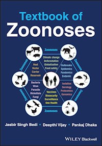
Textbook of Zoonoses
Comprehensive resource covering the aetiology, epidemiology and transmission cycle, clinical symptoms, diagnosis, and prevention and control strategies of the important zoonoses.
Zoonoses are the diseases which can spread from animals to humans. This book covers all important zoonoses that are prevalent in today’s world. As a modern learning resource, it incorporates recent scientific developments and concepts to give readers a complete overview of each zoonoses. Written by three well-qualified authors in academia, sample topics covered within the book include:
- Bacterial, viral, parasitic, rickettsial, fungal, prion, and foodborne zoonoses
- Aetiology and epidemiology of each zoonotic disease
- Clinical symptoms and diagnosis in animals and humans
- Treatment options, plus prevention and control strategies
- CDC classification of zoonotic agents and the WHO’s list of ‘neglected zoonoses’
Written for undergraduate and postgraduate students studying veterinary public health and epidemiology, Textbook of Zoonoses is also a helpful resource for other veterinary and medical professionals interested in public health and epidemiology.
Sie lesen das E-Book in den Legimi-Apps auf:
Seitenzahl: 754
Veröffentlichungsjahr: 2022
Ähnliche


Table of Contents
Cover
Title Page
Copyright Page
Dedication Page
Preface
How Did the Idea for Writing the Book Come Up?
Who Should Use this Book?
Book Content and Our Expectations
Acknowledgements
About the Authors
Introduction to Zoonoses
What are Zoonoses?
Overview on Zoonoses
Classification of Zoonoses
Major Transmission Routes of Zoonoses
References
Understanding Concepts and Terms Related to Zoonoses
Important Terms
Factors Responsible for the Emergence of Infectious Diseases [8]
References
Section 1: Bacterial Zoonoses
1 Anthrax
Etymology
Synonyms
Aetiology and Pathogen Characteristics
Pathogenesis and Virulence Factors
Transmission Cycle
Factors Affecting the Transmission of Anthrax
Anthrax in Animals
Anthrax in Humans
Laboratory Diagnosis
Vaccination
Treatment in Humans
Prevention and Control Measures
References
2 Brucellosis
Synonyms
Historical Context
Characteristics of the Organism
Survivability Factors
Pathogenesis
Transmission of Brucellosis in Animals
Transmission in Humans
Clinical Signs of Brucellosis in Animals
Disease in Humans
Diagnosis of Brucellosis
Vaccination and Treatment
Prevention and Control
References
3 Cat‐Scratch Disease
Aetiology
Historical Context
Disease Transmission
Disease in Cats
Disease in Humans
Diagnosis in Humans
Treatment
Prevention and Control
References
4 Glanders
Aetiology and Pathogen Characteristics
Historical Overview
Pathogenesis and Virulence Factors
Transmission Cycle
Disease in Animals
Disease in Humans
Laboratory Diagnosis
Treatment
Prevention and Control
References
5 Leptospirosis
Historical Overview
Aetiological Agent and Characteristics
Disease Transmission and Risk Factors
Pathogenesis
Disease in Animals
Disease in Humans
Laboratory Diagnosis
Vaccination and Treatment
Prevention and Control
References
6 Lyme Disease (or Lyme Borreliosis)
Aetiology
Historical Context
Epidemiology and Disease Transmission
Pathogenesis and Disease in Humans
Diagnosis
Treatment
Prevention and Control
References
7 Plague
Aetiology and Pathogen Characteristics
Historical Overview
Pathogenesis and Virulence Factors
Transmission Cycle
Disease in Animals and Humans
Laboratory Diagnosis
Treatment in Humans
Prevention and Control
References
8 Q Fever
Historical Context
Aetiological Agent and Epidemiological Characteristics
Risk Factors of
C. burnetii
Infection in Animals, Humans and Environment
Transmission Cycle
Disease in Animals and Humans
Diagnosis of Q Fever
Treatment in Animals and Humans
Prevention and Control Strategies
References
9 Tularaemia
Aetiology and Pathogen Characteristics
Historical Overview
Pathogenesis and Virulence Factors
Transmission Cycle
Tularaemia in Animals
Tularaemia in Humans
Laboratory Diagnosis
Vaccination and Treatment in Humans
Prevention and Control
References
10 Chlamydial Zoonoses
Species Affecting Humans
Species of Zoonotic Importance
Historical Context
Developmental Cycle and Pathogenesis
Mode of Transmission for Chlamydial Zoonoses
Disease in Birds, Animals and Humans
Diagnosis
Treatment
Prevention and Control
References
11 Zoonotic Tuberculosis
Epidemiology
Disease Transmission
Pathogenesis
Disease in Animals
Disease in Humans
Diagnosis
Vaccination and Treatment
Prevention and Control
References
12 Other Bacterial Zoonoses (including food‐borne pathogens) of Public Health Importance
Melioidosis
Tetanus
Dog Bite‐Transmitted Bacterial Pathogens (
Capnocytophaga canimorsus
and
Pasteurellas spp.
)
Rat Bite Fever Agents
Bacterial Food‐borne Pathogens
References
Section 2: Viral Zoonoses
Introduction
13 Crimean‐Congo Haemorrhagic Fever (CCHF)
Historical Context
Epidemiology and Transmission
Pathogenesis
Disease in Animals and Humans
Diagnosis
Treatment
Prevention and Control
References
14 Ebola Virus
Aetiological Agent
Historical Context
Epidemiology and Disease Transmission
Pathogenesis
Disease in Animals and Humans
Diagnosis
Treatment
Prevention and Control
References
15 Hantavirus
Historical Context
Epidemiology and Transmission
Pathogenesis
Disease in Animals and Humans
Diagnosis
Treatment
Prevention and Control
References
16 Influenza Viruses
Aetiological Agent
Historical Context
Epidemiology and Disease Transmission
Pathogenesis
Disease in Humans and Animals
Diagnosis
Treatment
Prevention and Control
References
17 Japanese Encephalitis
Historical Context
Epidemiology and Transmission
Disease in Animals and Humans
Diagnosis
Treatment
Prevention and Control
References
18 Nipah
Transmission Cycle
Disease in Humans and Animals
Diagnosis
Treatment
Prevention and Control
References
19 Rabies
Aetiology
Historical Context
Epidemiology and Transmission
Pathogenesis
Disease in Animals and Humans
Diagnosis
Treatment
Prevention and Control
References
20 Rift Valley Fever
Aetiology
Historical Context
Epidemiology and Transmission
Pathogenesis
Disease in Animals and Humans
Diagnosis
Treatment
Prevention and Control
References
21 West Nile Fever
Aetiological Agent
Historical Context
Epidemiology and Transmission
Pathogenesis
Disease in Animals and Humans
Diagnosis
Treatment
Prevention and Control
References
22 Yellow Fever
Historical Context
Epidemiology and Transmission
Pathogenesis
Disease in Animals and Humans
Diagnosis
Treatment
Prevention and Control
Conclusion
References
23 Zoonotic Coronaviruses
Aetiology
Zoonotic Origin of Coronaviruses
Emerging Zoonotic Coronaviruses
Transmission
Diagnosis
Treatment
Prevention and Control
References
24 Viral Haemorrhagic Fevers
Treatment
Prevention and Control
References
25 Other Zoonotic Viruses of Public Health Importance
26 Food‐borne Viral Zoonoses
Virus Characteristics and Transmission
Viral Gastroenteritis
Faeco‐orally Transmitted Hepatitis Viruses
Diagnosis of Food‐borne Viruses
Prevention and Control
References
Section 3: Parasitic Zoonoses
Introduction
27 Amoebiasis
Aetiology
Transmission Factors
Disease in Humans
Diagnosis
Treatment
Prevention and Control
References
28 Balantidiasis
Transmission
Disease in Humans
Diagnosis
Treatment
Prevention and Control
References
29 Cryptosporidiosis
Life Cycle
Disease Transmission
Disease in Animals
Disease in Humans
Diagnosis
Treatment
Prevention and Control
References
30 Cutaneous Larva Migrans
Epidemiology and Transmission
Disease in Animals
Disease in Humans
Diagnosis and Treatment
Prevention and Control
References
31 Diphyllobothriasis
Transmission Cycle
Clinical Signs in Humans
Diagnosis
Treatment
Prevention and Control
References
32 Echinococcosis
Epidemiology and Transmission
Disease in Animals
Disease in Humans
Diagnosis
Treatment
Prevention and Control
References
33 Giardiasis
Aetiology
Transmission Factors
Pathogenesis
Disease in Humans
Diagnosis
Treatment
Prevention and Control
References
34 Leishmaniasis
Epidemiology
Transmission
Clinical Signs
Diagnosis
Treatment
Prevention and Control
References
35 Sarcocystosis
Transmission Cycle
Clinical Signs
Diagnosis
Treatment
Prevention and Control
References
36 Schistosomiasis
Epidemiology
Transmission Cycle
Clinical Signs
Diagnosis
Treatment
Prevention and Control
References
37 Taeniasis/Cysticercosis Complex
Epidemiology and Transmission
Pathogenesis
Disease in Animals
Disease in Humans
Diagnosis
Treatment
Prevention and Control
References
38 Toxoplasmosis
Life Cycle
Transmission Route
Disease in Animals
Disease in Humans
Diagnosis
Treatment
Prevention and Control
References
39 Trichinellosis
Aetiology
Epidemiology and Disease Transmission
Disease in Animals and Humans
Diagnosis
Treatment
Prevention and Control
References
40 Trypanosomiasis
Human African Trypanosomiasis
American Trypanosomiasis
References
41 Visceral Larva Migrans
Epidemiology
Transmission Cycles
Disease in Animals
Disease in Humans
Diagnosis
Treatment
Prevention and Control
References
42 Other Parasitic Zoonoses of Public Health Importance
Angiostrongyliasis
Anisakiasis
Clonorchiasis
Dracunculiasis (Guinea Worm Disease)
Fasciolopsiasis
Paragonimiasis
Pentastomiasis
Primary Amoebic Meningoencephalitis or Amoebic Encephalitis
References
Section 4: Fungal Zoonoses
Introduction
43 Aspergillosis
Etiology
Epidemiology and Transmission
Disease in Animals
Disease in Humans
Diagnosis
Treatment
Prevention and Control
References
44 Blastomycosis
Etiology
Epidemiology
Transmission
Disease in Humans
Diagnosis
Treatment
Prevention and Control
References
45 Coccidioidomycosis
Etiology
Epidemiology and Transmission
Disease in Animals
Disease in Humans
Diagnosis
Treatment
Prevention and Control
References
46 Cryptococcosis
Aetiology
Epidemiology
Transmission
Disease in Animals
Disease in Humans
Diagnosis
Treatment
Prevention and Control
References
47 Dermatophytosis
Epidemiology and Transmission
Clinical Signs
Diagnosis
Treatment
Prevention and Control
References
48 Histoplasmosis
Epidemiology
Disease in Animals
Disease in Humans
Diagnosis
Treatment
Prevention and Control
References
49 Mucormycoses
Aetiology
Transmission
Disease in Humans
Diagnosis
Treatment
Prevention and Control
References
50 Sporotrichosis
Aetiology
Transmission
Disease in Animals
Disease in Humans
Diagnosis
Treatment
Prevention and Control
References
51 Other Important Fungal Infections
Section 5: Rickettsial Zoonoses
Introduction
Part A: Typhus Group
52 Epidemic Typhus
Synonyms
Aetiology
Epidemiology and Risk Factors
Clinical Signs
Treatment
Prevention and Control
References
53 Endemic Typhus
Synonyms
Aetiology
Epidemiology and Transmission
Clinical Signs
Treatment
Prevention and Control
References
Part B: Spotted Fever Group
54 Tick‐Borne Spotted Fever
Rocky Mountain Spotted Fever
Other Important Tick‐Borne Spotted Fever Rickettsioses
References
55 Flea‐Borne Spotted Fever
References
56 Mite‐Borne Spotted Fever (Rickettsial Pox)
References
Part C: Scrub Typhus
57 Scrub Typhus
Epidemiology and Transmission
Clinical Signs
Treatment
Prevention and Control
References
58 Diagnosis of Rickettsioses
Staining and Culture Techniques
Serological Tests
Molecular Methods
References
Section 6: Prion Diseases
59 Prion Diseases
Epidemiology and Transmission
Disease in Animals
Disease in Humans
Diagnosis
Treatment
Prevention and Control
References
Appendix 1: Important Global Health Days
Appendix 2: List of Important Zoonoses Related to Farm Animals and Pets
Appendix 3: Bioterrorism Agents
Category A
Category B
Category C
Index
End User License Agreement
List of Tables
Introduction to Zoonoses
Table I.1 Classification of zoonoses based on aetiological agents.
Table I.2 Classification and examples of metazoonoses.
Understanding Concepts and Terms Related to Zoonoses
Table 1 Brief description of various factors associated with emerging infect...
Chapter 1
Table 1.1 Differences between
B. anthracis
and other bacilli (anthracoid).
Chapter 2
Table 2.1 List of
Brucella
species and their natural hosts.
Chapter 12
Table 12.1 Food‐borne bacterial pathogens along with their disease characte...
Chapter 15
Table 15.1 Various genotypes of Hantan virus along with their host and geog...
Chapter 23
Table 23.1 Important characteristics of zoonotic coronaviruses.
Chapter 24
Table 24.1 Summary of various viral haemorrhagic fevers (VHFs). Based on [2,...
Chapter 26
Table 26.1 List of food‐borne viruses associated with human illnesses.
Part 3: Introduction
Table 1 Examples of emerging viral diseases in humans.
List of Illustrations
Understanding Concepts and Terms Related to Zoonoses
Figure 1 The important factors for emergence of zoonoses in humans.
Chapter 1
Figure 1.1 The transmission cycle of anthrax between animals and humans.
Chapter 2
Figure 2.1 An overview of the transmission cycle of brucellosis in animals a...
Chapter 5
Figure 5.1 An overview of the transmission cycle of leptospirosis in animals...
Chapter 7
Figure 7.1 An overview of the sylvatic and urban cycle of plague in humans....
Chapter 8
Figure 8.1 Major transmission routes of Q fever in humans.
Chapter 13
Figure 13.1 Transmission cycle of CCHF involving ticks, small mammals and bi...
Chapter 14
Figure 14.1 Transmission cycle of Ebola virus among bats, non‐human primates...
Chapter 16
Figure 16.1 Transmission cycle of avian influenza virus among reservoir bird...
Chapter 17
Figure 17.1 The natural and amplification transmission cycle of Japanese enc...
Chapter 18
Figure 18.1 Transmission pathways of Nipah virus during past major outbreaks...
Chapter 20
Figure 20.1 Transmission cycles of Rift Valley fever among
Aedes
spp. and
Cu
...
Chapter 22
Figure 22.1 The sylvatic, savannah and urban transmission cycles of the yell...
Chapter 29
Figure 29.1 Transmission of
Cryptosporidium
spp. parasites from faecal oocys...
Chapter 32
Figure 32.1 Transmission cycle of
Echinococcus granulosus
.
Chapter 34
Figure 34.1 The life cycle of
Leishmania
spp. in sand fly and natural hosts....
Chapter 35
Figure 35.1 Life cycle of
Sarcocystis
spp. in intermediate and definitive ho...
Chapter 37
Figure 37.1 Transmission cycle of taeniasis/cysticercosis complex in humans ...
Chapter 38
Figure 38.1 Transmission pathways of
Toxoplasma gondii
in humans.
Chapter 52
Figure 52.1 Transmission cycle of epidemic typhus.
Chapter 53
Figure 53.1 Transmission cycle of endemic typhus.
Chapter 57
Figure 57.1 Transmission cycle of scrub typhus.
Guide
Introduction to Zoonoses
Understanding Concepts and Terms Related to Zoonoses
Cover Page
Title Page
Copyright Page
Dedication Page
Preface
Acknowledgements
About the Authors
Table of Contents
Begin Reading
Appendix 1: Important Global Health Days
Appendix 2: List of Important Zoonoses Related to Farm Animals and Pets
Appendix 3: Bioterrorism Agents
Index
Wiley End User License Agreement
Pages
iii
iv
v
x
xi
xii
xiii
1
2
3
4
5
6
7
8
9
11
12
13
14
15
16
17
18
19
20
21
22
23
24
25
26
27
28
29
30
31
32
33
34
35
36
37
38
39
40
41
42
43
44
45
46
47
48
49
50
51
52
53
54
55
56
57
58
59
60
61
62
63
64
65
66
67
68
69
70
71
72
73
74
75
76
77
78
79
80
81
82
83
84
85
86
87
88
89
90
91
92
93
94
95
96
97
98
99
100
101
102
103
104
105
106
107
108
109
110
111
112
113
114
115
116
117
118
119
120
121
122
123
125
126
127
128
129
130
131
132
133
134
135
136
137
138
139
140
141
142
143
144
145
146
147
148
149
150
151
152
153
154
155
156
157
158
159
160
161
162
163
164
165
166
167
168
169
170
171
172
173
174
175
176
177
178
179
180
181
182
183
184
185
186
187
188
189
190
191
192
193
194
195
196
197
198
199
200
201
202
203
204
205
206
207
208
209
210
211
212
213
215
217
218
219
220
221
222
223
224
225
226
227
228
229
230
231
232
233
234
235
236
237
238
239
240
241
242
243
244
245
246
247
248
249
250
251
252
253
254
255
256
257
258
259
260
261
262
263
264
265
266
267
268
269
270
271
272
273
274
275
276
277
278
279
280
281
282
283
284
285
286
287
289
291
292
293
294
295
296
297
298
299
300
301
302
303
304
305
306
307
308
309
310
311
312
313
314
315
316
317
318
319
320
321
322
323
325
327
328
329
331
333
334
335
336
337
338
339
341
343
344
345
346
347
348
349
351
353
354
355
356
357
358
359
361
362
363
364
365
366
367
368
369
370
371
372
373
374
375
376
377
378
379
380
381
382
383
384
385
386
387
388
389
390
391
392
393
394
395
396
397
Textbook of Zoonoses
Jasbir Singh Bedi
Guru Angad Dev Veterinary and Animal Sciences University
Punjab, India
Deepthi Vijay
Kerala Veterinary and Animal Sciences University
Kerala, India
Pankaj Dhaka
Guru Angad Dev Veterinary and Animal Sciences University
Punjab, India
This first edition first published 2022© 2022 John Wiley & Sons Ltd
All rights reserved. No part of this publication may be reproduced, stored in a retrieval system, or transmitted, in any form or by any means, electronic, mechanical, photocopying, recording or otherwise, except as permitted by law. Advice on how to obtain permission to reuse material from this title is available at http://www.wiley.com/go/permissions.
The right of Jasbir Singh Bedi, Deepthi Vijay and Pankaj Dhaka to be identified as the authors of this work has been asserted in accordance with law.
Registered OfficesJohn Wiley & Sons, Inc., 111 River Street, Hoboken, NJ 07030, USAJohn Wiley & Sons Ltd, The Atrium, Southern Gate, Chichester, West Sussex, PO19 8SQ, UK
Editorial Office9600 Garsington Road, Oxford, OX4 2DQ, UK
For details of our global editorial offices, customer services, and more information about Wiley products visit us at www.wiley.com.
Wiley also publishes its books in a variety of electronic formats and by print‐on‐demand. Some content that appears in standard print versions of this book may not be available in other formats.
Limit of Liability/Disclaimer of WarrantyThe contents of this work are intended to further general scientific research, understanding, and discussion only and are not intended and should not be relied upon as recommending or promoting scientific method, diagnosis, or treatment by physicians for any particular patient. In view of ongoing research, equipment modifications, changes in governmental regulations, and the constant flow of information relating to the use of medicines, equipment, and devices, the reader is urged to review and evaluate the information provided in the package insert or instructions for each medicine, equipment, or device for, among other things, any changes in the instructions or indication of usage and for added warnings and precautions. While the publisher and authors have used their best efforts in preparing this work, they make no representations or warranties with respect to the accuracy or completeness of the contents of this work and specifically disclaim all warranties, including without limitation any implied warranties of merchantability or fitness for a particular purpose. No warranty may be created or extended by sales representatives, written sales materials or promotional statements for this work. The fact that an organization, website, or product is referred to in this work as a citation and/or potential source of further information does not mean that the publisher and authors endorse the information or services the organization, website, or product may provide or recommendations it may make. This work is sold with the understanding that the publisher is not engaged in rendering professional services. The advice and strategies contained herein may not be suitable for your situation. You should consult with a specialist where appropriate. Further, readers should be aware that websites listed in this work may have changed or disappeared between when this work was written and when it is read. Neither the publisher nor authors shall be liable for any loss of profit or any other commercial damages, including but not limited to special, incidental, consequential, or other damages.
Library of Congress Cataloging‐in‐Publication Data
Names: Bedi, Jasbir Singh, 1978‐ author. | Vijay, Deepthi, 1988‐ author. | Dhaka, Pankaj, 1988‐ author.Title: Textbook of zoonoses / Jasbir Singh Bedi, Deepthi Vijay, Pankaj Dhaka.Description: Hoboken, NJ : Wiley‐Blackwell, 2022. | Includes bibliographical references and index.Identifiers: LCCN 2022009992 (print) | LCCN 2022009993 (ebook) | ISBN 9781119809517 (paperback) | ISBN 9781119809524 (adobe pdf) | ISBN 9781119809531 (epub)Subjects: MESH: ZoonosesClassification: LCC RC113.5 (print) | LCC RC113.5 (ebook) | NLM WC 950 | DDC 616.95/9‐‐dc23/eng/20220323LC record available at https://lccn.loc.gov/2022009992LC ebook record available at https://lccn.loc.gov/2022009993
Cover Design: WileyCover Images: © KristinaVelickovic/Getty Images, carduus/Getty Images, CSA Images/Getty Images, Sunny_nsk/Shutterstock.com, Serkan OZBAY/Shutterstock.com
We dedicate this book to our colleagues and families who remain a constant source of inspiration and support throughout our life journey. We express our gratitude to GOD ALMIGHTY for blessing us with such wonderful companies.
Preface
Zoonoses are infections that are naturally transmissible between animals and humans. Zoonotic diseases need special attention, as most of the emerging infectious diseases of epidemic and pandemic potential belong to this category. Zoonoses have a substantial socioeconomic impact not only on the rural population of the world where the human–animal interface is quite porous, but also many of these infections are emerging due to the unsustainable expansion of our cities and other anthropogenic activities disturbing the biodiversity (e.g. deforestation, climate change, wars and conflicts, etc.).
How Did the Idea for Writing the Book Come Up?
There are few textbooks on zoonoses along with the information available on the websites of public health agencies. Our students and professional colleagues used to ask us ‘Where can we get the required information on all the relevant zoonoses in one place?’. Our attempts to address this query inspired us to collect and present this Textbook on Zoonoses in a logical format, covering the required information for each zoonosis. The COVID‐19 pandemic has made this the best possible time to write this book to further spread knowledge and awareness of zoonoses.
Who Should Use this Book?
Most zoonoses are multifaceted in origin, involving the interaction(s) of host(s) (both human and animal), agent and environment‐related factors. Therefore, the effective tackling of zoonoses needs a ‘One Health’ approach, where collaborations between various professionals can produce synergistic effects for efficient prevention and control.
We hope this textbook will be of help to all public health professionals, mainly veterinary and medical professionals, to inspire learning and development of expertise in the field of zoonoses.
Book Content and Our Expectations
The book has six sections on bacterial, viral, parasitic, fungal, rickettsial and prion zoonoses. Each chapter describes the aetiology, epidemiology, clinical symptoms in humans and animals, diagnosis, treatment options, and prevention and control strategies of the mentioned disease. By using this book as reference material, we hope that public health students and professionals across relevant disciplines will develop a deep appreciation of the epidemiological and clinical characteristics of various zoonoses, which will enable them to play a valuable part in the ‘One Health’ taskforce of regional, national and global importance.
Jasbir Singh Bedi
Deepthi Vijay
Pankaj Dhaka
Acknowledgements
We express our gratitude to our mentors at the Guru Angad Dev Veterinary and Animal Sciences University, Ludhiana; Kerala Veterinary and Animal Sciences University, Pookode; Indian Veterinary Research Institute, Bareilly; Rajiv Gandhi College of Veterinary and Animal Sciences, Pondicherry; and Royal Veterinary College, London, for giving us the solid professional ground on which we stand today.
This book stands on the shoulders of the knowledge imparted by the zoonoses and public health experts across the world, which we relied upon throughout the drafting of this textbook. We would also like to thank our families for patiently allowing us the time and wholeheartedly supporting us to finalise this text.
Jasbir Singh Bedi
Deepthi Vijay
Pankaj Dhaka
About the Authors
Dr Jasbir Singh Bedi is currently Director, Centre for One Health, Guru Angad Dev Veterinary and Animal Sciences University, Ludhiana, India. Dr Bedi has served as an academician and researcher for the last 20 years in the areas of zoonoses and veterinary public health, and has been associated with research projects on zoonoses, food safety and antimicrobial resistance.
Dr Deepthi Vijay is an Assistant Professor at the Department of Veterinary Public Health, College of Veterinary and Animal Sciences, Kerala Veterinary and Animal Sciences University, Mannuthy, Kerala. Dr Deepthi has 7 years’ in the academic areas of veterinary public health and epidemiology, with research expertise in various zoonoses and antimicrobial resistance in the animal health sector.
Dr Pankaj Dhaka is an Assistant Professor at the Centre for One Health, Guru Angad Dev Veterinary and Animal Sciences University, Ludhiana, India. Dr Dhaka is involved in the academic and research activities of undergraduate and postgraduate students in the areas of epidemiology of zoonoses, farm biosecurity and antimicrobial resistance.
Introduction to Zoonoses
What are Zoonoses?
The word ‘zoonoses’ is derived from the Greek words zōon meaning ‘animal’ and nosos means ‘disease’ (the singular is ‘zoonosis’ and plural is ‘zoonoses’).
The term ‘zoonoses’ was coined by Rudolf Virchow during his study on Trichinella in 1855, to indicate the infectious disease link between animal and human health [1]. As described by the World Health Organization (WHO), ‘A zoonosis is any disease or infection that is naturally transmissible from vertebrate animals to humans’ [2].
Overview on Zoonoses
Since the Agricultural Revolution, humans have been afflicted by zoonoses. The classic zoonoses, such as rabies, plague, leptospirosis, brucellosis, bovine tuberculosis, cysticercosis, echinococcosis, toxoplasmosis and yellow fever, have been well known for centuries and are still causing major socio‐economic effects in many parts of the globe. In recent years, new zoonotic entities (e.g. Lyme borreliosis, enterohaemorrhagic Escherichia coli, cryptosporidiosis, Ebola, Nipah, severe acute respiratory syndrome coronavirus (SARS‐CoV), Middle East respiratory syndrome (MERS), influenza viruses of animal origin (swine flu – H1N1), hantavirus, etc. are posing a serious threat to the globalised world. Among other issues, there is also concern regarding the potential ‘bio‐weaponisation’ of many of the zoonotic pathogens, some of which have been used this way historically (e.g. anthrax and glanders).
A wide variety of animal species, domesticated, peridomesticated, and wild, can act as reservoirs for these pathogens. Therefore, considering the wide variety of animal species involved and the often complex natural history of the pathogens concerned, effective surveillance, prevention and control of zoonotic diseases pose challenges to public health.
The awareness of zoonoses is very important, more especially among occupationally at‐risk groups like farmers, pet owners, veterinarians, etc. In this regard, ‘World Zoonoses Day’ is held every year on July 6. The day commemorates 6 July 1885, when the renowned microbiologist Louis Pasteur successfully administered the first vaccine against the rabies virus.
Classification of Zoonoses
As per the joint WHO/Food and Agricultural Organization (FAO) Expert Group on zoonoses, the zoonoses can be grouped into three categories.
Classification of zoonoses based on aetiological agents:
Zoonoses can be caused by a range of pathogens such as viruses, bacteria, fungi and parasites. In a study, out of the listed 1415 pathogens known to infect humans, 61% were found to be zoonotic
[3]
. The classification of zoonoses based on the category of aetiological agents is given in
Table I.1
.
Classification of zoonoses based on the reservoir host(s):
Zoonoses can be classified based on the reservoir host(s) and the life cycle of the infecting pathogen. The reservoir of an infectious agent is the habitat in which the agent normally lives, grows and multiplies. The reservoirs for zoonotic pathogens include humans, animals and the environment. Our incomplete understanding of the reservoirs can hamper the control of zoonoses (e.g. it is important to know the possible wildlife reservoirs of rabies in a given area). Based on reservoir hosts, zoonoses can be classified as follows.
Anthropozoonoses
: The zoonotic diseases which can be transmitted to humans from lower vertebrates. Therefore, these infections primarily affect animals but can be naturally transmitted to humans (e.g. rabies, brucellosis, Q fever, leptospirosis, ringworm, etc.).
Zooanthroponoses (also known as ‘reverse zoonotic disease transmission’)
: Those zoonotic diseases which can be transmitted to lower vertebrate animals from infected humans. Therefore, these infections are primarily of human origin (e.g. methicillin‐resistant
Staphylococcus aureus
,
Cryptosporidium parvum
,
Ascaris lumbricoides
, etc.).
Amphixenoses
: The zoonoses which are maintained in both humans and lower vertebrate animals, which may be transmitted in either direction (e.g.
Staphylococcus
infection,
E. coli
infection, salmonellosis, etc.).
Classification based on the transmission cycle:
The transmission of zoonotic pathogens can occur through reservoir hosts (e.g. bats shedding Nipah virus into date palm collection vessels), and in other instances, can be facilitated by intermediate hosts (e.g. Nipah virus infection from bats to pigs in Malaysia resulting in pig‐to‐pig and pig‐to‐human transmission by aerosol route) or via insect vectors (e.g. West Nile virus as a mosquito‐borne disease). Therefore, it is important to understand the transmission cycle of the pathogen for proper implementation of surveillance systems and control measures. Based on the requirement of intermediate host and inanimate objects, zoonoses can be categorised as follows.
Direct zoonoses:
Those zoonotic diseases which are perpetuated in nature by a single vertebrate species (e.g. anthrax, rabies, Q fever, etc.).
Cyclozoonoses:
Zoonotic diseases which require two or more vertebrate hosts to complete the transmission cycle. These can be further classified as follows.
Obligatory cyclozoonoses: The zoonotic diseases in which the involvement of humans as a host is compulsory to continue the transmission cycle (e.g. taeniasis).
Non‐obligatory cyclozoonoses: The zoonotic diseases in which humans are accidentally involved in the transmission cycle of the pathogen (e.g. hydatidosis).
Metazoonoses:
The zoonotic diseases which require both vertebrate and invertebrate hosts to continue their transmission cycle. This can be further classified as shown in
Table I.2
.
Saprozoonoses:
The zoonotic diseases which require an inanimate object(s) for the completion of the transmission cycle are known as saprozoonoses. These can be further classified as follows.
Saproanthropozoonoses: The zoonoses which can transfer from animals to humans through inanimate substances (e.g. erysipeloid).
Saproamphixenoses: The zoonoses which can be shared between humans and animals through inanimate objects (e.g. histoplasmosis).
Saprometanthropozoonoses: These zoonoses require vertebrate hosts and invertebrate hosts as well as inanimate objects for the completion of their life cycle (e.g. fascioliasis).
Table I.1 Classification of zoonoses based on aetiological agents.
Sl. No.
Type
Examples
1
Bacterial zoonoses
Anthrax, brucellosis, coxiellosis, plague, leptospirosis, tuberculosis, Lyme disease, zoonotic tuberculosis, etc.
2
Viral zoonoses
Rabies, yellow fever, Ebola, Japanese encephalitis, zoonotic coronaviruses, Nipah, Rift valley fever, etc.
3
Parasitic zoonoses
Toxoplasmosis, taeniasis, cryptosporidiosis, echinococcosis, trichinellosis, leishmaniasis, etc.
4
Fungal zoonoses
Aspergillosis, blastomycosis, coccidioidomycosis, cryptococcosis, histoplasmosis, etc.
5
Rickettsial zoonoses
Epidemic typhus, endemic typhus, scrub typhus, tick typhus, Rocky Mountain spotted fever, etc.
6
Prions
New variant Creutzfeldt–Jakob disease
(
nvCJD
)
Other Classifications
Classification According to the Ecosystem in which Pathogens Circulate [4]
Synanthropic zoonoses:
The zoonotic diseases which transmit through the urban (domestic) cycle where the sources of infection(s) are domestic and synanthropic animals (e.g. urban rabies, cat‐scratch disease and zoonotic ringworm through pets).
Exoanthropic zoonoses:
The zoonotic diseases which transmit through the sylvatic cycle in natural foci through feral or wild animals (e.g. arboviruses, wildlife rabies, Lyme disease and tularaemia).
Note: Some zoonoses can circulate in both urban and sylvatic cycles (e.g. yellow fever and Chagas disease).
Major Transmission Routes of Zoonoses
Generally, disease results from the interaction(s) of the host (person or animal), agent (e.g. bacteria, virus, parasite or fungi) and the environment (e.g. contaminated feed and/or water supply, dirty farm conditions). The diseases can be transmitted directly or indirectly. A disease can be transmitted directly from animal to human (i.e. direct transmission) (e.g. rabies through dog bite). Indirect transmission can occur through common vehicles such as contaminated air or water supply, or by vectors such as mosquitoes, or inanimate objects. Some of the important modes of transmission for zoonotic diseases are listed below.
Table I.2 Classification and examples of metazoonoses.
Type
Number of invertebrate hosts
Number of vertebrate hosts
Examples
I
1
1
Yellow fever, plague
II
2
1
Paragonimiasis
III
1
2
Clonorchiasis
IV
Transovarian transmission
Tick‐borne encephalitis
Direct contact of a susceptible host with infected animals (e.g. scabies, brucellosis, leptospirosis, etc.).
Direct transmission through animal bites (e.g. rabies) and scratches (e.g. cat‐scratch fever).
Transmission through contaminated animal food products, mainly due to improper food handling and inadequate cooking practices (e.g.
Salmonella
spp.,
Clostridium perfringens
,
E. coli
, etc.).
Faeco–oral transmission from animals to humans (e.g. salmonellosis,
E. coli
,
Toxoplasma gondii
, etc.).
Vector‐borne transmission:
Vectors such as mosquitoes, ticks, fleas and lice can transmit zoonotic diseases to humans (e.g. yellow fever, Kyasanur forest disease, plague, etc.).
Air‐borne transmission:
Air‐borne transmission results from the inhalation of small particles (droplet nuclei) which are considered to have diameters ≤5 μm (e.g. influenza viruses).
Indirect transmission through contaminated soil (e.g. roundworm eggs can survive for years in contaminated soil). Allowing the faeces to dry out and disintegrate contaminates the soil which increases the risk of exposure to pathogens
[5]
.
Indirect transmission through contaminated water sources (e.g.
Cryptosporidium
spp., cholera, rotavirus infection, leptospirosis, etc.).
Note: Some occupational groups (e.g. farmers, butchers, veterinarians) are at high risk of exposure to zoonotic pathogens due to their frequent exposure to livestock which may result in increased occurrence of transmission. Further, these high‐risk groups can also become carriers of zoonotic pathogens that may spread in the community.
References
1
Brown, C. (2003). Virchow revisited: Emerging zoonoses.
ASM News‐American Society for Microbiology
69 (10): 493–497.
2
World Health Organization (2020). Zoonoses.
www.who.int/topics/zoonoses/en/
3
Taylor, L.H., Latham, S.M., and Woolhouse, M.E. (2001). Risk factors for human disease emergence.
Philosophical Transactions of the Royal Society of London. Series B: Biological Sciences
356
(1411): 983–989.
4
Hubálek, Z. (2003). Emerging human infectious diseases: anthroponoses, zoonoses, and sapronoses.
Emerging Infectious Diseases
9
(3): 403.
5
Beeler, E., and May, M. (2011). The link between animal feces and zoonotic disease. LA County Department of Public Health. June–July 2011, pp. 4–5.
http://publichealth.lacounty.gov/vet/docs/Educ/AnimalFecesandDisease.pdf
)
Understanding Concepts and Terms Related to Zoonoses
Globally, zoonoses are responsible for severe socio‐economic losses, affecting global food security networks and thereby posing an increasing public health threat to our interconnected world. The endemic zoonoses are responsible for the majority of human cases of illness as well as for the reduction in livestock production in many parts of the world. In a study, 56 zoonoses were found to be responsible for around 2.5 billion cases of human illness and 2.7 million human deaths a year [1].
The emergence of novel zoonotic pathogens is one of the greatest challenges to global health security in the twenty‐first century. The importance of zoonotic diseases can be observed from the fact that out of 1415 species known to be pathogenic to humans, 61% (868/1415) are considered to be zoonotic. And, out of 175 pathogenic species which are considered to be ‘emerging’ pathogens, 75% (132/175) are considered zoonotic [2]. In general, viruses account for a significant proportion of emerging infectious diseases (EIDs), and the majority have zoonotic origin, including ebolaviruses, human immunodeficiency virus (HIV), hantaviruses, Hendra and Nipah viruses, severe acute respiratory syndrome (SARS) coronavirus, influenza A viruses and severe acute respiratory syndrome coronavirus 2 (SARS‐CoV‐2). These listed viruses are RNA viruses, which are considered as the primary aetiological agents of emerging infectious diseases (44% of total EIDs) due to their higher ability to infect new host species with exceptionally short generation times. The RNA viruses are also characterised by their rapid evolutionary rates due to the frequent error‐prone replication cycles [3].
Important Terms
Emerging Infectious Diseases
These are the diseases that have not occurred before (e.g. SARS in 2003, COVID‐19) or have occurred previously but affected only small numbers of people in isolated places but now are rapidly increasing in incidence or geographical range (e.g. Zika was discovered in 1947 but the major epidemic was in 2015–2016) or have occurred throughout the human history but only recently been recognised as a distinct disease due to infectious agent (e.g. the causative agent of Lyme disease was discovered in 1982) [4].
Re‐emerging Infectious Diseases
These are the diseases that once were major health problems globally or in a particular country, and then declined dramatically, but are again becoming health problems for a significant proportion of the population (e.g. malaria, rabies, cholera, tuberculosis) [4].
Neglected Zoonotic Diseases
Neglected zoonotic diseases are a subset of neglected tropical diseases. The term ‘neglected’ highlights that ‘these diseases affect mainly poor and marginalised populations in low‐resource settings’. Examples include rabies, echinococcosis, taeniasis/cysticercosis, schistosomiasis, etc. Addressing this group of diseases requires collaborative, cross‐sectoral efforts of human and animal health systems and a multidisciplinary approach that considers the complexities of the ecosystems where humans and animals coexist [5]. Ongoing efforts to establish the ‘One Health’ framework will be helpful in addressing these neglected zoonoses.
Transboundary Animal Diseases (TADs)
These may be defined as those epidemic diseases which are highly contagious or transmissible and have the potential for very rapid spread, irrespective of national borders, causing serious socio‐economic and possibly public health consequences [6]. Globalisation, land encroachment and climate change contribute to outbreaks of such animal diseases, some of which are transmissible to humans, such as brucellosis, bovine tuberculosis, parasitic illnesses, anthrax, bovine spongiform encephalopathy and influenza viruses [6].
Endemic, Epidemic and Pandemic Diseases
An
endemic
is defined as the habitual presence of a disease within a given geographic area. It may also refer to the usual occurrence of a given disease within an area (e.g. rabies and brucellosis in India).
An
epidemic
is defined as the occurrence of disease above the normal expectancy in a region/country (e.g. Ebola outbreak in West African countries during 2014).
A
pandemic
refers to a worldwide epidemic covering larger geographical regions (e.g. H1N1 outbreak in 2009; COVID‐19 outbreak of 2019–2021).
Public Health Emergencies of International Concern (PHEIC)
A PHEIC is defined in the International Health Regulations (2005) as ‘an extraordinary event which is determined to constitute a public health risk to other States through the international spread of disease and to potentially require a coordinated international response’ [7]. This definition implies a situation that:
is serious, sudden, unusual or unexpected
carries implications for public health beyond the affected state's national border
may require immediate international action.
Factors Responsible for the Emergence of Infectious Diseases [8]
Different determinants can contribute to the emergence of novel zoonotic agents. Among the factors that shape the emergence of zoonoses are human demographics and behaviour; technological developments, industrialisation and agricultural activities; unsustainable economic development and land use; international trade and travel; commerce‐related activities; military expeditions and wars; microbial adaptation and change; and breakdown of public health measures due to natural or man‐made calamities. Some examples of zoonoses emergence and responsible factors are described in Table 1 and Figure 1.
Table 1 Brief description of various factors associated with emerging infectious diseases.
Factors
Examples of specific factors
Examples of diseases
Ecological changes
Agricultural land use, depletion of human–wildlife interface, deforestation, changes in the ecosystem and associated biodiversity loss, and global climate change
Vector‐borne diseases (e.g. Zika, dengue, etc.), schistosomiasis, Rift Valley fever, scrub typhus, leptospirosis, Lyme disease, hantavirus pulmonary syndrome, etc.
Human demographics and behaviour
Rapid population growth and migration (movement from rural regions to cities); war or civil conflicts
Leptospirosis, HIV, dengue, malaria, cholera, etc.
International travel and trade
Globalisation leading to the worldwide movement of goods and people
Dissemination of mosquito vectors (e.g. dengue, malaria), rodent‐borne diseases, dissemination of O139
Vibrio cholerae
in many parts of the globe
Technological advancement and industrial influences
Globalisation of food supplies; changes in food processing pattern and packaging; drugs causing immunosuppression; widespread misuse of antibiotics and dissemination of resistant bugs
Food‐borne outbreaks of
E. coli
O157:H7 through contaminated beef, Creutzfeldt–Jakob disease from contaminated batches of human growth hormone
Microbial adaptation and changes
Microbial evolution, selection pressure on microbes and response to selection in the environment
The antibiotic‐resistant phenomenon in bacteria, ‘antigenic drift’ and ‘antigenic shift’ in segmented RNA viruses
Breakdown in public health measures
Curtailment or reduction in prevention programmes including vaccinations; inadequate sanitation and vector control measures
Extensively drug‐resistant tuberculosis; cholera in refugee camps during a natural disaster or war‐related breakdown in public health infrastructure
Figure 1 The important factors for emergence of zoonoses in humans.
References
1
Grace, D., Mutua, F., Ochungo, P., et al. (2012). Mapping of poverty and likely zoonoses hotspots. Zoonoses Project 4. Report to the UK Department for International Development. Nairobi, Kenya: ILRI2012.
https://hdl.handle.net/10568/21161
2
Taylor, L.H., Latham, S.M., and Woolhouse, M.E. (2001). Risk factors for human disease emergence.
Philosophical Transactions of the Royal Society of London. Series B: Biological Sciences
356
(1411): 983–989.
3
Woolhouse, M.E. and Gowtage‐Sequeria, S. (2005). Host range and emerging and re‐emerging pathogens.
Emerging Infectious Diseases
11
(12): 1842.
4
National Institutes of Health (2007). Understanding emerging and re‐emerging infectious diseases. National Institutes of Health, Bethesda, MD. www.ncbi.nlm.nih.gov/books/NBK20370/
5
World Health Organization (2021). Neglected zoonotic diseases.
www.who.int/neglected_diseases/zoonoses/infections_more/en/
6
Food and Agricultural Organization (FAO) (2021). Transboundary animal diseases.
www.fao.org/ag/againfo/programmes/en/empres/diseases.asp
7
World Health Organization (2021). IHR Procedures concerning public health emergencies of international concern (PHEIC).
www.who.int/ihr/procedures/pheic/en/
8
Morse, S.S. (2001). Factors in the emergence of infectious diseases. In:
Plagues and Politics
(ed. F. Mullan), 8–26. London: Palgrave Macmillan.
Section 1Bacterial Zoonoses
1Anthrax
Etymology
The word ‘anthrax’ is derived from the Greek word anthrakis which means ‘coal’. This is linked with the characteristic dark necrotic skin‐eschar in the cutaneous form of anthrax in humans.
Synonyms
Siberian plague, black bane, charbon, splenic fever, ragpicker's disease, hide porter's disease, wool sorters' disease, Cumberland disease, malignant pustule, malignant carbuncle and Milzbrand.
Aetiology and Pathogen Characteristics
Anthrax is an anthropozoonotic infection caused by Bacillus anthracis. The organism is a Gram‐positive, aerobic or facultative anaerobic, non‐motile, non‐haemolytic, spore‐forming, rod‐shaped bacteria. The organism develops a capsule in the body of the host.
Sporulation
Spores are the dormant form of bacteria that are highly resilient, with resistance to temperature extremes, drought and UV light, possibly due to the protection of DNA in a crystalline core. In the case of B. anthracis, sporulation may initiate due to poor nutrient availability and in the presence of oxygen. Some of the characteristics of B. anthracis include the following.
Spores can survive in dry soil for 60 years
[1]
; the longest reported survival of spores, i.e. 200 ± 50 years, is from bones retrieved during archaeological excavations at Kruger National Park in South Africa
[2]
.
The pathogen is categorised as a
Centers for Disease Control and Prevention
(
CDC
) ‘category A’ biological agent. The spores can be used as bioweapons due to their size of 2–6 microns diameter, which is an ideal size for impinging on the human lower respiratory tract. Moreover, anthrax spores lend themselves well to aerosolization.
Historical Overview on Anthrax
1834:
The first case of human anthrax was detected in 1834 in the USA, and in 1938, Delafond demonstrated the causal organism microscopically in the blood of animals.
1877:
Robert Koch discovered the anthrax bacillus and also hypothesised Koch postulates.
1881:
Louis Pasteur developed the first whole‐cell anthrax vaccine.
1930s:
Discovery of Sterne‐type vaccines. The vaccine is based on an avirulent non‐encapsulated strain 34F2 (pXO1
+ve
and pXO2
−ve
), which can stimulate a protective immune response. The Sterne strain is currently the predominant strain used for immunisation of domesticated animals against anthrax. It is administered to livestock in a dose containing up to 10 million viable spores.
1979:
Anthrax outbreak in Sverdlovsk (USSR) caused 61 deaths and 11 non‐fatal cases in 6 weeks. Some researchers concluded the outbreak could have resulted from the accidental spread of anthrax spores by the wind from a microbiology facility at the local military compound
[3]
.
2001:
Use of anthrax spores in a bioweapon attack in the USA by mailing of spores to seven locations, which resulted in 22 cases of anthrax (including five deaths)
[4]
.
2009:
The first outbreak of injectional anthrax was reported in heroin users in Scotland. The source of contamination was proposed to be goat skins that were used to transport the heroin
[5]
.
Pathogenesis and Virulence Factors
The bacterium B. anthracis is likely to be evolved from Bacillus cereus that acquired two extrachromosomal plasmids, pXO1 and pXO2, from the environment through lateral genetic transfer.
The plasmid pXO1 encodes tripartite toxin complex as follows.
Protective antigen
(
PA
, 83 kDa): This permits the entry of toxins into the host cell.
Oedema factor
(
EF
, 90 kDa): This is responsible for oedema toxin (PA + EF). Due to this toxin, the calmodulin‐dependent adenylate cyclase increases intracytoplasmic levels of cAMP that lead to alteration of water homeostasis which results in oedema. The oedema toxin can induce lethality in the host mainly by targeting hepatocytes.
Lethal factor
(
LF
, 89 kDa): This is responsible for lethal toxin (PA + LF). It is a zinc metalloprotease toxin that can cause the hyperinflammatory condition in macrophages by activating the oxidative burst pathway and release of reactive O
2
intermediates. It cleaves and inactivates
mitogen‐activated protein kinase kinase
s (
MAPKK
s) 1–4, 6 and 7, which play a crucial role in responses to diverse stimuli, such as mitogens, heat shock, proinflammatory cytokines and cellular stresses. It is responsible for the production of proinflammatory cytokines (TNF‐α and IL‐1β). The lethal toxin causes lethality by targeting the cardiovascular system, in particular cardiomyocytes and vascular smooth muscle cells.
The plasmid pXO2 encodes proteins that synthesise a poly‐γ‐D‐glutamic acid capsule which confers resistance to phagocytosis.
Transmission Cycle
Most mammals are susceptible to anthrax. The disease is most commonly seen in herbivores (e.g. cattle, sheep, goats) whereas pigs, equines, dogs and camels are reported to be moderately susceptible. The disease has also been commonly reported in wild animals (e.g. lion, hyena, elephant, jackal, giraffe, zebra, etc.).
Herbivores are considered the primary host for anthrax. Upon the death of the host, bacteria in the carcass are exposed to air through haemorrhages, opening of the carcass by scavengers, etc. On exposure, the bacteria sporulate and persist in the soil for prolonged periods which can be the source of infection to other animals or humans. The soil can act as a long‐term reservoir for spores of anthrax bacilli. In addition, regions with high humidity, alkaline soils and a high amount of organic matter are categorised as ‘incubator areas’ for the survival or persistence of anthrax spores. An overview of the transmission cycle of anthrax in animals and humans is provided in Figure 1.1.
Figure 1.1 The transmission cycle of anthrax between animals and humans.
Factors Affecting the Transmission of Anthrax
Transmission in Animals
Ingestion of contaminated fodder, water and processed feed (meat/bone meal, meat scraps).
Inhalation of spores during wallowing in contaminated water sources.
Climatic conditions may influence the animal's contact with spores.
Grazing closer to contaminated soil in dry periods when grass is sparse increases the chances of animal contact with spores.
Enforced grazing at restricted sites (contaminated areas/burial sites) when water sources become scarce is also considered an important risk factor.
Spiky grass and grits can cause orogastrointestinal lesions in animals which can be infected by germination of spores.
Calcium‐rich soils with neutral‐to‐alkaline pH can act as favourable sites for spore development. Such regions are also known as ‘anthrax belts’.
Note: Role of calcium in spore formation: calcium is integral to the dehydration of vegetative cell genome precursors, which is necessary for its effective long‐term storage in spore form.
Mechanical transmission of the pathogen can occur by biting flies (e.g.
Hippobosca
spp.,
Tabanus
spp.).
The use of contaminated surgical instruments for dehorning and docking may cause disease transmission.
Transmission in Humans
Animal products including meat, hide, hair or bone from infected animals can be heavily contaminated with anthrax spores, which can act as important sources for human infection. Anthrax is considered an occupational hazard among butchers, textile workers, wool industry workers, farmers, knackers, veterinarians, workers concerned with the processing of animal products (e.g. tannery) and laboratory workers.
Anthrax in Animals
The susceptibility and clinical signs of anthrax in different species of animals are described below.
Herbivores
(bovines, sheep, and goats): Herbivores generally exhibit per‐acute infection which may lead to sudden death. At death, blood exudes from the rectum and other natural openings of the animal. The blood of the infected dead animal does not clot and there is absence of rigor mortis in the carcass. It has been found that the blood of the infected animal may contain >10
8
bacilli/mL
[6]
.
Horses
: Equines mainly exhibit acute symptoms and die within 2–3 days of infection. In some animals, biting flies may transmit the pathogen and cause large oedematous lesions on breast, abdomen, neck and shoulders.
Pigs
: Pigs are more resistant to anthrax than bovines and mainly exhibit localised signs which include oedema of the throat, pharyngeal and cervical lymph nodes.
Dogs and cats:
Dogs and cats are considered to be resistant to anthrax. Dogs that have scavenged anthrax carcasses may suffer from severe inflammation and oedematous swelling of the throat, stomach, intestine, lips, jowl, tongue and gums.
Birds
: In birds, apoplectic type of death is observed due to anthrax whereas less acute cases may exhibit carbuncular lesions on comb or extremities.
Anthrax in Humans
The clinical forms of anthrax in humans are described below.
Cutaneous anthrax:
The cutaneous form of anthrax is responsible for 95% of global human cases and is mainly reported in developing countries following contact with infected animals and their products. Cutaneous anthrax usually develops 1–7 days after exposure, but incubation periods as long as 17 days have been reported
[7]
. The characteristic clinical signs are anthrax eschars on exposed regions of the body, i.e. face, neck, hands and wrists. Malignant oedema is a rare complication of the cutaneous form which is characterised by severe oedema, induration, multiple bullae and symptoms of shock (Note: The common description of this form as ‘malignant pustule’ is a misnomer because the lesion is not purulent and painless.)
Inhalation anthrax:
This occurs mainly due to inhalation of spores (size <5 μm) which reach the lower respiratory tract. The incubation period ranges from 1 to 60 days. The alveolar macrophages then phagocytise these spores and transport them to hilar and mediastinal lymph nodes, where they germinate, proliferate and spread systemically. There is also the possibility that spores gain entry to subepithelial and lymphatic tissues in the upper airways where germination occurs and vegetative forms can spread. The initial symptoms are fever, cough, myalgia, malaise, chest pain and acute respiratory distress. However, in the septicemic form, severe cases involve high fever, dyspnoea, cyanosis, haemorrhagic mediastinitis and effusion followed by rapid progression of shock. In untreated cases, the mortality rate is nearly 100%.
Gastrointestinal anthrax:
Gastrointestinal anthrax mainly occurs after consumption of contaminated meat from infected animals. The two forms of gastrointestinal anthrax are:
oropharyngeal form:
characterized by the development of oral or oesophageal ulcers followed by regional lymphadenopathy, oedema and sepsis
lower gastrointestinal form
: exhibits intestinal lesions mainly in the terminal ileum or caecum.
Injectional anthrax:
The heroin (drug)‐associated anthrax resulting from direct injection or injection under the skin, or ‘skin popping’, among
persons who inject drug
s (
PWID
s) is a distinct form of anthrax which was reported during the 2009–2010 outbreak in Scotland and England and again during 2012–2013 in northern Europe and Germany
[5]
.
Laboratory Diagnosis
Growth Media and Biochemical Characteristics of B. anthracis
Selective media:
Polymyxin‐lysozyme‐EDTA‐thallous
acetate (
PLET
) agar.
On blood agar:
Non‐haemolytic colonies with irregular borders.
In liquid medium:
Inverted fir tree appearance.
On nutrient agar:
Medusa head or comet tail appearance.
McFadyean reaction:
Polychrome methylene blue stain (blue bacilli with purple capsule).
Table 1.1 Differences between B. anthracis and other bacilli (anthracoid).
Characteristics
Anthrax
Other bacilli
Capsule
Present
Absent
Motility
Non‐motile
Motile
On blood agar
Non‐haemolytic
Haemolytic
Gelatin liquefaction
Slow
Rapid
Susceptibility to penicillin
Susceptible
Not susceptible
Susceptible to γ phage
Susceptible
Not susceptible
Animal pathogenicity
Pathogenic
Non‐pathogenic
Diagnostic Tests
Ascoli test:
This is a thermostable antigen precipitin test developed in 1911. It is an old method but is still used in several countries to detect residual antigens in animal tissue(s). It is not a highly specific test as the antigens being detected are shared by other
Bacillus
spp.
McFadyean reaction:
The McFadyean stain remains important for the rapid diagnosis of anthrax. It is a staining procedure for blood or tissue smears from dead animals. The capsular material of the organism can be detected by the McFadyean reaction which involves staining with polychrome methylene blue. The positive observation includes blue rods in a background of purple/pink‐stained capsular material.
Molecular diagnosis:
Polymerase chain reaction
(
PCR
) is commonly used to target the specific genes of the organism. In addition, molecular typing of the isolates can be carried out by appropriate tools including DNA microarrays,
pulsed‐field gel electrophoresis
(
PFGE
),
multilocus variable number tandem repeat analysis
(
MLVA
), etc.
Serology:
The commonly used serological procedure is
enzyme‐linked immunosorbent assay
(
ELISA
) in microwell plates coated with protective antigen (PA) and lethal factor (LF). Other tests including
direct fluorescent assay
(
DFA
) and
fluorescence resonance energy transfer
(
FRET
) assay can be used as per availability.
Vaccination
Animals
Globally, the Sterne strain 34F2 anthrax vaccine (non‐capsulating [pXO1+/pXO2−]) is used in animals. This vaccine affords immunological protection primarily due to antibodies specific for the protective antigen (PA). In vaccinated animals, observation of the withholding period for meat (i.e. 3–6 weeks before slaughter) is highly important for human consumption. There is no withholding period for milk in vaccinated animals.
Humans
In humans, immunisation with live spores has been limited to the former USSR and China. Other cell‐free human vaccines like Biothrax™ are available in the UK and USA.
Treatment in Humans
The drugs commonly used for anthrax treatment are ciprofloxacin and doxycycline (usually administered together) [8]. It is important to start oral antibiotics within 24 hours of exposure. Advocated antibiotics course durations are:
60 days – without vaccine
30 days – with three doses of vaccine.
Prevention and Control Measures
The epidemiology of anthrax involves livestock, wildlife, human and environmental components. This complex cycling of the pathogen makes anthrax an ideal example for discussion in the One Health context. Therefore, prevention and control measures should target the relevant chain of transmission. Some of the measures are listed below.
In Animals
Vaccination of livestock to generate herd immunity in endemic areas.
Restrict grazing on contaminated pastures.
Proper quarantine of imported animals.
Respect import bans from endemic areas.
Implementation of laws on prohibition of slaughter and consumption of meat and animal products from infected animals.
Adequate tracing and destruction of contaminated meat and animal products.
During an outbreak:
avoid opening or postmortem of ‘suspected’ dead animals
plug orifices of dead animals with cotton soaked in carbolic acid/lysol
safe disposal of the carcass as per the guidelines
disinfect the site of the dead animal with lysol or 3–5% formaldehyde
disinfect slaughter sites, processing factories and retail outlets as per the guidelines.
In Humans
Rapid detection and confirmation of cases by laboratory diagnostics is essential.
Robust surveillance and tracing of cases should be the priority in endemic areas.
Appropriate medical interventions during outbreaks.
Community education and awareness programmes for occupational risk groups.
Environment
Environmental contamination from stray or wild animal carcasses or even from soil disturbance over historic animal graves is possible. Therefore, extra care is required in endemic regions, especially in context with an extensive livestock production system.
References
1
Inglesby, T.V., O'Toole, T., Henderson, D.A. et al. (2002). Anthrax as a biological weapon, 2002: updated recommendations for management.
JAMA
287
(17): 2236–2252.
2
De Vos, V. and Turnbull, P.C. (2004). Anthrax. In:
Infectious Diseases of Livestock, with Special Reference to Southern Africa
, 2e, vol. 3 (eds. J.A. Coetzer, G.R. Thomson and R.C. Tustin), 1788–1818. Cape Town: Oxford University Press Southern Africa.
3
Meselson, M., Guillemin, J., Hugh‐Jones, M. et al. (1994). The Sverdlovsk anthrax outbreak of 1979.
Science
266
(5188): 1202–1208.
4
Jernigan, D.B., Raghunathan, P.L., Bell, B.P. et al. (2002). Investigation of bioterrorism‐related anthrax, United States, 2001: epidemiologic findings.
Emerging Infectious Diseases
8 (10): 1019.
5
Abbara, A., Brooks, T., Taylor, G.P. et al. (2014). Lessons for control of heroin‐associated anthrax in Europe from 2009–2010 outbreak case studies, London, UK.
Emerging Infectious Diseases
20 (7): –1115.
6
Baillie, L. and Read, T.D. (2001).
Bacillus anthracis
, a bug with attitude!
Current Opinion in Microbiology
4
(1): 78–81.
7
Centers for Disease Control and Prevention (2020). Travel‐Related Infectious Diseases. Chapter 4‐ Anthrax.
https://wwwnc.cdc.gov/travel/yellowbook/2020/travel‐related‐infectious‐diseases/anthrax
8
Turnbull, P.C.B. (ed.) (2008).
Anthrax in Humans and Animals
, 4e. World Health Organization
https://apps.who.int/iris/handle/10665/97503
.
2Brucellosis
Brucellosis, one of the most prevalent but neglected zoonoses worldwide, is caused by infection with Gram‐negative bacteria of the genus Brucella. The most common species causing brucellosis in humans are Brucella melitensis (main reservoirs: goats and sheep), Brucella abortus (main reservoirs: cattle/other Bovidae) and Brucella suis


