65,99 €
Mehr erfahren.
- Herausgeber: John Wiley & Sons
- Kategorie: Fachliteratur
- Sprache: Englisch
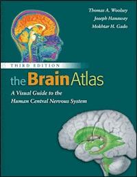
The Brain Atlas: A Visual Guide to the Human Central Nervous System integrates modern neuroscience with clinical practice and is now completely revised and updated for a Fourth Edition. * Each page uses direct labeling system, including an alphabetical list of terms for each image * Presents unrivaled treatment of brain pathways, with colored lines that clearly trace pathways over actual brain slices used earlier in the book * Over 400 high quality images, including multiple magnetic resonance images side-by-side with corresponding brain slices * Blood supply maps consistently and methodically presented with exhaustive depictions of arteries and blood territory maps next to each brain slice * Print edition comes with free access to Wiley companion digital edition accessible on any device, allowing the reader to make notes, bookmark, follow cross references, and download figures
Sie lesen das E-Book in den Legimi-Apps auf:
Seitenzahl: 225
Veröffentlichungsjahr: 2013
Ähnliche


Contents
Preface
Acknowledgments
PART I
Introduction
Overview
The Nervous System
Using This Book
Materials and Methods
References
PART II
The CNS and Its Blood Vessels
Cerebral Hemisphere and Brain Stem (1X); Sulci and Gyri (0.7X)–Lateral Aspect
Brain Stem, Diencephalon, Basal Ganglia, and Cerebellum (1X)–Anterolateral Aspect
Cerebellum (1X)–Superior Surface
Arteries to Spinal Cord (Diagrammatic)
Principal Fiber Bundles in Cerebral Hemisphere and Brain Stem (Semi-Schematic) (0.7X)–Lateral and Mesial Aspects
PART III
Brain Slices
Coronal Section Through Rostral Wall of Lateral Ventricle (1X) with Vessel Territories (0.7X)
Sagittal Section Through Superior, Middle, and Inferior Temporal Gyri (1X) with Vessel Territories (0.7X)
Axial Section Through Superior Caudate Nucleus (1X) with MRI (0.7X)
PART IV
Histological Sections
Horizontal Section Through Fastigial Nucleus (1.5X) with Vessel Territories
Transverse Section Through Superior Colliculus (3X) with Vessel Territories
Transverse Section Through Upper Cervical Level (10X) with Vessel Territories
Coronal Section Through Nucleus Accumbens (1.5X)
Coronal Section Through Optic Chiasm–top; Coronal Section Through Pituitary Stalk–bottom (4X)
Coronal Section Through Olfactory Trigone and Nucleus Basalis (5X)
Coronal Section Through Body of Hippocampus (5X)
PART V
Pathways
General Organization of Spinal Cord Gray Matter
Organization of the Thalamus
Organization of Hypothalamus
Touch and Position Sense Pathways: Posterior (Dorsal) Column/Medial Lemniscus and Trigeminal Main Sensory Nucleus
Vestibular Pathways
Corticospinal (Pyramidal) and Corticobulbar Pathways
Cerebellar Pathways: Somatic Afferents
Basal Ganglia Pathways
Arousal and Sleep Pathways
Hippocampal Pathways: Afferents
Amygdalar Pathways: Afferents
Hypothalamic Pathways: Afferents
Autonomic Pathways: Afferents
Cholinergic and Dopaminergic Pathways
Copyright © 2008 by John Wiley & Sons, Inc. All rights reserved.
Published by John Wiley & Sons, Inc., Hoboken, New Jersey.
Published simultaneously in Canada.
No part of this publication may be reproduced, stored in a retrieval system, or transmitted in any form or by any means, electronic, mechanical, photocopying, recording, scanning, or otherwise, except as permitted under Section 107 or 108 of the 1976 United States Copyright Act, without either the prior written permission of the Publisher, or authorization through payment of the appropriate per-copy fee to the Copyright Clearance Center, Inc., 222 Rosewood Drive, Danvers, MA 01923, 978-750-8400, fax 978-750-4470, or on the web at www.copyright.com. Requests to the Publisher for permission should be addressed to the Permissions Department, John Wiley & Sons, Inc., 111 River Street, Hoboken, NJ 07030, (201) 748-6011, fax (201)748-6008, e-mail: [email protected].
Limit of Liability/Disclaimer of Warranty: While the publisher and author have used their best efforts in preparing this book, they make no representations or warranties with respect to the accuracy or completeness of the contents of this book and specifically disclaim any implied warranties of merchantability or fitness for a particular purpose. No warranty may be created or extended by sales representatives or written sales materials. The advice and strategies contained herein may not be suitable for your situation. You should consult with a professional where appropriate. Neither the publisher nor author shall be liable for any loss of profit or any other commercial damages, including but not limited to special, incidental, consequential, or other damages.
For general information on our other products and services please contact our Customer Care Department within the U.S. at 877-762-2974, outside the U.S. at 317-572-3993 of fax 317-572-4002.
Wiley also publishes its books in a variety of electronic formats. Some content that appears in print, however, may not be available in electronic format.
Library of Congress Cataloging-in-Publication Data:
Woolsey, Thomas A.
The brain atlas: a visual guide to the human central nervous system / Thomas A. Woolsey,
Joseph Hanaway, Mokhtar H. Gado. —3rd ed.
p. cm.
Previous ed. cataloged under title.
Includes bibliographical references and index.
ISBN 978-0-470-08476-2
1. Brain — Anatomy — Atlases. 2. Central nervous system — Anatomy — Atlases. I. Hanaway, Joseph,
1933−II. Gado, Mokhtar H., 1931-III. Title.
QM455.B633 2003
611′.0022′2 — dc21
2002013579
Dedicated to
Clinton N. Woolsey
Jerzy E. Rose
David Bodian
W. Maxwell Cowan
Raymond D. Adams
C. Miller Fisher
James W. Bull
George du Boulay
Preface
The first edition of TheBrainAtlas:A VisualGuidetotheHumanCentralNervousSystem (1997) gave students of modern neuroscience an innovative combination of information on the human brain from many different sources. It included: dissections of the human brain and its blood vessels; photographs of human brain slices; MRI scans matched directly to those slices; maps of the brain territories supplied by major blood vessels; histological sections through the spinal cord, brain stem, thalamus, hypothalamus, and forebrain; and pathway diagrams rendered to convey the three-dimensional character of the principal connections of the brain. The first edition of TheBrainAtlas also was published in French and Italian. The second edition of TheBrainAtlas (2002) added several major improvements: simplified layout; a novel labeling system with direct labeling of all structures and all terms listed alphabetically on or opposite each page for rapid identification; reformatted pathway diagrams for improved clarity; and new illustrations to show cortical sulcal patterns and fiber bundles in the brain. Further, the second edition of TheBrainAtlas was spiral bound to lie flat to make it easier to use and more durable.
Many medical schools, universities, and colleges adopted the TheBrainAtlas for use in their neuroscience courses. The second edition was adopted by more institutions and is increasingly used as a resource in more undergraduate and graduate courses and health care training programs. Critical feedback from colleagues, instructors, medical, graduate, and undergraduate students, residents, and others has been given on both editions. These suggestions and the expertise of our publisher have guided the preparation of this third edition.
The third edition of TheBrainAtlas follows the successful organization, layout and design of the previous editions. However, there are a number of important improvements. First, newer imaging techniques have greatly improved in quality and detail, such that one is often hard pressed to distinguish radiological images from living human beings from those of brain slices prepared painstakingly from fixed brains. The third edition of The TheBrainAtlas has completely new radiological images of brain sections and blood vessels. Further, magnetic resonance images (MRI) have been added to illustrate the brain stem and spinal cord for comparison to the histological sections they match. Second, imaging is now fully integrated into research and clinical care so much so that the orientation conventions of radiology are now the standard. The third edition of TheBrainAtlas reflects this. The histological sections have been rotated 180° consistent with radiological conventions. In this orientation, axial images of the brain are conceptually continuous with the histological sections from the midbrain to the spinal cord. Accordingly, the order of the sections in Part III is now rearranged to flow directly from axial brain images into histological cross-sections through brain stem and spinal cord in Part IV. Third, this new edition of TheBrainAtlas includes several new and/or significantly revised illustrations in Part V that reflect advances in understanding the brain and its disorders. These are related to: a) the basal ganglia—substantia nigra, subthlamic nucleus, thalamus-cortical circuits; b) brain pathways for arousal and sleep; and c) pain pathways. Fourth, new diagrams integrating sections through the thalamus and hypothalamus illustrate the layout of cell groups and their connections in those structures. These are conceptually similar to the illustrations showing the organization of cranial nerve nuclei in the brain stem. Lastly, the third edition of TheBrainAtlas is printed so that complementary figures showing the organization of the brain stem cranial nerves and nuclei are on facing pages for direct comparison.
The third edition of TheBrainAtlas is designed to be an even easier to use, up-to-date learning resource for students of medicine and other health care professions (Physical Therapy, Occupational Therapy, Nursing), and students of biology, neuroscience, psychology and philosophy at all stages of their education. It is our hope that the third edition of TheBrainAtlas will continue to serve as an authoritative reference that is easy to use. This is important for anyone interested in the human brain, particularly resident and staff neuropathologists, neuroradiologists, neurologists, neurosurgeons and psychiatrists; selected other health care personnel; and neurobiologists, psychologists and cognitive scientists. Now is a remarkable epoch for the study of the human brain. It is our hope that the third edition of TheBrainAtlas will serve more people better as a field guide for those committed to these endeavors.
Acknowledgments
Assistance of the following persons is gratefully acknowledged. James Nelson, M.D., and Adrianne Noe, Ph.D., provided access to and assistance with the Yakovlev-Haleem slide collection in the National Museum of Health and Medicine. At Washington University, Martin M. Henegar, M.D., made some of the dissections; J. Gayle King helped with the cadavers and Walter Clermont photographed brain specimens.
Many undergraduate, medical and graduate students, resident trainees, and colleagues have used The BrainAtlas at Washington University and elsewhere. We thank them for valuable suggestions leading to improvements in this edition of TheBrainAtlas. In particular, Washington University colleagues Drs. Joel C. Geerling and Arthur D. Lowey provided important perspectives regarding the pathways related to arousal and sleep.
We particularly wish to thank Michele Ruschhaupt and her outstanding team at The Format Group, LLC, for the cover design, general aspects of the interior design, page layout, interior design and overall composition. Their attention to detail, timely work and pleasant disposition have been fantastic. Thom Moore and his colleagues at John Wiley & Sons, have worked hard to bring this book to a higher level. We thank them for their tireless efforts.
Support from the Spastic Paralysis Foundation of the Illinois–Eastern Iowa District of Kiwanis International and the John Simon Guggenheim Memorial Foundation to Dr. Woolsey is gratefully acknowledged. Cindy Woolsey and Nancy Hanaway remain truly tolerant, patient and supportive heroines who often suffered quietly.
PART I
PART I is an illustrated overview of The Brain Atlas. The main features of the central nervous system and the organization of this book are described. Sources of the specimens and the methods by which they were prepared and photographed are detailed. Key aspects of the various radiological techniques for the images included are outlined. Selected references are listed at the end.
Introduction
Overview
The Nervous System
Using This Book
Materials and Methods
References
Overview
The human nervous system is complex and sophisticated. It is the most remarkable system in biology. A major challenge for neuroscience, psychology, medicine, and, indeed, for civilization is to understand the nervous system at the same fundamental levels at which we now understand other organ systems. Early in the 21st century, only 50 years after the discovery of the genetic “alphabet,” the complete human genome has been mapped. Likewise, new knowledge about the brain and diseases that afflict the nervous system is exploding. One goal for future work on the human brain is to reach a level of detailed understanding similar to that now possible for the genome.
An anchor in this quest is information about the structure and organization of the central nervous system (CNS). The Brain Atlas: A Visual Guide to the Human Central Nervous System was prepared to help students and professionals understand the normal human brain and guide interpretation of clinical and experimental work.
Clear charts and maps of biological structures have been teaching aids from the earliest times. In the biological sciences, the first detailed and illustrated text based on direct observation was the De Fabrica Humani Corporis (1543) and its synoptic Epitome (1543) by Andreas Vesalius (1514–1564). Those books have been said to “mark the beginning of modern science.” Publication of the Fabrica also was a major landmark in book publishing. The highly popular Epitome was intended as a primer but served very much as a modern day atlas. Such works have evolved and today are used in the same way maps are used to plan travel and understand geographic relationships.
Lesen Sie weiter in der vollständigen Ausgabe!
Lesen Sie weiter in der vollständigen Ausgabe!
Lesen Sie weiter in der vollständigen Ausgabe!
Lesen Sie weiter in der vollständigen Ausgabe!
Lesen Sie weiter in der vollständigen Ausgabe!
Lesen Sie weiter in der vollständigen Ausgabe!
Lesen Sie weiter in der vollständigen Ausgabe!
Lesen Sie weiter in der vollständigen Ausgabe!
Lesen Sie weiter in der vollständigen Ausgabe!


