132,99 €
Mehr erfahren.
- Herausgeber: John Wiley & Sons
- Kategorie: Fachliteratur
- Sprache: Englisch
Unilateral posterior crossbite is a problem often seen in orthodontic practice, and properly understanding chewing patterns will lead to the most effective treatment program. Drawing on their research and available literature, Drs. Piancino and Kyrkanides present a fascinating look at chewing cycles and their role in the functional treatment of unilateral posterior crossbite. * Describes the physiology and pathology of chewing patterns and muscular activation in humans * Explains chewing patterns and muscular coordination, and their influence on the growth and harmony of the stomatognathic system * Clinical instruction for checking and correcting masticatory function and functional asymmetry in order to prevent the relapse of the malocclusion * Clinical cases walk readers through the treatment of seven crossbites
Sie lesen das E-Book in den Legimi-Apps auf:
Seitenzahl: 395
Veröffentlichungsjahr: 2016
Ähnliche


For my father and mother,
who taught me about intellectual integrity,
which is the cornerstone of this book.
For Stefano and Federica, for putting up with such a “busy mom.”
MGP
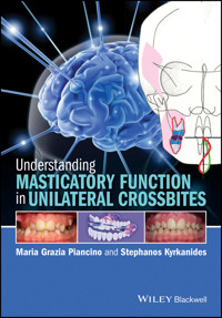
Understanding Masticatory Function in Unilateral Crossbites
Maria Grazia Piancino, MD, DDS, PhD
Researcher and Aggregate Professor – Orthognathodontics Dental School International Research Center Department of Surgical Sciences University of Turin Turin, Italy
Stephanos Kyrkanides, DDS, PhD
Dean College of Dentistry University of Kentucky Lexington, KY, USA
This edition first published 2016 © 2016 by John Wiley & Sons Inc.
Editorial offices:1606 Golden Aspen Drive, Suites 103 and 104, Ames, Iowa 50010, USA The Atrium, Southern Gate, Chichester, West Sussex, PO19 8SQ, UK 9600 Garsington Road, Oxford, OX4 2DQ, UK
For details of our global editorial offices, for customer services and for information about how to apply for permission to reuse the copyright material in this book please see our website at www.wiley.com/wiley-blackwell.
Authorization to photocopy items for internal or personal use, or the internal or personal use of specific clients, is granted by Blackwell Publishing, provided that the base fee is paid directly to the Copyright Clearance Center, 222 Rosewood Drive, Danvers, MA 01923. For those organizations that have been granted a photocopy license by CCC, a separate system of payments has been arranged. The fee codes for users of the Transactional Reporting Service are ISBN-13: 978-1-1189-7187-1/ 2016.
Designations used by companies to distinguish their products are often claimed as trademarks. All brand names and product names used in this book are trade names, service marks, trademarks or registered trademarks of their respective owners. The publisher is not associated with any product or vendor mentioned in this book.
The contents of this work are intended to further general scientific research, understanding, and discussion only and are not intended and should not be relied upon as recommending or promoting a specific method, diagnosis, or treatment by health science practitioners for any particular patient. The publisher and the author make no representations or warranties with respect to the accuracy or completeness of the contents of this work and specifically disclaim all warranties, including without limitation any implied warranties of fitness for a particular purpose. In view of ongoing research, equipment modifications, changes in governmental regulations, and the constant flow of information relating to the use of medicines, equipment, and devices, the reader is urged to review and evaluate the information provided in the package insert or instructions for each medicine, equipment, or device for, among other things, any changes in the instructions or indication of usage and for added warnings and precautions. Readers should consult with a specialist where appropriate. The fact that an organization or Website is referred to in this work as a citation and/or a potential source of further information does not mean that the author or the publisher endorses the information the organization or Website may provide or recommendations it may make. Further, readers should be aware that Internet Websites listed in this work may have changed or disappeared between when this work was written and when it is read. No warranty may be created or extended by any promotional statements for this work. Neither the publisher nor the author shall be liable for any damages arising herefrom.
ISBN: 9781118971871
A catalogue record for this book is available from the Library of Congress and the British Library.
Wiley also publishes its books in a variety of electronic formats. Some content that appears in print may not be available in electronic books.
Cover image of brain: © Getty Images/Kiyoshi Takahase Segundo
CONTENTS
Foreword
Preface
Masticatory Function as a Reference Point
A Consistent Reasoning from Diagnosis to Therapy
How to Use This Book
Acknowledgments
Chapter 1
Introductory Explanation of Masticatory Function
1.1 Introduction
1.2 The study of masticatory function
1.3 The evolution of electrognathography and electromyography
1.4 From the 1980s to today
1.5 Ready to start
References
Chapter 2
Physiology of Mastication: The Chewing Pattern and Masticatory Function
2.1 Introduction
2.2 Features of masticatory function
2.3 Terminology
2.4 The chewing pattern
2.5 The chewing pattern as an indicator of masticatory function
References
Chapter 3
Physiology of Mastication: Neuromuscular Control of Masticatory Function
3.1 Importance of the motor activity (Figure 3.1)
3.2 The nervous system (Figures 3.2 and 3.3)
3.3 Receptors in the stomatognathic system (Figure 3.5)
3.4 Reflex movements
3.5 Automatic movements (Figures 3.10 and 3.11)
3.6 Motor control: feedback and feed-forward (Figures 3.13 and 3.14)
3.7 Neuromuscular control (Figure 3.15)
3.8 Coordination of masticatory muscles during mastication
3.9 Neuromuscular adjustment to load (Figure 3.19)
References
Chapter 4
Alterations to Masticatory Function in Unilateral Crossbites
4.1 Introduction
4.2 Crossbite=neuromuscular syndrome (Figure 4.2)
4.2.4 Functional classification
4.3 Unilateral posterior crossbite
4.4 Alteration to masticatory function in unilateral posterior crossbite (Figure 4.13)
4.5 Anterior crossbite=neuromuscular syndrome (Figures 4.28 and 4.29)
References
Chapter 5
Therapy with Function Generating Bite Appliance: Actions and Effects on Malocclusion and Masticatory Function
5.1 Orthognathodontic therapy aimed to restore physiological neuromuscular equilibrium to the stomatognathic system
5.2 Dental–alveolar–basal actions and effects of function generating bite appliances
5.3 Therapy timing and duration and caries-free management
5.4 Compliance
References
Chapter 6
Cases
6.1 Introduction
6.2 Case 1: Right unilateral posterior crossbite (3 years, 9 months)
6.3 Case 2: Right unilateral posterior crossbite (6 years, 4 months)
6.4 Case 3: Positional crossbite (7 years, 6 months)
6.5 Case 4: Left unilateral posterior crossbite (7 years, 11 months)
6.6 Case 5: Right unilateral posterior crossbite (10 years, 3 months)
6.7 Case 6: Anterior open bite and left unilateral posterior crossbite (7 years, 9 months)
6.8 Case 7: Anterior open bite and bilateral posterior crossbite (7 years, 8 months)
Appendix: Terminology Update
Index
EULA
List of Illustrations
Chapter 1
Figure 1.1
Linking masticatory function with dental occlusion, cranial structure, and neuromuscular activity.
Figure 1.2
The stomatognathic system: relationships between dental occlusion, temporomandibular joint (TMJ) and neuromuscular control.
Source for the muscles:
redrawn from Neff (1999).
Source for the brain:
Purves
et al.
(2000). Reproduced with the permission of Sinauer Associates
Figure 1.3
Functional magnetic resonance has allowed the study of neural control during masticatory function. Source: Bracco et al. (2010). Reproduced with the permission of Maney Publishing
Figure 1.4
The beginning of masticatory function.
Figure 1.5
Multiple sensor array of Case Gnathic Replicator.
Source:
Lundeen and Gibbs (1982). Reproduced with permission from Elsevier.
Figure 1.6
Multiple sensor array of the Sirognathograph (Siemens, Germany).
Figure 1.7
Magnet of the Sirognathograph.
Figure 1.8
Sirognathograph.
Figure 1.9
Single chewing cycles superimposed, in the frontal (a) and sagittal (b) planes recorded with Sirognathograph and reproduced with a plotter.
Figure 1.10
Single chewing cycles superimposed in the frontal (a) and sagittal (b) planes.
Figure 1.11
The computer connected to the Sirognathograph.
Figure 1.12
The plotter connected to the Sirognathograph
Figure 1.13
First plots of mandibular kinetic movement in frontal, sagittal, and horizontal planes (top) aligned with electromyography envelope plotted versus the vertical jaw displacement, of right and left anterior temporalis muscles and right and left masseters (bottom). Red tracings: opening; blue tracings: closing.
Figure 1.14
First printing of plots of average chewing pattern and velocity versus the vertical jaw displacement. Red tracings: opening; blue tracing: closing.
Figure 1.15
Electromyograph preamplifier (Myotronics, Tukwila, WA, USA).
Figure 1.16
Multiple-sensor lightweight array of the kinesiograph (Myotronics, Tukwila, WA, USA).
Figure 1.17
K6-I kinesiograph (Myotronics, Tukwila, WA, USA).
Figure 1.18
Prints of mandibular kinetic movement in frontal and sagittal planes (
top
) aligned with electromyography recordings of right and left anterior temporalis muscles and right and left masseters (
bottom
) after one of the rewriting of the software. Green tracings: opening; red tracings: closing R right, L left.
Figure 1.19
K7-I kinesiograph (Myotronics, Tukwila, WA, USA).
Figure 1.20
Print of chewing patterns in frontal, sagittal, and horizontal planes of the program dedicated to the K7-I kinesiograph; the solid line (green for the opening pattern and red for the closing pattern) represents the average chewing cycle of three trials lasting 10 s each. The green and red shaded areas represent the standard deviation from the average cycle.
Figure 1.21
Print of an average chewing pattern in the frontal and sagittal planes (top); electromyography envelope plotted versus the vertical jaw displacement. The solid line (green for the opening pattern and red for the closing pattern) represents the average chewing cycle of three trials lasting 10 s each. The green and red shaded areas represent the standard deviation from the average cycle.
Figure 1.22
Multiple-sensor lightweight array of K7-I kinesiograph (Myotronics, Tukwila, WA, USA).
Figure 1.23
Magnet of the K7-I kinesiograph (Myotronics, Tukwila, WA, USA).
Figure 1.24
Print of the superimposed chewing cycles in the frontal plane: deliberate right-hand and left-hand mastication (
top
); free (alternate) mastication (
bottom
). Green tracings: opening; red tracings: closing.
Figure 1.25
Left unilateral posterior crossbite before correction in mixed dentition.
Figure 1.26
Dental occlusion after functional correction.
Chapter 2
Figure 2.1
The masticatory system. The stomatognatic system: relationships between dental occlusion, TMJ, and neuromuscular control.
Source for the muscles:
Neff (1999).
Source for the brain:
Purves
et al.
(2000). Reproduced with the permission of Sinauer Associates.
Figure 2.2
Physiological versus reverse chewing cycle. Green tracings: opening; red tracings: closing.
Figure 2.3
Motor control and peripheral inputs during masticatory function.
Figure 2.4
Overlapping chewing cycles in the frontal plane during chewing deliberately on the right (a) and left (b) side. Green tracings: opening; red tracings: closing.
Figure 2.5
Kinetics of the right first molar, on the bolus side – sagittal plane.
Source:
Redrawn from Lundeen and Gibbs (1982). Reproduced with permission from Elsevier.
Figure 2.6
Kinetics of the mandible during chewing on the right side – frontal plane. Central tracing: recording at the incisal point; (a) recording at the first molar point on the bolus side; (b) recording at the first molar point on the contralateral side.
Source:
Redrawn from Lundeen and Gibbs (1982). Reproduced with permission from Elsevier.
Figure 2.7
Kinetics of the left first molar on the contralateral chewing side – sagittal plane.
Source:
Redrawn from Lundeen and Gibbs (1982). Reproduced with permission from Elsevier.
Figure 2.8
Kinetics of the right (a) bolus side and left (b) contralateral condyle during chewing on the right side – sagittal plane. Sagittal condylar tracings follow different trajectories.
Source:
Redrawn from Lundeen and Gibbs (1982). Reproduced with permission from Elsevier.
Figure 2.9
Kinetics of the mandible and condyles during chewing on the right side – frontal plane. The condylar patterns are different between sides.
Source:
Redrawn from Lundeen and Gibbs (1982). Reproduced with permission from Elsevier.
Figure 2.10
Kinetics of the condyles in the sagittal plane (a: bolus side; b: contralateral side) and in the frontal plane (center of the image). Opening and closing tracing follow different trajectories.
Source:
Redrawn from Lundeen and Gibbs (1982). Reproduced with permission from Elsevier.
Figure 2.11
TMJ of a child with deciduous dentition (
left
) and of an adult with permanent dentition (
right
).
Figure 2.12
Sagittal condylar patterns during mastication in deciduous (
left
) and permanent (
right
) dentition.
Figure 2.13
The physiological chewing pattern during chewing deliberately on the right and on the left side (
top
) and during free-alternate mastication (
bottom
). Green tracings: opening; red tracings: closing.
Figure 2.14
Overlapping chewing cycles during chewing deliberately on the right and on the left side, showing the repeatability and variability of the masticatory movement. Green tracings: opening; red tracings: closing.
Figure 2.15
Representation of the direction of the tracing of closure: clockwise during chewing on the right side; anti-clockwise during chewing on the left side. Green tracings: opening; red tracings: closing.
Figure 2.16
Representation of the closing angle during chewing on the right side and on the left side. Green tracings: opening; red tracings: closing.
Figure 2.17
In physiological conditions, the mandible deviates laterally, toward the bolus side, and then, during closure, medially through the transcuspal and intercuspal phases of mastication.
Source:
Redrawn from A. Lewin (1985) with the permission of Quintessence Publishing.
Figure 2.18
Representation of the height and width of the chewing pattern during chewing on the right side and on the left side. Green tracings: opening; red tracings: closing.
Figure 2.19
Representation of the lateral shift of the chewing pattern during chewing on the right side and on the left side. Green tracings: opening; red tracings: closing.
Figure 2.20
Overlapping chewing patterns during chewing a soft bolus (
top
) and hard bolus (
bottom
) on the right and left side. Green tracings: opening; red tracings: closing
Chapter 3
Figure 3.1
Motor control of masticatory function. The stomatognathic system: relationships between dental occlusion, TMJ, and neuromuscular control.
Source for the muscles:
Redrawn from Neff (1999).
Source for the brain:
Redrawn from Purves
et al.
(2000). Reproduced with permission of Sinauer Associates.
Figure 3.2
Motor cortex.
Source:
Adapted from Purves
et al.
(2000). Reproduced with permission from Sinauer Associates.
Figure 3.3
Spinal cord. Source: Adapted from Purves et al. (2000). Reproduced with permission from Sinauer Associates.
Figure 3.4
Central nervous system.
Source:
Adapted from Purves
et al.
(2000). Reproduced with permission from Sinauer Associates.
Figure 3.5
Peripheral receptors of the stomatognathic system.
Source:
Adapted from Purves et al. (2000). Reproduced with permission from Sinauer Associates.
Figure 3.6
Peripheral inputs and central output.
Figure 3.7
Immunofluorescence reaction for PGP 9.5 in periodontal ligament of rat incisor. It is possible to observe a low PGP 9.5 immunostaining (red fluorescence) in the tooth-related part (TRP*), where it marks thin lines of nervous fibers; instead, in the bone-related part (BRP**) it is possible to observe a high PGP 9.5 immunofluorescence pattern that marks, prevalently, receptor endings of nervous fibers.
Source:
G. Anastasi and G. Cutroneo, University of Messina, Messina, Italy. Reproduced with permission from G. Anastasi and G. Cutroneo.
Figure 3.8
Different characteristics of the upper and lower molar periodontal mechanoreceptors in experimental studies in rats. Interestingly, the rapid-adapting receptors (black triangles) are more concentrated in the lower molars, which convey information from the moving bone, and the slow-adapting receptors are more concentrated in the maxillary molars. B: buccal; M: mesial; L: lingual; D: distal.
Source:
Tabata
et al.
(2006). Reproduced with permission from Elsevier.
Figure 3.9
Muscular receptors and motor control.
Figure 3.10
The inputs arising from the periodontal mechanoreceptors of teeth have a powerful influence on the motor control.
Figure 3.11
Continuous adaptability of the chewing pattern to the bolus type during chewing on the right side.
Figure 3.12
The motor control adapts to the peripheral inputs from the mechanoreceptors of teeth, which are influenced by occlusion and malocclusions.
Figure 3.13
The inputs arising from the periodontal mechanoreceptors of teeth, especially from the posterior region of the occlusion, have a powerful influence on the motor control of chewing.
Figure 3.14
The motor control adapts the chewing pattern to the peripheral inputs.
Top left: Source for the muscles:
Redrawn from Neff (1999).
Source for the brain:
Redrawn from Purves
et al.
(2000). Reproduced with permission of Sinauer Associates.
Figure 3.15
Representation of different groups of muscles of head and neck in different colors.
Source:
Redrawn and adapted from Neff (1999).
Figure 3.16
Schematic representation of anterior temporalis and masseter muscles.
Figure 3.17
Electromyography tracings of anterior temporalis and masseter during physiologic alternate chewing showing the neuromuscular balance between sides. RTA: right anterior temporalis, LTA: left anterior temporalis, RMM: right masseter muscle; LMM: left masseter muscle. Green tracings: opening; red tracings: closing.
Figure 3.18
The chewing pattern simultaneously recorded with the electromyography of both sides of the anterior temporalis and masseter muscles during physiologic alternate chewing showing the neuromuscular balance between sides. RTA: right anterior temporalis; LTA: left anterior temporalis; RMM: right masseter muscle; LMM: left masseter muscle. Green tracings: opening; red tracings: closing.
Figure 3.19
Adjustment to load of the chewing pattern and electromyography activity during chewing a soft and a hard bolus on the right side: the contralateral left masseter increases significantly more in comparison with that of the bolus side. Green tracings: opening; red tracings: closing.
Figure 3.20
Importance of forces of mastication and bolus consistency for the craniofacial bone development; the red arrows represents the transmission of muscular forces via the teeth during chewing to palate and zygomatic bones. Green tracings: opening; red tracings: closing.
Chapter 4
Figure 4.1
The masticatory system.
Source for the muscles:
Neff (1999).
Source for the brain:
Purves
et al.
(2000). Reproduced with the permission of Sinauer Associates.
Figure 4.2
Right and left unilateral posterior crossbite malocclusions: a neuromuscular syndrome.
Figure 4.3
Distribution of crossbites in Italian orthodontic population.
Figure 4.4
Distribution of right and left unilateral posterior crossbites in Italian orthodontic population
Source:
Piancino
et al.
(2006a). Reproduced with permission from Oxford University Press.
Figure 4.5
Tongue thrusting.
Figure 4.6
Thumb-sucking.
Figure 4.7
Anterior and posterior functional region of the occlusion. The diagram in two colours demonstrates the dual role of premolars, which provide support as well as dynamic control.
Source:
Slavicek (2002). Reproduced with permission from R. Slavicek.
Figure 4.8
Unilateral posterior crossbite involving multiple teeth (
top
) and one tooth (
bottom
).
Figure 4.9
Unilateral anterior (
top
) and canine (
bottom
) crossbite.
Figure 4.10
Positional or functional crossbite: occlusion in maximum intercuspation (a) and in centered position (b). Postero-anterior cephalometry (c: frontal plane): observe that in centered position the asymmetries, even if they are present, are not detectable. Representation of the deciduous stage of development (d: sagittal plane): the TMJ and the mandibular bone are clearly immature.
Source:
The representation of the deciduous stage has been redrawn from Slavicek (2002). Reproduced with permission from R. Slavicek.
Figure 4.11
Dento-alveolar crossbite: occlusion in maximum intercuspation (b); observe that the centered position is highly unstable (a). Postero-anterior cephalometry (c: frontal plane): both in intercuspation and centered position basal asymmetries are detectable; the dentoalveolar bone is clearly involved. Representation of the mixed dentition stage of development (d: sagittal plane): the TMJ and the mandibular bone are growing; if a posterior crossbite is still present in the mouth of the child, all the basal structures grow in an asymmetric way. This is compensation, not adaptation. See terminology.
Source:
The representation of the mixed dentition stage has been redrawn from Slavicek (2002). Reproduced with permission from R. Slavicek.
Figure 4.12
Basal crossbite: occlusion in maximal intercuspation (a); the centered position is not detectable. Postero-anterior cephalometry (b: frontal plane): the asymmetries are evident. Representation of the permanent dentition stage of development (c: sagittal plane): at the end of growth the compensatory, basal asymmetries to the posterior crossbite malocclusion are irreversible.
Source:
The representation of the permanent dentition has been redrawn from Slavicek (2002). Reproduced with permission from R. Slavicek.
Figure 4.13
Physiological (a) versus reverse (b) chewing pattern during chewing on the right side.
Figure 4.14
Physiological (a) pattern versus different type of reverse chewing cycles, with (b) and without (c) crossover of the tracings, during chewing on the left side.
Figure 4.15
Comparison between physiological (
top
) activation of masseters during chewing on the left side and altered coordination during reverse chewing cycles on the left crossbite side (
bottom
).
Figure 4.16
Comparison between physiological (
top
) activation of masseters during chewing on the right side and altered coordination during reverse chewing cycles on the right crossbite side (
bottom
). Green tracings: opening; red tracings: closing.
Figure 4.17
Comparison between physiological (
top
) activation of masseters during chewing on the left side and altered coordination during reverse chewing cycles on the left crossbite side (bottom).
Figure 4.18
Chewing patterns during chewing on the right crossbite side and on the left non-crossbite side. Observe the functional asymmetry of the chewing pattern and the reverse anomalous pattern on the crossbite side. Green tracings: opening; red tracings: closing.
Figure 4.19
Chewing patterns during chewing on the right non-crossbite side and on the left crossbite side. Observe the functional asymmetry of the chewing pattern and the reverse anomalous pattern on the crossbite side. Green tracings: opening; red tracings: closing.
Figure 4.20
Bilateral chewing patterns and EMG activation of the masseters during chewing on the right non-crossbite side and on the left crossbite side. Observe the functional asymmetry of the chewing pattern and the EMG asymmetrical activation between sides. Green tracings: opening; red tracings: closing.
Figure 4.21
Histological and immunohistochemical findings of right and left masseter muscles in right unilateral posterior crossbite. The muscular alterations on the crossbite side (right side) are evident. Green tracings: opening; red tracings: closing.
Source:
G. Cutroneo, University of Messina, Messina, Italy. Reproduced with permission from G. Cutroneo.
Figure 4.22
Immunohistochemical findings of right and left masseter muscles in right unilateral posterior crossbite using antibodies against sarcoglycans. The increased staining patterns of sarcoglycans on the crossbite side (right side) are evident. Green tracings: opening; red tracings: closing.
Source:
G. Cutroneo, University of Messina, Messina, Italy. Reproduced with permission from G. Cutroneo.
Figure 4.23
Tractography of the left and right masseter muscles obtained by diffusion tensor resonance magnetic imaging (DTRMI) scan in a patient with left posterior crossbite malocclusion. Observe the increased volume of the contralateral muscle (right masseter) compared with the crossbite side (left masseter).
Source:
G. Anastasi University of Messina, Messina, Italy. Reproduced with permission of G. Anastasi.
Figure 4.24
Top:
comparison of percentage of reverse chewing cycles during chewing on the crossbite side and on the healthy side, before and after therapy.
Center:
chewing pattern and EMG activity of a patient with right unilateral posterior crossbite before correction;
bottom:
after correction. Green tracings: opening; red tracings: closing.
Source:
Piancino
et al.
(2006). Reproduced with permission from Oxford University Press.
Figure 4.25
Comparison of chewing pattern in left posterior crossbite before and after therapy with function generating bite appliance. Green tracings: opening; red tracings: closing.
Figure 4.26
The percentage difference in masseter muscle activity between sides in the children with unilateral posterior crossbite (top) when chewing on the crossbite side was significantly (*) lower (yellow) compared with the difference observed for the control group (blue), and corresponded to the altered kinematics of the reverse chewing cycles. After therapy (bottom) it was similar for patients and controls.
Figure 4.27
Face and occlusion of a child aged 6 years and 4 months with right posterior crossbite.
Figure 4.28
Unilateral anterior crossbite=neuromuscular syndrome.
Figure 4.29
The functional aspects of anterior crossbite are different from that of posterior crossbite. The anterior region of the occlusion has a role of guidance of the mandible in the space, while the posterior regions are dedicated to support and mastication.
Source:
Redrawn from Slavicek (2002). Reproduced with permission from R. Slavicek.
Figure 4.30
Chewing patterns of a patient with anterior crossbite. The closure direction is not altered on both sides. Green tracings: opening; red tracings: closing.
Figure 4.31
Comparison of the prevalence of reverse chewing cycles between unilateral anterior and posterior crossbites. There is no difference, during chewing, between sides in anterior crossbites, whereas there is significant difference between sides in unilateral posterior crossbites. Moreover, there is no difference between the normal sides of anterior versus posterior crossbites, whereas there is a significant difference between the anterior versus posterior crossbite side.
Source:
Piancino
et al.
(2012). Reproduced with permission from Oxford University Press.
Chapter 5
Figure 5.1
FGB appliance with posterior bite planes.
Source:
Courtesy of P. Bracco, Piancino
et al.
(2006). Reproduced with permission from Oxford University Press.
Figure 5.2
FGB appliance with double anterior and posterior bite planes.
Source:
Courtesy of P. Bracco, Castroflorio
et al.
(2004). Reproduced with permission from John Wiley & Sons.
Figure 5.3
(a) The stomatognathic system. Relationships between dental occlusion, TMJ, and neuromuscular control. (b, c) Premature contacts cause deviation of the mandible, dislocation of the condyle, and muscular imbalance in the three planes of space. Source for the muscles: Neff (1999). Source for the brain: Purves et al. (2000). Reproduced with the permission of Sinauer Associates.
Figure 5.4
FGB appliance with posterior bite planes (
top
), with anterior and posterior bite planes (
middle
), and with double anterior and posterior bite planes (
bottom
).
Figure 5.5
FGB appliance components: (1) resilient stainless steel bite planes, (2) buccal shields, (3) expansion springs, (4) palatal button.
Figure 5.6
FGB appliance component: resilient stainless steel bite planes.
Figure 5.7
Old version of FGB appliance with resin bite planes (drawing and prototype). G: cheek; E: expansion springs; S: structural wire; P: palatal button; BO: occlusal plate (resin); V: buccal shield; d: dental point contact.
Source:
P. Bracco, Turin, Italy. Reproduced with permission from P. Bracco.
Figure 5.8
Old version of FGB appliance with the anterior metallic bite plane used in the posterior region.
Source:
P. Bracco, Turin, Italy. Reproduced with permission from P. Bracco.
Figure 5.9
Disengagement of the mandible and self-repositioning with FGB appliance during swallowing.
Source:
P. Bracco, Turin, Italy. Reproduced with permission from P. Bracco.
Figure 5.10
Adaptive growth of the TMJ during different developmental stages from deciduous dentition (a) to permanent dentition (b) (top). Functional balancing is important for harmonious growth of the joint and skeletal structures (c) (bottom).
Source for part (c):
Slavicek (2002). Reproduced with permission from R. Slavicek.
Figure 5.11
New, resilient stainless steel bites (
top
); bites after use with cupped signs (
bottom
).
Figure 5.12
Leveling of the occlusal plane before and after the use of FGB appliance in mixed dentition.
Figure 5.13
Axiography recording with metallic clutch.
Figure 5.14
Signs on the bite planes after use of FGB appliance looking like “gothic arches.”
Source:
Courtesy of P. Bracco.
Figure 5.15
FGB appliance component: buccal shields.
Figure 5.16
Detail of the buccal shield.
Figure 5.17
Action wedge of the buccal shield.
Source:
P. Bracco, Turin; Italy. Reproduced with permission from P. Bracco.
Figure 5.18
Asymmetrical action of the buccal shields for the correction of unilateral posterior crossbites.
Figure 5.19
Transmission of self-regulated muscle forces to expansion springs through buccal shields for the correction of unilateral posterior crossbites. Adapting the buccal shields in an asymmetrical way, it is possible to apply asymmetrical forces and achieve true asymmetrical action during the correction of the unilateral posterior crossbite. This is an asymmetrical malocclusion requiring the possibility of application of asymmetrical forces; this is only possible with extra-dental anchorage, as with the FGB appliance that progressively works with muscular anchorage (a–d). Observe the internal and external adaptation of the buccal shields.
Figure 5.20
Decompression space, represented in gray.
Figure 5.21
FGB appliance component: expansion springs.
Figure 5.22
Intermittent forces; activation of the appliance during swallowing. A: resilient stainless steel bite planes; B: palatal button; C: expansion springs.
Source:
Piancino
et al.
(2006).
Figure 5.23
The simultaneous actions of the FGB appliance realize bodily movement of the teeth. The decompression space is represented in grey.
Figure 5.24
Narrow and asymmetrical palatal vault before therapy (a); observe the expansion of the palatal vault, but especially the recovery of the asymmetry (b) after therapy with FGB appliance in mixed (
top
) and deciduous dentition (
bottom
). A: resilient stainless steel bite planes; B: palatal button; C: expansion springs.
Figure 5.25
FGB appliance component: palatal button.
Figure 5.26
The palatal button acts as an artificial bolus (a). Restoration of the tongue function and swallowing automatism (b).
Figure 5.27
Representation of the forces developed during mastication of hard boluses, influencing the bone growth and the activity of the palatal suture, through the dental roots, especially the roots of the upper first molars (
top
). FGB appliance action and effects develop similar forces during swallowing (
bottom
).
Figure 5.28
Interpremaxillary suture (a) and nasal suture (c) of rats fed a normal diet compared with rats fed a soft diet (b, d). Observe the less active and less developed suture of the soft diet group.
Source:
Engström
et al.
(1986). Reproduced with permission from Oxford University Press.
Figure 5.29
Intermittent, self-regulating forces are typical of the FGB appliance, which is activated during swallowing and phonation. A: resilient stainless steel bite planes; B: palatal button; C: expansion springs.
Source:
Piancino
et al.
(2006).
Figure 5.30
Recovery of the chewing pattern before/after therapy with FGB appliance of a patient with anterior open bite and left unilateral posterior crossbite.
Figure 5.31
Dynamic cephalometric analysis of hyperdivergent and hypodivergent craniofacial morphology with muscular and articular vector of forces represented.
Figure 5.32
Dynamic cephalometric analysis in the sagittal plane of a hyperdivergent craniofacial morphology with mandibular clockwise vector of forces represented in red.
Figure 5.33
Representation of counter-clockwise vector of forces (green) generated by the posterior stainless steel resilient bite planes of FGB in a hyperdivergent subject, counteracting the vectors on the TMJ.
Figure 5.34
Dynamic cephalometric analysis of hypodivergent subject with counter-clockwise vector of forces (red) (a); representation of clockwise vector of forces (green) generated by FGB anterior and posterior bite planes (b), lowering the TMJ overload.
Figure 5.35
Comparison before/after therapy of the axiography parameters of a group of young/developing patients (group A) with a group at the end of the growth period (group B). The only non-significant parameter is the asymmetry of tracing in the group at the end of the growth period (see text for explanation).
Chapter 6
Figure 6.1
Top:
images of the face;
bottom:
images of the occlusion (frontal and lateral views). The X-ray and cephalometric analyses were not performed owing to the very young age of the patient. They will be requested at a later stage in development.
Figure 6.2
Top:
maximum intercuspal position (on the left) and centered position with centered midlines (on the right). Observe the occlusal incongruence.
Bottom:
view of the FGB appliance in the mouth and out of the mouth (view of the lower side of the appliance on the left, and of the upper side on the right). As soon as the FGB appliance is in the mouth, the mandible is centered.
Figure 6.3
Images of the face (
top
) and of the occlusion (
bottom
) after 3 months of therapy with FGB.
Figure 6.4
Comparison of the face, occlusion, and palate before (
left
) and after (
right
) correction of the unilateral posterior crossbite. The intermolar distance measured with a caliper on the stonecasts shown in the image was 2.5 cm before and 2.9 cm after correction (increase of 4 mm).
Figure 6.5
Comparison of the occlusion before therapy (3.9 y), after correction (4.0 y), at 10 months (4.10 y) and at 21 months (5.9 y) follow-up (FGB appliance was worn during the night).
Figure 6.6
Top:
images of the face;
bottom:
images of the occlusion (frontal and lateral views).
Figure 6.7
Latero-lateral and postero-anterior teleradiography (
left
) and cephalometric tracings (
right
).
Figure 6.8
Maximal intercuspation (
top
) and orthopantomography (
bottom
).
Figure 6.9
Chewing cycles before therapy; observe the severe alteration of the pattern morphology during chewing on the crossbite side and the consequent serious functional asymmetry.
Figure 6.10
Maximal intercuspation (
top
) and FGB appliance in the mouth and out of the mouth (
bottom
). Observe the asymmetric activation of the buccal shields.
Figure 6.11
Images of the face (
top
) and of the occlusion (
bottom
) after 4 months of therapy with the FGB appliance.
Figure 6.12
Comparison of the face and occlusion before (on the left) and after correction of the unilateral posterior crossbite (on the right).
Figure 6.13
Comparison of the e-models of palate before and after correction of the right unilateral posterior crossbite.
Figure 6.14
Comparison of the chewing cycles before (
top
) and after correction of the right unilateral posterior crossbite (
bottom
). Observe the recovery of the functional symmetry.
Figure 6.15
Top:
images of the face;
bottom:
images of the occlusion (frontal and lateral views).
Figure 6.16
Top:
latero-lateral and postero-anterior teleradiography recorded with the mandible in centered position.
Bottom:
cephalometric tracings. Observe the hyperdivergent tendency.
Figure 6.17
Maximal intercuspation (
top
) and orthopantomography (
bottom
).
Figure 6.18
Comparison of intercuspal position (
top
) and centered position (
bottom
). Observe the cusp-to-cusp contacts in centered position.
Figure 6.19
Chewing cycles before therapy. Observe the severe alteration of the pattern morphology during chewing on both sides due to the occlusal instability represented by the different position of the functional intercuspation (see Section 2.3).
Figure 6.20
Images of the occlusion without (
top
) and with (
bottom
) the FGB appliance. When the appliance is in the mouth, the midlines are centered and the occlusion is protected from traumatic cusp-to-cusp contacts. The mandible is free to self-regulate the position in the three planes of space at any stage of therapy, thanks to the smooth stainless steel resilient bite planes. The self-regulated position of the mandible, free from dental contacts, enables the condyles to spontaneously find the most physiological position within the TMJ. In this way it is possible to avoid any type of joint compensation (see Section 2.3), as happens with traumatic and antiphysiological therapies.
Figure 6.21
Images of the face (
top
) and of the occlusion (
bottom
) after 4 months of therapy with the FGB appliance.
Figure 6.22
Comparison of the face, occlusion, and palate before and after correction of the unilateral posterior crossbite. The intermolar distance measured with a caliper on the stonecasts shown in the image was 2.9 cm before and 3.2 cm after correction (increase of 3 mm). Observe the arch form.
Figure 6.23
Chewing cycles before (
top
) and after therapy (
bottom
). Observe the recovery of the functional physiology and symmetry.
Figure 6.24
Comparison of the face, occlusion, and palate before therapy (
left
), after correction (
center
) and at 9.5 years of age (
right
). The intermolar distance measured with a caliper on the stonecasts shown in the image was 2.9 cm before, 3.2 cm after correction, and 3.35 cm at 2 years' follow-up (increase of 4.5 mm measured at the second deciduous molar). Observe the absence of relapse, the stability of the correction, and the continuous improvement of the intermolar distance and palatal shape.
Figure 6.25
Maximal intercuspation and orthopantomography before (
top
) and after correction (
bottom
).
Figure 6.26
Latero-lateral and postero-anterior teleradiography and cephalometric tracings before (
top
) and after (
bottom
) correction of unilateral posterior crossbite, at age 14 years and 2 months.
Figure 6.27
Occlusion at ages 14 years and 2 months and at 18 years.
Figure 6.28
Top:
images of the face;
bottom:
images of the occlusion (frontal and lateral views).
Figure 6.29
Top:
latero-lateral and postero-anterior teleradiography.
Bottom:
cephalometric tracings.
Figure 6.30
Maximal intercuspation and orthopantomography.
Figure 6.31
The chewing patterns before therapy. Observe the severe alteration of the pattern morphology during chewing on the crossbite side and the consequent serious functional asymmetry.
Figure 6.32
Images of the mouth without (
top
) and with the FGB appliance (
bottom
). When the appliance is in the mouth the mandible are centered. View of the lower side of the appliance (
right
) and of the upper side (
left
).
Figure 6.33
Images of the face (
top
) and of the occlusion (
bottom
) after 7 months of therapy with the FGB appliance.
Figure 6.34
Comparison of the face, occlusion, and palate before therapy and after correction. The intermolar distance measured with a caliper on the stonecasts shown in the image was 2.9 cm before and 3.5 cm after correction (increase of 6 mm).
Figure 6.35
Maximal intercuspation and orthopantomography before (
top
) and after therapy with FGB (
bottom
).
Figure 6.36
Chewing patterns before and after therapy. Observe the recovery of the functional physiology and symmetry of the chewing pattern.
Figure 6.37
Follow-up of the occlusion at ages 10 years and 6 months (
top
) and at 11 years and 3 months (
bottom
). The appliance FGB was worn as much as possible until the correction of crossbite and then at home and during the night.
Figure 6.38
Top:
images of the face;
bottom:
images of the occlusion (frontal and lateral views).
Figure 6.39
Top:
latero-lateral and postero-anterior teleradiography.
Bottom:
cephalometric tracings.
Figure 6.40
Top
: maximal intercuspation;
bottom:
orthopantomography.
Figure 6.41
Images of the mouth without (
top
) and with (
bottom
) the FGB appliance.
Figure 6.42
Images of the face (
top
) and of the occlusion (
bottom
) after 10 months of therapy with the FGB appliance.
Figure 6.43
Comparison of the face, occlusion, and palate before and after correction of the unilateral posterior crossbite. The intermolar distance measured with a caliper on the stonecasts shown in the image was 2.9 cm before and 3.3 cm after correction (increase of 4 mm).
Figure 6.44
Comparison of the occlusion and orthopantomography before and after therapy.
Figure 6.45
Images of the occlusion (
top
) and orthopantomography (
bottom
) at 2 years and 9 months' follow-up. Observe the parallelism of the roots due to the action of the resilient stainless steel bite planes.
Figure 6.46
Comparison of the face, occlusion, and palate before and after correction of the unilateral posterior crossbite and at 13 years of age. Observe the improvement of the shape of the upper arch. The intermolar distance measured with a calipers on the stonecasts shown in the image was 2.9 cm before, 3.3 cm after correction, and 3.6 cm at 2 years and 9 months' follow-up (increase of 7 mm).
Figure 6.47
Images of the face (
top
); images of the occlusion (
bottom
) (frontal and lateral views).
Figure 6.48
Top:
latero-lateral and postero-anterior teleradiography.
Bottom:
cephalometric tracings.
Figure 6.49
Maximal intercuspation and orthopantomography.
Figure 6.50
The chewing patterns before therapy of left posterior crossbite. Observe the severe alteration of the pattern morphology during chewing on the crossbite side (left side) and the consequent serious functional asymmetry.
Figure 6.51
The FGB appliance.
Figure 6.52
Images of the face (
top
) and of the occlusion (
bottom
) after 6 months of therapy with the FGB appliance.
Figure 6.53
Occlusion and orthopantomography before and after correction of the anterior open bite and left posterior crossbite.
Figure 6.54
The chewing patterns before and after correction. Observe the recovery of the functional symmetry and of the closure direction.
Figure 6.55
Image of face and occlusion at age 9 years and 5 months.
Figure 6.56
Latero-lateral and postero-anterior teleradiography and cephalometric tracings before (
top
) and at 10 years and10 months (
bottom
).
Figure 6.57
Face and occlusion at age 13 years and 6 months. In this case the aesthetic alignment has been obtained with a short period of fixed appliance (8 months).
Figure 6.58
Comparison of the face and occlusion before and after correction of the unilateral posterior crossbite and at age 13 years and 6 months.
Figure 6.59
Top
: images of the face;
bottom:
images of the occlusion (frontal and lateral views).
Figure 6.60
Top:
latero-lateral and postero-anterior teleradiography.
Bottom:
cephalometric tracings. Observe the hyperdivergent tendency.
Figure 6.61
Maximal intercuspation and orthopantomography.
Figure 6.62
The chewing patterns before therapy. Observe the reverse chewing patterns on both sides.
Figure 6.63
Images of the occlusion without (
top
) and with (
bottom
) the FGB appliance. Observe the alteration of the tongue function.
Figure 6.64
Images of the face (
top
) and of the occlusion (
bottom
) after 10 months of therapy with the FGB appliance.
Figure 6.65
Comparison of the face, occlusion, and palate before and after correction of the bilateral posterior crossbite and anterior open bite. The intermolar distance measured with a caliper on the stonecasts shown in the image was 2.7 cm before and 3.6 cm after correction (increase of 9 mm).
Figure 6.66
Image of occlusion at ages 11 years and 3 months (
top
), observe that both the upper right and left canine erupted in crossbite: they were easily and immediately corrected adapting the expansion spring, without any need of laboratory work and 12 years and 8 months (
bottom
).
Middle:
the FGB appliance.
Figure 6.67
Comparison of the face, occlusion, and palate before (
left
) and after therapy (
center
) and at years and follow up (
right
). The intermolar distance measured with a caliper on the stonecasts shown in the image was 2.7 cm before correction, 3.6 cm after correction, and 3.7 cm at 5 years follow-up (increase of 10 mm). Observe the improvement of the arch form.
Figure 6.68
Latero-lateral teleradiography before therapy (
left
), at 10 years (
center
), and at 12 years and 8 months (
right
).
Bottom:
cephalometric tracings.
Figure 6.69
Postero-anterior teleradiography before therapy (
left
), at 10 years (
center
), and at 12 years and 8 months (
right
).
Bottom:
cephalometric tracings.
Figure 6.70
Progressive reprogramming of the chewing motor control, with the arrows representing the direction of the pattern of closure progressively restored. Observe the progressive improvement of the symmetry of the chewing pattern.
Figure 6.71
Progressive reprogramming of the chewing motor control. Observe the progressive improvement of the chewing pattern.
Guide
Cover
Table of Contents
Preface
Pages
vii
viii
ix
x
xi
1
2
3
4
5
6
7
8
9
10
11
12
13
16
17
19
23
24
25
26
27
28
29
30
31
32
33
34
35
36
37
38
40
41
42
43
45
46
47
49
51
52
54
55
57
58
59
60
61
62
63
64
65
66
67
68
69
70
71
72
73
74
75
76
77
79
81
83
84
85
86
87
88
91
92
93
94
95
96
97
98
99
100
101
102
103
104
105
106
107
108
109
110
111
112
113
114
116
120
121
122
123
125
128
129
130
132
133
135
136
137
138
139
140
141
142
143
144
145
146
148
149
150
151
152
153
154
155
156
158
159
161
162
163
164
165
166
167
168
169
170
172
173
174
175
176
178
180
181
182
183
184
185
186
189
193
200
205
209
215
225
227
228
229
230
231
232
233
234
235
Foreword
This book's uniqueness lies in the fact that it studies malocclusion from a gnathological viewpoint, in terms of both diagnosis and treatment. The study of masticatory function includes gnathological principles that allow us to understand the functional aspects of malocclusion conditions and their effects on the entire stomatognathic system. A gnathological approach helps us understand the importance of selecting and applying treatments which respect overall physiological wellbeing.
Indeed, it is only through balanced and harmonious function that a developing system may grow healthily from a neurophysiological perspective, avoiding impairment and maintaining these results constantly throughout adulthood. Therefore, the final goal of early orthognathodontic treatment is to achieve (via the teeth) a rebalancing of function by following gnathological principles. The study of theories forwarded by respected scholars (from Gisi onwards) of the structure and function of the masticatory system and craniomaxillofacial area was extremely important for us.
During our studies we had the chance to understand the works of, as well as spend time with, B. McCollum, Ch. Stuart, H. Stallard, and P. K. Thomas (nicknamed “the immortals of Loma Linda University”), along with R. Lee, S. P. Ramfjord, T. Lundeen, and B. Jankelson. A special mention goes to R. Slavicek of the University of Vienna and A. Lewin of the University of Witwatersrand in Johannesburg, South Africa. The ideas and teachings of these scholars (on whose work our own is founded) were fundamental in the writing of this book about crossbite, thanks to their smart understanding of the risks and collateral effects as well as neuromuscular implications of the condition.
This is a matter studied and discussed by many but still deserving of further research, owing to the important and irreversible gnathological implications that we have learnt to identify thanks to the aforementioned experts. The knowledge gathered by research into mastication and then applied to the use of orthognathodontic devices before/after treatment has been essential in understanding the effect of occlusion on chewing cycles and the importance of respecting gnathological principles when selecting therapies.
The understanding and respect of these principles allowed the perfection over time of a therapy that for many years gave even better results than we could have predicted. Thanks to the study of masticatory function, many of these results became clearer, and they are described in this book in a precise and logical way (despite the complexity of the topic).


