Erhalten Sie Zugang zu diesem und mehr als 300000 Büchern ab EUR 5,99 monatlich.
- Herausgeber: Quintessence Publishing Co, Inc
- Kategorie: Fachliteratur
- Sprache: Englisch
Treatment planning zygoma implants and understanding their indications can be complicated. This text takes the guesswork out of zygoma implant therapy by teaching readers to understand the anatomical, biomechanical, and prosthetic aspects of zygoma implant therapy and cross-arch stabilization. Emphasis is given to the basic biomechanical principles underlying appropriate treatment methods and prosthetic care. Avoid surgical and prosthetic complications and achieve patient satisfaction by following the outlined principles and protocols. Tips for coordinating implant treatment from start to finish are included.
Sie lesen das E-Book in den Legimi-Apps auf:
Seitenzahl: 497
Veröffentlichungsjahr: 2025
Das E-Book (TTS) können Sie hören im Abo „Legimi Premium” in Legimi-Apps auf:
Ähnliche


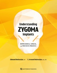
Understanding Zygoma Implants
Biomechanical, Surgical, and Prosthetic Principles
One book, one tree: In support of reforestation worldwide and to address the climate crisis, for every book sold Quintessence Publishing will plant a tree (https://onetreeplanted.org/).
Library of Congress Cataloging-in-Publication Data
Names: Bedrossian, Edmond, editor. | Bedrossian, E. Armand (Edmond Armand), editor.
Title: Understanding zygoma implants : biomechanical, surgical, and prosthetic principles / edited by Edmond Bedrossian and E. Armand Bedrossian.
Description: Batavia, IL : Quintessence Publishing Co, Inc, [2024] | Includes bibliographical references and index. | Summary: “This text takes the guesswork out of zygoma implant therapy by teaching readers to understand the anatomical, biomechanical, and prosthetic aspects of zygoma implant placement and cross-arch stabilization”-- Provided by publisher.
Identifiers: LCCN 2024007406 | EISBN 9781647242213
Subjects: MESH: Zygoma--surgery | Dental Implantation, Endosseous | Zygoma--anatomy & histology | Maxilla--surgery | Dental Prosthesis, Implant-Supported
Classification: LCC RK667.I45 | NLM WU 640 | DDC 617.6/93--dc23/eng/20240401
LC record available at https://lccn.loc.gov/2024007406
A CIP record for this book is available from the British Library.
ISBN: 9781647241957
© 2024 Quintessence Publishing Co, Inc
Quintessence Publishing Co, Inc
411 N Raddant Road
Batavia, IL 60510
www.quintessence-publishing.com
5 4 3 2 1
All rights reserved. This book or any part thereof may not be reproduced, stored in a retrieval system, or transmitted in any form or by any means, electronic, mechanical, photocopying, or otherwise, without prior written permission of the publisher.
Editor: Kristen Clark
Production: Angelina Schmelter
Design: Sue Zubek
CONTENTS
Preface
Contributors
1/HISTORY OF ZYGOMA IMPLANTS AND THEIR INDICATIONS
2/ORAL HEALTH–RELATED QUALITY OF LIFE
3/MAXILLOFACIAL ANATOMY AND BONE FOR IMPLANT PLACEMENT
4/BIOMECHANICS OF ZYGOMA IMPLANTS AND BONE ANCHORAGE
5/ZYGOMA IMPLANT DESIGN
6/TREATMENT PLANNING FOR IMPLANT-SUPPORTED FIXED PROSTHESES IN EDENTULOUS PATIENTS
7/FULL-ARCH PROSTHESES AND IMPLANT LOADING
8/INTRASINUS VERSUS EXTRASINUS IMPLANT PLACEMENT
9/SURGICAL TECHNIQUES: ZYGOMA AND QUAD ZYGOMA PROCEDURES
10/ZYGOMA IMPLANTS FOR UNILATERAL MAXILLARY EDENTULISM
11/DIGITAL WORKFLOW FOR DEFINITIVE PROSTHESIS FABRICATION
12/ALTERNATIVE PROSTHETIC WORKFLOWS AND A QUAD ZYGOMA CASE REVISION
13/PROSTHETIC COMPLICATIONS
14/SURGICAL AND POSTSURGICAL COMPLICATIONS
15/COORDINATING TREATMENT
Index
PREFACE
This book is dedicated posthumously to Professor Per-Ingvar Brånemark in honor of his vision for developing surgical protocols and armamentaria that allow for the predictable rehabilitation of patients with maxillary defects, whether congenital or acquired. His lifetime dedication to patient care not only helped to restore form and function to this group of patients but, more importantly, improved their quality of life and reintroduced them to society. I therefore wish to remind my colleagues, as well as current and future residents, of the fundamental principles that Brånemark lived by:
Interdisciplinary management to allow for enlightened treatment planning.
Simplification and identification of adequate versus optimal treatment concepts.
Predictability achieved via documentation and evidence-based treatment concepts.
The patient is paramount—listen to the needs and demands of your patient.
Brånemark lived by these principles and mentored all who were fortunate enough to have known him. Each chapter in this book is written to honor him by upholding these principles.
Treating maxillectomy patients and patients with severe maxillary resorption is one of the most challenging maxillofacial reconstruction procedures. The introduction of the zygoma implant allowed practitioners to reconstruct these difficult defects without extensive grafting, a treatment dubbed the “graftless concept.” Over the past 25 years, clinicians using zygoma implants have followed the ad modum Brånemark technique, which has resulted in clinical success rivaling conventional axial implants, with multiple systematic reviews reporting long-term success rates of 98%.
The popularity and acceptance of the zygoma implant for the treatment of the completely edentulous maxilla is both exciting and worrisome. Preserving the technique described by Brånemark is critical for the continuance of predictable long-term outcomes with zygoma implants. To emphasize the need for maintaining the technique as described by Brånemark, I have recruited leading clinicians experienced in the treatment of patients with advanced maxillary resorption to contribute to this textbook. I have asked them to share their thoughts and their contemporary approach to executing the Brånemark technique.
The title of this book emphasizes the need to fully understand zygoma implants, including the biomechanical, surgical, and prosthetic principles that have made the use of this implant in maxillary reconstructions extremely predictable. It has been a great joy to coauthor this textbook with my son and prosthodontist, Armand, who led the team of contributors in emphasizing the role of the digital workflow (DWF) in maxillary reconstruction.
The DWF is governed by a complete understanding of the fundamental surgical and prosthetic principles. The digilog concept of implementing fundamental, evidence-based principles while using contemporary DWFs is discussed in depth, both for planning and implementing extensive maxillary reconstructions using the zygoma concept.
When navigating modern surgical education, we must always rely on the fundamental principles that have advanced the predictability of restoration using the zygoma implant—the best interests of the patient come first.
ACKNOWLEDGMENTS
I want to express my great appreciation to my wife, Jasmine, whose support enabled me to write and organize this textbook immediately following completion of my other Quintessence book, The Immediacy Concept. I also want to recognize all the contributors to this textbook, as well as the corporate support from Quintessence Publishing and Straumann, who have facilitated communication of the principles described in this text to all our colleagues. Without their dedication, this book would not have been possible.
CONTRIBUTORS
Fawaz Alzoubi, BDM, BMS, MA, EdD
Associate Professor
Faculty of Dentistry
Kuwait University
Kuwait City, Kuwait
Edmond Bedrossian, DDS
Director of Advanced Implant Reconstruction
Clinical Professor
Department of Oral and Maxillofacial Surgery
Arthur A. Dugoni School of Dentistry
Chairman
ITI Scholarship Center
University of the Pacific
Private Practice
San Francisco, California
E. Armand Bedrossian, DDS, MSD
Affiliate Professor
Department of Prosthodontics
University of Washington
Seattle, Washington
Private Practice
San Francisco, California
John B. Brunski, PhD
Senior Research Engineer
Division of Plastic and Reconstructive Surgery
Department of Surgery
Stanford University School of Medicine
Stanford, California
Professor Emeritus
Department of Biomedical Engineering
Rensselaer Polytechnic Institute
Troy, New York
Konstantinos Chochlidakis, DDS
Associate Professor and Program Director, Prosthodontics
Eastman Institute for Oral Health
University of Rochester
Rochester, New York
Carlo Ercoli, DDS, MBA
Chairperson of Prosthodontics
University of Rochester Medical Center
Rochester, New York
Christopher A. Gurries, DDS
Private Practice
San Francisco, California
Panos Papaspyridakos, DDS, MS, PhD
Associate Professor
Tufts University School of Dental Medicine
Boston, Massachusetts
Constantinus Politis, MD, DDS, MM, MHM, PhD
Head of the Department of Oral and Maxillofacial Surgery
University Hospitals Leuven
Leuven, Belgium
Lambert J. Stumpel, DDS
Private Practice
San Francisco, California
Jean Baptiste Verdino, DDS, MS
Private Practice
Hyères, France
Luc Vrielinck, MD, DDS
Oral and Maxillofacial Surgeon
Private Practice
Genk, Belgium
1 /
History of Zygoma Implants and Their Indications
EDMOND BEDROSSIAN
Rehabilitating patients who have suffered traumatic events or undergone oncologic resections that resulted in total or partial loss of the maxilla is one of the most challenging reconstructive dental procedures to perform. The lack of bone volume to support a conventional removable prosthesis is a major obstacle, and even in cases where obturators can be fabricated using the residual undercuts in the patient’s anatomy, the results are generally less than optimal. Placing conventional dental implants is impossible in these patients due to the severe damage or resection of the maxillary alveolar bone. To stabilize prostheses in these patients, P-I Brånemark instead considered a distant implant anchorage site—the zygoma (Fig 1-1).
Fig 1-1/ The zygoma (blue shading) forms a portion of the roof of the maxillary sinus and the lateral border of the orbit.
This chapter reviews the history of the zygoma implant according to its indications, from its original purpose to treat patients suffering from traumatic maxillary bone defects or resections to its contemporary uses in patients with severely resorbed maxillae.
Traumatic Defects
At one time, a tissue-supported removable prosthesis was the only treatment option for the dental rehabilitation of many patients who had undergone significant midfacial trauma (Fig 1-2). However, due to the difficulty of establishing an adequate peripheral seal, the reliable retention of these prostheses was often not possible, resulting in compromised function and esthetics. To enhance prosthesis retention, P-I Brånemark began exploring the possibility of anchoring endosteal implants in the surrounding stable maxillofacial skeletal bones. For this group of patients, the zygoma was the distant anchorage site of choice for endosseous implants.
Fig 1-2/ 3D reconstruction of a patient with significant right midfacial trauma and failed bone grafting.
Initially, conventional 3.75-mm machined titanium implants 8 to 10 mm in length were placed in the zygoma according to a two-stage surgical protocol (Fig 1-3). A waiting period of 6 months was allowed for implant osseointegration prior to uncovering the implants to begin the prosthetic phase of rehabilitation. Long, custom abutments were fabricated to allow maxillofacial prosthodontists to bring the implant platform closer to the occlusal plane.
Fig 1-3/ Placement of two endosteal implants with cover screws in the remaining portion of the right zygoma.
The placement of conventional machined titanium implants in maxillectomy patients was also reported by Thomas Weischer et al in 1997.1 As seen in Fig 1-4a, three endosteal implants were placed in the patient’s right maxillary alveolar bone, along with two endosseous implants in the left zygoma. After osseointegration of all implants, the implants in the left zygoma were connected with a titanium bar and used to retain the overlying prosthesis (Figs 1-4b and 1-4c).
Fig 1-4/(a) A patient with a left maxillectomy. Two endosseous implants were placed in the left zygoma. (b and c) Splinted implants were used to retain the overlying removable prosthesis.
In many patients treated according to these methods, however, the outcome was that the implant-abutment junction was too far from the abutment-prosthesis junction, creating a long cantilever. Although the prostheses were tissue supported and only implant retained, in general the clinical outcomes were associated with frequent loosening and fracturing of the prosthesis and the custom-made abutment screws. The difficulty of replacing the fractured abutment screws made this treatment unpredictable, and a very high level of maintenance was also required.
The logical solution to this type of complication was exchanging the conventional implants with long, custom-made abutments for single-piece implants—hence, the birth of the zygoma implant. Figure 1-5 shows the use of conventional implants in the patient’s right zygoma as well as experimentation with a zygoma implant in the patient’s left zygoma. The extended length of the zygoma implant replaces the need for a long custom abutment and brings the restorative platform of the implant into the proper occlusal plane. Eliminating long custom abutments resulted in fewer mechanical complications and more predictable restorations with easier maintenance for this group of patients.
Fig 1-5/ The extended length of the zygoma implant eliminates the need for long, custom-made abutments.
Brånemark did not report the use of the new zygoma implant until 10 years after treating the first patient in 1989; the first publication on the use of the zygoma implant was not until 2000.2 This study reported the predictable treatment outcomes found with zygoma implants, both in the patient who received the newly designed zygoma implant and in patients treated earlier according to a zygoma concept. The first course to introduce the zygoma concept to North America was held in 1996, and the technique was quickly adopted by a select group of maxillofacial surgeons, who found the same positive outcomes as reported in the 2000 paper.
The zygoma implant proved to be invaluable for treating patients who had suffered trauma to the midface. Figure 1-6 shows an example of one of these patients. He had suffered a gunshot wound to his right midface, resulting in partial avulsion of the right hemimaxilla. The lack of hard tissue support in the maxillary right posterior quadrant created difficulty in retaining a maxillary partial denture, even with the assistance of splinted conventional implants in the anterior maxilla. To facilitate the retention and function of the partial denture, a zygoma implant was placed distal to the existing implants in the intact zygomatic bone. This allowed for the extension of the implant-retained bar for a more stable partial denture.
Fig 1-6/(a and b) Loss of the right maxilla and inadequate prosthesis retention even with three conventional implants and a bar-retained partial denture. (c to e) Views at the 8-year follow-up after the placement of a zygoma implant that was used to extend the original bar for better retention of the partial denture.
Oncologic Defects
Zygoma implant therapy has also become a predictable treatment option for patients who have undergone oncologic resections involving partial or total maxillectomy (Figs 1-7 and 1-8). Clinical success with the zygoma implant concept led to the need for establishing specific protocols for treating patients with maxillofacial defects. In 2012, Bedrossian and Brånemark reported systematic treatment planning protocols for the rehabilitation of patients with various maxillofacial defects.3 The number and distribution of implants for the reconstruction of orbital defects, nasal defects, and hemifacial resections were outlined, and the authors emphasized the importance of interdisciplinary collaboration between the oncologic (primary) surgeon and the reconstructive (maxillofacial) surgeon to ensure retention of the zygoma when possible.
Fig 1-7/ Model showing double zygoma implants used for the reconstruction of a partial maxillectomy.
Fig 1-8/ Single zygoma implant used to rehabilitate a patient with a maxillofacial defect.
It is important that oncologic surgeons treating patients with neoplastic midface lesions critically evaluate whether the zygomatic bone can be spared from the resection field. In cases where the zygoma is resected due to a cancerous lesion or when the rehabilitation of the patient is not adequately considered preoperatively and the uninvolved zygoma is partially or totally resected, the resultant lack of zygomatic bone prohibits the rehabilitation of the patient with any reliable retentive prosthesis, as seen in Fig 1-9.
Fig 1-9/(a and b) Resection of the right zygomatic bone limits the ability to predictably rehabilitate this patient.
Congenital Defects
The positive functional and esthetic outcomes of zygoma implants used for the treatment of traumatic and oncologic lesions led to the consideration of expanding zygoma implant indications. It is encouraging to see that the indications for zygoma implants now include the treatment of congenital defects affecting the maxilla. Examples of the congenital defects that can be treated with zygoma implants are listed here:
Cleft palate
Ectodermal dysplasia
Hypoplastic maxilla with anodontia
Cleft palate
Zygoma implants have been adopted by maxillofacial prosthodontists to aid in the predictable retention of maxillary obturators in patients with cleft palate defects (Fig 1-10). The quad zygoma concept for the retention of obturators in patients with more extensive hard palate clefts is also a favored treatment option over conventional obturators and hard and soft tissue grafting procedures (Fig 1-11).
Fig 1-10/(a to c) Zygoma implants used to support implant-retained obturators for patients with cleft palates.
Fig 1-11/(a to c) Quad zygoma concept used to retain a large obturator.
Ectodermal dysplasia
For patients with ectodermal dysplasia, the complete or partial absence of tooth buds results in the inadequate development of the maxillary and mandibular alveolar bones. The minimal development of the alveolar bone does not provide favorable topography to support a conventional tissue-supported removable prosthesis.
To improve the comfort and masticatory function of these patients, as well as to enhance the facial soft tissue drape and support, endosteal implants to support a fixed prosthesis can be considered. An example of this treatment concept can be seen in a 23-year-old man who presented with ectodermal dysplasia (Figs 1-12a and 1-12b). The existing maxillary and mandibular removable prostheses were not functional, and the failing remaining dentition qualified this patient for treatment with zygoma implants. The patient was treated in one surgical appointment, with removal of the remaining maxillary and mandibular dentition, placement of the implants, and immediate loading of the implants with fixed, implant-supported provisional prostheses (Figs 1-12c and 1-12d).
Fig 1-12/(a and b) The preoperative presentation of a patient with ectodermal dysplasia. (c and d) The postoperative fixed prostheses.
Hypoplastic maxilla with anodontia
A 21-year-old man presented with a congenitally hypoplastic maxilla as well as anodontia. He had never had any type of dental rehabilitation because retention and function with a maxillary or mandibular removable prosthesis had been impossible (Figs 1-13a to 1-13d). To address the vertical and anteroposterior (AP) maxillary bone deficiencies, two options for treatment planning were possible: a staged grafting procedure, which would take between 24 and 30 months to complete, or a graftless procedure to compensate for the vertical and horizontal bone deficits in one surgical appointment. The patient chose the graftless option and was treated with quad zygoma implants in the maxilla as well as tilted implants in the mandible, with immediate loading of both jaws. After 6 months of osseointegration, the final zirconia hybrid prostheses compensated for the vertical, horizontal, and AP maxillary deficits (Figs 1-13e to 1-13h).
Fig 1-13/(a to d) Pre-operative presentation of a 21-year-old patient with a hypoplastic maxilla. (e to h) Final rehabilitation establishing the missing vertical, AP, and horizontal bone volume of the hypoplastic maxilla.
Severely Resorbed Maxillae
Today, zygoma implants are the preferred treatment option over grafting procedures for the rehabilitation of patients with severely resorbed maxillae.
Establishing posterior support in an edentulous maxilla is challenging because edentulous maxillae have a unique anatomical presentation that may prevent the placement of the appropriate number and distribution of implants within the alveolar ridge (Fig 1-14). The maxillary sinuses and the position of the nasal floor in the anterior maxilla limit the vertical volume of alveolar bone available for implant placement (Fig 1-15). The frontal view of the completely resorbed maxillary alveolar bone with 3D radiography is shown in Fig 1-16. Complete resorption of the maxillary alveolus leaves a shell of the maxilla proper, with large air spaces of the maxillary sinus and nasal cavity. Placing conventional implants without extensive bone grafting is therefore not possible.
Fig 1-14/ An 85-year-old woman with complete maxillary alveolar resorption.
Fig 1-15/ Illustration of the complete resorption of the maxillary alveolar bone.
Fig 1-16/(a and b) Complete resorption of the maxillary alveolus.
Prior to zygoma implant therapy, patients with bone deficits had to undergo extensive grafting procedures with autogenous bone harvested from the calvaria or ilium. These grafting procedures are completed in multiple steps, and patients are often hesitant to receive this treatment for several reasons:
Two surgical sites (the graft donor and recipient sites) are required.
Multiple-day hospitalization is required.
Patients cannot use a provisional prosthesis during the healing period.
Various procedures to reconstruct bone volume have been reported in the literature. Composite grafts can be used to provide a tooth anchorage system.4,5 Le Fort I osteotomies and iliac block grafts have also been used to reestablish bone volume for the placement of implants.6–8 Alternative procedures to increase bone volume in the posterior maxilla, such as sinus elevation grafting procedures, have also been advocated, and implant success within grafted sinuses has been reported as 90%.9
Tolman and Keller have also described grafting of the maxilla with delayed implant placement and delayed loading, with 87% and 95% implant and prosthetic survival rates, respectively.10,11 Rasmusson et al, using autogenous inlay, onlay, and/or Le Fort I procedures, reported success rates of 80% for grafting with delayed implant placement and 77% for grafting with simultaneous implant placement.12 Others have also reported using various bone grafting techniques to reconstruct resorbed maxillae.5–8 The disadvantage of maxillary onlay grafts or the grafting of the maxillary sinus with simultaneous implant placement is the inability to perform immediate implant loading.
In the early 1990s, the author was accustomed to working with the Brånemark horseshoe graft, which involved using a large block of the patient’s iliac bone to reconstruct the missing horseshoe-shaped maxillary alveolar bone (Fig 1-17). The bone graft (Fig 1-17a) was positioned with an anterior cantilevered trajectory as well as with horizontal cantilevers in an attempt to reestablish the posterior and palatal resorption pattern of the maxillary alveolar bone (Fig 1-17b). The graft was stabilized with specific fixation tools developed for this procedure; implants were placed according to a two-stage delayed loading protocol in conjunction with simultaneous bilateral sinus grafting procedures to create additional bone volume in the posterior maxilla (Figs 1-17c and 1-17d). Although this procedure restored the form and function of the maxilla (Fig 1-17e), patients with severely resorbed maxillae were reluctant to undergo these grafting procedures due to the disadvantages mentioned previously, including the extended length of the total treatment time as well as the associated morbidity.
Fig 1-17/(a) Autogenous iliac bone harvested for the reconstruction of the maxillary alveolus. (b) The vertical, posterior, and horizontal bone deficiencies are compensated for by the bone graft. (c and d) Stabilizing the graft with endosteal implants. (e) Final maxillary rehabilitation using the Brånemark horseshoe graft and a hybrid prosthesis supported by six axial implants.
The disadvantages of grafting procedures limit patient case acceptance of surgical reconstruction via this approach. Poor outcomes reported in the literature for immediately loaded grafted implants9 have encouraged clinicians to adopt a graftless approach using tilted or zygoma implants instead of performing sinus grafting with immediate or delayed implant placement. Placing endosteal implants and compensating for the missing anatomy (the maxilla and/or the maxillary alveolus) with a hybrid prosthesis can be completed in one procedure with decreased morbidity, resulting in greater patient acceptance.
In contemporary implant practice, it is well accepted that the prosthetic reconstruction of a surgical defect is more predictable than a surgical reconstruction. Graftless procedures, where the missing alveolar bone is replaced with pink acrylic or porcelain, are more predictable than grafting procedures. Even for patients with complete maxillary alveolar resorption, the quad zygoma concept, involving the placement of two zygoma implants in each zygoma, allows for the stabilization and retention of a fixed hybrid prosthesis, eliminating the need for bone grafting (Fig 1-18).
Fig 1-18/(a and b) The 19-year follow-up of a patient treated with the quad zygoma concept.
Classification of resorbed maxillae
In 2008, Bedrossian et al introduced a systematic treatment planning protocol for the evaluation of patients with maxillary edentulism or maxillary terminal dentition.13 Under this treatment planning protocol, the maxilla was divided into three zones. Zone 1 encompassed the bone between the canines, Zone 2 represented the bone available in the premolar region, and Zone 3 consisted of the bone in the molar region (Fig 1-19). The classification remains the same regardless of the degree of maxillary alveolar resorption (Fig 1-20). The decision to place axial, tilted, zygoma, or quad zygoma implants to support a fixed maxillary hybrid prosthesis was made by evaluating the presence or lack of bone in the various zones:
If the bone in all zones was present, axial implants were placed.
If bone in Zones 1 and 2 was present, axial implants were placed in the anterior maxilla, and tilted implants were placed in the posterior maxilla.
If bone was present in only Zone 1, axial implants were placed in the anterior maxilla, and zygoma implants were placed in the posterior maxilla.
Finally, in cases of complete maxillary resorption, four zygoma implants were placed, two in each zygoma.
Fig 1-19/ The zones of a maxilla that is not resorbed.
Fig 1-20/ The zones of the maxilla in the presence of complete maxillary alveolar resorption.
More recently, to avoid extensive bone grafting procedures, tilted implants have been recommended for the rehabilitation of patients with bone available in Zones 1 and 2.14–16 Using tilted implants requires the presence of vertical and horizontal bone volume in the first premolar region as well as the presence of at least 3 to 4 mm of residual bone volume in the second molar region, as shown in Fig 1-21.
Fig 1-21/ Tilted implant concept to establish posterior support.
For patients with extensive anterior maxillary sinuses extending forward into the premolar region (lack of bone in Zone 2), zygoma implants have become a common treatment, used according to a graftless concept to allow for immediate loading on the day of implant placement (Fig 1-22).
Fig 1-22/ The presence of residual anterior maxillary bone will allow for the placement of axial implants in the anterior maxilla and zygoma implants in the posterior maxilla, without the need for grafting.
Zygoma implants allow for the establishment of posterior maxillary support by using the limited crestal alveolar bone in the premolar region to stabilize the platform of the implant and the zygoma for establishing initial stability of the apical portion of the implant, thus allowing for quad cortical stabilization of the zygoma implant (Fig 1-23).
Fig 1-23/(a to d) Quad-cortical stabilization of the zygoma implant.
Success of Zygoma Implants
Zygoma implants have been studied internationally, with reported success rates between 94% and 100%.17–21 In 2004, Brånemark et al reported a cumulative survival rate (CSR) of 94% in their initial retrospective study of 52 patients treated with zygoma implants according to a two-stage delayed loading protocol.20 In 2010, Bedrossian et al reported on the 7-year prospective follow-up of patients treated using the immediate loading protocol with a CSR of 97.2%.22 In 2014, Aparicio et al reported on a 10-year prospective study using the immediate loading protocol with a similar CSR of 97.71%.23
Zygoma implants can also be used to rehabilitate patients in whom other treatment options have failed. Bedrossian introduced the rescue implant concept in 2011, which involves using zygoma implants to treat cases of failed sinus grafting procedures or failed tilted implants.24 This concept allows for continuity of care of the patient without major interruption in their treatment.
In recent years, the popularity of zygoma implants has increased due to favorable systematic reviews. Chrcanovic and Abreu reported a 96.7% success rate over a 12-year follow-up period,25 and Goiato et al reported a 97.86% success rate for 1,541 zygoma implants.26
Conclusion
The remainder of this book addresses the details of the concepts mentioned in this chapter, including the biomechanical principles, treatment planning protocols, and surgical procedures for zygoma implants used in various indications, as well as the digital workflows for prosthesis fabrication and complication prevention and management. Various case studies illustrate the covered concepts. By following the scientific principles outlined by Brånemark and the confirmed biomechanical principles of zygoma implants placed under functional loads, clinicians can achieve the same positive results and long-term successes that are reported in the literature.
References
1.Weischer T, Schettler D, Mohr C. Titanium implants in the zygoma as retaining elements after hemimaxillectomy. Int J Oral Maxillofac Implants 1997;12:211–214.
2.Higuchi KW. The zygomatic fixture: An alternative approach for implant anchorage in the posterior maxilla. Ann R Australas Coll Dent Surg 2000;15:28–33.
3.Bedrossian E, Brånemark PI. Systematic treatment planning protocol for patients with maxillofacial defects: Avoiding living a life of seclusion and depression. Atlas Oral Maxillofac Surg Clin North Am 2012;20:135–158.
4.Adell R, Lekholm U, Rockler B, Brånemark PI. A 15-year study of osseointegrated implants in the treatment of the edentulous jaw. Int J Oral Surg 1981;10:387–416.
5.Breine U, Brånemark PI. Reconstruction of alveolar jaw bone. An experimental and clinical study of immediate and preformed autologous bone grafts in combination with osseointegrated implants. Scand J Plast Reconstr Surg 1980;14:23–48.
6.Isaksson S, Ekfeldt A, Alberius P, Blomqvist JE. Early results from reconstruction of severely atrophic (class VI) maxillas by immediate endosseous implants in conjunction with bone grafting and Le Fort I osteotomy. J Oral Maxillofac Surg 1993;22:144–148.
7.Adell R, Lekholm U, Gröndahl K, Brånemark PI, Lindström J, Jacobsson M. Reconstruction of severely resorbed edentulous maxillae using osseointegrated fixtures in immediate autogenous grafts. Int J Oral Maxillofac Implants 1990;5:233–246.
8.Isaksson S, Alberius P. Maxillary alveolar ridge augmentation with onlay bone-grafts and immediate endosseous implants. J Craniomaxillofac Surg 1992;20:2–7.
9.Jensen OT, Shulman LB, Block MS, Iacono VJ. Report of the Sinus Consensus Conference of 1996. Int J Oral Maxillofac Implants 1998;13(suppl):11–45.
10.Tolman DE. Reconstructive procedures with endosseous implants in grafted bone: A review of the literature. Int J Oral Maxillofac Implants 1995;10:275–294.
11.Keller E, Tolman DE, Eckert SE. Maxillary antral-nasal inlay autogenous bone graft reconstruction of compromised maxillae: A 12-year retrospective study. Int J Oral Maxillofac Implants 1999;14:707–721.
12.Rasmusson L, Meredith N, Cho IH, Sennerby L. The influence of simultaneous versus delayed placement on stability of titanium implants in onlay bone grafts. A histologic and biomechanic study in the rabbit. Int J Oral Maxillofac Surg 1999;28:224–231.
13.Bedrossian E, Sullivan RM, Fortin Y, Malo P, Indresano T. Fixed-prosthetic implant restoration of the edentulous maxilla: A systematic pretreatment evaluation method. J Oral Maxillofac Surg 2008;66:112–122.
14.Fortin Y, Sullivan RM, Rangert BR. The Marius implant bridge: Surgical and prosthetic rehabilitation for the completely edentulous upper jaw with moderate to severe resorption: A 5-year retrospective clinical study. Clin Implant Dent Relat Res 2002;4:69–77.
15.Krekmanov L, Kahn M, Rangert B, Linström H. Tilting of posterior mandibular and maxillary implants for improved prosthesis support. Int J Oral Maxillofac Implants 2000;15:405–414.
16.Aparicio C, Perales P, Rangert B. Tilted implants as an alternative to maxillary sinus grafting: A clinical, radiologic, and periotest study. Clin Implant Dent Relat Res 2001;3:39–49.
17.Stevenson AR, Austin BW. Zygomatic fixtures—The Sydney experience. Ann R Australas Coll Dent Surg 2000;15:337–339.
18.Bedrossian E, Stumpel L 3rd, Beckely ML, Indresano T. The zygomatic implant: Preliminary data on treatment of severely resorbed maxillae. A clinical report. Int J Oral Maxillofac Implants 2002;17:861–865.
19.Malevez C, Abarca M, Durdu F, Daelemans P. Clinical outcome of 103 consecutive zygomatic implants: A 6-48 month follow-up study. Clin Oral Implants Res 2004;115:18–22.
20.Brånemark PI, Gröndahl K, Ohrnell LO, et al. Zygoma fixture in the management of advanced atrophy of the maxilla: Technique and long-term results. Scand J Reconstr Surg Hand Surg 2004;38:70–85.
21.Aparicio C, Ouazzani W, Garcia R, Arevalo X, Muela R, Fortes V. A prospective clinical study on titanium implants in the zygomatic arch for prosthetic rehabilitation of the atrophic edentulous maxilla with a follow-up of 6 months to 5 years. Clin Implant Dent Relat Res 2006;8:114–122.
22.Bedrossian E. Rehabilitation of the edentulous maxilla with the zygoma concept: A 7-year prospective study. Int J Oral Maxillofac Implants 2010;25:1213–1221.
23.Aparicio C, Manresa C, Francisco K, et al. The long-term use of zygomatic implants: A 10-year clinical and radiographic report. Clin Implant Dent Relat Res 2014;16:447–459.
24.Bedrossian E. Rescue implant concept: The expanded use of the zygoma implant in the graftless solutions. Oral Maxillofac Surg Clin North Am 2011;23:257–276.
25.Chrcanovic BR, Abreu MH. Survival and complications of zygomatic implants: A systematic review. Oral Maxillofac Surg 2013;17:81–93.
26.Goiato MC, Pellizzer EP, Moreno A, et al. Implants in the zygomatic bone for maxillary prosthetic rehabilitation: A systematic review. Int J Oral Maxillofac Surg 2014;43:748–757.
2 /
Oral Health–Related Quality of Life
EDMOND BEDROSSIAN / FAWAZ ALZOUBI
Traditionally, the results of dental implant treatment were described in terms of implant-related outcomes (survival, success, marginal bone loss [MBL], peri-implant health, and esthetics) and prosthesis-related outcomes (survival and technical complications).1 However, a more comprehensive, evidence-based evaluation of treatment involves consideration of the patient-related outcomes.1–5 The American Dental Association (ADA) defines evidence-based dentistry (EBD) as “an approach to oral healthcare that requires the judicious integration of systematic assessments of clinically relevant scientific evidence relating to the patient’s oral and medical condition and history with the dentist’s clinical expertise and the patient’s treatment needs and preferences.”6 Hence, evaluating patient-related outcomes regarding treatment is crucial.
Measuring Oral Health–Related Quality of Life
Optimizing patient-related outcomes with oral rehabilitation is a significant goal.7 Two patient-related measurements are traditionally used in the literature: (1) the impact of the treatment on patient quality of life and (2) patient satisfaction.1 Improving quality of life is one of the main objectives of any medical treatment.
Oral health–related quality of life (OHRQoL) is integral to an individual’s well-being.3 OHRQoL is a multidimensional construct involving functional, psychologic, and social factors related to oral health, as well as related pain or discomfort, that affect the overall well-being of a person.8 A conventional validated measure to evaluate OHRQoL within the field of dentistry and oral sciences is the Oral Health Impact Profile (OHIP).9–12 This instrument covers several domains related to OHRQoL:
Functional limitation
Physical pain
Psychologic discomfort
Physical disability
Psychologic disability
Social disability
Subjective facets such as patient satisfaction also need to be assessed to complement objectively measured factors. Mastication, esthetics, comfort, taste, speech, and ease of prosthesis maintenance all impact satisfaction.13 In addition, psychologic factors such as self-esteem and social impact need to be considered.13 Patient satisfaction is generally evaluated with visual analog scales.14,15
OHRQoL and edentulous patients
Complete edentulism is a condition that is expected to become more prevalent in the future,16 and it is correlated with many comorbid health care factors.17 Oral function and esthetics that affect the daily activities and the psychologic well-being of this patient population significantly influence quality of life.18–21 The loss of various orofacial structures and function22 results in significantly reduced chewing efficiency,23 smaller chewing cycles,24 and reduced muscle activity and bite force,25 limiting the food choices and affecting the nutritional intake of these patients.3,26–28 Furthermore, there is an emotional impact related to the perception of aging and trauma in this population29,30 and a significant psychologic impact due to reduced self-confidence as a result of altered facial form.29,31 It is not surprising that OHRQoL and satisfaction are reduced in this patient population.32–36
Zygoma Implants and OHRQoL
Implant-supported fixed complete dentures (ISFCDs) are of great benefit for meeting the expectations of edentulous patients regarding acceptable oral function, esthetics, and improved self-esteem, leading to improved OHRQoL and patient satisfaction.37–41 ISFCDs using conventional root-form dental implants are widely documented as a valid option to treat completely edentulous patients,42–44 but some cases represent specific challenges and restrictions associated with bone availability. Treatments for cases involving severe atrophy can generally be divided into two main types: grafting solutions and graftless solutions.45–53
With grafting solutions, regenerative procedures are performed to regain lost hard and soft tissue so that conventional root-form dental implants can be placed and restored with an ISFCD.45 With graftless solutions, the approach is to bypass regenerative procedures and use nonconventional implants to support an ISFCD. Defects resulting from the severe atrophy associated with these cases are usually restored with prosthetic options, such as pink porcelain or acrylic, as opposed to surgical interventions.
The zygoma implant is a graftless solution for managing severely atrophic maxillae.44,49,54–56 It is a widely documented option with high survival and success rates in the short-, intermediate-, and long-term.41,49,54–64 Nonetheless, some controversies and misconceptions regarding its impact on OHRQoL and patient satisfaction in terms of the following factors still need to be clarified:
Palatal implant emergence and its effects on speech
Oral hygiene maintenance
Esthetic outcomes
Treatment cost
Invasiveness of the procedure
All these factors play an important role when assessing treatment impact on OHRQoL and patient satisfaction. Misconceptions about these aspects of treatment might contribute to the assumption that this treatment might negatively impact patient-related outcomes, which is contrary to the available evidence.
Patient satisfaction
The impact of zygoma implants on OHRQoL and patient satisfaction has been studied extensively.1,65–71 Patients receiving zygoma implants have reported patient-related outcomes similar to those reported by patients receiving grafting and/or conventional alternatives, with a high impact on OHRQoL and patient satisfaction.1,65–67,70–72 In fact, some studies report that patient-related outcomes—as reflected by OHRQoL and patient satisfaction—were superior in patients receiving zygomatic implants when compared to patients receiving four conventional implants.68,69
In one study, maximum bite force, OHRQoL, and patient satisfaction were evaluated in patients receiving graftless solutions including zygoma implants. The study reported a high positive impact on quality of life and high patient satisfaction, with an average maximum bite force of 108 N in the anterior and 206 N in the posterior aspects of the prosthesis.70 The study also reported that patient-related outcomes seemed to be better when patients initially presented as completely edentulous, as opposed to having some terminal dentition.70 Patient satisfaction and OHRQoL outcomes also seem to be correlated, meaning that when the impact on improving OHRQoL is high, patient satisfaction is also high.70 Furthermore, maximum bite force and masticatory function also seem to be positively correlated with patient-related outcomes.45,61,70–73
Palatal emergence and its effects on speech
Concerns surrounding the palatal emergence of zygoma implant platforms (Fig 2-1) can be diminished by developing a proper understanding of the anatomy of atrophic maxillae. Severely atrophic maxillae are resorbed both in the transverse and axial dimensions. In other words, the maxilla does not resorb in only a vertical direction but also in a horizontal direction, with the alveolar process shifting medially toward the midline (Fig 2-2). As a result of this centripetal resorption pattern,65 maxillae in severely atrophic jaws seem to be smaller in size and constricted toward the medial aspect.
Fig 2-1/ Palatal position of the screw access holes due to palatal resorption of the alveolus.
Fig 2-2/ Vertical (a) and horizontal (b) resorption pattern of the maxilla.
Typically, zygoma implants engage the alveolar process in the maxilla and then the zygomatic bone. Hence, the implants follow the trajectory of the alveolar process, which is palatalized due to the resorption pattern. In other words, the final implant is not in a “palatal” position so much as it is simply following the trajectory of the alveolar process. Due to the local anatomy, this palatal trajectory is similar whether zygoma implants or conventional implants are placed in a severely atrophic maxilla.
Even if zygoma implants were in a palatal position, as some may consider them to be, measures related to parameters that might be affected by this palatal implant position (such as speech and maintaining oral hygiene) do not seem to be affected, as reported by patient-centered studies,45,51,71 which have also shown lower rates of peri-implantitis with zygoma implants than with conventional treatment approaches.78
Oral hygiene maintenance
When compared to implant-retained removable overdentures, zygoma implants supporting an ISFCD facilitated better oral hygiene maintenance in a report on two cases.79 This may be due to the fact that implant-supported removable overdentures undergo significant prosthesis rotation, resulting in a pivoting phenomenon that is associated with stomatitis.80,81 Furthermore, limited salivary flow around the peri-implant site may lead to an acidic environment beneath the prosthesis.82 Finally, oral hygiene measures around ISFCDs might not be influenced by the surgical intervention as much as they are by the prosthesis design. A prosthesis with an ovate intaglio surface is required to facilitate oral hygiene regardless of the surgical intervention that is used (Fig 2-3).83 For patients with severely atrophic maxillae who have been rehabilitated with an ISFCD, vents may be added to the prosthesis to aid hygiene access (Fig 2-4).
Fig 2-3/ Soft tissue changes after replacing a prosthesis with a poorly designed intaglio surface (a) with a provisional prosthesis with an ovate pontic intaglio surface (b). (Reprinted with permission from Alzoubi and Wong.83)
Fig 2-4/(a) Buccal vents around abutments designed to facilitate oral hygiene.(b) Slough ways to facilitate threading dental floss.
Esthetic outcomes
Another concern when rehabilitating severely atrophic maxillae is the degree of lip support gained from ISFCDs. Lip support is maintained by the anteroposterior (AP) position of the incisors and the projection of the anterior maxilla (Fig 2-5). One of the disadvantages of some fixed prostheses is an inability to achieve acceptable lip support, as evaluated according to nasolabial angle norms.80,84 However, proper positioning of the anterior teeth and contouring of the pink acrylic/porcelain of a hybrid prosthesis can make achieving proper nasolabial angles and esthetic lip projection predictable (Fig 2-6).
Fig 2-5/ Lip support is a function of the AP position of the anterior teeth.
Fig 2-6/ Proper contouring of the prosthesis flange mimics the profile of the missing alveolus.
Communication with the patient is also critical within this context. When given the option between a removable prosthesis with acceptable lip support and a fixed prosthesis with unacceptable lip support, patients may still choose the fixed solution given its many other advantages.
Treatment cost
One argument for favoring removable implant-supported overdentures over fixed prostheses is the cost of treatment. In some satisfaction and quality of life studies, the cost of treatment was considered as a factor when measuring these outcomes. The higher initial cost of ISFCDs may have resulted in more favorable results with implant-supported removable overdentures. If both treatments were performed excluding cost as a factor, however, or if a long-term cost-effective analysis were performed, the results may be different.85,86
Invasiveness of the procedure
When considering the invasiveness of zygoma implant procedures, the invasiveness of alternative solutions should also be considered within the context of the severely atrophic maxilla. This will aid in a fair comparison and individualized assessment. Each procedure is meant for specific cases with specific indications, and no treatment should be considered as a routine solution that will work for every edentulous patient.
Within this individualized assessment, risks and benefits need to be carefully considered. In severely atrophic maxillae, there is not a noninvasive surgical approach available to rehabilitate patients with an ISFCD. Invasiveness is relative, and options need to be carefully assessed, with the least invasive solution with the maximum benefits and minimal risks selected. When compared to conventional solutions such as advanced grafting procedures, zygoma implants are less invasive and involve a shorter duration of treatment that does not involve wearing poorly adapted removable complete dentures that compromise function and esthetics.41,54,71–87
Zygoma Implants Versus Grafting Procedures
Traditionally, severely atrophic maxillae were treated with advanced regenerative procedures to restore lost hard and soft tissue, and then conventional implants were placed to support an ISFCD.45,47 Autogenous bone grafting (in the form of block grafting and/or sinus elevation procedures), either alone or combined with a Le Fort osteotomy, is usually involved in this form of treatment.46,65,90,91 This entails harvesting a large amount of bone from extraoral donor sites.69 Grafting thus raises concerns related to donor and host site morbidity, the duration of treatment, and the success of this relatively complex procedure, which requires skillful training.65,67,71,92 Furthermore, immediate loading is not possible, which leads to a challenging transitional phase during healing that may last up to 6 months.71,93 The additional cost of these procedures may also be a significant factor.70
In one study, patient-related outcomes were compared between patients receiving zygoma implants and patients who underwent advanced grafting procedures. The study authors concluded that zygoma implants were a valid alternative to grafting and that they had a high positive impact on quality of life and patient satisfaction, recommending it as a first-line treatment option.65
As mentioned previously, treatment options need to be carefully evaluated according to an individualized assessment strategy and careful case selection that weighs risks and benefits. The most minimally invasive method that provides acceptable patient-related outcomes should be the treatment of choice to achieve the ultimate objective of restoring patient-related outcomes including esthetics and function.94,95 The selected minimally invasive method should also be financially feasible96; avoid the need for advanced grafting procedures97; result in minimal postoperative morbidity such as pain,98 swelling,99 and bleeding100; and limit treatment duration.102 Furthermore, immediate loading not only presents an obvious advantage in terms of improving the transitional healing phase for patients102,103 but also provides feedback regarding prosthesis design, which can be used to improve the final prosthesis.
Keeping all these factors in mind, zygoma implants can fulfill requirements for all parameters of OHRQoL, making them a reasonable option for the treatment of severely atrophic maxillae compared to more extensive and invasive procedures.45,65,70,71,104,105
Treatment of congenital conditions
Finally, special consideration needs to be given to cases involving syndromic patients. With syndromic cases involving premature tooth loss at an early age (such as Papillon-Lafèvre syndrome and Chediak-Higashi syndrome), quality of life is significantly impacted.106 Zygoma implants can be especially advantageous for these patients and lead to improvement in their overall quality of life and satisfaction.79,83
Surgical and prosthetic rehabilitation is extremely challenging in patients with advanced bone resorption resulting from early tooth loss. It is impossible to provide removable complete dentures with clinically acceptable parameters of stability, support, and retention, and given the young age of these patients, removable options are also not acceptable due to the psychosocial impact.83 Advanced bone grafting procedures can be considered, but the prognosis is questionable in immunocompromised patients. Furthermore, the negative factors associated with advanced grafting procedures mentioned earlier also need to be considered.
Not only do these cases involve surgical challenges, but they also entail prosthetic challenges. Premature tooth loss in growing individuals will lead to inappropriate vertical facial growth because alveolar and dental growth contribute significantly to facial growth. The result is compromised lower facial height. This issue must be addressed during the diagnosis, planning, and execution phases of treatment to rehabilitate patients and restore acceptable facial dimension norms.
Zygoma implants can be an acceptable alternative to manage these cases and restore quality of life and patient satisfaction.79,83 They allow for the reconstruction of existing composite defects with a fixed hybrid prosthesis.44 The physiologic reintroduction of the internal loading forces during function allows for the maintenance of the residual alveolar bone and improved psychosocial benefits, making zygoma implants a beneficial option for reconstructing moderate to advanced maxillary resorption in this group of patients.
Conclusion
It is evident from the literature, as well as the experience of the authors, that zygoma implants used to treat moderate to extreme maxillary resorption are well accepted by most patients. Reports on palatal implant emergence and subsequent difficulty with speech have been shown to be exaggerated. The ease of oral hygiene maintenance, the positive esthetic outcomes, the lower treatment costs, and the reduced invasiveness of zygoma implant procedures compared to grafting alternatives are a testament to the satisfaction of most patients treated according to the zygoma concept.
References
1.Pommer B, Mailath-Pokorny G, Haas R, Busenlechner D, Fürhauser R, Watzek G. Patients’ preferences towards minimally invasive treatment alternatives for implant rehabilitation of edentulous jaws. Eur J Oral Implantol 2014;7(suppl 2):S91–S109.
2.Marshall S, Haywood K, Fitzpatrick R. Impact of patient-reported outcome measures on routine practice: A structured review. J Eval Clin Pract 2006;12:559–568.
3.Petricevic N, Celebic A, Rener-Sitar K. Improvement of patient’s satisfaction and oral health-related quality of life by the implant and prosthodontic treatment. In: Virdi MS (ed). Oral Health Care: Pros-thodontics, Periodontology, Biology, Research and Systemic Conditions. InTech, 2012:25–52.
4.Hayes C. The use of patient based outcome measures in clinical decision making. Community Dent Health 1998;15:19–21.
5.Sánchez-Siles M, Ballester-Ferrandis JF, Salazar-Sánchez N, Gómez-García FJ, Moraleja-Ruiz R, Camacho-Alonso F. Long-term evaluation of quality of life and satisfaction between implant bar overdentures and conventional complete dentures: A 23 years retrospective study. Clin Implant Dent Relat Res 2018;20:208–214.
6.American Dental Association. ADA Policy Statement On Evidence-Based Dentistry. https://www.ada.org/en/resources/research/science-and-research-institute/evidence-based-dental-research#:~:text=ADA%20Policy%20Statement%20on%20Evidence,condition%20and%20history%2C%20with%20the. Accessed 15 November 2023.
7.Al-Omiri M, Hantash RA, Al-Wahadni A. Satisfaction with dental implants: A literature review. Implant Dent 2005;14:399–406.
8.Ingelehart MR, Bagramian RA. Oral health-related quality of life: An introduction. In: Ingelehart MR, Bagramian RA (eds). Oral Health-Related Quality of Life. Quintessence, 2002:1–6.
9.Allen PF, McMillan AS, Walshaw D, Locker D. A comparison of the validity of generic- and disease-specific measures in the assessment of oral health-related quality of life. Community Dent Oral Epidemiol 1999;27:344–352.
10.Allen PF, McMillan AS. A longitudinal study of quality of life outcomes in older adults requesting implant prostheses and complete removable dentures. Clin Oral Implants Res 2003;14:173–179.
11.Heydecke G, Locker D, Awad MA, Lund JP, Feine JS. Oral and general health-related quality of life with conventional and implant dentures. Community Dent Oral Epidemiol 2003;31:161–168.
12.De Kok IJ, Chang KH, Lu TS, Cooper LF. Comparison of three-implant-supported fixed dentures and two-implant-retained overdentures in the edentulous mandible: A pilot study of treatment efficacy and patient satisfaction. Int J Oral Maxillofac Implants 2011;26:415–426.
13.Assunção WG, Zardo GG, Delben JA, Barão VA. Comparing the efficacy of mandibular implant-retained overdentures and conventional dentures among elderly edentulous patients: Satisfaction and quality of life. Gerodontology 2007;24:235–238.
14.Sanchez-Siles M, Muñoz-Cámara D, Salazar-Sánchez N, Ballester-Ferrandis J, Camacho-Alonso F. Incidence of peri-implantitis and oral quality of life in patients rehabilitated with different neck designs: A 10-year retrospective study. J Craniomaxillofac Surg 2015;43:2168–2174.
15.Dellepiane E, Pera F, Zunino P, Mugno MG, Pesce P, Menini M. Patient satisfaction and comfort after a full-arch immediate loaded prosthesis. J Oral Implantol 2020;46:540–549.
16.Douglass CW, Shih A, Ostry L. Will there be a need for complete dentures in the United States in 2020? J Prosthet Dent 2002;87:5–8.
17.Felton DA. Edentulism and comorbid factors. J Prosthodont 2009;18:88–96.
18.Emami E, de Souza RF, Kabawat M, Feine JS. The impact of edentulism on oral and general health. Int J Dent 2013;2013:498305.
19.Müller F, Naharro M, Carlsson GE. What are the prevalence and incidence of tooth loss in the adult and elderly population in Europe? Clin Oral Implants Res 2007;18(suppl):S2–S14.
20.Gift HC, Redford M. Oral health and the quality of life. Clin Geriatr Med 1992;8:673–683.
21.MacEntee MI, Walton JN. The economics of complete dentures and implant-related services: A framework for analysis and preliminary outcomes. J Prosthet Dent 79:24:1998.
22.Polzer I, Schimmel M, Müller F, Biffar R. Edentulism as part of the general health problems of elderly adults. Int Dent J 2010;60:143–155.
23.Fontijn-Tekamp FA, Slagter AP, Van Der Bilt A, et al. Biting and chewing in overdentures, full dentures, and natural dentitions. J Dent Res 2000;79:1519–1524.
24.Piancino MG, Farina D, Talpone F, et al. Surface EMG of jaw-elevator muscles and chewing pattern in complete denture wearers. J Oral Rehabil 2005;32:863–870.
25.Raustia AM, Salonen MA, Pyhtinen J. Evaluation of masticatory muscles of edentulous patients by computed tomography and electromyography. J Oral Rehabil 1996;23:11–16.
26.Millwood J, Heath MR. Food choice by older people: The use of semi-structured interviews with open and closed questions. Gerodontology 2000;17:25–32.
27.Scott BJ, Leung KC, McMillan AS, Davis DM, Fiske J. A transcultural perspective on the emotional effect of tooth loss in complete denture wearers. Int J Prosthodont 2001;14:461–465.
28.van Waas MA. Determinants of dissatisfaction with dentures: A multiple regression analysis. J Prosthet Dent 1990;64:569–572.
29.Fiske J, Davis DM, Frances C, Gelbier S. The emotional effects of tooth loss in edentulous people. Br Dent J 1998;184:90–93.
30.Friedman N, Landesman HM, Wexler M. The influences of fear, anxiety, and depression on the patient’s adaptive responses to complete dentures. Part I. J Prosthet Dent 1987;58:687–689.
31.Davis DM, Fiske J, Scott B, Radford DR. The emotional effects of tooth loss: A preliminary quantitative study. Br Dent J 2000;188:503–506.
32.Atchison KA, Dolan TA. Development of the geriatric oral health assessment index. J Dent Educ 1990;54:680–687.
33.Steele JG, Sanders AE, Slade GD, et al. How do age and tooth loss affect oral health impacts and quality of life? A study comparing two national samples. Community Dent Oral Epidemiol 2004;32:107–114.
34.Koshino H, Hirai T, Ishijima T, Tsukagoshi H, Ishigami T, Tanaka Y. Quality of life and masticatory function in denture wearers. J Oral Rehabil 2006;33:323–329.
35.Locker D, Jokovic A. Using subjective oral health status indicators to screen for dental care needs in older adults. Community Dent Oral Epidemiol 1996;24:398–402.
36.Raghoebar GM, Meijer HJ, van ’t Hof M, Stegenga B, Vissink A. A randomized prospective clinical trial on the effectiveness of three treatment modalities for patients with lower denture problems: A 10 year follow-up study on patient satisfaction. Int J Oral Maxillofac Surg 2003;32:498–503.
37.Assunção WG, Barão VA, Delben JA, Gomes EA, Tabata LF. A comparison of patient satisfaction between treatment with conventional complete dentures and overdentures in the elderly: A literature review. Gerodontology 2010;27:154–162.
38.Strassburger C, Kerschbaum T, Heydecke G. Influence of implant and conventional prostheses on satisfaction and quality of life: A literature review. Part 2: Qualitative analysis and evaluation of the studies. Int J Prosthodont 2006;19:339–348.
39.Williams BH, Ochiai KT, Hojo S, Nishimura R, Caputo AA. Retention of maxillary implant overdenture bars of different designs. J Prosthet Dent 2001;86:603–607.
40.Mumcu G, Inanc N, Ergun T, et al. Oral health related quality of life is affected by disease activity in Behçet’s disease. Oral Dis 2006;12:145–151.
41.Alzoubi F, Bedrossian E, Wong A, Farrell D, Park C, Indresano T. Outcomes assessment of treating completely edentulous patients with a fixed implant-supported profile prosthesis utilizing a graftless approach. Part 1: Clinically related outcomes. Int J Oral Maxillofac Implants 2017;32:897–903.
42.Brånemark PI, Svensson B, van Steenberghe D: Ten-year survival rates of fixed prostheses on four or six implants ad modum Brånemark in full edentulism. Clin Oral Implants Res 1995;6:227–231.
43.Maló P, Rangert B, Nobre M: All-on-4 immediate-function concept with Brånemark System implants for completely edentulous maxillae: A 1-year retrospective clinical study. Clin Implant Dent Relat Res 2005;7(suppl):S88–S94.
44.Bedrossian E, Sullivan RM, Fortin Y, Maló P, Indresano T. Fixed-prosthetic implant restoration of the edentulous maxilla: A systematic pretreatment evaluation method. J Oral Maxillofac Surg 2008;66:112–122.
45.Candel-Marti E, Peñarrocha-Oltra D, Peñarrocha-Diago M, Peñarrocha-Diago M. Satisfaction and quality of life with palatal positioned implants in severely atrophic maxillae versus conventional implants supporting fixed full-arch prostheses. Med Oral Patol Oral Cir Bucal 2015;20:e751–e756.
46.Del Fabbro M, Testori T, Francetti L, Weinstein R. Systematic review of survival rates for implants placed in the grafted maxillary sinus. Int J Periodontics Restorative Dent 2004;24:565–577.
47.Peñarrocha M, García B, Martí E, Boronat A. Rehabilitation of severely atrophic maxillae with fixed implant-supported prostheses using zygomatic implants placed using the sinus slot technique: Clinical report on a series of 21 patients. Int J Oral Maxillofac Implants 2007;22:645–650.
48.Peñarrocha M, Carrillo C, Boronat A, Peñarrocha M. Retrospective study of 68 implants placed in the pterygomaxillary region using drills and osteotomes. Int J Oral Maxillofac Implants 2009;24:720–726.
49.Mattsson T, Köndell PA, Gynther GW, Fredholm U, Bolin A. Implant treatment without bone grafting in severely resorbed edentulous maxillae. J Oral Maxillofac Surg 1999;57:281–287.
50.Brånemark PI, Adell R, Albrektsson T, Lekholm U, Lindström J, Rockler B. An experimental and clinical study of osseointegrated implants penetrating the nasal cavity and maxillary sinus. J Oral Maxillofac Surg 1984;42:497–505.
51.Peñarrocha M, Carrillo C, Boronat A, Balaguer J, Peñarrocha M. Palatal positioning of implants in severely resorbed edentulous maxillae. Int J Oral Maxillofac Implants 2009;24:527–533.
52.Peñarrocha-Oltra D, Candel-Martí E, Peñarrocha-Diago M, Martínez-González JM, Aragoneses JM, Peñarrocha-Diago M. Palatal positioning of implants in severely atrophic edentulous maxillae: Five-year cross-sectional retrospective follow-up study. Int J Oral Maxillofac Implants 2013;28:1140–1146.
53.Peñarrocha M, Carrillo C, Boronat A, Peñarrocha M. Maximum use of the anterior maxillary buttress in severe maxillary atrophy with tilted, palatally positioned implants: A preliminary study. Int J Oral Maxillofac Implants 2010;25:813–820.
54.Bedrossian E, Stumpel L 3rd, Beckely ML, Indresano T. The zygomatic implant: Preliminary data on treatment of severely resorbed maxillae. A clinical report. Int J Oral Maxillofac Implants 2002;17:861–865.
55.Bedrossian E, Stumpel LJ 3rd. Immediate stabilization at stage II of zygomatic implants: Rationale and technique. J Prosthet Dent 2001;86:10–14.
56.Malevez C, Abarca M, Durdu F, Daelemans P. Clinical outcome of 103 consecutive zygomatic implants: A 6-48 months follow-up study. Clin Oral Implants Res 2004;15:18–22.
57.Al-Thobity AM, Wolfinger GJ, Balshi SF, Flinton RJ, Balshi TJ. Zygomatic implants as a rehabilitation approach for a severely deficient maxilla. Int J Oral Maxillofac Implants 2014;29:e283–e289.
58.Mozzati M, Monfrin SB, Pedretti G, Schierano G, Bassi F. Immediate loading of maxillary fixed prostheses retained by zygomatic and conventional implants: 24-month preliminary data for a series of clinical case reports. Int J Oral Maxillofac Implants 2008;23:308–314.
59.Kuabara MR, Ferreira EJ, Gulinelli JL, Paz LG. Rehabilitation with zygomatic implants: A treatment option for the atrophic edentulous maxilla—9-year follow-up. Quintessence Int 2010;41:9–12.
60.Maló P, Nobre MA, Lopes A, Ferro A, Moss S. Five-year outcome of a retrospective cohort study on the rehabilitation of completely edentulous atrophic maxillae with immediately loaded zygomatic implants placed extra-maxillary. Eur J Oral Implantol 2014;7:267–281.
61.Wang F, Monje A, Lin GH, et al. Reliability of four zygomatic implant-supported prostheses for the rehabilitation of the atrophic maxilla: A systematic review. Int J Oral Maxillofac Implants 2015;30:293–298.
62.Chrcanovic BR, Abreu MH. Survival and complications of zygomatic implants: A systematic review. Oral Maxillofac Surg 2013;17:81–93.
63.Goiato MC, Pellizzer EP, Moreno A, Gennari-Filho H, et al. Implants in the zygomatic bone for maxillary prosthetic rehabilitation: A systematic review. Int J Oral Maxillofac Surg 2014;43:748–757.
64.Candel-Martí E, Carrillo-García C, Peñarrocha-Oltra D, Peñarrocha-Diago M. Rehabilitation of atrophic posterior maxilla with zygomatic implants: Review. J Oral Implantol 2012;38:653–657.
65.Laventure A, Lauwers L, Nicot R, Kyheng M, Ferri J, Raoul G. Autogenous bone grafting with conventional implants vs zygomatic implants for atrophic maxillae: A retrospective study of the oral health-related quality of life. J Stomatol Oral Maxillofac Surg 2022;123:e782–e789.
66.Peñarrocha, M, Carrillo C, Boronat A, Martí E. Level of satisfaction in patients with maxillary full-arch fixed prostheses: Zygomatic versus conventional implants. Int J Oral Maxillofac Implants 2007;22:769–773.
67.Davó R, Pons O. 5-year outcome of cross-arch prostheses supported by four immediately loaded zygomatic implants: A prospective case series. Eur J Oral Implantol 2015;8:169–174.
68.El Salam SEA, El Khashab MA. Zygomatic implants may improve quality of life and satisfaction in patients with atrophied maxilla. J Evid Based Dent Pract 2022;22:101729.
69.Fernández-Ruiz JA, Sánchez-Siles M, Guerrero-Sánchez Y, Pato-Mourelo J, Camacho-Alonso F. Evaluation of quality of life and satisfaction in patients with fixed prostheses on zygomatic implants compared with the all-on-four concept: A prospective randomized clinical study. Int J Environ Res Public Health 2021;18:3426.
70.Alzoubi F, Bedrossian E, Wong A, Ferrell D, Park C, Indresano T. Outcomes assessment of treating completely edentulous patients with a fixed implant-supported profile prosthesis utilizing a graftless approach. Part 2: Patient-related outcomes. Int J Oral Maxillofac Implants 2017;32:1080–1085.
71.Sartori EM, Padovan LE, de Mattias Sartori IA, Ribeiro Jr PD, de Souza Carvalho AC, Goiato MC. Evaluation of satisfaction of patients rehabilitated with zygomatic fixtures. J Oral Maxillofac Surg 2012;70:314–319.
72.Ahlgren F, Storksen K, Tornes K. A study of 25 zygomatic dental implants with 11 to 49 months’ follow-up after loading. Int J Oral Maxillofac Implants 2006;21:421.
73.Fontijn-Tekamp FA, Slagter AP, van’t Hof MA, Geertman ME, Kalk W. Bite forces with mandibular implant-retained overdentures. J Dent Res 1998;77:1832–1839.
74.Geckili O, Bilhan H, Mumcu E, Tuncer N. The influence of maximum bite force on patient satisfaction and quality of life of patients wearing mandibular implant overdentures. J Oral Implantol 2012;38:271–277.
75.van Kampen FMC, van der Bilt A, Cune MS, Bosman F. The influence of various attachment types in mandibular implant-retained overdentures on maximum bite force and EMG. J Dent Res 2002;81:170–173.
76.Serra CM, Manns AE. Bite force measurements with hard and soft bite surfaces. J Oral Rehabil 2013;40:563–568.
77.Feine JS, de Grandmont P, Boudrias P, et al. Within-subject comparisons of implant-supported mandibular prostheses: Choice of prosthesis. J Dent Res 1994;73:1105–1111.
78.


