49,99 €
Mehr erfahren.
- Herausgeber: John Wiley & Sons
- Kategorie: Fachliteratur
- Sprache: Englisch
A clear-cut introduction to the technique and applications of x-ray absorption spectroscopy
X-ray Absorption Spectroscopy is being applied to a widening set of disciplines. Applications started with solid state physics and grew to materials science, chemistry, biochemistry and geology. Now, they cut across engineering materials, environmental science and national heritage — providing very detailed and useful information facilitating understanding and development of materials. This practical guide helps investigators choose the right experiment, carry it out properly and analyze the data to give the best reliable result. It gives readers insights to extract what they need from the world of large-scale experimental facilities like synchrotrons, which seem distant to many laboratory scientists.
X-ray Absorption Spectroscopy for the Chemical and Materials Sciences seeks to educate readers about the strengths and limitations of the techniques, including their accessibility. Presented in six sections, it offers chapters that cover: an introduction to X-ray absorption fine structure XAFS; the basis of XAFS; X-ray sources; experimental methods; data analysis and simulation methods; and case studies.
- A no-nonsense introduction to the technique and applications of x-ray absorption spectroscopy
- Features Questions to support learning through the book
- Relevant to all working on synchrotron sources and applications in physics, materials, environment/geology and biomedical materials
- Four-color representation allows easy interpretation of images and data for the reader
X-ray Absorption Spectroscopy for the Chemical and Materials Sciences is aimed at Masters-level and PhD students embarking on X-ray spectroscopy projects as well as scientists in areas of materials characterization.
Sie lesen das E-Book in den Legimi-Apps auf:
Seitenzahl: 327
Veröffentlichungsjahr: 2017
Ähnliche


Table of Contents
Cover
Title Page
About the Author
Preface
Acknowledgments
Glossary and Abbreviations
1 Introduction to X‐Ray Absorption Fine Structure (XAFS)
1.1 Materials: Texture and Order
1.2 Absorption and Emission of X‐Rays
1.3 XANES and EXAFS
1.4 Information Content
1.5 Using X‐Ray Sources as They Were
1.6 Using Light Sources Now and To Be
1.7 Questions
References
2 Basis of XAFS
2.1 Interactions of X‐Rays With Matter
2.2 Secondary Emissions
2.3 Effects of Polarization
2.4 Questions
References
3 X‐Ray Sources and Beamlines
3.1 Storage Rings
3.2 Other Sources
3.3 Beamline Architecture
3.4 Effect of Photon Energy on Experiment Design
3.5 Questions
References
4 Experimental Methods
4.1 Sample Characteristics
4.2 Scanning Modes
4.3 Detection Methods
4.4 Spatial Resolution
4.5 Combining Techniques
4.6 X‐Ray Free Electron Lasers (XFELs)
4.7 Questions
References
5 Data Analysis and Simulation Methods
5.1 Background Subtraction
5.2 Compositional Analysis
5.3 Structural Analysis
5.4 Present To Future Opportunities
5.5 Questions
References
6 Case Studies
6.1 Chemical Processing
6.2 Functional Materials
6.3 Imaging on Natural, Environmental, and Heritage Materials
6.4 Questions
References
Index
End User License Agreement
List of Tables
Chapter 02
Table 2.1 Absorption edges and their corresponding electron configurations.
Table 2.2 X‐ray absorption edge energies and emission energies for copper.
Table 2.3 The energies and line widths (eV) of the
K, L,
and
M
absorption edges of molybdenum.
Chapter 03
Table 3.1 Crystal planes used in XAFS spectrometers and the energy ranges accessible with a wide monochromator scan range.
Table 3.2 Monochromator crystal planes showing the energy steps (eV) per millidegree of rotation at a series of Bragg angles (°), the Darwin width of the plane and the broadening due to core‐hole lifetime of a nearby
K
edge (energy in keV).
Chapter 04
Table 4.1 Energies (keV) of edges and emissions of the
K
absorption of the elements of the first long period and of potential Z‐1 filters for fluorescence measurements. Gray shading indicates degree of problems monitoring with the emission line using the suggested filter.
Table 4.2 Energies (keV) of edges and emissions of the
L
3
absorption of elements of the third long period and potential filters for fluorescence measurements. Gray shading indicates degree of problems monitoring with the emission line using the suggested filter.
Table 4.3 Examples of crystal reflections and diffraction angles required the measure the x‐ray emission spectra of some emissions of
3d
elements (as for I20, Diamond).
List of Illustrations
Chapter 01
Figure 1.1 The normalized W
L
3
edge x‐ray absorption spectrum of a solution of (NBu
4
)
2
[WO
4
] (10 mM) in acetonitrile.
Figure 1.2 Successive photographs of a stone entering a lake depicting initial excitation, hole formation, and wave development.
Figure 1.3 The hole and wave created by a diving coot (top) and the interaction of the wave with a neighbor (bottom).
Chapter 02
Figure 2.1 Interaction of x‐rays with materials.
Figure 2.2 W
L
3
XAS of (NBu
4
)
2
[WO
4
] (10 mM) in CH
3
CN
Figure 2.3 Transitions involved in a) XANES and b) EXAFS spectrum features.
Figure 2.4 The XANES regions at the a)
L
3
and b)
L
1
absorption edge of (NBu
4
)
2
[WO
4
] (inset) (10 mM) in CH
3
CN solution (10 mM) recorded in transmission
Figure 2.5 Low‐lying vacant states as calculated for [WO
4
]
2−
in aqueous solution using Spartan’16 using the ωB97X‐D/6‐31G* method. a) one of the
e
set and b) one of the
t
2
set.
Figure 2.6 Cr
K
edge XANES of Cr metal, [Cr
o
(CO)
6
], Cr
III
2
O
3
and K
2
[Cr
VI
O
4
]
Figure 2.7 Low‐lying vacant states as calculated for two chromium complexes in aqueous solution using Spartan’16 using the ωB97X‐D/6‐31G* method. a) [CrO
4
]
2−
and b) [Cr(OH
2
)
6
]
3+
.
Figure 2.8 Typical electron mean free path in a material—modeled with silicon.
Figure 2.9 Energy level diagrams showing the effect of increasing the
Z
eff
from that of the reference material (center): a) when the transition is localized on the absorbing atom and b) if there is significant charge transfer.
Figure 2.10 Sulfur
K
edge XANES of a series of compounds of different oxidation state
Figure 2.11 Low‐lying vacant states as calculated for two sulfate ions in aqueous solution using Spartan’16 using the ωB97X‐D/6‐31G* method. a) [SO
4
]
2−
and b) [SO
3
]
2−
.
Figure 2.12 a) The photoelectron outgoing wave and b) with back scattering from a neighboring atom.
Figure 2.13 Steps to identify EXAFS, χ(
k
). a) Experimental x‐ray absorption spectrum of the
L
3
edge of (NBu
4
)
2
[WO
4
] in MeCN solution, showing pre‐edge background and post‐edge background, b) the resulting χ(
k
), c) the
k
2
weighted EXAFS,
k
2
.
χ(
k
) and d) the magnitude (solid), real part (dashed) and imaginary part (dotted) of the Fourier transform of
k
2
.
χ(
k
)
Figure 2.14 a) The oscillations calculated for an interatomic distance,
R
, of 1.8 (solid line) and 2.1 Å (dashed line), taking
N
as 1 and 2σ
2
as 0.005 Å
2
. b) Oscillations calculated for 2σ
2
as 0.005 (solid line) and 0.015 Å
2
(dashed line), taking
N
as 1 and
R
as 2.1 Å.
Figure 2.15 a) The
k
3
χ(
k) of the W
L
3
edge of (NBu
4
)
2
[W
6
O
19
] (inset) and b) the magnitude of the Fourier transform of this pattern
Figure 2.16 a) Creation of a core hole and b) its relaxation through x‐ray emission.
Figure 2.17 a) Creation of a core hole and b) its relaxation through Auger electron emission.
Figure 2.18 A x‐ray resonant inelastic scattering process or Resonant x‐ray Raman. a)
L
3
edge absorption and b)
L
3
M
1
emission.
Figure 2.19 Example of the XMCD effect (dotted) at the
L
3
and
L
2
edges of an iron oxide. XAFS with right (solid) and left (dashed) polarized light. Recorded by total electron yield on a thin film sample
Chapter 03
Figure 3.1 a). Three‐dimensional model of Diamond showing the positions of the linear accelerator, the booster synchrotron, storage ring, beamlines, hutches. and control cabin. b). An aerial view of the Diamond site that can be correlated with the schematic source
Figure 3.2 Plots of the brightness output from a bending magnet. Plots a) and b) are per the Advanced Light Source (ALS):
E
e
1.9 GeV, Current 400 mA, B 1.27 T and c) and d) as per the Advanced Photon Source (APS):
E
e
7.0 GeV, Current 100 mA, B 0.6 T. The dashed lines are for a 5 T magnet at the ALS.
Figure 3.3 The output of the 27‐mm period undulator (2 m) on I18 of Diamond operating at 300 mA. a) The harmonics from a 7 mm gap. b) The output curves derived with optimized gap scans.
Figure 3.4 Schematic of the components of the Microfocus Spectroscopy beamline, I18, at Diamond with a 27‐mm period in vacuum undulator as source
Figure 3.5 A helical undulator, as on beamline I06 at Diamond a) schematic of the magnetic array, b) the magnetic array of an undulator assembly.
Figure 3.6 Schematic for an early XAFS beamline.
I(o), I(t),
and
I(r)
detect the x‐ray flux before the sample, after the sample and after the reference material.
Figure 3.7 Angular divergence of a beamline with a source to slit distance of L and a vertical slit width of
s
v
and a source size of σ
v
.
Figure 3.8 Energy resolution expected for Si(111) and Si(311) crystals divided into the Darwin width [9] contribution and from beam divergence from a 900 μm vertical source size and pre‐monochromator slits of 1 and 0.1 mm set at 18 m from the
Source:
a) energy range available between 5 and 75° and b) for 2 to 12 keV.
Figure 3.9 Reflectivity of nickel and rhodium mirrors set at two angles. Surface roughness set at 1.5 Å RMS.
Figure 3.10 Photon energies attained by different Bragg angles for Si(111) (full line) and Si(311) (dashed) and a InSb(111) (shorter dashed).
Figure 3.11 Double crystal monochromator.
Figure 3.12 Geometric arrangement for a Laue monochromator.
Figure 3.13 Darwin width of the a) Si(111) (solid line) and b) Si(311) (dashed line) crystals compared with the core‐hole lifetimes (eV) of the
K
() and
L
3
() absorption edges as a function of photon energy (keV).
Figure 3.14 Transmission calculated through a sample of air (100 mm), water (2 mm), silicon nitride (1 mm), and 0.1 mm of iron (0.1 mm).
Chapter 04
Figure 4.1 Attenuation length of dry air as a function of energy (250–10,000 eV).
Figure 4.2 Transmission of a polyimide film (25 µm) between 200 and 4200 eV.
Figure 4.3 Regions of a XAFS spectrum, shown for the W
L
3
edge of (NBu
4
)
2
[W
6
O
19
] in CH
3
CN solution
Figure 4.4 Schematic representation of energy dispersive XAFS using a Bragg monochromator.
Figure 4.5 a) A configuration for measuring XAFS spectra in transmission and b) the components of an ion chamber.
Figure 4.6 The absorption efficiency of a 10 cm path length of He, N
2
, Ar, and Kr at 380 torr pressure (2–30 keV).
Figure 4.7 Formation of a depletion layer in a
pn
junction of silicon‐based semiconductors and the effect on the energies of the top of the valence band (
E
v
), the Fermi level, and the bottom of the conduction band (
E
c
) due to electron diffusion and a reverse voltage.
Figure 4.8 Absorption efficiency of silicon at different thicknesses (2–30 keV).
Figure 4.9 a) Relative signal intensity on
I
t
and
I
0
ion chambers as a function of sample absorbance, μ, if 90% and 20% absorbing, respectively. b) Relative signal/noise (S/N) versus sample absorbance.
Figure 4.10 Attenuation length (µm) at 50 eV above the absorption edges of compounds of formula Na
2
EO
4
at normal density (E = S, Cr, Fe, W, Se, and Mo).
Figure 4.11 Effect of pinholes on the x‐ray transmission through a solid sample.
Figure 4.12 Comparison of the transmission of a sample of Na
2
CrO
4
above the Cr
K
edge (ignoring EXAFS features) for a sample with a uniform thickness (15 µm) packed at a density of 1.4 gcm
−3
with one of 20 µm thickness with 25% of the sample area containing pin holes.
Figure 4.13 Attenuation length of aqueous solutions (50 mM) of elements (P –Po) at the onset of their
K
and
L
3
absorption edges of energy less than 30 keV.
Figure 4.14 The change is x‐ray absorbance of aqueous solutions (50 mM) of elements (P–Po) at their
K
and
L
3
absorption edges of energy less than 30 keV. a) for a sample thickness of the attenuation length and b) per µm pathlength.
Figure 4.15 Conversion electron/ion yield detection.
Figure 4.16 Counts measured from a nickel‐containing sample with a multi‐element germanium detector at 90° to the x‐ray path
Figure 4.17 The fluorescence yield following absorption at the
K
and
L
3
edges as a function of atomic number.
Figure 4.18 Configuration for measuring XAFS in fluorescence in addition to transmission.
Figure 4.19 X‐ray transmission of a 1 µm zinc “sample” with the effect of a 10 µm copper foil as a filter.
Figure 4.20 Schematic of a filter and Soller slit assembly in front of a fluorescence detector.
Figure 4.21 64‐element germanium detector on I20
Figure 4.22 Fluorescence and scatter signals from a Ni‐containing sample with a multi‐channel analyzer (MCA) highlighting the energy window of a XAFS scan and also of selective
Kα
monitoring
Figure 4.23 Events related to two x‐ray photons of different energy impacting on a diode in an energy discriminating detector.
Figure 4.24 Sample orientation variation in a fluorescence measurement.
Figure 4.25 Johann geometry for energy resolution of the emission from a sample using a point detector.
Figure 4.26 View over mount for three‐analyzer crystals through the helium shroud to the sample position
Figure 4.27
Kα
1
x‐ray emission spectra of Cu, Cu
2
O, and CuO recorded with a four‐bounce Si(111) monochromator for
I
0
and three Si(444) XES analyzer crystals
Figure 4.28
K
edge HERFD of copper oxides detected using the
Kα
1
emission line of copper
Figure 4.29
K
emission lines of copper metal
Figure 4.30 Cu
K
edge HERFD of copper foil measured with emissions from
Kα
1
(8047.8 eV, 4 scans) and
Kβ
1,3
(8905.3 eV, 12 scans)
Figure 4.31
Kα
1
emission spectra of CuO with three excitation energies. The XES with the two lower energy excitations are amplified by a factor of 10.
Figure 4.32
Kα
1
RIXS spectra of CuO plotted with 32 contours of
I
f
/I
0
. Axes used are the excitation and a) emission or b) transfer energies.
Figure 4.33
Kα
1
RIXS spectra with 32 contours of
I
f
/I
0
of two Cu(I) samples. a) Cu
2
O b) [Cu(dpm)
2
]PF
6
(dpm = 2,9‐dimethyl‐1,10‐phenanthroline).
Figure 4.34 Inelastic scattering spectra Left: a) of graphite at 60° excited at 8900 eV, b) of graphite at 60° excited at 8900 eV, c) of diamond at 60° excited at 8400 eV. Right: Extraction of the C
K
edge EXAFS of diamond.
Figure 4.35 Schematic of instrumentation for carrying out x‐ray excited optical luminescence (XEOL).
Figure 4.36 Comparison of the optically detected (OD) (700 nm emission) and transmission Ge
K
edge EXAFS (top) and Fourier transform (bottom) of nanocrystalline germanium prepared by laser ablation (LP‐PLA).
Figure 4.37 Schematic of a soft x‐ray full‐field transmission x‐ray microscope.
Figure 4.38 Transmission calculated for a 1 µm sample of ethanol in the C, N, and O
K
edge regions.
Figure 4.39 Slice of a three‐dimensional construct of a neuron‐like mammalian cell taken by cryogenic full field transmission x‐ray microscopy using 500 eV radiation.
Figure 4.40 a) XAFS with right (I
+
) and left (I
−
) circular polarization and electron yield detection and the resulting XMCD signal of the Mn
L
2/3
edges of La
0.7
Ca
0.3
MnO
3
on BaTiO
3
at 210K and an applied magnetic field of 0.5. b) A zero field Mn PEEM image at ~150 K; image resolution 50 nm.
Figure 4.41 Images of a sliver of wood from the Tudor warship
Mary Rose
. a) Optical image. b) Sulfur fluorescence map with excitation at 2473.1 eV (upper) and above 2482.1 eV (lower). c) Fe fluorescence map of Fe(II) (upper, amplified 4X) and total Fe (lower).
Figure 4.42 Schematic of the initial layout for beamline I14 at Diamond.
Figure 4.43 Schematic of the sample area of a scanning x‐ray microscope.
Figure 4.44 a) Absorption contrast image of a sol‐gel product from the reaction of Si(NHMe)
4
with NH
3
observed above the silicon
K
‐edge (1850 eV). b) Si
K
edge XANES of the imaged particle
Figure 4.45 Schematic of beamline I09 at Diamond.
Figure 4.46 Installation of a curved multi‐element solid‐state detector for wide‐angle x‐ray scattering on an XAFS beamline.
Figure 4.47 Schematic time sequence for a laser‐pump, XAFS‐probe experiment.
Figure 4.48 Liquid jet sampling system for time‐resolved XAFS (SLS, X10DA).
Figure 4.49 Schematic of a van Hamos geometry for an energy‐dispersive x‐ray emission spectrometer.
Chapter 05
Figure 5.1 Input into arriving at chemical structures from XAFS measurements.
Figure 5.2 a) Experimental x‐ray absorption spectrum of the
L
3
edge of (NBu
4
)
2
[WO
4
] in MeCN solution showing pre‐ and post‐edge backgrounds. b) normalized x‐ray absorption.
Figure 5.3 Experimental x‐ray absorption spectrum of the
L
3
edge of (NBu
4
)
2
[WO
4
] in MeCN and the first derivative with energy.
Figure 5.4 Experimental x‐ray absorption spectrum of the
L
3
edge of (NBu
4
)
2
[WO
4
] in MeCN showing pre‐ and post‐edge baselines and the post‐edge background.
Figure 5.5 Fourier transform components of the
k
3
χ(
k) of the W
L
3
edge of (NBu
4
)
2
[W
6
O
19
].
Figure 5.6
In situ
energy‐dispersive Rh
K
edge XAFS of Rh/γ‐Al
2
O
3
under H
2
/He and O
2
/He.
Figure 5.7 Cr
K
edge XAFS of the reaction of [CrCl
3
(PPh
2
N
i
PrPPh
2
)(THF)] with AlMe
3
in toluene. a) Comparison of spectra obtained after 1 min, 5 min, and the steady state. b) Comparison of the spectrum after 5 minutes reaction time with a 50:50 mixture of the spectra after 1 minute and the steady state.
Figure 5.8 Activation of 0.5%Rh–5%Cr on mesoporous SiO
2
under H
2
/He. a) Rh
K
edge XANES of the sample at 305
K
under He with that of Rh standards. b) Proportions of Rh(0) and Rh(III) derived from least squares analyses during heating under H
2
/He.
Figure 5.9 Activation of 0.5%Rh–5%Cr on mesoporous SiO
2
under H
2
/He, Rh
K
edge spectra. a) the first three eigenvalues of the principal component analysis of the series of 21 spectra. b) comparison of the target transform with the spectrum of Rh foil.
Figure 5.10 A calcium carbonate granule excreted from the earthworm
Lumbricus terestris
. a) optical microscope image showing region of μXANES and μXRD study b) the component spectra identify from the μXANES mapping: bottom component 1, vaterite; middle component 2, calcite; top component 3, mixture c) phase map: dark gray vaterite, light gray calcite.
Figure 5.11 Shapes of potentials a) a muffin‐tin and b) the ionization potential map calculated for [Cr(CO)
6
] by density functional theory (Spartan’16).
Figure 5.12 Calculated (FEFF)
k
‐weighted EXAFS for two Ni‐O shells. Top 2.0 and 2.1 Å; bottom 2.0 and 2.2 Å.
Figure 5.13 Estimation of bond angles in [NiBr
2
{PPh
2
(C
2
H
4
)PPh
2
}] from EXAFS analysis using the Ni and Br
K
edges.
Figure 5.14 Scattering pathways for a linear M‐C‐O unit: single (left), double (center), and triple (right).
Figure 5.15 Calculated
k‐
weighted Co
K
edge EXAFS of a Co‐C‐O unit (Co‐C 1.85 Å, C‐O 1.15 Å) with different bond angles: 180° (top), 150° (center), and 120° (bottom) (using FEFF).
Figure 5.16 Top: four complexes illustrating the characteristics of multiple scattering. Middle: the most important multiple scattering contributions to the Ni
K
edge EXAFS of complex
B
. Bottom: the important scattering pathways to the EXAFS of the metal edges of octahedral aquo complexes in solution.
Figure 5.17 Fourier transforms of the calculated
k‐
weighted Ni
K
edge EXAFS of a Br‐Ni‐Br unit (Ni‐Br 2.298 Å) with different bond angles: 180° (top) and 120° (bottom) (using FEFF).
Figure 5.18 Calculated energy profile (DFT) of the W‐Cl distance in the adduct [W(CO)
5
(ClC
6
H
11
)].
Figure 5.19 Calculated (FEFF)
k‐
weighted Ni
K
edge EXAFS for Ni‐F (
N
= 1) and Ni‐C (
N
= 2) shells (
N
= 1) Both with bond length of 2.0 Å).
Figure 5.20 Calculated (FEFF) K weighted Ni
K
edge for a) Ni‐X, all at a Ni‐Cl distance (2.14 Å), b) Ni‐Ni (2.50 Å) with Ni‐Pd (2.63 Å), c) Ni‐Ni (2.50 Å) with Ni‐Pt (2.63 Å), d) Ni‐C (2.00 Å) with Ni‐Pt (2.63 Å).
Figure 5.21 Mo
K
edge a) EXAFS and b) Fourier transform of [N(PPh
3
)
2
]
3
[Mo
12
PO
40
] (100 mM) in acetonitrile.
Figure 5.22 Cr
K
edge XANES from the reaction of [CrCl
3
(PPN)(THF)] {PPN = (PPh
2
)
2
N
i
Pr}, B(C
6
F
5
)
3
, and AlMe
3
in toluene. Comparison of experimental spectrum calculated spectra of two possible models.
Figure 5.23 Fe
K
edge pre‐edge features calculated for [Fe(CN)
6
]
n−
, n = 3,4 using ORCA.
Figure 5.24 Calculated N
K
edge of NH
4
NO
3
with FEFF9 and StoBe.
Figure 5.25 Effect of the parameters contributing to the appearance of the L
2,3
absorption edges of
3d
0
ions in an octahedral field. Top: with
2p
spin‐orbit coupling; center with multiplet calculations, or including an
O
h
crystal field; bottom, including both.
Figure 5.26 Fe
L
2,3
XAS and XMCD of an iron oxide showing the peaks A, B, and C. XAS absorbance taken as the mean of positive and negative polarization values.
Figure 5.27 Fe
K
edge VtC emission of [{μ‐S(CH
2
)
3
S}{HFe(CO)
3
Ni(dppe)}]
+
showing experiment, with a 3σ scaling bar, and calculations for two Fe‐H distances using ORCA.
Figure 5.28 Experimental and calculated spectra of [OsCl(bipy)
2
(CO)]
+
a) Os
L
3
HERFD, b) Os
Lα
1
RIXS, experiment (left) calculations using the ADF DFT method (right).
Figure 5.29 Mo
Lα
1
RIXS of Na
2
MoO
4
. a) experimental, b) calculated with FEFF9, c) calculated with CTM4XAS multiplet code.
Chapter 06
Figure 6.1 X‐ray transmission of two path lengths of ethanol, with energies of selected
3d
,
4d,
and
5d
absorption edges.
Figure 6.2 A heatable cells with inlets for air sensitive solutions.
Figure 6.3 The Rh
K
edge XAFS of 2–3 mM [Rh(acac)(CO)
2
]/PEt
3
in a mixture of oct‐1‐ene, hydrogen, carbon monoxide in (c) l‐CO
2
and scCO
2
at Rh/P = 1 and 3.
Figure 6.4 Reaction scheme for the activation of a rhodium catalyst for the hydroformylation of octene in scCO
2
.
Figure 6.5 Arrangement for observing a frozen solution by fluorescence detection. Cooling is by an Oxford Instruments Cryostream with an outlet temperature of 100
K
.
Figure 6.6 A stopped‐flow cell (Biologic) for transmission XAFS and simultaneous uv‐visible spectroscopy mediated by optical fibers.
Figure 6.7 Reaction scheme for the inner‐sphere electron transfer reaction between [IrCl
6
]
2−
and [Co(CN)
5
]
3−
.
Figure 6.8 Ir
L
3
edge EXAFS from the reaction of [IrCl
6
]
2−
with [Co(CN)
5
]
3−
(80 mM) using a stopped‐flow system (ID24, ESRF). a) EXAFS of the time series, b) expansion of a), c) Fourier transforms.
Figure 6.9 Arylation of imidazole by phenylboronic acid with the copper catalyst.
Figure 6.10 Observed reactions between the Cu catalyst in Figure 6.9 with the catalysis reagents in aqueous NMP (N‐methylpyrrolidone).
Figure 6.11 Energy dispersive Cu
K
edge XAFS of the reaction: [Cu(OH)(TMEDA)]
2
Cl
2
], imidazole and PhB(OH)
2
. Maximum after ∼30 s (arrow 1), after which the features decrease again (arrow 2).
Figure 6.12 Photoreaction scheme for [Cu
I
(dmp)
2
]
+
in acetonitrile solution. Laser excitation creates transient MLCT excited state in which the Cu
II
excited state that flattens and may also form an exciplex with a solvent molecule.
Figure 6.13 The Cu
K
edge XANES of the ground state and laser excited [Cu
I
(dmp)
2
]PF
6
(2 mM) in acetonitrile solution. Irradiation via a Nd/LF laser, λ = 527 nm, 1mJ/pulse, 5 ps FWHM) (APS, 11ID‐D).
Figure 6.14 a) Fe
K
edge XANES spectrum of the LS state of [Fe
II
(bpy)
3
]
2+
(circles) and of the HS state at 50 ps time. (b) Transient XANES spectrum at 50 ps (red dots) and at 300 fs (blue stars) time delays.
Figure 6.15 X‐ray transmission of two path lengths of SiO
2
(ρ = 2.27 gcm
−3
), with energies of
3d
,
4d,
and
5d
absorption edges.
Figure 6.16 X‐ray transmission of SiO
2
(ρ = 2.27 gcm
−3
) of 10 µm path length, with energies of selected
K
,
L
3
,
and
M
5
absorption edges (1–4.5 keV).
Figure 6.17 Al
K
edge XANES of a) α‐Al
2
O
3
(black) and Na‐Y (red), and b) NH
4
‐Y (black) and H‐Y zeolites during heating and cooling in a steamed de‐alumination.
Figure 6.18
In situ
reactor cell for gas‐solid reactions, including heterogeneous catalysis. The temperature control uses a hot‐air blower mediated by an
in situ
thermocouple. Gas composition is controlled by switching valves and mass flow controllers, and the output analyzed by mass spectrometry (multiple ion monitoring). The window for a nine‐element Ge fluorescence detector is in view.
Figure 6.19 Combined diffuse reflectance Fourier transform infrared spectroscopy (DRIFTS) and transmission XAFS cell, showing IR detector. Gas composition is controlled by switching valves and lass flow controllers, and the output analyzed by mass spectrometry (multiple ion monitoring).
Figure 6.20 Gas switching between 5% O
2
/He and 5% CO/He at 573
K
. Plot of changes in the Rh
K
edge XANES intensity at 23250 eV, CO
2
concentration, and the IR bands of linear and bridged CO sites on 4 wt% Rh/γ‐Al
2
O
3
.
Figure 6.21 Image of a 1‐mm x 1‐mm region of a bed of a catalyst (2.5 wt% Rh‐ 2.5 wt% Pt/Al
2
O
3
) using x‐rays at 11596 eV. The oxidized (light) and reduced (dark) regions are distinguished by x‐ray transmission at the Pt
L
3
white line.
Figure 6.22 Circularly polarized Cu
L
2,3
edges of 1 ML CuPc/Ag(100) at 6
K
showing observed spectra with different circular polarization directions, and the difference spectra, with theoretical simulations on the basis of
2p
6
3d
9
2p
5
3d
10
transitions. Inset shows the spectra before background removal.
Figure 6.23 XMCD component at the Zn
K
edge for the ferromagnetic portion of ligand‐capped ZnO nanoparticles recorded at 5
K
and different applied magnetic fields; Capping ligands: top TOPO {OP(
n
‐C
8
H
17
)
3
}; bottom THIOL = (C
12
H
25
SH).
Figure 6.24 Plots of the Fe
L
2,3
edges of 9 magnetosomes (averaged) in magnetotactic vibrio strain MV‐1 recorded with the circular polarization parallel and antiparallel to magnetite.
Figure 6.25 Mapping of a metamorphic rock of quartz, garnet, phengite, chlorite, and oxides. a) Total Fe content, b) proportion of Fe(III), c)
c
axis mapping of chlorite, and d) the proportion of Fe(III) in the chlorite.
Figure 6.26 S
K
edge XANES of the surface layer of a plank from the
Stora Sofia
, showing the experimental and fitted spectra and second derivative.
Figure 6.27 S
K
edge XANES as a function of depth from samples of an oak stem post, close to and distance from an original iron fixing.
Figure 6.28 Left: Sb
K
emission map of a region of the painting
Patch of Grass
by Vincent van Gogh. Right: Sb
K
edge XANES of regions of the painting compared to those of Naples yellow and antimony white.
Figure 6.29 Structure of complex C.
Figure 6.30 Rh K‐edge XAFS of Rh/alumina in the presence of three gases.
Guide
Cover
Table of Contents
Begin Reading
Pages
iii
iv
ix
xi
xiii
xv
xvi
1
2
3
4
5
6
7
8
9
10
11
12
13
14
15
16
17
18
19
20
21
22
23
24
25
26
27
28
29
30
31
32
33
34
35
36
37
38
39
40
41
42
43
44
45
46
47
48
49
50
51
52
53
54
55
56
57
58
59
60
61
62
63
64
65
66
67
68
69
70
71
72
73
74
75
76
77
78
79
80
81
82
83
84
85
86
87
88
89
90
91
92
93
94
95
96
97
98
99
100
101
102
103
104
105
106
107
108
109
110
111
112
113
114
115
117
118
119
120
121
122
123
124
125
126
127
128
129
130
131
132
133
134
135
136
137
138
139
140
141
142
143
144
145
146
147
148
149
150
151
152
153
154
155
156
157
158
159
160
161
163
164
165
166
167
168
169
170
171
172
173
174
175
176
177
178
179
180
181
182
183
184
185
186
187
188
189
190
191
192
193
194
195
196
197
198
199
200
201
202
205
X‐Ray Absorption Spectroscopy for the Chemical and Materials Sciences
John Evans
Professor Emeritus, University of Southampton, UKVisiting Scientist, Diamond Light Source, UK
This edition first published 2018© 2018 John Wiley & Sons Ltd
All rights reserved. No part of this publication may be reproduced, stored in a retrieval system, or transmitted, in any form or by any means, electronic, mechanical, photocopying, recording or otherwise, except as permitted by law. Advice on how to obtain permission to reuse material from this title is available at http://www.wiley.com/go/permissions.
The right of John Evans to be identified as the author of this work has been asserted in accordance with law.
Registered Office(s)John Wiley & Sons, Inc., 111 River Street, Hoboken, NJ 07030, USAJohn Wiley & Sons Ltd, The Atrium, Southern Gate, Chichester, West Sussex, PO19 8SQ, UK
Editorial OfficeThe Atrium, Southern Gate, Chichester, West Sussex, PO19 8SQ, UK
For details of our global editorial offices, customer services, and more information about Wiley products visit us at www.wiley.com.
Wiley also publishes its books in a variety of electronic formats and by print‐on‐demand. Some content that appears in standard print versions of this book may not be available in other formats.
Limit of Liability/Disclaimer of WarrantyIn view of ongoing research, equipment modifications, changes in governmental regulations, and the constant flow of information relating to the use of experimental reagents, equipment, and devices, the reader is urged to review and evaluate the information provided in the package insert or instructions for each chemical, piece of equipment, reagent, or device for, among other things, any changes in the instructions or indication of usage and for added warnings and precautions. While the publisher and authors have used their best efforts in preparing this work, they make no representations or warranties with respect to the accuracy or completeness of the contents of this work and specifically disclaim all warranties, including without limitation any implied warranties of merchantability or fitness for a particular purpose. No warranty may be created or extended by sales representatives, written sales materials or promotional statements for this work. The fact that an organization, website, or product is referred to in this work as a citation and/or potential source of further information does not mean that the publisher and authors endorse the information or services the organization, website, or product may provide or recommendations it may make. This work is sold with the understanding that the publisher is not engaged in rendering professional services. The advice and strategies contained herein may not be suitable for your situation. You should consult with a specialist where appropriate. Further, readers should be aware that websites listed in this work may have changed or disappeared between when this work was written and when it is read. Neither the publisher nor authors shall be liable for any loss of profit or any other commercial damages, including but not limited to special, incidental, consequential, or other damages.
Library of Congress Cataloging‐in‐Publication Data
Names: Evans, John, 1949 June 2‐ author.Title: X‐ray absorption spectroscopy for the chemical and materials sciences / Professor, John Evans, Chemistry, University of Southampton, UK, Diamond Light Source, UK.Description: First edition. | Hoboken, NJ : Wiley, [2018] | Includes bibliographical references and index. |Identifiers: LCCN 2017024956 (print) | LCCN 2017027875 (ebook) | ISBN 9781118676172 (pdf) | ISBN 9781118676189 (epub) | ISBN 9781119990918 (hardback) | ISBN 9781119990901 (paperback)Subjects: LCSH: X‐ray spectroscopy.Classification: LCC QD96.X2 (ebook) | LCC QD96.X2 E93 2018 (print) | DDC 543/.62–dc23LC record available at https://lccn.loc.gov/2017024956
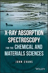
Cover Design: WileyCover Image: Sound Waves on Water: © Sunny/Getty Images; Duck Images provided courtesy of John Evans
About the Author
John Evans hails from Newcastle upon Tyne. He studied Chemistry at Imperial College, London, and carried out his PhD at the University of Cambridge supervised by Lord (Jack) Lewis and Brian Johnson. His postdoctoral research was at Princeton University, with Jack Norton, and then with ICI and Royal Society Pickering Research Fellowships back at Cambridge. He moved with the Pickering Fellowship to Southampton in 1976, became a lecturer in 1978, and a professor in 1990. He is now an emeritus professor there. He was science program advisor at the Diamond Light Source Ltd from 2002 to 2007. His experience in applying XAFS spectroscopy to chemical problems extends over 35 years; his research group has carried out experiments at the SRS, ESRF, SLS, Hasylab, Diamond, and APS.
Preface
This is a textbook aimed at master’s‐level students, including fourth‐year UK MSci degrees, of the chemical and related sciences suitable as an introductory text for PhD students embarking on x‐ray absorption fine structure (XAFS) spectroscopy. The background should also appeal to established scientists from other fields (environmental, life, and engineering sciences), wishing to assess the potential of x‐ray spectroscopy for their science. The chapters progress initially through the history and principles of XAFS. The next two chapters deal with experimental design: first, light sources and beamlines and then at the experimental station itself. Chapter 5 provides the background to the methods of extracting and using the results in materials and chemical analyses. The final chapter provides a series of case studies to illustrate a variety of applications. Each chapter concludes with a set of problems. There is a strong emphasis on the need to make the right choices for experimental design, and guidance provided to do so.
John Evans
Southampton UK
April 2017
Acknowledgments
I wish to thank all the members of my former research group for their talents and dedication in pursuing some optimistic experiments for 24/7 periods with food of varying desirability. Much of the developments came with collaborations that extended beyond a single position and with staff members from other institutions: Neville Greaves, Andy Dent, Sofia Diaz‐Moreno, Norman Binsted, Trevor Gauntlett, Fred Mosselmans, Judith Corker, Steven Fiddy, Mark Newton, Moniek Tromp, Peter Wells, and Stuart Bartlett. Judith’s loss to leukemia in 1998 remains a deep sadness. The book builds on the immense expertise of those who design, construct, develop, and operate these great accelerator‐based light sources. Advances in science, technology medicine, and cultural heritage owe much to them.
In the writing of the book, I have been helped greatly by staff at Diamond and colleagues for providing raw data and graphics. Special thanks go to Stuart Bartlett, Andrew Hector, Fred Mosselmans, Sofia Diaz‐Moreno, Roberto Boada Romero, Sarnjeet Dhesi, and Liz Duke. I am grateful, too, for the support of the CEOs of Diamond Light Source, Gerd Materlik, and Andrew Harrison, and also from EPSRC in the form of the Dynamic Structural Science and Catalysis Hub consortia at the Research Complex at Harwell. I am grateful for the confidence shown in this project by Jenny Cossham at Wiley and the continued patience of the staff at Wiley through the years. Inevitably, this has impacted on my family the most. Without the support of my wife, Hilary, and our daughters, Beccy and Lisa, and their families, this would not have reached fruition.
Glossary and Abbreviations
Absorption edge
Rapid increase in absorption with increasing energy
AEY
Auger electron yield
APD
Avalanche photodiode
Auger process
Relaxation of a core‐hole via electron emission
CCD
Charge‐coupled device
CEE
Constant emission energy
CIE
Constant incident energy
Compton scattering
Inelastic scattering
Debye‐Waller
Factor describing disorder in interatomic distances
DFT
Density functional theory
EDE
Energy dispersive EXAFS
EDX
Energy dispersive x‐ray spectroscopy
EXAFS
Extended x‐ray absorption fine structure
FEL
Free electron laser
FT
Fourier transform
FY
Fluorescence yield
FZL
Fresnel zone plate
HARPES
Hard x‐ray photoelectron spectroscopy
HERFD
High‐energy resolution fluorescence detection
IV
In vacuum
KB
Kirkpatrick‐Baez (mirrors)
MLL
Multilayer Laue lens
NEXAFS
Near‐edge x‐ray fine structure
NIXS
Nonresonant Inelastic x‐ray Scattering
OD
Optically detected
PCA
Principal component analysis
QEXAFS
Quick extended x‐ray absorption fine structure
Rayleigh scattering
Elastic scattering
REXS
Resonant x‐ray Emission Spectroscopy
RIXS
Resonant Inelastic x‐ray scattering or spectroscopy
SR
Synchrotron radiation
STXM
Scanning transmission x‐ray microscopy
TEY
Total electron yield
TXM
Transmission x‐ray microscopy
VtC
Valence to core
X‐PEEM
X‐ray photoelectron emission microscopy
XAFS
X‐ray absorption fine structure
XANES
X‐ray absorption near‐edge structure
XAS
X‐ray absorption spectroscopy
XEOL
X‐ray excited optical luminescence
XES
X‐ray emission spectroscopy
XFEL
X‐ray free electron laser
XMCD
X‐ray magnetic circular dichroism
XMLD
X‐ray magnetic linear dichroism
XRS
(Inelastic) X‐ray Raman Scattering
1Introduction to X‐Ray Absorption Fine Structure (XAFS)
1.1 Materials: Texture and Order
Today, research laboratories have powerful techniques for establishing the chemical nature and structure of pure materials. Our view of chemical structure is formed around the results of x‐ray diffraction, recorded from single crystals or from polycrystalline powders. Structures in the liquid phase can be inferred from expectations for bond lengths and angles derived from crystallography; to do so, information is gathered about the local symmetry, atomic connectivity, and proximity in the material derived from structurally sensitive spectroscopies, particularly nuclear magnetic resonance (NMR) and infrared (IR) and Raman vibrational spectroscopies.


