129,99 €
Mehr erfahren.
- Herausgeber: Wiley-VCH GmbH
- Kategorie: Wissenschaft und neue Technologien
- Sprache: Englisch
Provides comprehensive coverage on using X-ray fluorescence for laboratory applications
This book focuses on the practical aspects of X-ray fluorescence (XRF) spectroscopy and discusses the requirements for a successful sample analysis, such as sample preparation, measurement techniques and calibration, as well as the quality of the analysis results.
X-Ray Fluorescence Spectroscopy for Laboratory Applications begins with a short overview of the physical fundamentals of the generation of X-rays and their interaction with the sample material, followed by a presentation of the different methods of sample preparation in dependence on the quality of the source material and the objective of the measurement. After a short description of the different available equipment types and their respective performance, the book provides in-depth information on the choice of the optimal measurement conditions and the processing of the measurement results. It covers instrument types for XRF; acquisition and evaluation of X-Ray spectra; analytical errors; analysis of homogeneous materials, powders, and liquids; special applications of XRF; process control and automation.
- An important resource for the analytical chemist, providing concrete guidelines and support for everyday analyses
- Focuses on daily laboratory work with commercially available devices
- Offers a unique compilation of knowledge and best practices from equipment manufacturers and users
- Covers the entire work process: sample preparation, the actual measurement, data processing, assessment of uncertainty, and accuracy of the obtained results
X-Ray Fluorescence Spectroscopy for Laboratory Applications appeals to analytical chemists, analytical laboratories, materials scientists, environmental chemists, chemical engineers, biotechnologists, and pharma engineers.
Sie lesen das E-Book in den Legimi-Apps auf:
Seitenzahl: 848
Veröffentlichungsjahr: 2021
Ähnliche


Table of Contents
Cover
Title Page
Copyright
Preface
List of Abbreviations and Symbols
About the Authors
1 Introduction
2 Principles of X-ray Spectrometry
2.1 Analytical Performance
2.2 X-ray Radiation and Their Interaction
2.3 The Development of X-ray Spectrometry
2.4 Carrying Out an Analysis
3 Sample Preparation
3.1 Objectives of Sample Preparation
3.2 Preparation Techniques
3.3 Preparation of Compact and Homogeneous Materials
3.4 Small Parts Materials
3.5 Liquid Samples
3.6 Biological Materials
3.7 Small Particles, Dust, and Aerosols
4 XRF Instrument Types
4.1 General Design of an X-ray Spectrometer
4.2 Comparison of Wavelength- and Energy-Dispersive X-Ray Spectrometers
4.3 Type of Instruments
4.4 Commercially Available Instrument Types
5 Measurement and Evaluation of X-ray Spectra
5.1 Information Content of the Spectra
5.2 Procedural Steps to Execute a Measurement
5.3 Selecting the Measurement Conditions
5.4 Determination of Peak Intensity
5.5 Quantification Models
5.6 Characterization of Layered Materials
5.7 Chemometric Methods for Material Characterization
5.8 Creation of an Application
6 Analytical Errors
6.1 General Considerations
6.2 Types of Errors
6.3 Accounting for Systematic Errors
6.4 Recording of Error Information
7 Other Element Analytical Methods
7.1 Overview
7.2 Atomic Absorption Spectrometry (AAS)
7.3 Optical Emission Spectrometry
7.4 Mass Spectrometry (MS)
7.5 X-Ray Spectrometry by Particle Excitation (SEM-EDS, PIXE)
7.6 Comparison of Methods
8 Radiation Protection
8.1 Basic Principles
8.2 Effects of Ionizing Radiation on Human Tissue
8.3 Natural Radiation Exposure
8.4 Radiation Protection Regulations
9 Analysis of Homogeneous Solid Samples
9.1 Iron Alloys
9.2 Ni–Fe–Co Alloys
9.3 Copper Alloys
9.4 Aluminum Alloys
9.5 Special Metals
9.6 Precious Metals
9.7 Glass Material
9.8 Polymers
9.9 Abrasion Analysis
10 Analysis of Powder Samples
10.1 Geological Samples
10.2 Ores
10.3 Soils and Sewage Sludges
10.4 Quartz Sand
10.5 Cement
10.6 Coal and Coke
10.7 Ferroalloys
10.8 Slags
10.9 Ceramics and Refractory Materials
10.10 Dusts
10.11 Food
10.12 Pharmaceuticals
10.13 Secondary Fuels
11 Analysis of Liquids
11.1 Multielement Analysis of Liquids
11.2 Fuels and Oils
11.3 Trace Analysis in Liquids
11.4 Special Preparation Techniques for Liquid Samples
12 Trace Analysis Using Total Reflection X-Ray Fluorescence
12.1 Special Features of TXRF
12.2 Sample Preparation for TXRF
12.3 Evaluation of the Spectra
12.4 Typical Applications of the TXRF
13 Nonhomogeneous Samples
13.1 Measurement Modes
13.2 Instrument Requirements
13.3 Data Evaluation
14 Coating Analysis
14.1 Analytical Task
14.2 Sample Handling
14.3 Measurement Technology
14.4 The Analysis Examples of Coated Samples
15 Spot Analyses
15.1 Particle Analyses
15.2 Identification of Inclusions
15.3 Material Identification with Handheld Instruments
15.4 Determination of Toxic Elements in Consumer Products: RoHS Monitoring
15.5 Toxic Elements in Toys: Toys Standard
16 Analysis of Element Distributions
16.1 General Remarks
16.2 Measurement Conditions
16.3 Geology
16.4 Electronics
16.5 Archeometric Investigations
16.6 Homogeneity Tests
17 Special Applications of the XRF
17.1 High-Throughput Screening and Combinatorial Analysis
17.2 Chemometric Spectral Evaluation
17.3 High-Resolution Spectroscopy for Speciation Analysis
18 Process Control and Automation
18.1 General Objectives
18.2 Off-Line and At-Line Analysis
18.3 In-Line and On-Line Analysis
19 Quality Management and Validation
19.1 Motivation
19.2 Validation
Appendix A: Tables
Appendix B: Important Information
B.1 Coordinates of Main Manufacturers of Instruments and Preparation Tools
B.2 Main Suppliers of Standard Materials
B.3 Important Websites
B.4 Laws and Acts, Which Are Important for X-Ray Fluorescence
References
Index
End User License Agreement
List of Tables
Chapter 2
Table 2.1 Approximate relative line intensities of main X-ray lines.
Table 2.2 Comparison of line designations for the main X-ray lines.
Chapter 3
Table 3.1 Preparation technologies for solid samples.
Table 3.2 Information depth for different fluorescence lines in various mater...
Table 3.3 Information depth and accumulated intensity in the case of 10 wt% N...
Table 3.4 Estimations of the sample volume and mass that contribute to the me...
Table 3.5 Contaminations in the range of traces by preparation tools (main co...
Table 3.6 Steps for processing and sampling of small part materials.
Table 3.7 Mills for fine grinding of powder-like material.
Table 3.8 Binders and additives for pressed pellets.
Table 3.9 Flux agents for different applications.
Table 3.10 Sample–flux ratios for typical materials in grams (valid for diame...
Table 3.11 Window materials for sample cups for liquid samples.
Chapter 4
Table 4.1 Comparison of wavelength- and energy-dispersive X-ray spectrometer ...
Table 4.2 Often used crystals for wavelength-dispersive spectrometry.
Table 4.3 Instrument setup parameters.
Table 4.4 Products offered by instrument manufacturers for different instrume...
Chapter 5
Table 5.1 Contributions to the Cr intensity by primary, secondary, and tertia...
Table 5.2 Relative statistical error as a function of the number of counts.
Table 5.3 Substances suitable for standard addition.
Table 5.4 Analytical uncertainties for different sample qualities, measuring ...
Table 5.5 Settings for the preparation of an analytical method.
Chapter 6
Table 6.1 Results of repeated intensity measurements.
Table 6.2 Student's
t
-distribution in dependence of the probability
p
and the ...
Table 6.3 Addition of error contributions.
Table 6.4 Number of significant figures typical for X-ray fluorescence.
Chapter 7
Table 7.1 Comparison of methods for element analyses.
Chapter 8
Table 8.1 Relative sensitivities of different body parts.
Table 8.2 Radiation levels depending on dose.
Chapter 9
Table 9.1 Mean deviations for a cast iron calibration.
Table 9.2 Typical analytical performance of WD spectrometers for low-alloy st...
Table 9.3 Analytical performance of WD spectrometers for stainless steel.
Table 9.4 Analytical performance of WD spectrometers for tool steel.
Table 9.5 Analytical performance of WD spectrometers for the analysis of Al a...
Table 9.6 Composition of eutectics of soft solders.
Table 9.7 Analytical performance for the analyses of soft solders.
Table 9.8 Accuracies for the analysis of gold alloys.
Table 9.9 Relative standard deviations of repeated measurements depending on ...
Table 9.10 Typical composition and measurement uncertainties of flat glasses.
Table 9.11 Polymers and their approximate composition and mass fractions in w...
Table 9.12 Correlation coefficients
R
2
for different energy resolutions.
Table 9.13 Mass fractions of hydrocarbon groups and H, N, and O for PUR and A...
Chapter 10
Table 10.1 Detection limits for some heavy elements using K-radiation.
Table 10.2 Maximum loads of soils and sewage sludge.
Table 10.3 Typical compositions of components for the cement production in wt...
Table 10.4 Relative standard deviations of repeated measurements for samples ...
Table 10.5 Concentrations of typical ferroalloys in wt%.
Table 10.6 Digestion procedures for different Ferroalloys.
Table 10.7 Analyses results of ferro-silicon using powder samples in wt%.
Table 10.8 Relative standard deviation
σ
rel
of repeated measurements of s...
Table 10.9 Mass fractions ranges of refractory materials.
Table 10.10 Results of repeated measurements on a refractory.
Table 10.11 Average limits for toxic elements in animal food.
Table 10.12 Comparison between calibration results and limits of detection fo...
Table 10.13 Maximal amount of intake of toxic elements per day in micrograms.
Table 10.14 Reference values for the content of heavy metals in secondary fue...
Table 10.15 Typical line overlaps for toxic elements.
Table 10.16 Results of repeated measurements for secondary fuels.
Table 10.17 Reproducibility and trueness of the analyses of toxic liquids.
Chapter 11
Table 11.1 Limit of detection for
S
in fuels depending on the measuring method...
Table 11.2 Repeatability and reproducibility limits for the detection of sulf...
Table 11.3 Requirements according to ASTM D 4927-05 and ASTM D6481.
Table 11.4 Analytical performance for additives with a WD spectrometer.
Chapter 12
Table 12.1 Preparation procedures for TXRF.
Table 12.2 Concentration limits of elements in drinking water.
Table 12.3 Quantification results of the fresh water sample (NIST SRM 1640).
Table 12.4 TXRF results of blood serum compared with certified values.
Chapter 13
Table 13.1 Properties of energy-dispersive detectors.
Chapter 14
Table 14.1 Detector influence on analytical results.
Table 14.2 Coating thickness results of an Au/Ni/Cu layer system evaluated in...
Table 14.3 Absolute and relative standard deviations of 10 repeated measureme...
Table 14.4 Selection of reference standards and their uncertainties (95%).
Table 14.5 Measurement results of a CIGS layer system.
Table 14.6 Characterization of a SnPb/Ni/AgPb/ceramic system.
Chapter 15
Table 15.1 Maximum concentrations for toxic substances according to RoHS.
Table 15.2 Maximum loads according to Toy Regulation (BGBl., II, Nr. 203/2011...
Table 15.3 Maximum soluble migrated element in ppm (mg/kg) for surface coatin...
Chapter 16
Table 16.1 Concentrations at the measuring points in wt%.
Chapter 19
Table 19.1 Parameters in dependence of the task for checking.
Table 19.2 Determination of the parameter range of application, linearity, an...
Appendix A
Table A.1 Periodic table of elements with atomic number, atomic weight, and d...
Table A.2 Atomic weight and density (in g/cm
3
) of all elements.
Table A.3 Energies of absorption edges (keV).
Table A.4 K-line energies (keV).
Table A.5 L-line energies (keV).
Table A.6 M-line energies (keV).
Table A.7 Wavelengths of K-lines (nm).
Table A.8 Wavelengths of L-lines (nm).
Table A.9 Wavelengths of M-lines (nm).
Table A.10 Common line interferences, energies in keV.
Table A.11 Fluorescence yields.
Table A.12 Transfer factors between elements and oxides.
List of Illustrations
Chapter 2
Figure 2.1 Parts of the continuous spectrum of an X-ray tube.
Figure 2.2 Line energy as a function of the atomic number.
Figure 2.3 Electron transitions with the corresponding line names.
Figure 2.4 Fluorescence yield as a function of the atomic number.
Figure 2.5 Contributions to the attenuation coefficient for X-radiation of (...
Figure 2.6 Elastic and inelastic scattering coefficients for selected elemen...
Figure 2.7 (a) Influence of the sample type on the intensity of the scattere...
Figure 2.8 Spectra of PMMA, Al, Ti, and Ni demonstrating the different scatt...
Figure 2.9 PW 1540 from Philips.
Figure 2.10 Analyst 0700 from KEVEX.
Figure 2.11 Steps for an analytical procedure.
Chapter 3
Figure 3.1 Information depth of fluorescence radiation.
Figure 3.2 Information depth for different matrices.
Figure 3.3 Cd intensities measured on polymer samples of different thickness...
Figure 3.4 Analyzed volume limited by the measurement geometry.
Figure 3.5 Specimen generated by sampling from a melt.
Figure 3.6 Influence of surface roughness on the measured fluorescence inten...
Figure 3.7 Dependence of fluorescence intensity on grinding time.
Figure 3.8 Dependence of fluorescence intensity on the morphology of the sam...
Figure 3.9 Press die for to produce pellets from small-sized materials.
Figure 3.10 Pressed pellet (a) and cross section of a pressed pellet in bind...
Figure 3.11 Influence of grain size distribution and different hardness of s...
Figure 3.12 Calibration curves for the same powder material prepared as pres...
Figure 3.13 Arrangement of layers of melting agent and sample in the fusion ...
Figure 3.14 (a) Pt crucibles over a gas burner. (b) Temperature profile of t...
Figure 3.15 Cooling curve to produce glass pellets.
Figure 3.16 Glassy (a) and milky, too quickly cooled fusion bead (b).
Figure 3.17 Sample cup for the analysis of liquids.
Figure 3.18 Transmission of low-energy radiation of sample cup films.
Chapter 4
Figure 4.1 Energy resolution of different ED spectrometer (blue) and WD spec...
Figure 4.2 Spectrum of stainless steel, measured by WDS and EDS: (a) low-ene...
Figure 4.3 Influence of the selected lattice plane (a) and the collimator si...
Figure 4.4 Spectra of a gold alloy measured with a proportional counter and ...
Figure 4.5 Energy dependence of energy resolution of ED-detectors (FWHM, ful...
Figure 4.6 Detection efficiency of prop-counter and scintillation counter.
Figure 4.7 Detection efficiency of ED-detectors.
Figure 4.8 Throughput of ED-detectors and their influence on the detector re...
Figure 4.9 Peak shape changes for very high count rates of WD spectrometers....
Figure 4.10 Effect of the count rate on the peak-to-background ratios.
Figure 4.11 Comparison of the ratios of the relative standard deviation (rel...
Figure 4.12 Low-energy spectral background due to a Compton-induced escape e...
Figure 4.13 Escape and pile-up peak of Ti radiation (
y
-axis in square root s...
Figure 4.14 Pt spectrum measured without and with Al filter (
y
-axis in squar...
Figure 4.15 Shelf and tail contributions to a Cu line (
y
-axis in square root...
Figure 4.16 Principle layout of EDS instruments (
MCA
,
multichannel analyzer
)...
Figure 4.17 Handheld instruments from various manufacturers: (a) XL5, Thermo...
Figure 4.18 Portable instruments of different manufacturers: (a) FXL, Thermo...
Figure 4.19 Various energy-dispersive spectrometers: (a) Epsilon 3, Panalyti...
Figure 4.20 Principle layout of WDS instruments.
Figure 4.21 A selection of wavelength-dispersive X-ray spectrometers: (a) Ze...
Figure 4.22 A selection of multichannel X-ray spectrometers: (a) S8 Lion, Br...
Figure 4.23 Principle setup of a TXRF instrument (MCA, multichannel analyzer...
Figure 4.24 Total reflection X-ray spectrometers: (a) TXRF-310, Rigaku Corp....
Figure 4.25 Layout of an X-ray spectrometer with monoenergetic excitation.
Figure 4.26 Spectrum of an oil sample excited with Mo radiation.
Figure 4.27 Measurement geometry for the excitation with polarized radiation...
Figure 4.28 Energy-dispersive instruments using polarized radiation: (a) XEP...
Figure 4.29 Basic layouts of position-sensitive X-ray spectrometers with a c...
Figure 4.30 Coating thickness instruments with collimators: (a) XDAL, Helmut...
Figure 4.31 Instruments utilizing X-ray optics for beam focusing: (a) M4 Tor...
Figure 4.32 Macro-X-ray fluorescence spectrometer for the investigation of l...
Figure 4.33 Confocal geometry for the investigation of 3D elemental distribu...
Figure 4.34 Grazing incident measurement geometry.
Figure 4.35 Grazing exit measurement geometry.
Chapter 5
Figure 5.1 Characteristics of the Fe intensity for the Cr–Fe binary material...
Figure 5.2 Characteristics of the Fe intensity for the Fe–Ni binary material...
Figure 5.3 Characteristics of the Fe intensity for the Cr–Fe–Ni material sys...
Figure 5.4 Excitation spectra for different tube parameters (
y
-axis in squar...
Figure 5.5 Influencing the measured spectrum with an Al filter.
Figure 5.6 Reduction of diffraction peaks and the scattered Rh-L radiation w...
Figure 5.7 Transmission of X-rays in different measurement atmospheres.
Figure 5.8 Standard deviation and error of repeated measurements.
Figure 5.9 Energies of X-ray lines.
Figure 5.10 Frequency distribution of X-ray lines as a function of the atomi...
Figure 5.11 Typical line overlaps for energy-dispersive spectrometers.
Figure 5.12 Methods for the determination of the peak intensity: (a) WDS ins...
Figure 5.13 Spectrum of a tool steel sample fitted with pure element peaks (...
Figure 5.14 Calibration curves based on net intensities (continuous line) an...
Figure 5.15 Setting of background points.
Figure 5.16 Background fit of an energy-dispersive measured spectrum (
y
-axis...
Figure 5.17 Calculation and use of a calibration curve.
Figure 5.18 Bracketing method for highly accurate analysis with two differen...
Figure 5.19 Representation of the trueness of an analysis method: (a) compar...
Figure 5.20 Determination of the limit of detection from the calibration fun...
Figure 5.21 Deviations of the gold concentrations measured with SDD and prop...
Figure 5.22 Comparison of the accuracy of standard-based and standard-free m...
Figure 5.23 Typical calibration curves for the emission and absorption signa...
Figure 5.24 Measurable layer thickness ranges for certain element groups and...
Figure 5.25 Corrected calibration curve using two reference samples.
Figure 5.26 Scheme of a principal component analysis.
Chapter 6
Figure 6.1 Error distribution in the analytical process.
Figure 6.2 Target model for the illustration of random and systematic errors...
Figure 6.3 Results of repeated measurement with a high (a) and lower (b) sta...
Figure 6.4 Specification limits of a process.
Figure 6.5 Gaussian distribution of measuring results.
Figure 6.6 Gaussian distributions with different standard deviations.
Figure 6.7 Statistical distribution around the expected value and shifted by...
Figure 6.8 Total measurement uncertainties of analytical results.
Chapter 7
Figure 7.1 Setup of an atomic absorption spectrometer.
Figure 7.2 Principal setup of an arc-spark spectrometer.
Figure 7.3 Basic setup of an optical emission spectrometer with excitation b...
Figure 7.4 Basic setup of a laser-induced breakdown spectrometer.
Figure 7.5 Basic setup of a mass spectrometer.
Figure 7.6 Excitation cross-sections of different elements for monoenergetic...
Chapter 8
Figure 8.1 Absorption of radiation in a material layer.
Figure 8.2 Half-width of organic tissues for different energies.
Chapter 9
Figure 9.1 Pressed pellet of metals chips in a ring of steel.
Figure 9.2 Change in measured Cr content as a function of grinding passes.
Figure 9.3 Influence of acids on the measured signal of Cu.
Figure 9.4 Spectra of high-purity gold with varying small silver contents (
y
Figure 9.5 Change of the boron intensity in a boron-silicate glass for sever...
Figure 9.6 Matrix influence of different polymers on the Br-K intensities (
A
...
Figure 9.7 Elastic and inelastic scattered tube radiation of various pure po...
Figure 9.8 Comparison between mass fractions of hydrogen (a), carbon (b), an...
Figure 9.9 Sample carriers for abrasion analyses: corundum disk for conventi...
Figure 9.10 Relative differences for a bulk and an abrasive analysis of stee...
Chapter 10
Figure 10.1 Limits of detection for geological material, measured with an ED...
Figure 10.2 Limits of detection for element oxides in coal for EDS instrumen...
Figure 10.3 Ferroalloy with different grain size.
Figure 10.4 Crucible for remelting ferroalloys.
Figure 10.5 Process scheme for the digestion of ferro-silicon.
Figure 10.6 Sampling of a slag sample from the melt with a sampling probe (a...
Figure 10.7 Spectra of a blank and a loaded filter.
Figure 10.8 Element distribution along the trace of collected dust.
Figure 10.9 Examples of raw secondary fuels (a) and crushed material (b).
Figure 10.10 Pellets produced at different temperatures have different stren...
Chapter 11
Figure 11.1 Plastic film with a drop of a solution (a), spectra of a solutio...
Figure 11.2 LiTRap-sample holder, not used (a) and with metal particles from...
Figure 11.3 Samples after complexation: a Chelex
®
-100-pellet with synth...
Chapter 12
Figure 12.1 Spectra of water fleas measured at conventional excitation geome...
Figure 12.2 Element-dependent sensitivities of a TXRF spectrometer.
Figure 12.3 Critical sample thicknesses for neglecting the matrix interactio...
Figure 12.4
Secondary electron microscope
(
SEM
) image of a TXRF sample (drie...
Figure 12.5 Spectra of different amounts of fine-grained Zn-particles. esc, ...
Figure 12.6 Limits of detection in aqueous solutions.
Figure 12.7 Spectrum of NIST SRM 1640 – fresh water.
Figure 12.8 Analysis results for Hg determined with and without EDTA-complex...
Figure 12.9 TXRF spectrum of an environmental aerosol sample, particle size ...
Figure 12.10 Calibration curve for platinum in blood serum.
Figure 12.11 TXRF spectrum of lung tissue of a foundry worker.
Figure 12.12 Depth profiles of As ions in a Si wafer determined with TXRF, R...
Chapter 14
Figure 14.1 Calibration curves for Au-layers for both Au-L- and Au-M-fluores...
Figure 14.2 Calibration curve for Al layer thickness based on Fe absorption....
Figure 14.3 Results of coating thickness measurements in relative and emissi...
Figure 14.4 0.2 mm wide Au/Ni/Cu coated area on PCB using a spot size of 0.1...
Figure 14.5 Simulated spectra of a 5 μm NiP/Fe system with different P conte...
Figure 14.6 Comparison of given and analyzed P contents in NiP layers.
Figure 14.7 Spectrum of an Au–Pd–Ni layer system.
Figure 14.8 Design of CIGS solar cells.
Figure 14.9 Spectrum of a SnPb solder system.
Figure 14.10 Layer structure form a vertical and tilted polished section.
Figure 14.11 Element distribution in a cross section (a) and along a line pe...
Figure 14.12 Spectra of a CIGS layer system measured with grazing incidence....
Figure 14.13 Intensity profiles of the components of the CIGS layer system f...
Figure 14.14 Distribution of pigment layers on both sides of a parchment (a)...
Chapter 15
Figure 15.1 Example of a metal particle; the analysis spot is marked.
Figure 15.2 Sample cup with particles.
Figure 15.3 Spectra of two particles of the same material but of different s...
Figure 15.4 Scores for different principal components for measurements on gl...
Figure 15.5 Spectra of a PE specimen with toxic elements close to the RoHS l...
Figure 15.6 Peak overlap of Br, As, Hg, and Pb.
Chapter 16
Figure 16.1 Typical geological samples for distribution analyses: (a) bore c...
Figure 16.2 Sum spectrum of the complete mapping (
y
-axis in square root scal...
Figure 16.3 Element distributions of the bore core for (a) Si, (b) Ca, (c) T...
Figure 16.4 Superposed element distribution of the pitchblende including ins...
Figure 16.5 Element distributions of the overlapped As-Kα and Pb-Lα-lines (a...
Figure 16.6 Sum and maximum pixel spectrum of the pitchblende (a) (
y
-axis in...
Figure 16.7 Element distributions of thin sections measured with different s...
Figure 16.8 (a) Photograph of a PCB of a mobile phone, (b) distribution of s...
Figure 16.9 Image of a coin of 2 Reichsmark minted in Munich (D) (a) and spe...
Figure 16.10 Distribution of Ag (a) and Cu (b) of a 2 Reichsmark coin.
Figure 16.11 St. Jerome with the Bible. (a) Photograph of the painting. (b) ...
Figure 16.12 Element distributions of a glass specimen: (a) Si distribution,...
Figure 16.13 Relative standard deviations of repeated measurements on the sa...
Chapter 17
Figure 17.1 High-throughput screening for the development of a catalyst.
Figure 17.2 (a) Sample carrier with 10
4
samples and (b) relations between ag...
Figure 17.3 Energy-dispersive measured X-ray spectra of few sodium salts....
Figure 17.4 Score plot of the principal components of different chemicals....
Figure 17.5 S-Kβ spectra of different sulfur compounds.
Figure 17.6 Al-Kβ spectra of Al nitride and Al oxide.
Figure 17.7 Calibration curve for the concentration ratio of Al
2
O
3
/AlN by us...
Figure 17.8 Si-Kβ spectra of SiO
2
and SiC.
Chapter 18
Figure 18.1 Automated laboratory with instruments for sample preparation and...
Figure 18.2 Scheme of an automated blast furnace laboratory for the analysis...
Figure 18.3 Scheme of a preparation automat for oxidic materials.
Figure 18.4 Pressed pellet from powder material in a ring of steel as a resu...
Figure 18.5 Scheme of an automated fusion automat with inductive heated fusi...
Figure 18.6 Fusion automat with two inductive heated fusion instruments.
Figure 18.7 External (a) and internal (b) view of an automated sample prepar...
Figure 18.8 Robot during loading of a resistance-heated fusion furnace (a) a...
Figure 18.9 Scheme of an in-line analyzer for bulk material.
Figure 18.10 Scheme of an on-line analyzer for liquids.
Figure 18.11 Measuring heads for a continuous monitoring of material flow. (...
Chapter 19
Figure 19.1 Elements of a quality management system.
Figure 19.2 Scheme for the connection of secondary references to SI units.
Figure 19.3 Scheme of a validation process.
Figure 19.4 Methods for a validation.
Figure 19.5 Determination of the parameter's precision and trueness.
Figure 19.6 Influence of deviations of the measurement to the determination ...
Guide
Cover Page
Table of Contents
Begin Reading
Pages
iii
iv
xvii
xviii
xix
xx
xxi
xxiii
1
2
3
4
5
7
8
9
10
11
12
13
14
15
16
17
18
19
20
21
22
23
24
25
26
27
28
29
31
32
33
34
35
36
37
38
39
40
41
42
43
44
45
46
47
48
49
50
51
52
53
54
55
56
57
58
59
60
61
62
63
64
65
66
67
68
69
70
71
72
73
74
75
76
77
78
79
80
81
82
83
84
85
86
87
88
89
90
91
92
93
94
95
96
97
98
99
100
101
102
103
104
105
106
107
108
109
110
111
112
113
114
115
116
117
118
119
120
121
122
123
124
125
126
127
128
129
130
131
132
133
134
135
136
137
138
139
140
141
142
143
144
145
146
147
149
150
151
152
153
154
155
156
157
158
159
160
161
162
163
164
165
167
168
169
170
171
172
173
174
175
177
178
179
180
181
183
184
185
186
187
188
189
190
191
192
193
194
195
196
197
198
199
200
201
202
203
204
205
206
207
208
209
210
211
213
214
215
216
217
218
219
220
221
222
223
224
225
226
227
228
229
230
231
232
233
234
235
236
237
238
239
240
241
242
243
244
245
246
247
248
249
250
251
252
253
254
255
256
257
258
259
260
261
262
263
264
265
267
268
269
270
271
272
273
274
275
276
277
278
279
280
281
282
283
284
285
286
287
288
289
290
291
292
293
294
295
296
297
298
299
300
301
302
303
304
305
306
307
308
309
310
311
312
313
314
315
316
317
318
319
320
321
322
323
324
325
326
327
328
329
330
331
332
333
334
335
336
337
338
339
340
341
342
343
344
345
346
347
348
349
350
351
352
353
355
356
357
358
359
360
361
362
363
364
365
366
367
368
369
370
371
372
373
374
375
376
377
379
380
381
382
383
384
385
386
387
388
389
390
391
392
393
394
395
396
397
398
399
400
401
402
403
404
405
406
407
408
409
410
411
412
413
414
415
416
417
418
419
420
421
422
423
424
425
426
427
428
429
430
431
432
433
434
435
436
437
438
439
440
441
442
443
444
445
446
447
448
449
450
451
452
453
454
455
456
457
458
459
460
461
462
463
464
465
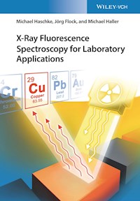
X-ray Fluorescence Spectroscopy for Laboratory Applications
Michael Haschke
Jörg Flock
Michael Haller
Authors
Dr. Michael HaschkeGünter Allee 1115345 EggersdorfGermany
Dr. Jörg FlockThyssen Krupp Stahl AGKaiser-Wilhelm-Str. 10047166 DuisburgGermany
Dipl.-Min. Michael HallerCrossRoads Scientific LLC.MiddletownCTUnited States
All books published by Wiley-VCH are carefully produced. Nevertheless, authors, editors, and publisher do not warrant the information contained in these books, including this book, to be free of errors. Readers are advised to keep in mind that statements, data, illustrations, procedural details or other items may inadvertently be inaccurate.
Library of Congress Card No.:applied for
British Library Cataloguing-in-Publication DataA catalogue record for this book is available from the British Library.
Bibliographic information published by the Deutsche NationalbibliothekThe Deutsche Nationalbibliothek lists this publication in the Deutsche Nationalbibliografie; detailed bibliographic data are available on the Internet at <http://dnb.d-nb.de>.
© 2021 Wiley-VCH GmbH, Boschstr. 12, 69469 Weinheim, Germany
All rights reserved (including those of translation into other languages). No part of this book may be reproduced in any form – by photoprinting, microfilm, or any other means – nor transmitted or translated into a machine language without written permission from the publishers. Registered names, trademarks, etc. used in this book, even when not specifically marked as such, are not to be considered unprotected by law.
Print ISBN: 978-3-527-34463-5ePDF ISBN: 978-3-527-81660-6ePub ISBN: 978-3-527-81662-0oBook ISBN: 978-3-527-81663-7
Cover Design Formgeber, Mannheim, Germany
Preface
The discovery of X-rays by Wilhelm Conrad Röntgen dates back to nearly 125 years. Despite their “age,” the research and discoveries that have been made in the past and today make X-rays one of the most powerful analytical tools available today. The discovery of this part of the electromagnetic spectrum has seen many applications. First, used for medical purposes by Röntgen himself, who found that the newly discovered rays can penetrate and at the same time can be absorbed by different types of matter. Therefore, it was now possible to image the human body. Later, Max von Laue showed that they are a higher-frequency part of the electromagnetic spectrum where natural light is also a part of. This led to his work on diffraction of X-rays, which to this day is used to investigate the crystalline and amorphous structure of solids. Finally, Moseley found that every element emitted characteristic X-ray radiation that can be used to determine the qualitative and quantitative elemental composition of materials of different types. This is the application that is most interesting for us – the spectroscopists.
X-ray fluorescence spectroscopy has now developed into an analytical technique that, due to its robustness and flexibility, can be used in almost all scientific areas, in research, and above all in quality control in industrial production. The technique has become so powerful because of its ability to analyze many different material types, having a wide range of elements and concentrations. The sample preparation is mostly simple or even not required, therefore making it possible to fully automate the entire analysis process.
Even when one has many years of experience with the method, specific expertise is still required to achieve reliable results, especially since the applications for X-ray fluorescence spectrometry have been significantly expanded in recent years due to new components being developed and becoming commercially available for X-ray spectrometers. Examples of such are X-ray optics, new types of detectors, and the availability of powerful computing technique and software solutions.
A combination of theoretical knowledge and practical know-how about the materials to be examined, the best practices for sample preparation, and the most suitable measuring instrument at optimal measurement conditions is required to make the most precise and true analysis results. The intention of this book is to summarize the experiences of the analytical community working with X-ray fluorescence. For many years this community has convened at global annual user meetings and conferences for X-ray fluorescence spectrometry, covering all types of applications of X-ray spectroscopy. The work presented at these meetings shares new developments in the field of device technology and provides information on the analytical capabilities of X-ray fluorescence for known applications, for instance, in the analysis of metallurgical or mineralogical samples. Interesting new applications are presented as well. These experiences are the basis of this book in which we tried to summarize but also to preserve this knowledge.
The book is addressed to current and future users of X-ray fluorescence analysis. It strives to provide suggestions and examples on how to use X-ray fluorescence and what kind of results can be expected, as well as advice on suitable preparation techniques and measurement conditions. Accordingly, in addition to the method-specific basics, the book contains information about the essential preparation techniques and a variety of material-specific applications that can serve as the basis for your own current and future measurement concepts.
Many inspiring discussions and joint projects with numerous users and instrument manufacturers have been included in this book. The authors want to especially thank Prof. A. Janßen, Ms.Sc. S. Hanning and many other colleagues not mentioned here for their support of this work. Our special acknowledgement goes to Dr. A. von Bohlen, he not only supported the project by a lot of discussions but also provided our work with elaborated information about conditions and applications for total reflection X-ray spectrometry.
Michael Haller dedicates his work in this book to the late Dr. Volker Röβiger. Friend, mentor, and true Renaissance man, his enthusiasm and kindness were an inspiration to all who had the privilege to know him.
Finally, thanks to the publisher Wiley-VCH for their support and smooth completion of this project.
We hope that this book will inspire and fascinate all readers using X-rays as an analytical tool.
February 2020
Michael Haschke, Jörg Flock, Michael HallerEggersdorf, Schwerte and Middletown
List of Abbreviations and Symbols
AAS
atomic absorption spectrometry
AES
atomic emission spectrometry
BG
background
c
velocity of light
C
det
detector capacitance
CR
counting rate
CRM
certified reference material
d
d-spacing of the scattering lattice, thickness
d
information
information depth
D
dose
e
charge of an electron
E
energy
E
C
energy of the absorption edge
ED
energy dispersive
EDS
energy-dispersive spectrometry
ENC
electronic noise contribution
EPMA
electron probe microanalysis
EXAFS
extended X-ray absorption fine structure
f
functional relation, degrees of freedom
F
Fano factor
FOM
figure of merit
FT-IR
Fourier transform infrared
FWHM
full width at half maximum
G
gain
GEXE
grazing exit X-ray emission
GIXE
grazing incident X-ray emission
h
Planck's constant
HOPG
highly oriented pyrolyzed graphite
HTS
high throughput analysis
i
current
I
intensity
I
o
primary intensity
I
d
leakage current
I
scat
scattered intensity
ICP
inductively coupled plasma spectrometry
k
factor of confidence, proportionally coefficient
keV
kiloelectron volt
LA-ICP
laser ablation inductively coupled plasma spectrometry
LIBS
laser-induced breakdown spectrometry
LN
2
liquid nitrogen
LOD
limit of detection
LOI
loss of ignition
m
atomic mass
m
0
mass of an electron
μ-XRF
micro-X-ray fluorescence analysis
M
matrix interaction
MC
Monte Carlo
MCA
multichannel analyzer
MPS
maximum pixel spectrum
MS
mass spectrometry
MXRF
macro-X-ray fluorescence analysis
n
refraction index, number (channels, pixels), order of diffraction
N
number of photons
NAA
neutron activation analysis
OES
optical emission spectrometry
p
probability
PC
proportional counter
PCA
principal component analysis
PCB
printed circuit board
PIXE
proton-induced X-ray emission
PMI
positive material identification
Q
mass per area
R
intensity ratio
R
2
correlation coefficient
RM
reference material
RoHS
Restriction of Hazardous Substances
SDD
silicon drift detector
SIMS
secondary ion mass spectrometry
SML
synthetic multilayer
SRM
standard reference material
t
thickness of a layer, measurement time
T
temperature
TXRF
total reflection X-ray fluorescence spectrometry
U
voltage, uncertainty
v
measured value
VPD
vapor phase deposition
w
mass fraction
WD
wavelength dispersive
WDS
wavelength-dispersive spectrometry
XANES
X-ray absorption near edge structure
XPS
X-ray induced photoelectron spectrometry
XRF
X-ray fluorescence
Z
atomic number
Δ
difference
η
efficiency
ε
energy for generation of a charge carrier
κ
overlapping factor
λ
wavelength
μ
mass attenuation coefficient
ρ
density of the absorbing material
ϑ
scatter angle
σ
standard deviation, scattering coefficient
Θ
angle
τ
shaping time, linear absorption coefficient
ψ
incidence/take-off angle
ω
fluorescence yield
Ω
captured angle
About the Authors
Dr. Michael Haschke has worked for more than 35 years in several companies in the field of product management for the development of new products and the market introduction of new methods in X-ray fluorescence. These were mainly instruments in the field of energy-dispersive spectroscopy. During the market introduction it was every time necessary to deal with competitional element analysis methods but also with the new applications. He, therefore, has both knowledge in the field of X-ray fluorescence and analysis method and for the wide range of applications for X-ray fluorescence.
Dr. Jörg Flock was for many years the head of the central laboratory of ThyssenKrupp Steel AG and therefore familiar with several analytical methods, in particular with X-ray fluorescence spectroscopy. He has a lot of practical knowledge for the analysis of various sample qualities.
Michael Haller, M.S., has been using X-rays as an analytical tool for over 30 years, first in X-ray crystallography and then later in the development and application of polycapillary X-ray optics. During the majority of his career, he has developed new applications for coating thickness instruments in industrial process control. In 2018 he became co-owner of CrossRoads Scientific, a company specializing in the development of analytical X-ray software.
1Introduction
X-ray spectrometry has been known as a method for element analyses for more than 70 years and can be regarded as a routine method since the 1960s. This means that there is a broad range of instruments available, and numerous analytical tasks are carried out routinely by X-ray fluorescence (XRF) analysis. For example, XRF is used for the characterization of metallic or geological materials or for analyses of solid or liquid fuels despite the fact that other elemental analytical methods have been developed and are readily available for these applications. Among them are optical emission spectrometry with excitation both by sparks and by inductively coupled plasmas and mass spectrometry. The high importance of using XRF is due to the fact that one can achieve very high precision over a wide concentration range. XRF also requires little effort with sample preparation and the method can be automated.
Especially in the last 15–20 years, XRF has experienced a new boom mainly because the technology has further developed, and new fields of applications could be opened up. These include, among others, the analysis of layered materials and high-resolution position-sensitive analysis. This was made possible by the availability of new components for X-ray spectrometers.
The development of high-resolution energy-dispersive detectors with good count rate capability now allows precision measurements also with energy-dispersive spectrometers. The simultaneous detection of a wide energy range over a large solid angle made possible with these detectors allows not only short measuring times but also special excitation geometries. It is therefore now possible to achieve higher sensitivities in the detection of traces; further, the fluorescence radiation of small surface areas can be detected with sufficient intensity.
The development of various X-ray optics allows shaping of the primary X-ray beam and thus the concentration of high excitation intensity on small sample surfaces; this development was the key to opening up new applications in the field for a spatially resolved analysis.
These developments have significantly expanded the range of applications of XRF analysis.
However, the most important influence in the further development of XRF into a routine method was the advances in data processing technology. These made it possible to automate instrument control as well as the evaluation of measurement data. Not only was it possible to reduce subjective influences by a manual operator but also the processes during instrument control and measurement data acquisition could largely be automated and made more effective. The evaluation of the measurement data, such as the peak area calculation in case of overlapping peaks, or the calculation procedures for the quantification could be expanded and significantly refined by the available computing power.
These improvements have been particularly important because X-rays strongly interact with the sample matrix, which requires complex correction procedures. Nevertheless, in contrast to other analytical methods, the physics of these interactions is very well understood and can be exactly modeled mathematically. Consequently, in principle, standard-less analysis is possible, which again requires a high computing effort.
As a result of these developments, new methodical possibilities for XRF emerged, combined with an expansion of their field of applications. For this reason, it seems to be meaningful to carry out an up-to-date compilation of the applications currently being processed by XRF, in combination with a discussion of both the necessary sample preparation and instrument-related efforts and the achievable analytical performance. There are several very good books available, which however, due to their date of publication, have not been able to take into account the developments of the last 15–20 years (Erhardt 1989; Hahn-Weinheimer et al. 2012) or they do not adequately address frequently used routine applications, in particular in industrial analyses (Beckhoff et al. 2006; van Grieken and Markowicz 2002).
The goal of this book is to focus on the practical aspects of the various applications of XRF. This leads to the discussion of the requirements necessary for the analysis of the very different sample qualities, such as the type of sample preparation, the available measurement technique or the required calibration samples, as well as the type and quality of the results to be expected with these efforts. This appeared to be important, in particular, because XRF is often used in many laboratories, but methodical studies are carried out only in very few of them.
This leads to the application aspects often not being understood very well. Consequently, the analytical results are accepted without scrutinizing the influence of sample state, preparation methods, and measurement parameters. This becomes especially true because complete results are often available as the outcome of an instrumental analysis and their quality cannot be correctly comprehended.
In order to assure the quality of the applications and their results, the analyst must critically question all aspects of the test method. For this purpose, a basic understanding of the influences of sample condition, preparation methods, measurement parameters, and evaluation models used on the quality of the analytical result is imperative.
Therefore, we are deliberately focusing on the daily laboratory work with commercially available instruments. On the other hand, the interesting but not routine applications of the method utilizing synchrotron radiation excitation are not addressed. Nevertheless, methodical developments obtained on a synchrotron are often incorporated into laboratory analysis, such as micro-X-ray fluorescence (μ-XRF) or applications with grazing beam geometry. However, this book treats only laboratory applications. If any of these newly developed methods have been implemented into special laboratory instruments these are also presented as examples.
Despite the focus on the various applications, a brief introduction to the fundamentals of X-ray spectrometry and a comprehensive presentation of the basic steps for a complete analysis are required in order to be able to relate in the following discussion of the individual applications.
The book therefore starts with a discussion of the analytical capability of X-ray spectrometry in Chapter 2. The most important relations that describe the generation of the characteristic radiation are presented, and the individual steps in the execution of an analysis follow, along with a brief characterization of their influence on the analysis result. Deeper descriptions of the physical bases are comprehensively given in other publications (e.g. Erhardt 1989; Hahn-Weinheimer et al. 2012; van Grieken and Markowicz 2002; Beckhoff et al. 2006).
In Chapter 3, the various sample preparation procedures typical for X-ray spectrometry are presented and their influence on the precision and trueness of the analyses is discussed. Even though the sample preparation is generally regarded as being very simple for XRF, it is important to carry it out carefully, appropriate to the expectations of the analysis result.
In Chapter 4, the different types of X-ray spectrometers are discussed. On the one hand, the general differences and application characteristics of wavelength-dispersive and energy-dispersive instruments are examined; on the other hand, the different instrument types as well as the instruments currently available on the market are presented.
In Chapter 5, the essential steps for the measurement of a spectrum are reviewed, in particular, the optimum selection of the measurement parameters and the steps for the evaluation of the measured data. The first step is the determination of the intensities of the fluorescence peaks, where different procedures are used for wavelength- and energy-dispersive spectrometers. Then quantification models and factors concerning the consideration of matrix interaction, both in the analysis of homogeneous samples and in the characterization of layers, are presented. Here, a comprehensive and detailed description of the theory of X-ray spectrometry is not required, since a series of detailed papers are available (see, for example, Hahn-Weinheimer et al. 2012; Jenkins et al. 1981; Lachance and Claisse 1994; Mantler 2006) and only very few new ideas have been added in the last few years. In this chapter, further possibilities for the evaluation of spectra are presented, in which the individual spectral components are not considered separately, but the spectrum as a whole is evaluated by means of chemometric methods.
Chapter 6 is devoted to the discussion of the classification, determination, and evaluation of errors. The achievable analytical precision of XRF is determined by the errors. In addition to the traditional treatment of errors with the Gaussian error model, the principle of measurement uncertainty is also discussed. This chapter is intended to qualify the expectations of an analytical result.
In Chapters 7 and 8, a brief comparison is made with other element analysis methods, in particular atomic absorption and emission spectrometry as well as mass spectrometry. The fundamentals of radiation protection when dealing with X-ray radiation, in particular when carrying out X-ray analysis experiments, are compiled as well.
Based on these fundamentals, various applications of XRF, which have been used already over a long time period or were introduced recently, are presented and discussed. The presentation here is carried out according to the different sample qualities or according to the analytical question.
Typical XRF applications are discussed first. Chapter 9 discusses the analysis of homogeneous solid samples, such as various metallic materials, glasses, or plastics. Chapter 10 describes the investigation of powdered samples, such as geological samples, soil, building materials, slags, and dusts.
In Chapter 11, the different possibilities for analyzing liquids are presented, either by direct analysis or, for example, by different enrichment procedures to answer specific analytical questions. Applications with total reflection XRF (TXRF) are dealt with in Chapter 12. Here, in addition to ultra-trace analyses of liquids the analysis of very small sample quantities is the focus.
Descriptions of the analysis of nonhomogeneous materials cover a wide range of analytical questions. This concerns inhomogeneities normal to the sample surface, i.e. the characterization of layered materials (Chapter 14) along with their different applications, as well as inhomogeneities in the sample plane and the analysis of irregularly shaped samples (Chapter 15). In this case, only small sample areas are to be analyzed, which means a point analysis has to be carried out. This is important when identifying particles or inclusions as also when analyzing inhomogeneous materials.
Handheld instruments are increasingly being used for element analysis. Based on this fact, the applications that up to now have been typical for this instrument technology are presented in Section 15.4. It was possible to increase the efficiency of this type of instruments significantly in recent years due to the miniaturization of all hardware assemblies. In this way, their range of measurement applications could be expanded continuously. An important factor for that expansion is the possibility for “on-site” analysis, i.e. materials for analysis are no longer required to be taken to a laboratory. However, the quality of the analyses is not as high, mainly because of a very simplified or even completely missing sample preparation, undefined sample geometry, or contamination in the measuring environment.
A further important field of application of spatially resolved analysis, the determination of element distributions, is dealt with in Chapter 16. This method allows not only the analysis of small areas on structured materials, but also the investigation of their element distributions and therefore their more detailed characterization. Presentation of the examples for the distribution analysis is carried out according to the different analytical tasks. For example, the analysis of geological samples and of electronic assemblies as well as homogeneity tests of reference samples is presented.
A specific problem is the analysis of archeological objects, because they cannot be modified by preparation due to their uniqueness. Examples for such analytical tasks are discussed in Section 16.5.
In Chapter 17, special applications of the XRF analysis are described. This implies the high-throughput analysis (HTS) for the characterization of small sample quantities, chemometric spectral evaluation with the resulting possibilities for material characterization, as well as speciation analysis.
Chapter 18 presents the requirements and conditions for the use of the XRF in process analysis, with particular attention to the automation of sample preparation. Requirements and possibilities of automated analyses are given, but the associated problems are pointed out as well.
Finally, in a brief discussion in Chapter 19 the assurance of the quality of analyses by means of a corresponding quality management system in test laboratories and also the requirements for the validation of test methods are mentioned.
All these applications are intended to demonstrate the wide range of possible measurement applications of XRF as well as the analytical performance that can be achieved.
At the end of the book, in Appendix A numerical data required for X-ray spectrometry is compiled in a comprehensive set of tables, and in Appendix B important references with information on instrument manufacturers, basic literature for the field of XRF spectrometry, important websites, as well as magazines, standards, and laws that help readers quickly find the right information and contacts for solving their analytical tasks can be found.
2Principles of X-ray Spectrometry
2.1 Analytical Performance
X-ray analysis has been established as an important method for element analysis. Already since Moseley's discovery in 1913 that the energies of the X-ray lines of individual elements differ from each other and depend on the square of the atomic number of the emitting atoms, preconditions for using this method for element analyses were given. However, it took several years until the first usable equipment for routine analyses was available. In the 1930s the first laboratory instruments were available, but they were not yet suited for routine analyses.
To this purpose, various instrumental prerequisites had to be developed, such as an effective excitation source with sufficient intensity and high stability, the supply of dispersive elements, i.e. crystals with high reflectivity and sufficient size, and then also synthetic multilayers with larger d-spacings, detectors with sufficient counting capacity, instruments that allow for simple and safe operation, in particular for sample positioning, and later also for radiation protection, possibilities for measurement in vacuum, as well as the effective recording of the measurement data and their evaluation. The first applications focused on the identification of the elements present in a sample, i.e. a purely qualitative analysis. In this way, some elements could even be discovered, such as hafnium in 1923 (Coster, v.Hevesy), rhenium in 1925 (Tacke 1925), and technetium in 1947 (Perrier and Segrè 1947).
However, very soon the mass fractions of the different elements in the examined materials became interesting, mainly for the quantitative assessment of the investigated materials.
After the availability of the first commercial equipment, very fast development and distribution of X-ray spectrometry began. The following characteristics of X-ray spectrometry have undoubtedly been of assistance in this process:
X-ray spectra have much less lines per element than optical spectra. This means that the lines in the spectrum are easy to identify and due to the corresponding small number of line interferences the requirements for the spectrometer resolution are not very high.
All important parameters for the spectrometry depend on the atomic number of the considered element, which significantly supports the interpretation and evaluation of the spectra.
The analysis can be carried out on very different sample qualities. Both compact solid samples and powder samples as also liquids and layered materials can be directly examined.
The analysis is nondestructive, i.e. the material to be examined is not consumed or changed by the analysis. Therefore, the sample is available for further or repeated examinations.
A large element and concentration range is covered. All elements except very light elements can be analyzed. The detectable element contents range from a few milligrams per kilogram to pure elements, i.e. at least 5 orders of magnitude. In cases of specific excitation geometries, instrument designs, or preparation methods, the detection limits can even be lowered to the sub-milligram per kilogram range.
The development of novel components for X-ray analysis, such as X-ray optics and
energy-dispersive
(
ED
) detectors, initialized a strong dynamic in the development of new methodical possibilities. In recent years, therefore, a clear extension of the application range of X-ray spectrometry could be observed.
The analytical performance of X-ray fluorescence spectrometry (XRF), however, is characterized by further properties, which, in some cases, have a limiting character.
The analysis can be carried out with very high precision because the statistical error can be kept very small due to the high measurable intensities. Typical analytical errors for the analysis of homogeneous samples are between 0.3 and 0.5 rel. wt%. With corresponding methods, these limits can be even further reduced.
The analytical accuracy can be influenced by the type of sample preparation, the selection of measurement conditions, the measurement sequences, and the effort on data processing.
The abovementioned high accuracies can be achieved only by comparative measurements with samples of exactly known composition, i.e. by calibrations using reference samples or primary substances (pure substances).
The strong matrix dependence of the method can be considered as a limiting factor. This means that the element intensities have a nonlinear dependence on the sample composition. This makes quantifications more difficult and complex.
During the development of X-ray spectrometry, it has been found that most of the interactions of both the incident radiation and the fluorescence radiation with the sample are physically very well understood and mathematically describable.
This means that X-ray spectrometry is a very well understood analytical method, which now can be used even standard-less, i.e. quantifications are possible without the use of reference samples, only based on fundamental parameters such as absorption cross sections, transition probabilities, fluorescence yields, and others. This widely reduces the effort for the analyses of unknown samples, but it can also reduce the accuracy of an analysis. In particular, exact knowledge of the fundamental parameters and of the measuring geometry is required for high accuracy. On the other hand, in the case of inaccurate knowledge of these parameters, the analysis accuracy is limited.
Typically, the analyzed sample volume is not very large. It is determined by the size of the area under investigation and the information depth, i.e. the thickness of the material that can be penetrated by the excited fluorescence radiation and contributes therefore to the measurement signal. For correct analysis, this volume should be representative of the material to be characterized.
The size of the excited area can be easily adjusted and depends substantially on the homogeneity of the sample. The depth of penetration depends on the energy of the fluorescence radiation of the investigated element as well as on its absorption in the sample, i.e. from the composition of the matrix of the sample.
X-ray fluorescence is known as a nondestructive analysis method that is capable of analyzing materials in various aggregate states, i.e. liquids, solid samples, or powders. Nevertheless, in many cases modifications of the material to be examined may be necessary for the analysis. These preparation procedures may be necessary, for example,
to adjust the material to be investigated to the instrument geometry, for example, by detaching parts from a larger piece of the material, by filling loose powder into a sample cup, or by pressing it into tablets;
to generate a sufficient representativity of the analyzed sample volume for the entire sample material, for example, by producing a planar sample surface or by cleaning the surface from contaminations; or
to avoid or reduce the influence of inhomogeneities of the sample material on the analysis result, for example, by homogenization through grinding, by dilution, or by the manufacturing of fusion beads.


