28,99 €
Mehr erfahren.
- Herausgeber: John Wiley & Sons
- Kategorie: Fachliteratur
- Sprache: Englisch
Highly Commended at the British Medical Association Book Awards 2016
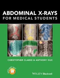
Abdominal X-rays for Medical Students is a comprehensive resource offering guidance on reading, presenting and interpreting abdominal radiographs. Suitable for medical students, junior doctors, nurses and trainee radiographers, this brand new title is clearly illustrated using a unique colour overlay system to present the main pathologies and to highlight the abnormalities in abdomen x-rays.
Abdominal X-rays for Medical Students:
- Covers the key knowledge and skills necessary for practical use
- Provides an effective and memorable way to analyse and present abdominal radiographs - the unique 'ABCDE' system as developed by the authors
- Presents each radiograph twice, side by side: the first as seen in the clinical setting, and the second with the pathology clearly highlighted
- Includes self-assessment to test knowledge and presentation technique
With a systematic approach covering both the analysis of radiographs and next steps mirroring the clinical setting and context, Abdominal X-rays for Medical Students is a succinct and up-to-date overview of the principles and practice of this important topic.
Sie lesen das E-Book in den Legimi-Apps auf:
Seitenzahl: 188
Veröffentlichungsjahr: 2015
Ähnliche


CONTENTS
Cover
Series page
Title page
Copyright page
Preface
Acknowledgements
Learning objectives checklist
Part 1
About X-rays
What are X-rays?
How are X-rays produced?
How do X-rays make an image?
How are X-ray images (radiographs) stored?
Radiation hazards
The Ionising Radiation (Medical Exposure) Regulations
In women of reproductive age
Indications for an abdominal X-ray
Abdominal X-ray views
AP Supine abdominal X-ray
Other views
Radiograph quality
Inclusion
Exposure
Normal anatomy on an abdominal X-ray
Right and left (Figure 7)
Quadrants and regions (Figure 8)
Abdominal viscera 1 (Figure 9)
Abdominal viscera 2 (Figure 10)
Skeletal structures (Figure 11)
Pelvis (Figure 12)
Lung bases (may be visualised at the top of the abdomen) (Figure 13)
Bowel 1 (Figure 14)
Bowel 2 (Figure 15)
Presenting an abdominal radiograph
Be systematic!
Part 2
Overview of the ABCDE of abdominal radiographs
A – Air in the wrong place
B – Bowel
C – Calcification
D – Disability (bones and solid organs)
E – Everything else
A
Pneumoperitoneum (gas in the peritoneal cavity)
Pneumoretroperitoneum (gas in the retroperitoneal space)
Pneumobilia (gas in the biliary tree)
Portal venous gas (gas in the portal vein)
B
Dilated small bowel
Dilated large bowel
Volvulus
Dilated stomach
Hernia
Bowel wall inflammation
Faecal loading
Faecal impaction
C
Gallstones in the gallbladder (cholelithiasis)
Renal stones (urolithiasis)
Bladder stones
Nephrocalcinosis
Pancreatic calcification
Adrenal calcification
Abdominal aortic aneurysm (AAA) calcification
Fetus
Calcified structures of little clinical significance
D
Pelvic fractures – 3 Polo rings test
Sclerotic and lucent bone lesions
Spine pathology
Solid organ enlargement
E
Medical and surgical objects (iatrogenic)
Foreign bodies
Lung bases
Self-assessment questions
Self-assessment answers
Glossary
Index
End User License Agreement
List of Illustrations
Chapter 01
Figure 1: The electromagnetic spectrum (Freq is short for frequency).
Figure 2: X-ray production.
Figure 3: The spectrum of tissues of different densities as seen on a conventional radiograph. The radiograph example shows the left lumbar region of a patient who swallowed a battery.
Chapter 03
Figure 4: Anterior–posterior (AP) supine abdominal X-ray.
Chapter 04
Figure 5: A normal abdominal radiograph showing the superior aspect of the liver (1), superior aspect of the spleen (2) and lateral abdominal walls (3) marked with dashed white lines. The pubic symphysis (4) is marked with a white circle (although ideally I would also like to see the inferior aspect of the pubic symphysis).
Figure 6: An underexposed abdominal radiograph demonstrating poor visualisation of the spine. It is more difficult to make out the bowel gas and the diagnostic value of this radiograph may be somewhat limited.
Lesen Sie weiter in der vollständigen Ausgabe!
Lesen Sie weiter in der vollständigen Ausgabe!
Lesen Sie weiter in der vollständigen Ausgabe!
Lesen Sie weiter in der vollständigen Ausgabe!
Lesen Sie weiter in der vollständigen Ausgabe!
Lesen Sie weiter in der vollständigen Ausgabe!
Lesen Sie weiter in der vollständigen Ausgabe!
Lesen Sie weiter in der vollständigen Ausgabe!
Lesen Sie weiter in der vollständigen Ausgabe!
Lesen Sie weiter in der vollständigen Ausgabe!
Lesen Sie weiter in der vollständigen Ausgabe!
Lesen Sie weiter in der vollständigen Ausgabe!
Lesen Sie weiter in der vollständigen Ausgabe!
Lesen Sie weiter in der vollständigen Ausgabe!
Lesen Sie weiter in der vollständigen Ausgabe!
Lesen Sie weiter in der vollständigen Ausgabe!
Lesen Sie weiter in der vollständigen Ausgabe!
Lesen Sie weiter in der vollständigen Ausgabe!
Lesen Sie weiter in der vollständigen Ausgabe!
Lesen Sie weiter in der vollständigen Ausgabe!
Lesen Sie weiter in der vollständigen Ausgabe!
Lesen Sie weiter in der vollständigen Ausgabe!
Lesen Sie weiter in der vollständigen Ausgabe!
Lesen Sie weiter in der vollständigen Ausgabe!
Lesen Sie weiter in der vollständigen Ausgabe!


