108,99 €
Mehr erfahren.
- Herausgeber: John Wiley & Sons
- Kategorie: Fachliteratur
- Sprache: Englisch
Jubilee edition of the classic text first published in 1963
Anaesthetists require a particularly specialized knowledge of anatomy
The anaesthetist must know intimately the respiratory passages, the major veins and the peripheral
nerves to deliver safe and effective pain control.
As one of the great teachers of anatomy, Professor Harold Ellis is eminently qualified to elegantly
provide the anatomical detail required of anaesthetists. Modern approaches to practice, including
the use of imaging to guide anaesthetic practice, add further depth to the fine full-colour anatomical
illustrations.
Designed for anaesthetists, Anatomy for Anaesthetists covers:
• The Respiratory Pathway, Lungs, Thoracic Wall and Diaphragm
• The Heart and Great Veins of the Neck
• The Peripheral Nerves
• The Autonomic Nervous System
• The Cranial Nerves
• The Anatomy of Pain
Clinical Notes throughout provide the clinical context for the anatomical detail. Designed for trainees, but of continuing relevance to practicing anaesthetists, and now in its Golden Jubilee edition, Anatomy for Anaesthetists provides a central pillar of anaesthetic knowledge.
Sie lesen das E-Book in den Legimi-Apps auf:
Seitenzahl: 469
Veröffentlichungsjahr: 2013
Ähnliche


Contents
Cover
Title Page
Copyright
Preface to the Ninth (Jubilee) Edition
Foreword to the First Edition
Acknowledgements to the Ninth (Jubilee) Edition
Part 1: The Respiratory Pathway, Lungs, Thoracic Wall and Diaphragm
The mouth
The nose
The pharynx
The larynx
The trachea
The main bronchi
The pleura
The intercostal spaces
The lungs
The diaphragm
Part 2: The Heart and Great Veins of the Neck
The pericardium
The heart
Developmental anatomy
The great veins of the neck
Part 3: The Vertebral Canal and its Contents
The vertebrae and sacrum
The spinal meninges
The spinal cord
Part 4: The Peripheral Nerves
The spinal nerves
The cervical plexus
The brachial plexus
The thoracic nerves
The lumbar plexus
The sacral and coccygeal plexuses
Part 5: The Autonomic Nervous System
Introduction
The sympathetic system
The parasympathetic system
Part 6: The Cranial Nerves
Introduction
The olfactory nerve (I)
The optic nerve (II)
The oculomotor nerve (III)
The trochlear nerve (IV)
The trigeminal nerve (V)
The abducent nerve (VI)
The facial nerve (VII)
The auditory (vestibulocochlear) nerve (VIII)
The glossopharyngeal nerve (IX)
The vagus nerve (X)
The accessory nerve (XI)
The hypoglossal nerve (XII)
Part 7: The thoracic inlet
The thoracic inlet
The antecubital fossa
The orbit and its contents
The abdominal wall
Part 8: The Anatomy of Pain
Introduction
Classification of pain
Peripheral receptors and afferent fibres
The spinal cord and central projections
Modulation of pain signals
The autonomic nervous system and pain
Index
This edition first published 2014 © 1963, 1969, 1977, 1983, 1988, 1993, 1997, 2004 by Blackwell Science Ltd. 2014 © John Wiley & Sons, Ltd
Registered office:John Wiley & Sons, Ltd, The Atrium, Southern Gate, Chichester, West Sussex, PO19 8SQ, UK
Editorial offices:9600 Garsington Road, Oxford, OX4 2DQ, UK 111 River Street, Hoboken, NJ 07030-5774, USA
For details of our global editorial offices, for customer services and for information about how to apply for permission to reuse the copyright material in this book please see our website at www.wiley.com/wiley-blackwell
The right of the author to be identified as the author of this work has been asserted in accordance with the UK Copyright, Designs and Patents Act 1988.
All rights reserved. No part of this publication may be reproduced, stored in a retrieval system, or transmitted, in any form or by any means, electronic, mechanical, photocopying, recording or otherwise, except as permitted by the UK Copyright, Designs and Patents Act 1988, without the prior permission of the publisher.
Designations used by companies to distinguish their products are often claimed as trademarks. All brand names and product names used in this book are trade names, service marks, trademarks or registered trademarks of their respective owners. The publisher is not associated with any product or vendor mentioned in this book. It is sold on the understanding that the publisher is not engaged in rendering professional services. If professional advice or other expert assistance is required, the services of a competent professional should be sought.
The contents of this work are intended to further general scientific research, understanding, and discussion only and are not intended and should not be relied upon as recommending or promoting a specific method, diagnosis, or treatment by health science practitioners for any particular patient. The publisher and the author make no representations or warranties with respect to the accuracy or completeness of the contents of this work and specifically disclaim all warranties, including without limitation any implied warranties of fitness for a particular purpose. In view of ongoing research, equipment modifications, changes in governmental regulations, and the constant flow of information relating to the use of medicines, equipment, and devices, the reader is urged to review and evaluate the information provided in the package insert or instructions for each medicine, equipment, or device for, among other things, any changes in the instructions or indication of usage and for added warnings and precautions. Readers should consult with a specialist where appropriate. The fact that an organization or Website is referred to in this work as a citation and/or a potential source of further information does not mean that the author or the publisher endorses the information the organization or Website may provide or recommendations it may make. Further, readers should be aware that Internet Websites listed in this work may have changed or disappeared between when this work was written and when it is read. No warranty may be created or extended by any promotional statements for this work. Neither the publisher nor the author shall be liable for any damages arising herefrom.
Library of Congress Cataloging-in-Publication Data
Ellis, Harold, 1926– author. Anatomy for anaesthetists / Harold Ellis, Andrew Lawson. -- Ninth edition. p. ; cm. Includes bibliographical references and index. ISBN 978-1-118-37598-3 (cloth : alk. paper) -- ISBN 978-1-118-37594-5 -- ISBN 978-1-118-37595-2 (emobi) -- ISBN 978-1-118-37596-9 (epdf) -- ISBN 978-1-118-37597-6 (epub) I. Lawson, Andrew (Medical ethicist), author. II. Title. [DNLM: 1. Anatomy. 2. Anesthesia. QS 4] QM23.2 611′.0024617--dc23 2013024798
A catalogue record for this book is available from the British Library.
Wiley also publishes its books in a variety of electronic formats. Some content that appears in print may not be available in electronic books.
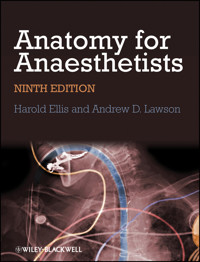
Cover image: Science Photo Library image: © POGEE/SCIENCE PHOTO LIBRARY Color-enhanced x-ray showing endotracheal intubation Cover design by Meaden Creative
Preface to the Ninth (Jubilee) Edition
I little thought, when I wrote the Introduction to the first edition of Anatomy for Anaesthetists for its publication in 1963, that one day I would be composing the introduction to its ninth (Jubilee) edition in 2013! The place of anatomy in anaesthetic practice is often considered merely as a prerequisite for the safe practice of local anaesthetic blocks. However, it is also important in understanding the anatomy of the airways, the functions of the lung and circulation, monitoring of neuromuscular blocks, long-term pain control and many other aspects of practical anaesthesia. This book is not intended to be a textbook of regional anaesthetic techniques; there are many excellent texts that cover this important field. However, it is a textbook written for anaesthetists, keeping in mind the special requirements of their daily practice. The anaesthetist requires a particularly specialized knowledge of anatomy. Some regions of the body, for example the respiratory passages, the major veins and the peripheral nerves, the anaesthetist must know with an intimacy of detail that rivals or even exceeds that of the surgeon; other areas can be all but ignored.
The first edition was written in collaboration with that talented medical artist, the late Margaret McLarty. I was then joined by my anaesthetic colleague at Westminster Medical School, Professor Stanley Feldman, as co-author. Dr William Harrop-Griffiths contributed much help on peripheral nerve blocks to the eighth edition. I am now delighted to have Dr Andrew Lawson as my collaborator. He has already added the important chapter on the anatomy of pain to the last two editions. He is not only an anaesthetist but also an expert in pain management and brings his expertise in this field in describing important clinical applications of anatomy to this aspect of anaesthetic practice.
In this ninth edition, we have carefully revised and expanded the text and have added new illustrations, including modern imaging techniques that are of particular interest to the anaesthetist. We hope this book will continue to serve anaesthetists as it has done over the past 50 years.
Harold Ellis July 2013
Foreword to the First Edition
The anaesthetist faced with higher examinations is confronted with the problem of how far he should delve into the many related specialities; and owing to constant change of emphasis, the answers will never be final. Professor Ellis, as a surgeon who has interested himself in our speciality for a number of years, here sets out boldly the anatomy he believes the present-day young anaesthetist should be familiar with when confronting the examiners. The choice of material, and its presentation, are full of commonsense, so that the book is attractive to the established anaesthetist in his daily work, and for occasional reading.
I have had the privilege of working with both authors for a number of years, and I can think of no pair better fitted to highlight the essentials of anatomy for the anaesthetist. Professor Ellis is an outstanding teacher, and in the field of medical artists Miss McLarty is distinguished by her flair for stressing points of anatomical importance to the clinician. I persuaded them to collaborate on a series of articles which recently appeared in Anaesthesia. Though these have been added to considerably they form the basis of this book.
Professor Sir Robert Macintosh
Introduction to the First Edition
The anaesthetist requires a peculiarly specialized knowledge of anatomy. Some regions of the body – the nerve pathways and respiratory passages for example – he must know with an intimate detail which rivals that of the surgeon; other areas he may all but ignore. As far as we know this book is the first to be designed with his particular needs in mind. It should prove of value to examination candidates and we hope also to the practical anaesthetist in his day to day work.
Harold Ellis, London Margaret McLarty, Oxford 1963
Acknowledgements to the Ninth (Jubilee) Edition
The first two editions of this textbook were prepared in collaboration with that skilled medical artist Miss Margaret McLarty. The illustrations for the sixth edition were almost all drawn or redrafted by Rachel Chesterton; we thank her for the excellent way in which they have been executed. Further illustrations for the seventh, eighth and this edition were prepared by Jane Fallows with great skill. Some of the figures have been reproduced from Clinical Anatomy, 13th edition. Thanks to Dr Charles Gaucci, Dr Ron Cooper, Dr Nick Morgan Hughes and Dr Vlademir Gorelov for providing images for use in this book.
Part 1
The Respiratory Pathway, Lungs, Thoracic Wall and Diaphragm
The mouth
The mouth is made up of the vestibule and the mouth cavity, the former communicating with the latter through the aperture of the mouth.
The vestibule is formed by the lips and cheeks without, and by the gums and teeth within. An important feature is the opening of the parotid duct on a small papilla opposite the 2nd upper molar tooth. Normally the walls of the vestibule are kept together by the tone of the facial muscles; a characteristic feature of a facial (VII) nerve paralysis is that the cheek falls away from the teeth and gums, enabling food and drink to collect in, and dribble out of, the now patulous vestibule.
The mouth cavity (Fig. 1) is bounded by the alveolar arch of the maxilla and the mandible, and teeth in front, the hard and soft palate above, the anterior two-thirds of the tongue and the reflection of its mucosa forwards onto the mandible below, and the oropharyngeal isthmus behind.
Fig. 1 View of the open mouth with the tongue depressed.
The mucosa of the floor of the mouth between the tongue and mandible bears the median frenulum linguae, on either side of which are the orifices of the submandibular salivary glands (Fig. 2). Backwards and outwards from these ducts extend the sublingual folds that cover the sublingual glands on each side (Fig. 3); the majority of the ducts of these glands open as a series of tiny orifices along the overlying fold, but some drain into the duct of the submandibular gland (Wharton's duct).
Fig. 2 View of the open mouth with the tongue elevated.
Fig. 3 Coronal section through the floor of the mouth.
Inspect your mouth in a mirror. Elevate your tongue, then press on one or the other side onto your submandibular gland beneath the angle of the jaw. You will see a jet of saliva emerge from the orifice of the submandibular duct at the tip of the sublingual fold. While about it, pull your cheek laterally with a finger, press on your parotid gland on that side and observe a jet of saliva emerge from the parotid duct, which lies at the level of your 2nd upper molar tooth.
The palate
The hard palate is made up of the palatine processes of the maxillae and the horizontal plates of the palatine bones. The mucous membrane covering the hard palate is peculiar in that the stratified squamous mucosa is closely connected to the underlying periosteum, so that the two dissect away at operation as a single sheet termed the mucoperiosteum. This is thin in the midline, but thicker more laterally owing to the presence of numerous small palatine salivary glands, an uncommon but well-recognized site for the development of mixed salivary tumours.
The soft palate hangs like a curtain suspended from the posterior edge of the hard palate. Its free border bears the uvula centrally and blends on either side with the pharyngeal wall. The anterior aspect of this curtain faces the mouth cavity and is covered by a stratified squamous epithelium. The posterior aspect is part of the nasopharynx and is lined by a ciliated columnar epithelium under which is a thick stratum of mucous and serous glands embedded in lymphoid tissue.
The ‘skeleton’ of the soft palate is a tough fibrous sheet termed the palatine aponeurosis, which is attached to the posterior edge of the hard palate. The aponeurosis is continuous on each side with the tendon of tensor palati and may, in fact, represent an expansion of this tendon.
Lesen Sie weiter in der vollständigen Ausgabe!
Lesen Sie weiter in der vollständigen Ausgabe!
Lesen Sie weiter in der vollständigen Ausgabe!
Lesen Sie weiter in der vollständigen Ausgabe!
Lesen Sie weiter in der vollständigen Ausgabe!
Lesen Sie weiter in der vollständigen Ausgabe!
Lesen Sie weiter in der vollständigen Ausgabe!
Lesen Sie weiter in der vollständigen Ausgabe!


