Erhalten Sie Zugang zu diesem und mehr als 300000 Büchern ab EUR 5,99 monatlich.
- Herausgeber: The History Press
- Kategorie: Lebensstil
- Sprache: Englisch
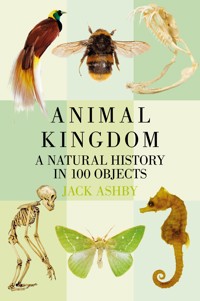
From a single beginning, countless millions of stories from the animal kingdom have – and continue to – run their course. Museum objects allow us to investigate some of those stories. Animal Kingdom journeys through both the evolutionary history of animals, and the ways that people have interpreted them in museums. Animals in museums are not only representatives of their entire species, but they also tell us something about the time in which they were collected. They provide windows into the past as well as data for the present. They embody centuries of natural ecosystems and human cultures. Through a selection of 100 objects, telling 100 stories, this beautifully illustrated book explores the diversity of animal life over the past 600 million years, and delves into some of the most exciting mechanisms in evolution. By understanding some of the key stories of how nature operates, we can gain amazing insight into the systems underlying life itself.
Sie lesen das E-Book in den Legimi-Apps auf:
Seitenzahl: 511
Veröffentlichungsjahr: 2017
Das E-Book (TTS) können Sie hören im Abo „Legimi Premium” in Legimi-Apps auf:
Ähnliche


First published 2017
The History Press
The Mill, Brimscombe Port
Stroud, Gloucestershire, GL5 2QG
www.thehistorypress.co.uk
© Jack Ashby, 2017
The right of Jack Ashby to be identified as the Author of this work has been asserted in accordance with the Copyright, Designs and Patents Act 1988.
All rights reserved. No part of this book may be reprinted or reproduced or utilised in any form or by any electronic, mechanical or other means, now known or hereafter invented, including photocopying and recording, or in any information storage or retrieval system, without the permission in writing from the Publishers.
British Library Cataloguing in Publication Data.
A catalogue record for this book is available from the British Library.
EPUB ISBN 978 0 7509 8613 7
Typesetting and origination by The History Press
Printed in Turkey by Imak
eBook converted by Geethik Technologies
Contents
Introduction
1 Platypus
Part 1: Understanding Diversity
2 Elephant Ear Sponge
3 Sea Anemones
4 Comb Jelly
5 Parasitic Flatworm
6 Bootlace Worm
7 King Ragworm
8 King Scallop
9 Bryozoan
10 Brachiopod
11 Velvet Worms
12 Dragonfly
13 Roundworms
14 Penis Worm
15 Sea Potato
16 Graptolites
17 Lancelets
18 Sea Squirts
19 Chimpanzee
Part 2: Life’s Turning Points
20 Ottoia The Penis Worm
21 Jawless Fishes
22 Cookie-Cutter Shark
23 Common Carp
24 Coelacanth
25 Ichthyostega
26 Wellesaurus: A Triassic Amphibian-Relative
27 Diadectes: A Close Amniote Relative
28 Lystrosaurus
29 Horse Relatives
Part 3: Natural Histories
30 Spiny Stick Insect
31 Giant Deer
32 Lesser Bird-of-Paradise
33 Garden Snail
34 Bed Bugs
35 Seahorses
36 Gorillas
37 Walrus
38 Dugong
39 Box Jellyfish
40 Striped Possum
41 Slow Worm
42 Narrow-Bordered Bee Hawkmoth
43 Heliconius Butterflies
44 Amphisbaenian: A ‘Worm-Lizard’
45 Essex Emerald Moth
46 Nine-Banded Armadillo
47 Crab-Eating Macaque
48 Megaladapis: The Koala Lemur
49 Hoolock Gibbon
50 North American Beaver
51 Hippopotamus
52 Alligator
53 Gaboon Viper
54 Narwhal
55 Bushmaster
56 Electric Ray
57 Cuttlefish
58 Blue Sea Slug
59 Caterpillar With Parasitoid Fungus
60 Bumblebee
61 Sponge Crab
62 Common Cuckoo
63 Horseshoe Crab
64 New Zealand Native Frog
65 Kangaroo
66 Koala
67 Cane Toad
68 Portuguese Man O’ War
69 Mantis Shrimp
70 Surinam Toad
71 Olm
72 Leaf-Cutter Ant
73 Aphids
74 Eyed Hawkmoth x Poplar Hawkmoth Hybrid
75 Archaeopteryx
76 House Mice
77 Elephant Bird
78 Thylacine
79 Tasmanian Devil
80 Tree Frogs
81 Marine Iguana
82 Crescent Nailtail Wallaby
83 Trilobites
84 Woolly Mammoth
85 Collie And King Charles Spaniel
Part 4: Displaying Nature
86 Domestic Pig
87 Entomology Collection
88 James Bond’s Hutia
89 Short-Beaked Echidna
90 African Rock Python
91 Hypsilophodon
92 Dodos
93 The Micrarium
94 Fruit Flies
95 Grass Snake
96 Domestic Cat
97 Copse Snail
98 Red Panda
99 Human
100 Antarctic Limpets
Selected References and Further Reading
Acknowledgements
Image Credits
Introduction
My home contains many skulls, fossils, rocks, shells, bones, and teeth. Most are reminders of trips I’ve taken and things I’ve seen – natural souvenirs from my life as a zoologist. The same items also exist in museum collections, but those are not souvenirs. When objects are put in museums they become specimens, imbued with some intrinsic and intangible value they did not have before they arrived there. They become exemplars of a group – representatives of their species or kind, setting a standard for a particular animal from a particular place at a particular time. By contrast, I think of my objects at home more as knick-knacks, there for my own enjoyment. They can tell stories, but they are just my stories.
Objects in museums tell different stories. And what we can learn about the animal kingdom from museum specimens is also very different from what we can learn watching animals in the wild. In my ecological fieldwork in Australia I am lucky enough to have engaged with thousands of animals at extremely close proximity. These kind of encounters – and the scientific research they are part of – help us understand how ecosystems work and how animals behave in the wild. The stories we gain from these experiences enable us to see nature in action, as a vibrant, impossibly complex series of living interactions.
Encounters with specimens from dead animals in museums give us a different view. These meetings happen on our own terms, and allow us to ask and answer different questions. I have seen many more living Tasmanian devils than I have dead museum specimens, for instance, but I feel like I have learned more about them, or at least different things about them, by close study of their remains in museums. Natural history objects allow us to investigate the evolutionary history of animals, and seek to find not just what they do, but how they do it.
Research in the wild and in the museum are both critical pillars of natural history; neither tells the whole story on its own. We can only go so far in studying a skeleton or a carcass to imagine how the animal is adapted to moving through its environment, for example. Similarly brief encounters with animals on their own terms do not tell us everything we seek to know about where a species came from and how it came to be.
Museum specimens also allow us to study animals that have been dead for centuries. New technologies are developed to answer questions that weren’t even imagined when the animals were first collected. Examining fossils allows us to understand species that have been dead for millions of years. We can use them to build a picture of worlds long disappeared, and how the earth came to be the way it is now.
Objects can tell the general stories about their kind, but also the specific stories that they alone were characters in. They are at once specimens and individuals – museum objects were collected, traded and used by people: their stories are historical as well as scientific. Investigating museum collections tells us something about the time in which they were collected. They provide windows into the past as well as data for the present. These historic repositories represent centuries of ecosystems and cultures. Even though they are just dead animals, they reflect local, national and global politics, and the societies they are linked with.
The UK alone holds well over 100 million natural history objects in its public museums. The global collection runs into the billions. The scale of these numbers is impossible to conceive of, and this is just one of the reasons why only a minuscule proportion of museum collections are on public display – usually less than 1 per cent. The functions of objects on display are very different to the collections held in storerooms, and this is one thing that I’ll be exploring in the final section of the book.
Animals are everywhere in the human world: they are used to advertise pretty much everything, from banks to toilet paper, and cars to cereals. They are mascots for sports teams and logos for airlines. The way they are represented usually links to some reflection of their natural history. As a result every single person walks down the street with an extraordinary knowledge of nature, even if they don’t think so themselves. This means unlocking some level of understanding of the natural world is easy. Museums can help us access some of this ingrained expertise, by sparking connections between what people already know and the amazing, rare objects in museum collections.
What I have attempted to do with this book is to select 100 museum specimens to tell 100 stories from the animal kingdom: 100 objects, 100 stories. It is not a history in the sense that it doesn’t have a beginning, a middle and an end, and on the whole it doesn’t run chronologically. The animal kingdom doesn’t have one history. From a single beginning, countless millions of histories have – and continue to – run their course. They intertwine constantly, some of them end abruptly and some fizzle out.
The book is arranged into four sections, each telling a different kind of story. The first explores the diversity of life over the past 600 million years or so, which is mainly the story of the invertebrates (animals without backbones; vertebrates are animals with backbones). Although information about natural history is everywhere in the human world, the coverage is not even. Large and charismatic species get the lion’s share of the limelight. Even though they outnumber vertebrates by more than twenty to one, invertebrates are given a raw deal in natural history museums, television programmes and books. I’ll admit that I am also somewhat guilty of this (as mammals are my passion), but the first section of this book attempts to show most of the ways there are to be an invertebrate as I go through many of the major divisions of the animal kingdom.
The Turning Points section tells the story of evolution from the perspective of us mammals. As I explain, this is only one of the countless routes through the branches of the tree of life. It begins with the sudden appearance of most of the key groups of animals a little over half a billion years ago, and explores the appearance of different kinds of fish. Our evolutionary history involves fishes without jaws and without hard skeletons, a huge diversity of bony fishes, the colonisation of the land, the end to a reliance on reproducing in water and responses to massively changing ecosystems. We can trace our relationships with fishes, amphibians and reptiles, and understand how the world’s mammalian fauna came to be the way it is today.
The third section is by far the biggest. It explores the animal kingdom by investigating some of the most exciting mechanisms in evolution. By understanding some of the key stories of how nature operates, we can gain an amazing insight into the workings of the world. The objects in this Natural Histories section reveal some of the astonishing systems underlying life. Nature is truly incredible, and with the objects in this section I have tried to highlight some of the genuinely astounding ways it works.
Finally, in the Displaying Nature section I hold a lens up to museums, one of the key windows we have onto the natural world. I ask questions about how the decisions that have been made in the creation and curation of museums throughout their history influence the way we see animals today. Museums are not sterile or natural places, but human inventions. As such the biases of human nature and politics can affect our interpretation of nature.
All of the objects in this book come from the collection which I manage – the Grant Museum of Zoology at University College London. However, in truth they could have come from any museum with a natural history collection. Aside from the individual historical stories that objects can tell, and perhaps a few genuinely rare ‘hero objects’, to some level of approximation all natural history museums have examples of more or less the same stuff, it’s just that the collections vary in size. I could have chosen any number of objects to illustrate these 100 stories, as every species and every individual is a result of its deep ancestry, subject to the same evolutionary pressures as everything else.
To set the scene I have chosen one object to kick things off, before we get into the four separate sections. I wanted to illustrate how the book works, with an object and a story. It explains one of the key concepts that underlies how evolution works, and uses my favourite animal to do it.
1 Platypus
TAXIDERMY SPECIMEN
Evolution Can Only Work With What It Has Got
Science is supposed to be unbiased, but scientists are not. We have our favourites and our passions, and mine is the platypus. I could have used this bizarre Australian to illustrate any number of nature’s mechanisms or incredible stories in the history of zoology, but I wanted to use it to demonstrate on a very simple idea about how evolution works, as a means to begin the book: Evolution can only work with what it has got.
One thing that evolution had at the end of the Triassic period, a little over 200 million years ago, was the synapsids – a group of reptile-like animals which would give rise to mammals. Triassic synapsids had a lot of characteristics that we might call reptilian. This is because they shared a recent evolutionary history with the groups that would become crocodiles, dinosaurs, lizards and turtles. They did things that reptiles do, like laying eggs and walking with bent elbows and knees, with their limbs held out at right angles to their bodies (or at least partially).
This is the ancestral frame that evolution had to work with. All new developments would have to start from here. This means that the first mammals would have shared a lot of characteristics with their reptilian forebears.
Platypuses are often described as ‘primitive’ because throughout their long evolutionary history they have retained these ancient ancestral features. Platypuses, and their closest living relatives the echidnas, still walk with bent elbows and knees, and still lay eggs, just like their Triassic relatives.
Platypuses are mammals, and laying eggs might not seem like a very mammalian thing to do. Many people are under the impression that all mammals give birth to live young. There are a few unique defining characteristics of all living mammals, but live-birthing is not one of them. They include the presence of hair, three inner-ear bones, a specialised ankle joint and a single bone forming either side of the lower jaw (reptiles have seven), and the ability to produce milk (mammary is the root of the word mammal). Platypuses have all of these features. (Platypuses do not truly suckle their young, though, as they don’t have nipples. However, they do have mammary glands: the milk kind of sweats out of them, and the babies lap it up.)
Together platypuses and echidnas form the order Monotremata, so named for their cloaca: a single (monos) hole (trema) for all their urogenitary functions. Like reptiles, they do all their defecating, urinating, mating and birthing through this one single opening. It is these reptilian features that have earned platypuses their ‘primitive’ reputation. However, on to this ancient template platypuses have added some of the most advanced characteristics of any mammal. This is the reason why I get so excited about platypuses.
First, male platypuses are among the only venomous mammals. They have a horny spur on their heels attached to a venom gland, which is used during sexual competition. (The venom is said to be excruciating and long-lasting for humans.) The males of many species have developed myriad ways to secure sexual success, from giant antlers to absurd decoration. Platypuses are the only mammal to use venom to fight off rivals.
Second, they are one of only a tiny number of mammals known to be capable of electroreception (the others are their close relatives the echidnas, and one species of dolphin): they can detect electricity through their bills. Platypuses are restricted to the lakes and rivers of Tasmania and eastern Australia, where they are reasonably common. They use their power of electroreception to hunt worms and tiny crustaceans. Muscular movement in animals is controlled by electrical impulses. Little is known about platypus feeding, but it is assumed that platypuses can locate their prey buried under silt by sensing these electrical impulses with their bills. They gather food up in little pouches at the base of the bill and then mash it up (adult platypuses have no teeth) with their rubbery bills when on the surface.
In the history of the platypus, evolution started with a ‘primitive’, reptile-like template. Without changing much of this ancient frame, incredibly advanced features were added. This demonstrates that a modern species can never itself be called ‘primitive’ – only the characteristics it has inherited from its ancestors. Neither can a species be considered ‘advanced’, as although it may have evolved some brand new tricks, it still retains those basic elements that have stood the test of time and are found to be still useful now.
When Europeans first discovered platypuses at the end of the eighteenth century they caused quite a stir. The first specimens to reach England – preserved in barrels of brandy or as dried skins – were thought to be hoaxes, made from at least three animals sewn together. There are (probably apocryphal) stories that the earliest specimen to reach what is now the Natural History Museum in London has visible plier marks on its bill, where the curator tried to wrench it off to demonstrate it had been sewn together as a fake. Indeed, in the Grant Museum of Zoology, where I work, there is a bill that has been detached from its body in a crude manner that implies it cannot have been carefully dissected away, which may be the result of similar treatment.
With its bizarre mix of characteristics, both novel and ancient, the platypus did not fit into the existing animal groups that scientists had conceived, and there was great debate over where to place it. Eventually, entirely new groups were created to accommodate it.
Taxonomy aside, it took nearly a century to settle the argument over whether it truly did lay eggs. Perhaps the religiously minded scientific elite took exception to the idea that something which was clearly a mammal could do something so reptilian – something that might drag our noble class down into the mud and slime.
Robert Grant – the founder of the Grant Museum and an early proponent of evolution (he had a huge influence on sparking Darwin’s thinking) – was a strong supporter of egg-laying in platypuses. Perhaps he used this taxidermy specimen in his search for evidence.
Part 1
Understanding Diversity
PUTTING ANIMALS IN ORDER
The human desire to classify is perhaps at its strongest when it comes to natural history. From our childhood years we are taught to put the animals we encounter in museums, living rooms and the natural environment into discrete categories. At school and on television we are taught the differences between groups like amphibians and fish.
Thomas Henry Huxley, one of the greatest biologists of the nineteenth century and a man who can take a lot of credit for making Darwin’s work accepted by the Victorian scientific community, said this:
To a person uninstructed in natural history, his country or sea-side stroll is a walk through a gallery filled with wonderful works of art, nine-tenths of which have their faces turned to the wall. Teach him something of natural history, and you place in his hands a catalogue of those which are worth turning around.1
He was right. Knowing what you are looking at – in both nature and the art gallery in Huxley’s analogy – is half the joy. But we can’t become ‘instructed’ in everything. The world is an endless purveyor of wonders too numerable to memorise. To make sense of the 1.5 million species so far described (and there may be 100 million undescribed species), natural historians have to come up with a system for arranging them and information about them.
This proved tricky until 1735, when Swedish botanist Carl Linnaeus proposed a system for putting species into hierarchical groups, and it stuck. In today’s terms, a rat can be a rat, a rodent, a mammal, a vertebrate and an animal all at once. Such taxonomic thinking is really important for how we understand the world and our place in it, as each of these terms comes with implicit information about how they relate to other groups. It neatly puts the world into boxes. Although it certainly wasn’t Linnaeus’ intention (he believed that studying nature would reveal the divine order of God’s creation), hierarchical taxonomies tell us a lot about an animal’s evolutionary history, as by their nature they show what came from what. This information is real and truthful, but, as is often said, a bee doesn’t care that it is a bee. Taxonomy – the science of putting things into groups – is a rigid human construct that is forced on top of the cacophonous uncertainty of the real wild world.
Comb jelly illustrations from Kunstformen der Natur (Art Forms of Nature), a 1904 book by German biologist Ernst Haeckel (see object 18).
One of the central tenets of modern taxonomy is that every group has to include, by definition, all of the groups that evolve from it. So rats did not stop being mammals when the rodent group branched off the evolutionary tree. Every branch on the tree of life is considered to be a member of all its parent branches.
This means, for example, there can be no definition of fishes that does not include everything that evolved from fishes. Following this logic, you could argue that as amphibians evolved from fishes, amphibians are fishes. Mammals evolved from animals that evolved from fishes, so mammals are fishes. We are fish. While every biologist knows this conundrum, and that there is no biological definition for what most people consider ‘fishes’, they decide not to worry about it because it’s helpful to think about living swimming ‘fishes’ as a group. Taxonomy is useful and makes a lot of sense, until it doesn’t.
Similarly we have all been taught that the animal world can be divided into vertebrates and invertebrates. This is a very handy division, but it suffers from the same problem as ‘fishes’: as vertebrates evolved from invertebrates, every single species of animal there has ever been is an invertebrate. Given that this means that animal and invertebrate are taxonomically synonymous, we just have to agree to ignore that and carry on as normal.
The boxes that taxonomy forces around the natural world come in a set sequence of ever-more specific groupings. The largest in common use is kingdom, and this book focusses on just one of them – the animals. The simplest hierarchy of groups goes like this:
Kingdom
Phylum
Class
Order
Family
Genus
Species
And for us, that hierarchy would look like this:
Kingdom: Animal
Phylum: Chordate
Class: Mammal
Order: Primates
Family: Great apes
Genus: Homo
Species: Homo sapiens
Below the kingdom level, the next sub-grouping is phylum (plural: phyla). There are commonly held to be thirty-five animal phyla. This first section of the book makes an attempt to give coverage to some of the diversity of life that doesn’t enjoy a lot of limelight. I could have chosen to dedicate a chapter to each of the phyla, but I haven’t.
That’s because evolution has produced many different ways of being a worm: about half of the phyla are distinctly wormish. I apologise to worm-fanciers out there, but given that many of these phyla contain rather few species, and that I have a lot more to say about the less vermicular animals of the world, I opted not to give them the proportional coverage you may think they deserve.
Instead, here follow eighteen different objects representing eighteen of the major groupings of the animal kingdom. It hopefully demonstrates most of the key ways there are of being an animal.
A few of which are worms.
1 Huxley, T.H., Lay Sermons, Addresses and Reviews (D. Appleton: New York, 1876)
2 Elephant Ear Sponge
DRY SPECIMEN
Sponges: The Poriferans
For most of the history of life all organisms were single cells. A giant leap was made when dividing cells didn’t separate from one another and became integrated to form a simple multicellular organism. Only then could huge steps be made to diversify the shapes, sizes and behaviours we see in the biological world today.
Sponges are among the oldest known animal fossils, dating back to the late Precambrian period, 580 million years ago. They branched off extremely early in the history of animals, separating them from the groups that have far more complex structures. They have one of the most simple body plans of all animals. They only have a small number of different types of cell, and these are not organised into properly differentiated tissues. They do not have any organs.
Because they are so unlike all other animals, until the 1820s sponges were believed to be plants. Robert Edmond Grant, the founder of the Grant Museum of Zoology, was a radical thinker who subscribed to ‘transmutationism’: the idea that species change over time – what we now call evolution. Grant spent several years studying and publishing on sponges and other simple invertebrates. He believed that they could provide insights to the roots of the tree of life.
Much of what we now know about sponges is down to Robert Grant. He noted that they passed out a fluid waste, which was evidence that they were digesting something – a key defining feature of animals. He also found that although sponges were not known to move, their reproductive cells could. Together this helped prove that sponges were in fact simple animals, not plants. He also came up with the name that unites all sponges into a single phylum: Porifera – ‘the animals that bear pores’.
Sponges live in all aquatic habitats, from the poles to the equator. They are extremely successful filter feeders, built as a network of channels lined with cells called ‘collar cells’. These have a single hair-like flagellum (like the tail on a human sperm) that they beat to form a current, sucking water through pores, along the channels and then out through one or a few large exit holes. In a single day a sponge can pass an amount of water equivalent to 20,000 times its own volume.
Sponges come in three classes, distinguished by the crystal structures called spicules that hold them together and essentially form their skeletons. Demosponges are the largest group and includes the elephant ear sponge, pictured on p. 17, and bath sponges. Demosponges have either fibres made of a collagen protein called spongin, spicules made of a silica mineral, or both. Calcareous sponges have crystals made of calcium carbonate, and the glass sponges have spicules made of silica crystals (but different to the ones demosponges have). This last group can form beautifully regular lattices of silica – particularly in Venus’ flower basket sponges (opposite) – which makes it difficult to picture where exactly the ‘wet’ part of the animals’ cells go. They are animals that are naturally formed of 90 per cent glass.
Venus’ flower basket – one of the glass sponges that are made almost entirely of silica.
In the nineteenth century it was discovered that sponges’ collar cells are strikingly similar to a group of single-celled organisms called choanoflagellates. This has led many zoologists to believe that choanoflagellates are animals’ closest relatives. In this case, sponges would be the most primitive group of animals, having evolved from a choanoflagellate-like ancestor.
Sponges do not have many of the features seen in other animals: they are not symmetrical in any way, and as well as lacking organised tissues they don’t have muscles, sense organs, nerves or a mouth. They don’t have respiratory, circulatory or kidney-like systems, but all parts of their bodies are very close to water so they can do fine without them. Gases and waste products can simply diffuse in and out using their water pumping system. It’s hard to rule out the possibility that sponges’ simplicity has evolved from more complex animals. Rather than never having evolved them, instead they may have lost some of these key features because living passively on the seabed does not require a very complex way of life. Interestingly, sponges have been found to have the genes that in other animals control some components of nerve impulses, but sponges do not have any nerves.
Because of the simplicity of sponge structures, they can reproduce simply by breaking apart. A fragment knocked off by waves or fishes has everything it needs to grow into a new sponge. They can reproduce asexually (without involving reproductive cells) by budding off a mini-replica, or by producing a gemmule. This is a little package of cells packed into a solid spore-like structure. Gemmules are particularly common among freshwater sponges which are more susceptible to drying out or freezing. Sponges can also reproduce sexually, and most are hermaphrodites, i.e. each individual can produce both male and female reproductive cells. Some species release eggs and then sperm into the water separately, so they can fertilise or be fertilised out in the open water. Others just release sperm, which are then trapped in other sponges by their filter feeding mechanisms.
3 Sea Anemones
TRAMOND WAX MODELS
Jelly With A Sting: The Cnidarians
Many of the world’s jelly-textured animals are grouped together in the phylum Cnidaria. This name takes its root from the Greek word for nettle – a reference to the fact that all cnidarians use stinging cells for hunting and defence. Anemones, corals, jellyfish and hydras are all cnidarians. They are a diverse, global group of over 12,000 species, all of which are aquatic (apart from some extremely unusual parasitic forms), but only twenty of them live in freshwater.
Wax models of snakelocks anemone, mottled anemone and Scolanthus callimorphus made by the nineteenth-century Parisian anatomical model-makers Maison Tramond.
Hard corals, like this brain coral, produce stony skeletons out of calcium carbonate that they extract from the water.
Cnidarians are the only animals visible from space. You can’t make out individual corals from a space station, but together millions of them have built Australia’s Great Barrier Reef – the world’s biggest living structure. Corals are responsible for creating entire ecosystems upon which untold numbers of other organisms rely. Coral reefs are some of the most diverse spots on earth: most cnidarians may be small, but they have a truly global impact.
Reefs are built of calcium carbonate structures deposited by corals which can take on a huge diversity of forms, from round corals, flat corals, branching corals and pipe corals. Each ‘lump’ of coral is built by hundreds or thousands of tiny individual coral animals called polyps that form colonies of clones, stuck together and working together as if they were a single animal (see chapter 68 for more on colonial living). Many species of coral have photosynthetic algae living inside them, producing nutrients for the coral in return for a safe place to live.
Despite the range of shapes that coral structures take on, up close the individual polyps display the features that make them recognisable as cnidarians. The corals, sea anemones, hydras and jellyfish are united by having a mouth (which also functions as their anus) surrounded by tentacles – with stinging cells called cnidocytes – and radial symmetry: when looked on from above the same pattern is seen if they are rotated.
The typical cnidarian life cycle alternates between two stages: a sedentary polyp (picture an anemone), which is basically a hollow tube fixed to a solid surface at one end, with a mouth at the top; and a free-living medusa phase (picture a jellyfish), which has formed by flattening the polyp’s tube into a disk, but with the mouth at the bottom. Some cnidarians have mixed life cycles with phases as each, some are only ever polyps, and some are only ever medusae.
Soft-bodied cnidarians, unsurprisingly, do not have a particularly recognisable fossil record. Definite, hard cnidarians are known from 400–500 million years ago, but many people have speculated that some of the most enigmatic and oldest animal fossils known are cnidarians, from the Precambrian Ediacaran period, well over 600 million years ago.
Along with sponges, comb jellies and the microscopic plate-like placozoans, cnidarians sit outside of the main grouping of animals, the Bilataria, which contains everything else. Bilatarians, named for their bilateral symmetry (they are roughly symmetrical around a midline), are animals with a front and back and top and bottom. Their sensory organs are concentrated around the mouth in a head (a feature not seen in cnidarians, which have a mouth but no head), and the digestive tract empties out of an anus – an evolutionary step up from using one’s mouth to defecate.
In any case cnidarians and the comb jellies represent a step up in animal complexity from the sponges. Cnidarian cells are organised into true tissues, but they do not have any organs, which are structures built from a combination of tissues working together. They have muscles, nerves and senses, as well as a mouth and a digestive cavity. As such they are more closely related to humans (and the rest of the Bilataria) than they are to sponges, and together cnidarians, comb jellies and bilatarians form a group called the Eumetazoa.
Cnidarians do not have circulatory or excretory systems, and no central nervous system. Their nerves are arranged into simple networks that control the contractions of their muscles and process the information from their sensory organs (which can include the detection of chemical signals, pressure, gravitational pull and light – see box jellyfish eyes, chapter 39) This may sound like a rather simple arrangement, but it has worked extremely well for the Cnidaria; they have survived for hundreds of millions of years and are critical elements of many aquatic ecosystems.
The key characteristic features of the group – the nematocyst stinging structure – are an amazing feat of engineering. When triggered, a coiled harpooning filament from within the cell fires into the victim at incredible speed – around 2m per second. This is astonishing considering it is generated by just part of a single cell. They then release a potent neurotoxin that acts on the nervous system to paralyse the victim. When this tactic is used in hunting rather than defence, the muscles in the tentacles then contract to pull the paralysed prey towards the mouth.
4 Comb Jelly
MICROSCOPE SLIDE
Comb Jellies: The Ctenophores
Pickling and other means of museum preservation do not do justice to this group of gelatinous, fragile and beautiful marine invertebrates. In life they are cloudy sacs of jelly covered in bands of fine hairs called cilia. The way that their cilia refract light as they beat in synchronous waves produces iridescent rainbow flashes, and furthermore they can even produce their own light from bioluminescent cells within their bodies.
Comb jellies form the phylum Ctenophora. They are small – most are just a few centimetres long, though they can range in size from just a few millimetres to just over a metre – and there are only 100–200 described species across all marine environments, but due to their role in the food chain as both significant predators and also as prey they have an important ecological impact on their habitats.
Aside from the larval stages of a couple of species, all comb jellies are predators. They swim very slowly, by beating their eight ‘combs’ of cilia down the length of their bodies. They are in fact the largest animals to swim using cilia. Moving like this, without using any kind of paddle or flipper, means that they can get very close to their prey without alerting them by disturbing the water in a way that a conventional swimmer would.
Microscope slide of the comb jelly Callianira sp.
The comb jelly Beroe sp. feeds mainly on other species of comb jelly.
Many species hunt using a pair of tentacles covered not with stinging cells, like cnidarians, but with little spots of glue. When an animal gets stuck on these they unwind a long spiral thread that ensnares the victim in a tangled gluey trap, which the comb jelly then reels into towards their mouths.
Others simply engulf their prey by shutting their mouths around them, and in a few species special cilia in the mouth are used as ‘pseudo-teeth’ for biting gelatinous prey such as other comb jellies. Their mouths open into a large digestive cavity where they break down their prey. Unlike cnidarians, which just have a mouth, comb jellies have small anal pores at the other end of their bodies. Some waste is ejected through these tiny openings, but most of it just comes back out of the mouth.
This is one of the many lines of evidence that have been used to try and establish where they fit on the tree of life. It is possible that the anal pores of comb jellies are evolutionarily linked to the true through-gut that bilatarian animals have (where all food comes in the mouth and all solid waste out of the anus).
It’s unclear whether the anal pores do suggest that comb jellies represent another step in increased complexity in the evolution of animals. Because of the simplicity of their bodies, the paucity of useful fossils of the right age and the uncertainties of extrapolating genetic comparisons over half a billion years or more, it’s very difficult to make firm conclusions about the early stages of animal evolution. Often the different tools used by evolutionary biologists suggest different answers to one another.
Comb jellies almost certainly fall outside the huge animal group that is the Bilataria, but their exact relationship to that group and to the cnidarians and the sponges is highly contentious. Because of their similarities to cnidarians they have often been placed in a group together, but these features would appear to have evolved independently and these two groups of jelly-like animals are not closely related.
Some people believe that they may be the very earliest group to split off on the first animal branch of the tree of life (even before sponges and the microscopic plate-like Placozoa). When the comb jelly genome was mapped, scientists at the University of Florida found that they lack many of the genes that are critical for the development of nerves in all other animals. This is rather surprising: comb jellies do have nerves, but now we don’t know how they build them. They were then found to lack all but one of the neurotransmitters that other animals use. The authors of the study claim that this suggests that nervous systems in comb jellies evolved separately from all other animals, meaning that nerves evolved twice.
Many evolutionary biologists have found this hard to believe, as the steps that were required to develop a network of communicating nerves were extremely complex and unlikely. For them to have appeared twice in the history of life is even more unlikely.
It’s incredible that any fossils of these bags of fluid have ever been found. However, there are definite comb jelly fossils from 380 million years ago, during the Devonian period, but the group definitely predates that. There are some possible contenders in the famous Burgess Shale of Canada (a fossil site that contains species that represent some of the earliest members of many major animal groups (see chapter 20)) from around 500 million years ago, but they differ slightly in structure from modern species.
All in all, a final answer on how these simple creatures fit into the story of animals is still beyond our grasp.
5 Parasitic Flatworm
MICROSCOPE SLIDE
Flatworms: The Platyhelminthes
The phylum that includes flukes, tapeworms and free-living flatworms – Platyhelminthes – contains worms with a wide diversity of lifestyles and adaptations. Flukes and tapeworms are parasitic, with some species living inside their hosts, some on the surface. Many species need to infest more than one species to complete their lifecycles and go through a very variable number of different developmental stages along the way. Free-living flatworms live in water or moist environments.
The features that unite them all are not easily spotted. When discussing this phylum nearly every phrase comes with the line ‘in some species’. They are restricted to aquatic environments: water, the wet films coating soil particles and leaf litter, and the wet internal environments of other animals, as parasites (around 80 per cent of the group are parasitic).
Platyhelminthes are collectively called flatworms, and their flatness is an adaptation to aid the transfer of gases across the body wall to and from the outside (which is why they are restricted to damp habitats – water is required to dissolve oxygen and carbon dioxide in order to cross cell membranes). This is necessary, since they possess neither circulatory nor respiratory systems.
The majority do not have a gut either. This is a common adaptation associated with parasitism – internal parasites live in a world opposite to our own – they are bathed in their food and absorb nutrients from the outside of their body; their ‘skin’ is adapted accordingly. By contrast we absorb food from within our bodies, via our guts.
The guts in the species that do have them are one-way bags. The mouth is often located in the centre of their underside, and food comes in and waste goes out of that same mouth. Sometimes their stomachs come out of their mouths too: many free-living species can turn their stomachs inside out to swallow up prey outside of their bodies. Within their bodies, the gut can have many extending fingers branching off of the centre so that nutrients can reach every part of the body. Rather than being allied with those animal groups that fall outside of the Bilataria which also lack an anus, platyhelminthes are believed to have decreased in complexity in their evolutionary history, and lost the through gut secondarily.
Free-living species swim by beating their cilia and by muscular undulation of the body, rather like a flying carpet. They are predatory, feeding on insects, molluscs and other worms. They have a range of adaptations for capturing prey, including wrestling them by wrapping their muscular bodies around them, coating them in a gluey or toxic mucus and stabbing them with their penis-sword. They may begin to digest their prey outside their bodies by secreting enzymes, before sucking up the resultant soup.
Microscope slide of the fish parasite Isoparorchis sp. – a diginean flatworm.
Tools for adherence are commonplace among platyhelminthes. Some have structures called duoglands which excrete a glue to stick them to surfaces to stop them from being washed away by fast currents (they also exude a solvent to dissolve the glue to allow them to move). Some have vacuum suckers to attach themselves to prey in the case of predators, or hosts in the case of parasites. Tapeworm heads have complex hooks and suckers to lock them into the wall of their host’s gut. Aside from that part, nearly the whole tapeworm body is dedicated to reproduction. The chances of making it through all the stages of their multi-host life cycles are slim, so parents have to stack the odds by creating thousands of fertilised eggs to ensure that at least some of their offspring survive to reproduce themselves.
These life cycles can be astonishingly complex. The liver fluke Fasciola hepatica, for example, has to go through stages living in a snail, a human (or sheep or cow) and freshwater in order to reproduce. Eggs are passed out of a mammalian host in their faeces, and if they end up in water they hatch into a larval stage. They then actively seek out specific species of lymnaeid snail to act as intermediate hosts. They burrow into the snail and metamorphose again into a cyst that buds into multiple larvae of a different kind, which feed on the snail’s tissues. Once matured into another life stage, they bore out of the snail back into the water. These larvae then swim to some vegetation and drop their tails, forming a new kind of cyst. There they wait to be eaten by a mammal (many human infections have been linked to the consumption of watercress).
Once inside the mammal host, the cysts burst to release juvenile worms which burrow through the gut, on to the liver, and eventually move to the bile ducts where they settle. Here they sexually mature and eventually reproduce. Fertilised eggs are ultimately passed out in the faeces to begin the cycle again. Other parasites can require three species of host, and some modify the behaviour of their hosts to increase their chances of reaching the next ‘level’ (as with the zombie death-grip fungus, chapter 59).
Platyhelminthes have a wide variety of sexual tactics too. Some of the free-living flatworms are famous for ‘penis fencing’ – attempting to stab their hermaphroditic partner with their penis-swords: whoever wins the swordfight gets to inject the other with sperm. Many hermaphroditic parasites release sperm into their host’s system to be absorbed by another member of the same species living there (though they are capable of self-fertilising if no mates are available).
Schistosoma, a major human parasite responsible for the disease schistosomiasis (also called bilharzia) has male and female forms, but once paired the female lives enclosed in a groove along the male’s body. A fish flatworm parasite Diplozoon paradoxum has taken relationships a step further and permanently fuses to its partner sharing a body cavity with no obvious dividing line. It is said to be the world’s most monogamous relationship.
6 Bootlace Worm
PRESERVED SPECIMEN
Ribbon Worms: The Nemerteans
What is it about worms that so many people find distasteful? A sea slug (see chapter 58) that is brightly adorned with vivid stripes of green, yellow, blue or purple would be considered beautiful, or even cute. So why when a similarly patterned animal is elongated into a worm-shape does it become monstrous? Many ribbon worms – members of the phylum Nemertea – are wormy browns and beiges, but there are a lot of extraordinarily patterned ones too. Like the sea slugs, these bright colours signal to would-be predators that these species carry nasty toxins and it would be unwise to attempt to eat them. Such pigmented warnings are called aposematic colouration and are discussed in depth with other objects later on (see chapter 42).
One nemertean from the North Sea is purported to be the world’s longest known animal: the 55m-long bootlace worm. Specimens of this length have been reported but not been formally registered in museum collections, but 30m examples are regularly found (for reference the largest recorded length for a blue whale is 33.5m). Worms of this size are pretty astonishing considering they are only 0.5 to 1cm thick.
When disturbed, bootlace worms can quickly retract themselves over metres of surface to shelter in a crevice. Being so extendable obviously makes measuring them difficult. Despite being named exactly 99 years after the birth of the father of modern taxonomy Carl Linnaeus, in 1806, its scientific name Lineus longissimus appears not to be a misspelled homage to him. It’s hard to know whether Linnaeus would be disappointed by that.
Most ribbon worms are highly capable predators or scavengers, found mostly in the sea but also in rivers, lakes and damp environments on land. Bivalve molluscs, annelid worms and crustaceans fall victim to their unusual feeding techniques. The group is characterised by a muscular trunk-like proboscis that can reach almost the length of their entire bodies. It is shot out in order to catch food. The proboscis can simply grab and wrestle their prey into the mouth, or coat their victims in mucus string and entangle them before drawing them in. Some have a stabbing needle in their proboscis which probably helps to convey toxins into the victim’s body.
A preserved bootlace worm Lineus longissimus, the species that may be the longest animal in the world.
Nemerteans feed either by ingesting the whole animal or by sucking out its body contents through a hole it makes in their surface. Interestingly the proboscis isn’t actually anatomically associated with the mouth or digestive tract: it is squeezed out of a fluid-filled sack running above the gut. The contraction of rings of muscles around the sack shoots the proboscis out like toothpaste out of a tube. A very long and stretchy muscle then pulls it back in. Those that live on land can also use their proboscis to pull themselves along.
Unlike the animals that we’ve explored so far in this book, ribbon worms have a true through gut with a mouth at one end and an anus at the other. It seems sensible to speculate that having a gut in which food passes all the way along the body in one direction would allow for the extraordinary body lengths seen in this group. It would be hard to imagine having to pass waste back from the closed end of a 50m-long gut back up to the mouth for expulsion.
They also have a circulatory system: they have blood vessels but no heart. Blood is pumped by the contraction of muscles around the body squeezing it along the vessels by constriction. This is more or less how our own veins work (our heart is only used to pump blood through arteries), but unlike in vertebrates, ribbon worm blood doesn’t necessarily only flow in one direction.
Ribbon worms have a well-developed nervous system with clusters of coordinating nerve cells (ganglia) that form a brain, and longitudinal nerve cords. Together these control the muscles that run below their ‘skin’, and receive and interpret information from clusters of simple eye spots (which can tell light from dark but can’t form images) and chemosensory organs in the species that have them.
Despite these organ systems adding significant levels of complexity to these worms’ bodies, nemerteans can reproduce asexually by simply breaking into pieces (which can also happen through damage from would-be predators or by environmental action). The broken sections can then regenerate into new individuals. However the majority of nemertean reproduction is done sexually by breeding between two separate sexes, mostly by releasing eggs and sperm to meet in the outside world.
With regards to their evolutionary relationships, although they are superficially similar to flatworms they are not closely related. Their similarities probably arose because they have independently evolved similar features to live in similar niches, or because they have both retained ancient characteristics from a distant shared ancestor. Molecular and anatomical comparisons suggest nermerteans are more closely related to annelids and molluscs.
7 King Ragworm
PRESERVED SPECIMEN
Segmented Worms: The Annelids
Earthworms, bristle worms, leeches, peanut worms, beard worms and spoon worms all belong to the phylum Annelida. They are a diverse group of 15–20,000 species that run the full gamut of animal ecological roles. There are herbivores; predators; composters; soil-, grit-, and sand-eaters; those that derive nutrients from hydrothermal vents; scavengers; internal and external parasites and filter-feeders.
Describing annelids is quite difficult: across their diversity of forms and lifestyles they have no major unique characteristics that define them as a group. They are known as the ‘segmented worms’, but segmentation is a feature that arose in their ancestors and was inherited by them – as such it is not unique to them (and indeed many modern species have all but lost the appearance of segmented bodies). At best, annelids can be defined by a specific combination of features, each of which is shared with other groups.
Segmentation is a neat evolutionary trick whereby the same basic anatomical layout is repeated again and again down the length of the body. Each segment has its own set of organs (although some, such as the nerves, muscles and blood vessels run through the partitions between segments) – even its own local brain. This allows for diversification and specialisation, as different parts of the animal can take on new forms and functions without compromising the overall requirements of the animal. The rearmost section contains the anus, and the segment at the front is the head. Owing to the complex sensory stimuli processed by many annelids, they have a complex brain at the head end, along with sensory receptors.
Segmentation also helps fluid-filled worm-shaped animals to move. By working the muscles in one segment, fluid is squeezed into the next one. Coordinating such muscular constrictions allows the worm to push forward and also to burrow. It also provides some degree of insurance against damage – if a few segments are injured the rest of them can take up the slack without the whole animal significantly suffering.
Annelids have an incredible range of kinds of eye, including light/dark sensors, complex eyes with retinas and lenses, and even compound eyes comparable to insects. They have some other structures that are poorly understood but are believed to also react to light. Some of their eyes are found in the head, associated with the brain as in most eyed animals, but they have them in other places too, such as down the length of their trunk or on the structures that project out of their bodies.
Historically, most annelids were placed into one of three taxonomic groups: polychaetes, oligochaetes and leeches, but the boundaries between these groups have become a bit fuzzy. The polychaetes – or bristle worms – have very obviously segmented bodies covered in bristles (chaetae) made of the complex polysaccharide chitin (which also forms insect exoskeletons – their hard outer coverings), and each segment has extensions coming out sideways. These ‘parapodia’ (meaning ‘side-feet’) are important in locomotion: sometimes they are used as paddles for swimming, but most often polychaetes walk on them. Polychaetes can have several tentacles on their heads to assist with finding food, with eyes, structures for detecting gravity and chemosensory organs. They can sport serious-looking jaws, as in this specimen of the omnivorous king ragworm, and in other species these can be strengthened with metal compounds and also inject venom.
The oligochaetes, typified by the humble earthworm, have neither prominent sensory organs nor parapodia, and few chaetae. The raised structure near the head of an earthworm – the saddle or clitellum – is a feature that unites this group. It is used to make a mucus cocoon that detaches and closes up to protect growing embryos as they develop.
Finally, the group that contains the leeches (the Hirudinea, closely related to oligochaetes) deserves some attention, as its members have some incredible adaptations to their bloodsucking lifestyles.
There seems to be inherently more respect given to predators than parasites. When a velvet worm (which is not an annelid) spews strings of glue all over its prey, it is considered spectacular and impressive, earning airtime on high-budget natural history documentaries of the highest calibre (chapter 11). Compare that with the distain heaped upon leeches, who are at least decent enough not to kill their prey.
Most leeches have a sucker at each end of their body, to allow them to attach themselves firmly to another animal during each step of their caterpillar-like walk. It isn’t precisely known how leeches detect their next victim, but terrestrial species could well be using vibrations, carbon dioxide or heat given off by vertebrate hosts. They attach themselves to something like a leaf and stretch out waiting to hitch on as something passes. However they do it, it pays to walk at the head of a group in leech-ridden habitats, as the stimuli given off by the group leader are likely to alert the leech to position itself to catch the people walking behind.
Once on board their host they find a secluded spot where they are not likely to be brushed off or spotted (like the groin or underarm) and get to work sawing a hole with their three serrated blade-like mouthparts. They inject chemicals to widen the blood vessels and to hold back clotting, as well as anaesthetics to avoid being noticed, and antibiotics.
Their blood meals, by their nature, already contain a lot of nutrients that can be absorbed directly by the leech, but leeches give a home to a community of symbiotic bacteria that digest other elements of the blood for them.
Leeches also show a wide variety of parental care behaviours, including carrying their eggs and young – even feeding them as they grow. This group of incredible parasites should be applauded rather than reviled (though I may withdraw that statement next time I’m back among them in the Australian rainforest).
Perhaps because of their abundance in the environment (which increases the odds of being preserved) or because of their collagen cuticles and chaetae, annelids have a better-established fossil record than other soft-bodied animals. Fossils confidently held to be annelids date back to the Cambrian period, 520 million years ago, but they almost certainly predate that.
The leech Helobdella stagnalis preserved on a microscope slide with the young it had been carrying on its body.
8 King Scallop
TRAMOND WAX MODEL
Success With Shells And Suckers: The Molluscs
We live in a mollusc-rich world. To date, over 117,000 living species have been described within the phylum Mollusca. Gastropod molluscs, the group that includes snails, slugs and sea slugs, number an incredible 100,000 species. That’s twice the number of vertebrates that have been described (and no doubt we’ve been focussing our efforts disproportionately on the vertebrates, so that discrepancy is likely to be even greater in reality).
Given that most molluscs have a hard shell, they have left us a rich fossil record to explore. Most of the groups alive today had arisen by 530 million years ago, in the Cambrian period, and there are even fossils from the enigmatic Ediacaran fauna, dating to 635 million years ago, that could be early members of Mollusca.
For much of their bewilderingly long history molluscs have been extremely diverse and abundant, just as they are today. While the Mesozoic era – comprising the Triassic, Jurassic and Cretaceous periods – is known as the ‘age of reptiles’ because of the impressive array of dinosaur, pterosaur, ichthyosaur and plesiosaur fossils of this age, there are those who roll their eyes at humanity’s obsession with giant reptilian teeth and claws and insist this era should be known as the ‘age of molluscs’.
At that time there were innumerable swimming shelled molluscs – the extinct ammonites and belemnites – as well as a dizzying diversity of molluscs from groups still living today. Molluscs even formed entire reef systems during the Cretaceous: the shells of rudist bivalves (also now extinct) could build reefs in seas that were too warm for corals to survive.
It’s not just the Mesozoic that mollusc-fans think should be named in their honour. There are also those who insist the Ordovician period (485–443 million years ago), the Silurian period (443–419 million years ago) or even the whole of the last 500 million years should be dubbed the ‘age of molluscs’. We now live in what’s called the ‘age of mammals’ – the Cenozoic period – which is rather unfair on the molluscs, which outnumber mammal species by more than twenty to one (not to mention the more than 900,000 species of insects that have been described).
Modern molluscs include the gastropods, the bivalves (those with two parts to their shell like the scallop pictured on the previous page, oysters and mussels), the cephalopods (squids, octopuses and nautili, all with large eyes and complex brains), chitons (which resemble flat slugs protected by eight shielding plates) and tusk shells.
On the face of it there is not a lot that appears to unite these disparate groups, but the general molluscan body plan includes a tough integument or ‘skin’ called a mantle on their upper and lateral surfaces, and a single muscular foot that is used for locomotion. The mantle can secrete calcium carbonate crystals and a protein called conchiolin to bind them. In most cases this forms a protective shell around the animal’s organs, but in some groups the shell has either been lost or positioned internally. Most molluscs also have an unusual feeding apparatus called a radula, which is a ribbon-like structure covered in teeth made of the tough polymer chitin. The radula moves like a conveyor belt to rasp away at whatever the animal is eating.
An octopus with its arms spread. Its mouth is in the centre with a sharp bird-like beak.


