59,99 €
Mehr erfahren.
- Herausgeber: Thieme
- Kategorie: Fachliteratur
- Sprache: Englisch
The definitive pocket guide to ear acupuncture - now in a Second Edition!
Written by one of the world's leading experts on Western auriculotherapy, Ear Acupuncture -- now in a fully updated Second Edition -- provides readers with succinct descriptions of acupuncture points and practical advice on how to incorporate ear acupuncture treatment strategies and techniques into daily practice.
Based on the work of auriculotherapy masters Nogier and Bahr, each practical two-page unit is comprised of concise text on the left-hand side supplemented by clearly labeled line drawings on the right. Localization points indicated in color depict each specific reflex zone. In cases where localization is particularly difficult, helpful enlargements of the region in question allow for easy identification.
Features
- 360 high-quality drawings demonstrate key points and treatment options
- Detailed coverage of nine new points, including the anger point, shen men point, super omega point, and more
- Numerous cross-referenced indications tables allow quick access to needed information
This user-friendly guide is the ideal choice for students or teachers of acupuncture and pain management or for anyone involved in the practice of complementary medicine.
Das E-Book können Sie in Legimi-Apps oder einer beliebigen App lesen, die das folgende Format unterstützen:
Seitenzahl: 252
Veröffentlichungsjahr: 2011
Ähnliche


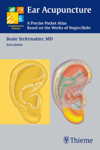
Ear Acupuncture
A Precise Pocket Atlas Based on the Works of Nogier/Bahr
Beate Strittmatter, MD Chief Instructor German Academy of Acupuncture Physician in Private Practice Saarbrücken, Germany
2nd edition
267 illustrations
Thieme Stuttgart · New York
Library of Congress Cataloging-in-Publication Data
Strittmatter, Beate, 1956-
[Taschenatlas Ohrakupunktur nach Nogier/Bahr. English]
Ear acupuncture: a precise pocket atlas based on the works of Nogier/Bahr/Beate Strittmatter; [translator, Ursula Vielkind; illustrator, Beate Strittmatter]. — 2nd ed.
p.; cm.
This book is a revised new edition based on the 4th German edition of Taschenatlas Ohrakupunktur nach Nogier/Bahr, published and copyrighted 2008 by Georg Thieme Verlag, Stuttgart, Germany.
Includes indexes.
ISBN 978-3-13-131962-3 (alk. paper)
1. Ear—Acupuncture—Atlases. I. Title.
[DNLM: 1. Acupuncture, Ear--Atlases. WB 17]
RM184.S77613 2010615.8’92—dc22
2010033135
Translator: Ursula Vielkind, PhD, Dundas, Ontario, Canada
Illustrator: Dr. Beate Strittmatter
© 2011 Georg Thieme Verlag,
Rüdigerstraβe 14, 70469 Stuttgart,
Germany
http://www.thieme.de
Thieme New York, 333 Seventh Avenue,
New York, NY 10001, USA
http://www.thieme.com
Cover design: Thieme Publishing Group
Typesetting by Primustype Hurler,
Notzingen, Germany
Printed in India by Gopsons Paper Limited,
New Delhi
ISBN 978-3-13-131962-3
1 2 3 4 5 6
Important note: Medicine is an ever-changing science undergoing continual development. Research and clinical experience are continually expanding our knowledge, in particular our knowledge of proper treatment and drug therapy. Insofar as this book mentions any dosage or application, readers may rest assured that the authors, editors, and publishers have made every effort to ensure that such references are in accordance with the state of knowledge at the time of production of the book.
Nevertheless, this does not involve, imply, or express any guarantee or responsibility on the part of the publishers in respect to any dosage instructions and forms of applications stated in the book. Every user is requested to examine carefully the manufacturers’ leaflets accompanying each drug and to check, if necessary in consultation with a physician or specialist, whether the dosage schedules mentioned therein or the contraindications stated by the manufacturers differ from the statements made in the present book. Such examination is particularly important with drugs that are either rarely used or have been newly released on the market. Every dosage schedule or every form of application used is entirely at the user’s own risk and responsibility. The authors and publishers request every user to report to the publishers any discrepancies or inaccuracies noticed. If errors in this work are found after publication, errata will be posted at www.thieme.com on the product description page.
Some of the product names, patents, and registered designs referred to in this book are in fact registered trademarks or proprietary names even though specific reference to this fact is not always made in the text. Therefore, the appearance of a name without designation as proprietary is not to be construed as a representation by the publisher that it is in the public domain.
This book, including all parts thereof, is legally protected by copyright. Any use, exploitation, or commercialization outside the narrow limits set by copyright legislation, without the publisher’s consent, is illegal and liable to prosecution. This applies in particular to photostat reproduction, copying, mimeographing, preparation of microfilms, and electronic data processing and storage.
Preface to the Second English Edition
The scientific foundations of body acupuncture have been established during the last 15 years. By contrast, ear acupuncture (auricular acupuncture, auriculotherapy) is still viewed by many as a therapeutic side issue, despite its immediate and long-lasting effect.
In a recent study representative of many scientific investigations, David Alimi at the University of Paris has demonstrated by functional MRI that auricular reflex zones are directly connected to the corresponding brain areas [1]. Needling the auricular point for the right thumb reached exactly the same brain area, in the precentral gyrus, as did direct stimulation of the right thumb (performed and measured separately prior to auricular acupuncture). The fact that Alimi was able to show by functional MRI such a precise and direct connection between an auricular reflex zone and the corresponding area in the brain is an impressive proof of the neurobiological effect of ear acupuncture. Furthermore, the fact that needling of the right ear stimulated the thumb area on the left half of the brain made it clear that the ear point representing a specific organ (or body part) must be needled on the same side of the body where the affected organ is.
Finally, ear acupuncture is becoming accepted throughout the world.
By initiating and conducting educational seminars and tutorials in the United States, Canada, and the United Arabic Emirates and by giving presentations at international congresses, I had the privilege to plant the seeds for a wider distribution of this method. The activities of the Auriculotherapy Certification Institute (ACI) of Dr. Terry Oleson in Los Angeles, USA (www.auriculotherapy.com), and also of the Canadian school of Dr. Muriel Agnes, who teaches the German version of auricular acupuncture (www.vitalprincipal.ca), have aroused the interest of many physicians, nurses, and acupuncturists. As a result, a considerable number of qualified auriculotherapists are now practicing in both the United States and Canada—much to the benefit of patients who do not get help from evidence-based conventional medicine. For several years now, interest in this special form of treatment has been steadily increasing among medical students and TCM instructors, even in China (Frank R. Bahr, MD, and coworkers).
The same picture is emerging everywhere around the world: those who get to know ear acupuncture are quickly filled with the same excitement that gripped me more than 25 years ago when I had the privilege of learning auricular acupuncture from Dr. Bahr when it was still in its infancy. Thanks to the unflagging research efforts of Dr. Bahr and coworkers, this system has since matured into a concept for specific, highly effective treatment of many diseases, particularly in patients resistant to conventional treatment.
After all, science is based on discovery, observation, and critical analysis, and thus science creates knowledge. In view of this, the scientific community can no longer ignore the fascinating discoveries in auricular acupuncture and is currently working on a model that may explain and monitor its effects.
When the first German edition of this pocket atlas was published in 2001, we did not expect it to meet with such success. By the time the fourth German edition appeared in 2007, it became obvious from the numerous positive responses coming from my colleagues that the basic concept of the book has been highly successful, both in didactical and practical terms. All other books showed maps with several reflex points on the same ear. In the medical practice, however, patients present with an “empty” ear—without any points for reference. In addition to overview maps, therefore, it seemed important to provide ear maps showing only one reflex point and accurately describe its localization. But such an approach required sufficient space, and I wish to thank the publisher once again for generously supporting such a paper-intensive presentation.
With this in mind, I wish you an interesting and informative reading. As always, I am grateful for any comments or criticism that will lead to reader-friendly improvements.
Beate Strittmatter
[1] Alimi D, Geissmann A, Gardeur D. Auricular Acupuncture Stimulation Measured on Functional Magnetic Resonance Imaging. Medical Acupuncture 2002;13(2):18–21.
Preface to the First English Edition
Acupuncture is a healing method, the value of which has been established through successful application over thousands of years. Unlike in the United States, in Europe acupuncture is widely used by physicians and is being taught in medical school and researched at universities. In Germany, Austria, and Switzerland alone, there are more than 20 000 physicians who apply a special form of acupuncture, namely, ear acupuncture (auriculotherapy).
The French physician, Paul Nogier, discovered this form of acupuncture 50 years ago and established its foundations. During the last 20 years, Frank R. Bahr, MD, and his coworkers consistently pursued the continuing development of ear acupuncture with respect to basic knowledge, application, and indication. In addition to the classical reflex points on the ear, other points (so-called Functional Points) have been found that are important in a physician’s practice: points for pain and addictions and those with psychotropic and/or druglike effects. The reflex zones of neurological structures, described only partially by Nogier, have been refined and mapped on the ear.
Another important development of practical implication is the opportunity to use ear acupuncture for identifying focal processes in the body, the diagnosis and therapy of which are often the key to success in difficult cases. Ear points indicating focal disturbances that can influence the disease process from far away (so-called Indicator Points) play a key role in ear acupuncture, thus setting it apart from other forms of acupuncture.
Ear acupuncture with its many applications is now well established as an efficient, inexpensive, and quick procedure that has very few side effects. The main indications are pain (migraine, musculoskeletal pain), allergic disorders, and all functional diseases, such as inflammation, proneness to infection, gastrointestinal disorders, and gynecological and medical disorders.
The idea for this book came up at the International Consensus Conference on Acupuncture, Auriculotherapy, and Auricular Medicine (ICCAAAM) in Las Vegas in 1999, when participants asked me for a book in English. Our host at that time was Terry Oleson, PhD, LA, chairman of the conference and also chairman of the Auriculotherapy Certification Institute (ACI). He, too, is actively involved in research and promotion of acupuncture in the United States. My work has been enriched in many ways through intense dialogue with him, also in connection with my educational activities at the ACI, and through his continuous and vivid interest in new developments and the integration of these methods into conventional medicine.
I was also encouraged by the workshops held subsequently (and now regularly) at the University of Miami at the invitation of Janet Konefal, PhD, Chief of Complementary Medicine, Department of Psychiatry and Behavioral Sciences. Dr. Konefal made it her task to promote ear acupuncture as a part of complementary medicine at university level, thus contributing to its spread all over the world.
The present Pocket Atlas of Ear Acupuncture is equally well suited as a concise textbook for beginners and as a compact reference book for more experienced practitioners. It was important for me to apply a didactic principle that makes it easy for the reader to learn and memorize: in addition to general maps of the ear that are easy to remember, each reflex zone or point is illustrated individually on an otherwise empty ear. This allows for quick and systematic orientation in daily practice.
My special thanks go to my teacher, Dr. Frank R. Bahr. Not only did I learn the method of ear acupuncture from him, but we both entered a mutually respectful collaboration in the continuous efforts to enrich ear acupuncture through new discoveries and therapeutic concepts. Dr. Bahr contributed considerably to the fact that ear acupuncture is now used throughout Europe. He helped to establish the method at universities and he now makes sure, through his persistent research activity, that this steadily growing healing method is developing further.
The translator of this book, Ursula Vielkind, PhD, Ontario, Canada, helped to improve the book through her scientific background and meticulous, discerning, and enthusiastic work. As an author, I have learned a lot during this collaboration.
My special thanks go to the publisher, especially to Liane Platt-Rohloff, PhD, for her support of this demanding project.
As so often before, I wish to thank my husband and children who supported me with endless patience and tolerance while I was working on this book, even during family vacations.
Fall 2002 Beate Strittmatter
Contents
1 Introduction
Zones of Projection
2 Projection of the Locomotor System
Auricular Innervation, Projection of Germ Layers
The Three Germ Layers: Ectoderm, Endoderm, Mesoderm
Projection of the Locomotor System
Locomotor System—Overview
Projection of the Vertebral Column/Thorax
Entire Vertebral Column
Structures of the Vertebral Column
Cervical Spine
Thoracic Spine
Lumbar Spine
C0/C1 (Atlanto-occipital Joint)
C7/T1
T12/L1
L5/S1 (Lumbosacral Transition)
Coccyx
Sacral Spine
Thorax—Overview
Ribs
Sternum
Clavicle
Trunk Muscles
Projection of the Pelvis
Pelvis—Overview
Pubic Bone
Sacroiliac Joint
Perineum
Projection of the Lower Limb
Entire Lower Limb
Hip Joint
Knee Joint
Ankle Joint
Buttock
Thigh
Lower Leg
Foot
Heel
Achilles Tendon
Toes
Projection of the Upper Limb
Upper Limb—Overview
Large Joints—Overview
Shoulder Joint
Acromioclavicular Joint
Elbow Joint
Wrist
Upper Arm
Forearm
Finger
Metacarpophalangeal Joint of Thumb
Carpometacarpal Joint of Thumb
3 Projection of the Internal Organs
Internal Organs—Overview
Projection of the Cardiovascular System
Heart
Blood Vessels
Lymph Vessels
Projection of the Respiratory System
Respiratory System—Overview
Lung
Bronchi
Trachea
Throat (Pharynx)
Projection of the Gastrointestinal System
Gastrointestinal System—Overview
Esophagus
Stomach
Duodenum
Jejunum/Ileum (Small Intestine)
Colon (Large Intestine)
Appendix
Rectum
Anus
Liver
Gall Bladder
Pancreas
Projection of the Immune System
Spleen
Thymus Gland
Tonsils
Lymph Nodes
Projection of the Urogenital System
Urogenital System—Overview
Kidney
Ureter
Urinary Bladder
Urethra
Uterus
Ovary/Testis
Vagina
Prostate/Fallopian Tube
Penis/Clitoris
Projection of the Endocrine System
Endocrine System—Overview
Thyroid Gland
Parathyroid Gland
Thymus Gland
Pancreas
Adrenal Gland (Cortisone Point)
4 Projection of the Head
Head—Overview
Bony Skull
Projection of the Temporomandibular Joint
Temporomandibular Joint
Projection of the Jaws and Teeth
Upper and Lower Jaws, Teeth
Projection of the Lips and Tongue
Lips
Oral Cavity, Tongue
Projection of the Tonsils and Throat
Tonsils
Throat (Pharynx)
Projection of the Eyes and Nose
Eye
Nose
Projection of the Paranasal Sinuses and Ear
Paranasal Sinuses
Ear
Projection of the Facial Muscles and Parotid Gland
Facial Muscles
Parotid Gland
Projection of the Occipital Trigger Points
Occipital Trigger Points
5 Projection of the Nervous System
Projection of the Central Nervous System
Central Nervous System—Overview
Cerebral Lobes—Overview
Cortex
Frontal Lobe
Parietal Lobe
Temporal Lobe
Auditory Line
Occipital Lobe
Corpus Callosum
Thalamus
Hypothalamus
Pituitary Gland
Pituitary Gland Points
Limbic System
Cerebellum
Brain Stem
Vomiting Point
Reticular Formation
Spinal Cord
Projection of the Cranial Nerves
Cranial Nerves—Overview
Olfactory Nerve (CN I)
Optic Nerve (CN II)
Oculomotor Nerve (CN III)
Trochlear Nerve (CN IV)
Trigeminal Nerve (CN V)
Abducens Nerve (CN VI)
Facial Nerve (CN VII)
Vestibulocochlear Nerve (CN VIII)
Glossopharyngeal Nerve (CN IX)
Vagus Nerve (CN X)
Accessory Nerve (CN XI)
Hypoglossal Nerve (CN XII)
Projection of the Autonomic Nervous System
Autonomic Nervous System—Overview
Sympathetic Nervous System—Overview
Parasympathetic Nervous System—Overview
Sympathetic Nuclei of Origin (Preganglionic Sympathetic Nerves)
Sympathetic Trunk (Paravertebral Chain of Sympathetic Ganglia, Postganglionic Sympathetic Nerves)
Superior Cervical Ganglion
Middle Cervical Ganglion
Inferior Cervical Ganglion (Stellate Ganglion)
Nervous Organ Points
Parasympathetic Nuclei of Origin
Prevertebral Ganglions—Overview
Bronchopulmonary Plexus
Cardiac Plexus
Celiac Plexus (Solar Plexus)
Superior Mesenteric Plexus
Inferior Mesenteric Plexus
Hypogastric Plexus
Peripheral Nervous System
6 Functional Points
Functional Points—Overview
Hormone and Metabolite Points
Hormone and Metabolic Points—Overview
Estrogen Point
Progesterone Point
Gonadotropin Point
ACTH Point
TSH Point
Prolactin Point
Endocrine Parathyroid Gland Point
Thyroid Gland Point
Thymus Gland Point
Endocrine Pancreas Point (Insulin Point)
Adrenal Gland Point (Cortisone Point)
Histamine Point (Allergy Point 1)
Prostaglandin E1 Point (PGE1 Point)
Prostaglandin E2 Point (PGE2 Point)
Interferon Point
Renin/Angiotensin Point
Beta-1-Receptor Point (Beta-Blocker Point According to Bahr)
Beta-2-Receptor Point (Beta-Mimetic Point According to Bahr)
Medication Analogue Points
Diazepam Analogue Point (Valium Point According to Bahr)
Barbiturate Analogue Point
Caffeine Analogue Point
Nervous Organ Points
Nervous Organ Points—Overview
Anger Point Syn. Nervous Liver Point
Psychotropic Points
Psychotropic Points—Overview
Master Omega Point (Bromazepam Analogue Point)
Omega Point I
Omega Point II
Depression Point
Anxiety Point
Worry Point
Anger Point
Nicotine Analogue Point According to Bahr
Aggression Point
Bridging Point According to Bahr
Frustration Point
Psychotherapy Point According to Bourdiol
Analgesic Points
Thalamus Point
Prostaglandin E1 Point (PGE1 Point)
Laterality Point According to Bahr
Shen Men Point
Pain Memory Points According to Bahr
Cardinal Points
Cardinal Points—Overview
Prostaglandin E1 Point (PGE1 Point)—Corresponds to Cardinal Point GB-41 of Body Acupuncture
Thymus Gland Point—Corresponds to Cardinal Point TB-5 of Body Acupuncture
Lung Zone—Corresponds to Cardinal Point LU-7 of Body Acupuncture
Diazepam Analogue Point (Valium Point According to Bahr)—Corresponds to Cardinal Point KI-6 of Body Acupuncture
Stellate Ganglion Point—Corresponds to Cardinal Point PC-6 of Body Acupuncture
Interferon Point—Corresponds to Cardinal Point SP-4 of Body Acupuncture
Retro-Celiac Plexus Point (Retro-Solar Plexus Point, Retro-Point Zero)—Corresponds to Cardinal Point SI-3 of Body Acupuncture
Pineal Gland Point—Corresponds to Cardinal Point BL-62 of Body Acupuncture
Energy Points
Master Point of Oscillation According to Bahr
Master Point of Regulation According to Bahr
Super Omega Point According to Bahr
Master Point of Qi Flow According to Bahr
Point Zero
Point GV-4
Focus Indicator Points
Focus Indicator Points According to Bahr—Overview
Histamine Point (Allergy Point 1)
Cyclophosphamide Analogue Point (Endoxan Point According to Bahr, Allergy Point 2)
Prostaglandin E1 Point (PGE1 Point)
Vitamin C Point
Laterality Point According to Bahr
Tissue Layer Control Points
Tissue Layer Control Points—Overview
Indicator Points for Vitamins and Trace Elements
Indicator Points for Vitamins According to Bahr—Overview
Indicator Points for Trace Elements According to Bahr—Overview
Important Silver Points
Important Silver Points on the Dominant Ear
Zone-Dominating Points
Zone-Dominating Points—Overview
Points Dominating Zone A (ZA Points)
Points Dominating Zone B (ZB Points)
Points Dominating Zone C (ZC Points)
Points Dominating Zone D (ZD Points)
Points Dominating Zone E (ZE Points)
Points Dominating Zone F (ZF Points)
Points Dominating Zone G (ZG Points)
7 Auxiliary Lines of Ear Acupuncture
Barbiturate Point / Caffeine Point
Barbiturate Analogue Point, Caffeine Analogue Point
Auxiliary Line through Omega Points
Omega Points
Aggression Point
Auxiliary Lines through Point Zero
Hormone Points
Cortisone Point, ACTH Point
Thymus Gland Point, Interferon Point, Laterality Point
Spleen Point, Diazepam Analogue Point
Depression Point, Tonsil Point, TMJ Point
Trigeminal Nerve
First Rib Point
First Rib Point, Stellate Ganglion Point
Beta-1-Receptor Point
Endocrine Thyroid Gland Point
Auxiliary Lines near the Tragus
Diazepam Analogue Point
Phosphate Analogue Point
Nicotine Analogue Point, Pineal Gland Point
Frustration Point
Interferon Point
Nose Point
Aggression Point
ACTH Point
Points of the Pituitary Gland Zone
Gonadotropin Point
Auxiliary Lines for Points of the Limbs
Points of the Hip Joint, Knee Joint, Ankle Joint
Wrist Point, Knee Joint Point
Points of the Shoulder Joint, Elbow Joint, Wrist
Kidney Point
Auxiliary Lines for Focus Indicator Points
Laterality Point
Cyclophosphamide Analogue Point (Allergy Point 2)
Histamine Point (Allergy Point 1), Vitamin C Point
8 Meridians on the Ear
LU/LI
Lung Meridian
Large Intestine Meridian
ST/SP
Stomach Meridian
Spleen Meridian
HT/SI
Heart Meridian
Small Intestine Meridian
BL/KI
Bladder Meridian
Kidney Meridian
PC/TB
Pericardium Meridian
Triple Burner Meridian
GB/LR
Gall Bladder Meridian
Liver Meridian
CV/GV
Conception Vessel (Ren Mai)
Governor Vessel (Du Mai)
9 Indications
Indications—Overview
Disorders of the Locomotor System
Headaches and Facial Pain
Allergic Disorders
Respiratory Disorders
Gastrointestinal Disorders
Inflammatory and Functional Intestinal Disorders
Cardiovascular Diseases
Disorders of the Urogenital System
Hormonal Disorders
Autonomic Symptoms
Mental Health Disorders
Addictions
Patient Information Leaflet: Permanent Needles
Appendix
Ear Topography
Projection Zones, Points, and Meridians on the Ear
Indications
Explanation of Symbols
Silver needle
Gold needle
Permanent needle
Hidden point locations
Note: If steel needles are used instead of gold and silver needles, always needle the Gold Point because this is the point that needs to be stimulated. For example, in case of the Diazepam Analogue Point, the Gold Point would be on the left ear in a right-handed person but on the right ear in a left-handed person.
Exemption: Anxiety Point, Worry Point, Aggression Point, Nicotine Analogue Point, Nervous Liver Point—here use Silver Point for steel needles (see also p. 271).
1 Introduction
2 Projection of the Locomotor System
3 Projection of the Internal Organs
4 Projection of the Head
5 Projection of the Nervous System
6 Functional Points
7 Auxiliary Lines of Ear Acupuncture
8 Meridians on the Ear
9 Indications
Appendix
Introduction
Ear acupuncture is an effective method for treating acute and chronic diseases without producing side effects. It therefore represents one of the most important complementary additions to conventional medicine today. Its main indication is certainly the treatment of pain, but a number of functional, organic, and mental disorders may be treated as well. Fortunately, this applies also to a number of diseases for which conventional medicine still has no cures to offer, for example, migraine and hay fever.
The origins of ear acupuncture can be traced back to the 4th century BC when Hippocrates tried to cure impotence by bloodletting at the ear. It is also known that pain has been treated in ancient Egypt by means of ear points. Throughout the centuries we find notes on similar treatments. The best-known document in the European region is certainly the painting “The Garden of Lust” by Hieronymus Bosch (17th century) which shows the Sciatica Point being pierced by a needle. A second needle held by the Satan pricks the two points known as External Genitals Point and Libido Point.
Ear acupuncture enjoyed a certain popularity also in China, but it fell into oblivion during the last centuries. Approximately 20 anterior and posterior ear points were known during the Tang dynasty (618–907 AD). The procedure then probably spread to Persia, Africa, India, and the Mediterranean area.
However, there is no evidence that a comprehensive reflex system on the auricle, complete with representations of the entire body, existed. We owe it to the French physician Paul Nogier that ear acupuncture has been rediscovered and that this happened in a way that provided astonishing opportunities for both diagnosis and therapy.
Nogier has been able to demonstrate that all organs of the body are represented as reflex zones on the outer ear (auricula or auricle).
The history of this rediscovery is as exciting as the entire therapeutic method itself. Around 1950, Nogier discovered in some of his patients certain scars at a specific site on the ear. The patients told him that they had been “treated” for back pain by applying a red-hot needle to this site of the ear, with the result that the pain disappeared. We owe it to Nogier’s unreserved objectivity that he followed up on this phenomenon and studied it. He was able to reproduce the phenomenon successfully and interpreted the site on the ear as reflex localization L5/S1.
Shortly thereafter he realized that, apart from this body area, all other sites or organs of the body must be projected on the ear. He also discovered how to demonstrate these sites: they showed an increased sensitivity to pressure if the corresponding region of the body was diseased. Subsequently, he recognized that active ear acupuncture points—those reflex points that indicate a pathological change in the body or are produced on the ear by such a pathology—have changed electrically (reduced resistance and increased conductance of the skin). This allows for objective measurements that are independent of the patient (electrical point finders with a double-ring electrode).
Step by step, Nogier established the auricular map used today. If the patient had a pathology (inflammation, pain, etc.), he only had to search for the corresponding electrically altered point on the ear. For example, to locate the reflex sites of large joints or skin areas, he would simply apply a pain stimulus in the respective body region; an active acupuncture point would immediately be created at the corresponding site on the ear and, hence, could be detected because of its sensitivity to pressure or its change in conductivity. (Even today, this is still an excellent method for practicing on the ear or identifying a point that corresponds to a distinct structure of the body.)
There are two basic lines of thought in ear acupuncture, the French school and the Chinese school. They differ in the specifications of point localizations and in their diagnostic and therapeutic approaches.
The present atlas describes the French school, the intellectual father and pioneer of which was Paul Nogier. His famous students include, among others, the French psychiatrist Bourdiol and the German physician Frank Bahr. The latter founded his own school in Germany and refined essential aspects of ear acupuncture (e.g., obstacles to therapy, focus diagnosis in patients resistant to therapy, projection of body meridians onto the ear, as well as various test procedures for medications, trace elements, vitamins, and homeopathic substances). The approach of the French school is rather pragmatic and corresponds to the pathophysiological knowledge of conventional medicine. The point localizations have distinct, anatomically oriented assignments to pathologies of the body. In addition, the unambiguous relationship of the ear points to the corresponding parts of the body can be clearly demonstrated by electrical changes (caused either by existing pathologies or by iatrogenic pain stimuli on the body, as described earlier).
On the one hand, these active points clearly indicate disorders of the body; on the other hand, they are also—via needle, laser, pressure massage—the therapeutic gateways to numerous diseases. This way, therapy can be applied in a controlled manner; that is, treatment is or should be preceded by the diagnosis of active points in order to provide information on the origin and type of the disease in question.
The present atlas describes the classic ear points, which are well-known and anatomically well-defined, as well as so-called Functional Points that influence the entire system (Nogier/Bahr). The book is not intended to replace hands-on courses and bedside teaching; it rather serves to deepen the knowledge and to provide a quick reference for the practice. For more information on ear acupuncture techniques in diagnosis and treatment, see B. Strittmatter, Overcoming Blockages to Healing—A Manual for Health Care Professionals, Thieme Publishers, Stuttgart—New York, 2004.
In a right-handed person, the right ear is the dominant one, and the left ear is non-dominant. In a left-handed person, the left ear is dominant, and the right ear is non-dominant. Our readers are already familiar with the topographic anatomy of the external ear (auricle). For quick reference, the auricular topography is nevertheless included at the end of the book: pp. 412/413. In this book, the most important points of each ear meridian are described together with their multiple functions. They carry the same names as the corresponding body acupuncture points. Points ending with “-1” are located close to the original point (e.g. TB-1-1 lies close to TB-1).Zones of Projection
The reflex zones and points on the auricle are arranged in such a way that they produce the inverted projection of an embryo. This image serves as a helpful memory tool when learning the points assigned to specific organs of the body.
The Inverted Embryo
The head of the embryo occupies the entire earlobe (lobule). This area is indeed the projection zone of the most important parts of the head (sensory organs, brain, temporomandibular joint, teeth, maxillary sinus, facial muscles). The embryo lies with its back (spinal cord) against the posterior margin of the auricle (descending helix)—here are the reflex zones of the sensory and motor portions of the spinal cord. The embryo covers the concha with its abdomen—here are the reflex zones of almost all intestinal organs, again in the upside-down arrangement. The embryo crosses its legs over the triangular fossa—this is the reflex zone of the lower limb.
As a rule, the sensory portions are found on the front of the ear (lateral surface of auricle), while the motor portions are found on the back of the ear (medial surface of auricle).
Ear Terraces
When memorizing the different groups of points, it is of great help to inspect or palpate the different planes of the auricle (ear terraces):
• The lower terrace (concha) contains the reflex zones of (almost) all internal organs.
• The terrace above (scapha, antihelix, triangular fossa) is occupied by the reflex zones of the entire locomotor system.
• The uppermost terrace, which is formed by the anterior, superior, and posterior margins of the auricle (helix), contains the reflex zones of the spinal cord.
Fig. 1.1 Zones of projection: the inverted embryo (homunculus)
The Three Germ Layers: Ectoderm, Endoderm, Mesoderm
The auricle is innervated by three nerves:
• The great auricular nerve (originating from the cervical plexus). It supplies the descending helix and the lobule. Here are the reflex zones of organs derived from the ectoderm (skin, nervous system).
• The auriculotemporal nerve (a branch of the trigeminal nerve). It supplies the ascending helix and superior helix as far as Darwin’s tubercle, the triangular fossa, the scapha, the antitragus, and the antihelix including the antihelical wall. Organs derived from the mesoderm are projected in this region of the ear (muscles, bones, ligaments, heart, kidney).
•The auricular branch of the vagus nerve. It supplies the concha—the projection zone of organs derived from the endoderm (internal organs, with the exception of kidney and heart). This zone extends to the projection zone of the sympathetic trunk where the concha merges into the antihelical wall.
This pattern of auricular innervation is easy to memorize and will be of great help when learning the reflex zones of the ear. Each of the three areas of innervation corresponds to one of the three germ layers (ectoderm, mesoderm, and endoderm) and is thus assigned to a clearly defined group of organs.
Remarkably, the representation of organs on the auricle follows this pattern in almost all cases—for example, the Kidney Point does not lie in the zone of the endoderm like the reflex points of other internal organs; it is found beneath the ascending helix in the zone of the mesoderm from which the kidney is derived.
Fig. 2.1 The three germ layers 1 Ectoderm 2 Endoderm 3 Mesoderm
Locomotor System—Overview
The locomotor system projects onto the upper terrace of the ear (scapha, triangular fossa). In principle, the sensory points lie on the front of the ear (lateral surface), while the motor points are on the back of the ear (medial surface). The forceps method according to Bahr is based on this arrangement:
A gold needle in the sensory point on the lateral surface, and a silver needle in the corresponding motor point on the medial surface.
Body parts, organs, or anatomical structures belonging to the same region project very close to each other onto the ear. For example: knee joint, popliteal artery, peroneal nerve.
Fig. 2.2 Projection of the locomotor system
Entire Vertebral Column
Location:
• The vertebral column, or spine, projects upside-down onto the entire antihelix.
• Start: Postantitragal fossa (Atlanto-occipital Joint Point, Point C0/C1).
• End:


