96,99 €
Mehr erfahren.
- Herausgeber: John Wiley & Sons
- Kategorie: Fachliteratur
- Sprache: Englisch
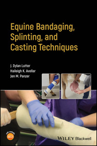
Equine Bandaging, Splinting, and Casting Techniques A practical reference manual dealing with a vital component of clinical practice in equine medicine The application of bandages, splints, and casts is an essential part of equine surgical and veterinary care. Traditionally, however, there have been few available resources wholly dedicated to application techniques. The result is that equine veterinary practitioners learn to bandage, splint, and cast on the job, with highly variable results; some practitioners are unwilling even to attempt a cast, spurning this valuable healing tool entirely rather than attempt it from an uncertain base of knowledge. Equine Bandaging, Splinting, and Casting Techniques offers the first comprehensive reference to this specific set of techniques and their applications in equine veterinary medicine. It promises to cultivate a rigorous, clinically tested, consistent standard of care that will improve patient outcomes and long-term owner costs. It is a must-own for any veterinary practitioner who works with equine patients. Equine Bandaging, Splinting, and Casting Techniques readers will also find: * Step-by-step organization guides reader smoothly through each process * Practical tips and advice for improving quality and appearance of bandages and casts * Over 270 color images, including clinical examples and radiographs, supplementing every listed technique Equine Bandaging, Splinting, and Casting Techniques is ideal for all veterinary practitioners, technicians, and students interested in equine care.
Sie lesen das E-Book in den Legimi-Apps auf:
Seitenzahl: 233
Veröffentlichungsjahr: 2024
Ähnliche


Table of Contents
Cover
Table of Contents
Title Page
Copyright Page
Preface
About the Companion Website
Section I: Application of Equine Bandages
1 Materials and Concepts for Bandage Application
Bandage Layers
Application of Bandage Materials
Sweat Bandages
Special Considerations for Bandage Application
Removing a Bandage
Bandage Management and Complications
Suggested Reading
2 Distal Limb Bandages
Hoof Bandages
Half Limb Bandages
3 Full Limb Bandages
Front Limb
Rear Limb
4 Carpal Bandages
5 Tarsal Bandages
6 Head and Neck Bandages
Ear Bandage
Head Pressure Bandage
Head Netting Bandage
7 Upper Limb and Body Bandages
Elastic Tape/Ether Bandage
Tie‐Over Bandage
Abdominal Wrap
Section II: Application of Equine Limb Splints
8 Materials and Concepts for Splint Application
Principles of Equine Limb Immobilization
Bandages for Splint Application
Splint Materials and Fabrication
Fracture First Aid
Fracture Regions
References
Suggested Reading
9 Distal Limb Splinting
Region I PVC Splinting – Phalanges and Distal Fetlock
10 Full Limb Splinting
Region II PVC Splinting – Metacarpus/Metatarsus and Carpus/Tarsus
Region III PVC Splinting – Radius, Ulna, and Tibia
Section III: Application of Equine Casts
11 Materials and Concepts for Cast Application
Casting Team
Patient Preparation for Cast Application
Limb Positioning for Cast Application
Cast Materials and Method of Application
Cast Management and Complications
Cast Removal
Suggested Reading
12 Hoof and Phalangeal Casts
Hoof Cast
Phalangeal Cast
13 Bandage Casts
14 Half Limb Casts
Anesthetized Half Limb Cast Application
Standing Half Limb Cast Application
Standing Cast Removal
15 Full Limb Casts
Index
End User License Agreement
List of Tables
Chapter 1
Table 1.1 Commonly used wound dressings and their classification.
Table 1.2 Commonly used materials for the secondary layer bandage.
Table 1.3 Commonly used materials for the tertiary layer bandage.
Table 1.4 Problems that may occur causing excessive damage, soiling, or moi...
Chapter 2
Table 2.1 Supply list for foot bandage.
Table 2.2 Supply list for half limb bandage.
Chapter 3
Table 3.1 Supply list for half limb bandage.
Chapter 4
Table 4.1 Supplies needed for a carpal bandage.
Chapter 5
Table 5.1 Supplies list for tarsal bandage.
Chapter 6
Table 6.1 Supplies list for head/neck bandage.
Chapter 7
Table 7.1 Materials needed for placement of a tie‐over bandage.
Table 7.2 Materials need for placement of an elastikon abdominal wrap banda...
Chapter 8
Table 8.1 Common splint lengths and their corresponding region for use in a...
Table 8.2 Emergency first aid medications and supplies.
Table 8.3 Supplies needed for a well‐stocked equine fracture kit. The quant...
Table 8.4 Fracture regions of the equine limb with corresponding structures...
Chapter 12
Table 12.1 Supply list for a phalangeal cast.
Chapter 13
Table 13.1 Materials list for a half limb bandage cast application.
Chapter 14
Table 14.1 Supply list for a half limb cast.
Chapter 15
Table 15.1 Supply list for a full limb cast.
List of Illustrations
Chapter 1
Figure 1.1 A telfa pad is a wound dressing commonly used as a primary layer ...
Figure 1.2 Any soft, conformable, and compressible material can be used as a...
Figure 1.3 The tertiary layer of a bandage may be composed of either one or ...
Figure 1.4 (a) Though it is tempting to allow this for minor wounds, cohesiv...
Figure 1.5 (a–c) If added security of the primary bandage layer is desired t...
Figure 1.6 The tertiary bandage layer must be applied with appropriate tensi...
Figure 1.7 (a, b) Each bandage layer should be wrapped in the same direction...
Figure 1.8 Despite there being no medical reason for it, the industry‐accept...
Figure 1.9 (a) When applying bandage material to the leg, the roll of materi...
Figure 1.10 This cohesive bandage material is being applied in the most acce...
Figure 1.11 (a) The tertiary layer of bandage material (brown gauze or vet w...
Figure 1.12 (a) Note the multiple unsafe conditions in this image. The handl...
Figure 1.13 Sweat bandage application. (a) The sweat or poultice has been sm...
Figure 1.14 Bandage‐dependent swelling. (a) A patient 14 days post surgery w...
Figure 1.15 Bandage removal with a scalpel blade. (a) How to safely hold a s...
Figure 1.16 This patient was bandaged with only cohesive bandage material di...
Figure 1.17 This patient presented for sloughing of the skin on the dorsal a...
Figure 1.18 (a) Skin pressure necrosis over the accessory carpal bone in a s...
Chapter 2
Figure 2.1 (a) Materials for a hoof bandage. (b) Materials for a foot soakin...
Figure 2.2 This soaking bandage is useful for soaking horses with hoof absce...
Figure 2.3 The duct tape pad is made by overlapping eight to nine strips in ...
Figure 2.4 A foot bandage may be applied directly over the soaking foot, sep...
Figure 2.5 Materials for a half limb bandage.
Figure 2.6 Positioning considerations for the cotton combine rolls in a half...
Figure 2.7 Application of a half limb bandage to a front limb. This illustra...
Figure 2.8 The appearance of a finished half limb bandage placed on a hind l...
Chapter 3
Figure 3.1 Materials needed for a full limb bandage are similar to those for...
Figure 3.2 Application of the full limb bandage to the front limb. (a) The f...
Figure 3.3 Application of the full limb bandage to the hind limb. (a) Positi...
Chapter 4
Figure 4.1 Materials for a carpal bandage.
Figure 4.2 Application of an equine carpal bandage. (a) After the primary co...
Chapter 5
Figure 5.1 Materials for placement of a tarsal bandage.
Figure 5.2 Application of an equine tarsal bandage. (a) A primary contact la...
Chapter 6
Figure 6.1 (a–c) Materials for placing a bandage on the head or neck.
Figure 6.2 An example of an ear bandage that may be used to help maintain th...
Figure 6.3 Application of a head pressure bandage. (a) A dressing is applied...
Figure 6.4 An example of a patient where a netting bandage would be useful. ...
Figure 6.5 The finished (a) front and (b) side appearance of a patient that ...
Chapter 7
Figure 7.1 Application of a simple elastic tape/ether bandage. (a) The banda...
Figure 7.2 Application of a tie‐over bandage on a large gluteal wound. (a) A...
Figure 7.3 Application of an elastic tape abdominal bandage. (a) Oftentimes ...
Chapter 8
Figure 8.1 Process for custom molding a splint from fiberglass cast material...
Figure 8.2 A carpal bandage cast has been placed on this patient to prevent ...
Figure 8.3 (a) The limb is bandaged with a well‐placed full limb bandage tha...
Figure 8.4 A full Robert Jones bandage has been applied to this horse's limb...
Figure 8.5 Divisions of the equine limb for splint application in the field ...
Chapter 9
Figure 9.1 Application and proper placement of dorsal and lateral splints in...
Figure 9.2 Application and proper placement of dorsal and lateral splints in...
Figure 9.3 Application of a distal limb Kimzey leg saver splint in the forel...
Chapter 10
Figure 10.1 Application and proper placement of a caudal and lateral splint ...
Figure 10.2 Application and proper placement of a caudal and lateral splint ...
Figure 10.3 Application of caudal and lateral splints for region III of the ...
Figure 10.4 Application of caudal and lateral splints for region III of the ...
Chapter 11
Figure 11.1 A tube cast has been applied to this patient in the form of a ba...
Figure 11.2 (a) The patient sustained a severely comminuted fracture of the ...
Figure 11.3 Examples of positioning a horse's limb for cast application. (a)...
Figure 11.4 (a) The length of stockinette is rolled to apply a double layer ...
Figure 11.5 Delta‐Dry (left) is a water‐resistant knit cast padding commonly...
Figure 11.6 (a) This section of cast material was not fully bonded together ...
Figure 11.7 Appearance and location of common cast sores. (a) This patient i...
Figure 11.8 Examples of cast windowing. (a) Here a window has been removed f...
Figure 11.9 Use of an oscillating cast saw for cast removal. (a) A full rear...
Figure 11.10 A half limb cast being removed in a standing patient and equipm...
Chapter 12
Figure 12.1 (a) A hoof wall resection has been performed on this hoof to rem...
Figure 12.2 (a) This horse has lacerated its lateral hoof capsule and corona...
Figure 12.3 Materials to have on hand for a phalangeal cast.
Figure 12.4 A common configuration of a heel bulb laceration that responds v...
Figure 12.5 Application of a phalangeal cast in a standing patient. The pati...
Figure 12.6 (a) To begin the casting procedure for the horse in Figure 12.5,...
Figure 12.8 (a) Once the cast padding has been placed for application of a p...
Figure 12.9 (a) Once the entire 5 in. roll of cast tape has been applied to ...
Chapter 13
Figure 13.1 Examples of wounds that would benefit from management with a ban...
Figure 13.2 Process for application of a half limb bandage cast. (a) To begi...
Figure 13.3 Process for re‐application of a carpal tube bandage cast. (a) He...
Chapter 14
Figure 14.1 Materials to have on hand for a half limb cast.
Figure 14.2 Application of a half limb cast in an anesthetized horse. (a) O...
Figure 14.3 (a) A different perspective of hind limb positioning on another...
Figure 14.4 Standing half limb cast application procedure. (a) This patient...
Figure 14.5 Standing cast removal of a half limb cast using a cast saw. Thi...
Chapter 15
Figure 15.1 Materials needed for a full limb cast.
Figure 15.2 Application of a full limb cast in the hind limb of an anestheti...
Figure 15.3 (a) Continuing the application of a full limb cast from Figure 1...
Guide
Cover Page
Title Page
Copyright Page
Preface
About the Companion Website
Table of Contents
Begin Reading
Index
WILEY END USER LICENSE AGREEMENT
Pages
iii
iv
ix
xi
1
3
4
5
6
7
8
9
10
11
12
13
14
15
16
17
18
19
20
21
22
23
24
25
26
27
28
29
30
31
32
33
34
35
36
37
38
39
40
41
42
43
44
45
46
47
48
49
50
51
52
53
54
55
56
57
58
59
60
61
62
63
64
65
66
67
68
69
71
72
73
74
75
76
77
78
79
80
81
82
83
84
85
86
87
88
89
90
91
92
93
94
95
97
99
100
101
102
103
104
105
106
107
108
109
110
111
112
113
114
115
116
117
118
119
120
121
122
123
125
126
127
128
129
130
131
132
133
134
135
136
137
139
141
142
143
144
145
146
147
148
149
150
151
152
153
154
155
156
157
159
160
161
162
163
164
165
166
167
168
169
170
171
172
173
174
175
176
177
179
180
181
182
183
184
185
186
187
188
189
190
191
192
193
194
195
196
197
198
199
200
201
202
203
205
206
207
208
209
210
211
212
Equine Bandaging, Splinting, and Casting Techniques
J Dylan Lutter, Haileigh Avellar, and Jen Panzer
Equine SurgeryDepartment of Clinical SciencesVeterinary Health CenterCollege of Veterinary Medicine Kansas State UniversityManhattan, KS, US
Copyright © 2024 by John Wiley & Sons, Inc. All rights reserved.
Published by John Wiley & Sons, Inc., Hoboken, New Jersey.Published simultaneously in Canada.
No part of this publication may be reproduced, stored in a retrieval system, or transmitted in any form or by any means, electronic, mechanical, photocopying, recording, scanning, or otherwise, except as permitted under Section 107 or 108 of the 1976 United States Copyright Act, without either the prior written permission of the Publisher, or authorization through payment of the appropriate per‐copy fee to the Copyright Clearance Center, Inc., 222 Rosewood Drive, Danvers, MA 01923, (978) 750‐8400, fax (978) 750‐4470, or on the web at www.copyright.com. Requests to the Publisher for permission should be addressed to the Permissions Department, John Wiley & Sons, Inc., 111 River Street, Hoboken, NJ 07030, (201) 748‐6011, fax (201) 748‐6008, or online at http://www.wiley.com/go/permission.
Trademarks: Wiley and the Wiley logo are trademarks or registered trademarks of John Wiley & Sons, Inc. and/or its affiliates in the United States and other countries and may not be used without written permission. All other trademarks are the property of their respective owners. John Wiley & Sons, Inc. is not associated with any product or vendor mentioned in this book.
Limit of Liability/Disclaimer of Warranty: While the publisher and author have used their best efforts in preparing this book, they make no representations or warranties with respect to the accuracy or completeness of the contents of this book and specifically disclaim any implied warranties of merchantability or fitness for a particular purpose. No warranty may be created or extended by sales representatives or written sales materials. The advice and strategies contained herein may not be suitable for your situation. You should consult with a professional where appropriate. Further, readers should be aware that websites listed in this work may have changed or disappeared between when this work was written and when it is read. Neither the publisher nor authors shall be liable for any loss of profit or any other commercial damages, including but not limited to special, incidental, consequential, or other damages.
For general information on our other products and services or for technical support, please contact our Customer Care Department within the United States at (800) 762‐2974, outside the United States at (317) 572‐3993 or fax (317) 572‐4002.
Wiley also publishes its books in a variety of electronic formats. Some content that appears in print may not be available in electronic formats. For more information about Wiley products, visit our web site at www.wiley.com.
Library of Congress Cataloging‐in‐Publication DataNames: Lutter, J Dylan, 1982– author. | Avellar, Haileigh, author. | Panzer, Jen, author.Title: Equine bandaging, splinting, and casting techniques / J Dylan Lutter, Haileigh Avellar, Jen Panzer.Description: Hoboken, New Jersey : Wiley Blackwell, [2024] | Includes index.Identifiers: LCCN 2024000512 (print) | LCCN 2024000513 (ebook) | ISBN 9781119841838 (paperback) | ISBN 9781119841852 (adobe pdf) | ISBN 9781119841845 (epub)Subjects: MESH: Horse Diseases–surgery | Fracture Fixation–veterinary | Extremities–injuries | Bandages–veterinary | Splints–veterinary | Casts, Surgical–veterinaryClassification: LCC SF959.F78 (print) | LCC SF959.F78 (ebook) | NLM SF 959.F78 | DDC 636.1/089705–dc23/eng/20240202LC record available at https://lccn.loc.gov/2024000512LC ebook record available at https://lccn.loc.gov/2024000513
Cover Design: WileyCover Image: Courtesy of J Dylan Lutter
Preface
I (JDL) first became interested in equine bandaging during my residency with the small project I published that qualified me to take the American College of Veterinary Surgeons' board exam. That project showed me how few resources there are in veterinary medicine about bandaging and more specifically the splinting of equine fractures. The majority of the articles written are review articles in which the authors state their personal experiences and reference the experiences of previous authors who have written other review articles or textbook chapters. At the time, there were no objective studies evaluating the effectiveness of the most commonly recommended fracture splinting techniques and very few studies investigating the effects of various bandaging techniques. There were also no available books dedicated to teaching the methods of bandaging, splinting, and casting equine limbs. Nearly all of the techniques used in these areas are passed down from person to person, each putting their own preferences and nuances onto those methods.
The purpose of this book is to address some of these deficiencies by providing step‐by‐step images with thorough descriptions of each step. It is not intended to be a reference text. Instead, it is written as a practical handbook that presents the authors' approach to equine bandaging, splinting, and casting.
About the Companion Website
This book is accompanied by a companion website.
www.wiley.com/go/lutter/1e
This website includes:
Figure PPTs
Section IApplication of Equine Bandages
1Materials and Concepts for Bandage Application
Bandages are frequently applied in equine veterinary medicine and are useful for a variety of purposes. Most commonly, bandages are applied to protect and manage the environment of a wound or surgical site while healing commences. At other times they are applied to help control/prevent edema, to provide compression and physical support to injured tissues, or to reduce the motion of a limb or region. A thorough discussion of the indications for and benefits of a bandage are well discussed elsewhere (see Suggested Reading at the end of this chapter) and are beyond the scope of this book. Suffice it to say that a properly applied bandage is generally beneficial to wound healing and greatly aids in the management of numerous equine disorders, but an inappropriately applied bandage may do far more harm than the issue for which that bandage was applied.
Bandage Layers
A bandage is composed of primary, secondary, and tertiary layers. Each of these layers serves a specific purpose and may be composed of a single kind or multiple types of material. In some instances a single material may serve the purpose of more than one layer. It is up to each clinician to evaluate the patient and the reasons for bandage application in order to determine which materials to use. In most cases a standard list of materials will be used, but in certain circumstances a material may be omitted, substituted, or added for a specific purpose. Additionally, the “standard bandage” will vary by where the veterinary practice is located, by practice type, client expectations, clinician preferences, and even by tradition. The bandage techniques shown in this book will represent one version of the “standard bandage” that can be broadly adopted regardless of these variables.
Primary Layer
The primary layer of a bandage is otherwise known as the contact layer or the wound dressing. The material applied in this layer directly interacts with the skin or wound below it. It may be used to deliver a medication, absorb exudate, debride tissue, or provide a sealed barrier. The most common material used as a primary layer in equine bandages is a telfa pad, which is classified as a non‐adherent, non‐occlusive, mildly absorptive wound dressing (Figure 1.1). Table 1.1 lists some commonly used dressings and their classifications. The effect of these dressings is more appropriate in a discussion of wound management and will not be further covered here.
Secondary Layer
The secondary layer is typically composed of a soft, conformable, compressible, and absorptive material (Figure 1.2). In wound management, this layer acts to absorb moisture (sweat) and wound exudate and creates a continuous barrier between the underlying tissue and the external environment while also holding any wound dressings in place. The secondary layer also reduces the contours of the limb, making it more cylindrical, and acts as a cushion to the limb due to its compressible nature. Both of these properties protect the underlying limb from external forces that may be applied to it. Most importantly, this layer protects and effectively distributes the forces applied to the limb by the tensioned materials of the tertiary bandage layer.
Figure 1.1 A telfa pad is a wound dressing commonly used as a primary layer of a bandage. It should be large enough to cover the wound and may either be (a) held in place with the bandager's hand while the secondary bandage layer is applied or (b) secured to the limb using inelastic woven gauze applied with no tension.
Table 1.1 Commonly used wound dressings and their classification.
Dressing type
Use
Gauze
Exudate absorption; superficial debridement if applied without a non‐adherent covering
Alginate
Exudate absorption; promotion of hemostatic, inflammatory, autolytic debridement, and proliferative stages of wound healing
Cellulose/hydrofiber
Promotion of moist wound healing/granulation; similar to alginate but not for use in bleeding wounds
Chitin/chitosan
For control of hemorrhage following initial wound occurrence
Hydrogel
Used in clean acute wounds that are non‐exudative/dry to promote autolytic wound debridement and initial moist wound healing; not for infected wounds
Hydrocolloid
Used in clean acute wounds during the inflammatory and early proliferative phases of wound healing; not for infected wounds
Foam
Exudate absorption and wound moisture maintenance in exudative wounds once the wound is filled with granulation tissue; promotes epithelization and wound contraction
Silicone
Fully occlusive wound dressing used to control and prevent occurrence of exuberant granulation tissue
Figure 1.2 Any soft, conformable, and compressible material can be used as a secondary layer of bandage. (a) Disposable materials such as loose roll cotton or CombiRoll® are convenient and commonly used when dealing with wounds that can thoroughly soil a bandage. (b) Reusable materials such as the bandage quilts shown or even soft towels can be used to reduce costs, but may be difficult to clean or become damaged before the cost is recouped.
Table 1.2 Commonly used materials for the secondary layer bandage.
Material
Use/function
Conform gauze
Woven gauze of various widths used to secure the wound dressing
Cotton cast padding
Easily torn thin sheet of cotton used in bandages to secure the wound dressing
Unlined cotton roll
Thick, compressible sheets of loose cotton that may be applied for bandage padding and exudate absorption. Easily torn and potentially messy to use, as cotton fibers may adhere to the wound
Lined cotton roll
Most commonly used material for the secondary bandage layer. Cotton roll with a thin cotton lining surrounding it to contain the cotton and provide additional strength/structure to the material
Quilt wrap
Washable cotton fabric containing a cushion layer that is washable and reusable
Conveniently, in equine practice a disposable cotton roll is commonly used. Other disposable items such as rolled gauze or cotton cast padding may be used if absorption is of little importance or if little to no tension will be applied to the tertiary layers. Non‐disposable items such as quilt wraps or even towels and cut blankets may be substituted in an emergency (Table 1.2).
Tertiary Layer
The tertiary layer of a bandage is typically composed of one or two different types of materials and has a primary purpose of providing compression (Figure 1.3). The first material applied after the secondary layer should be an inelastic, woven gauze. This inner tertiary layer provides most of the compression of the underlying bandage materials, holding the bandage together and helping to stiffen it. The second outer material is typically an extensible material that, when tension is applied to it, further compresses the underlying bandage materials and stiffens the bandage. The outer tertiary bandage material should be sturdier than the materials in the underlying layers and frequently has water‐resistant properties. Both of these characteristics add an environmental protection component to the purposes of this bandage layer. Most frequently elastic, cohesive bandage materials are used as the outer tertiary bandage layer (Figure 1.4).
On occasion, a clinician may choose to omit either the inner gauze material or the outer cohesive material of the tertiary layer. This is often done as a cost‐saving measure. However, in doing so the clinician must be aware of the trade‐offs and decide if it is worth the risk. These trade‐offs include reduced compression of the bandage material, a less stiff/sturdy bandage, reduced support to the underlying anatomy, and (in the case of omitting cohesive material) reduced environmental protection. Table 1.3 contains a partial list of the available materials used for the tertiary layer. Elastic non‐cohesive bandage materials, such as an ACE™ bandage or a “polo” wrap, can be used with or without brown gauze as the tertiary bandage layer. Because they are reusable they can provide a measure of cost saving, especially in cases where bandages are required for many days or weeks. However, these materials do not provide the same measure of bandage stiffness/sturdiness and environmental protection as a cohesive bandage material.
Figure 1.3 The tertiary layer of a bandage may be composed of either one or multiple different materials used in various combinations to add compression and stiffness to the bandage. (a) A standard bandage is shown using cohesive bandage material and woven brown gauze. (b) A polo wrap can be used to reduce bandage material waste, but can be less effective at staying in place and may need to be replaced more frequently. (c) If further compression or bandage stiffness is desired, additional material, such as elastic adhesive bandage (shown) or another cohesive bandage, may be applied on top of the standard cohesive bandage/brown gauze layer. The added benefit of these additional layers diminishes with each subsequent layer.
Figure 1.4 (a) Though it is tempting to allow this for minor wounds, cohesive bandage material should never contact skin. (b) It may constrict over time, restrict blood flow, and cause tissue damage or even skin sloughing.
Table 1.3 Commonly used materials for the tertiary layer bandage.
Material
Use/function
Woven brown gauze
Inelastic, easily torn woven gauze used immediately after the secondary padding layer to compress the padding and conform it to the leg
Cohesive bandage
Elastic self‐adherent conforming material used to compress the secondary bandage material layer, conform it to the leg, and provide a semi‐water‐resistant protective layer. May include latex or be latex free, and may come with adhesive glue applied. Should not be applied directly to skin
Adhesive elastic tape
Woven fabric tape with adhesive applied to secure and seal the ends of a bandage. Should only be applied to skin without applying tension
Polo wrap
Commonly used supportive leg wrap in the equine industry. May be used as a washable alternative to the cohesive bandage to compress and secure the secondary bandage layer. Typically does not result in as secure a bandage as the cohesive bandage


