119,99 €
Mehr erfahren.
- Herausgeber: Thieme
- Kategorie: Fachliteratur
- Sprache: Englisch
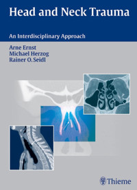
Written by an interdisciplinary team of experts in ear, nose, and throat trauma; oral and maxillofacial surgery; neurosurgery; and accident surgery, this book is a state-of-the-art manual on the diagnostic, treatment, and therapeutic techniques available to manage trauma injuries to the head and neck. This book features more than 240 illustrations, most in color, and step-by-step discussions of the initial management, evaluation, and examination of the patient, followed by a thorough collection of flow-charts and checklists. In each chapter the authors present the surgical anatomy, pathomechanism and classification, clinical signs and symptoms, functional tests, diagnostic imaging, as well as other appropriate diagnostic measures. Head and Neck Trauma covers everything from wound management to the latest surgical techniques and their complications. The authors also discuss the medical and technical aspects of trauma management, including antibiotic therapy, grafting and osteosynthesis materials, and the assessment of posttraumatic functional disorders. This beautifully illustrated reference belongs in every emergency room and trauma center library and is an essential tool for any medical professional treating patients with head and neck injuries.
Das E-Book können Sie in Legimi-Apps oder einer beliebigen App lesen, die das folgende Format unterstützen:
Seitenzahl: 401
Veröffentlichungsjahr: 2006
Ähnliche


Library of Congress Cataloging-in-Publication Data
Ernst, Arne, 1958-[Traumatologie des Kopf-Hals-Bereichs. English]Head and neck trauma/Arne Ernst, Michael Herzog,Rainer Ottis Seidl; with contributions by Karl-Ludwig Kiening,Andreas Unterberg, Ulrich W. Thomale.
p.; cm.
Includes index.ISBN-13: 978-3-13-140001-7 (GTV: alk. paper)ISBN-10: 3-13-140001-3 (GTV: alk. paper)ISBN-13: 978-1-58890-437-9 (TNY: alk. paper)ISBN-10: 1-58890-437-7 (TNY: alk. paper) 1. Neck—Wounds andinjuries. 2. Head—Wounds and injuries. I. Herzog, Michael, MD.II. Seidl, Rainer Ottis. III. Title.[DNLM: 1. Craniocerebral Trauma. 2. Neck Injuries.3. Reconstructive Surgical Procedures. WL 354 E71 t 2006]RD521.E76 2006617.5′1044–dc22
2006012337
This book is an authorized and revised translationof the German edition published and copyrighted 2004by Georg Thieme Verlag, Stuttgart, Germany.Title of the German edition: Traumatologie des Kopf-Hals-Bereiches
Translator: Stephanie Kramer, BA, Dipl Trans, IoL, Berlin
Illustrators: Peter Haller and Joachim Hormann, Stuttgart
2006 Georg Thieme Verlag,Rüdigerstrasse 14, 70469 Stuttgart, Germanyhttp://www.thieme.deThieme New York, 333 Seventh Avenue,New York, NY 10001, USAhttp://www.thieme.com
Typesetting by Sommer Druck, FeuchtwangenPrinted in Germany by Appl, Wemding10-ISBN 3-13-140001-3 (GTV)13-ISBN 978-3-13-140001-7 (GTV)10-ISBN 1-58890-437-7 (TNY)13-ISBN 978-1-58890-437-9 (TNY) 1 2 3 4 5 6
Important note: Medicine is an ever-changing science undergoing continual development. Research and clinical experience are continually expanding our knowledge, in particular our knowledge of proper treatment and drug therapy. Insofar as this book mentions any dosage or application, readers may rest assured that the authors, editors, and publishers have made every effort to ensure that such references are in accordance with the state of knowledge at the time of production of the book.
Nevertheless, this does not involve, imply, or express any guarantee or responsibility on the part of the publishers in respect to any dosage instructions and forms of applications stated in the book. Every user is requested to examine carefully the manufacturers’ leaflets accompanying each drug and to check, if necessary in consultation with a physician or specialist, whether the dosage schedules mentioned therein or the contraindications stated by the manufacturers differ from the statements made in the present book. Such examination is particularly important with drugs that are either rarely used or have been newly released on the market. Every dosage schedule or every form of application used is entirely at the user's own risk and responsibility. The authors and publishers request every user to report to the publishers any discrepancies or inaccuracies noticed. If errors in this work are found after publication, errata will be posted at www.thieme.com on the product description page.
Some of the product names, patents, and registered designs referred to in this book are in fact registered trademarks or proprietary names even though specific reference to this fact is not always made in the text. Therefore, the appearance of a name without designation as proprietary is not to be construed as a representation by the publisher that it is in the public domain.
This book, including all parts thereof, is legally protected by copyright. Any use, exploitation, or commercialization outside the narrow limits set by copyright legislation, without the publisher's consent, is illegal and liable to prosecution. This applies in particular to photostat reproduction, copying, mimeographing, preparation of microfilms, and electronic data processing and storage.
Preface
The treatment of patients with head and neck trauma requires a highly professional, fast, and multidisciplinary approach. There might be certain regional differences around the globe, but in most countries anesthesiologists, trauma surgeons, neurosurgeons, ear, nose, and throat (ENT) specialists, and oral and maxillofacial surgeons (OMS) are primarily involved.
While initial management is usually limited to identifying and treating life-threatening conditions, later steps in the adequate management of the consequences of head and neck trauma generally belong to only a few specialized fields. The present book is the product of a long-standing, clinical cooperation between the Departments of Otorhinolaryngology and Maxillo-Facial Surgery at the Berlin Trauma Center (UKB), with an additional neurosurgical contribution.
The book is intended to serve as a guide, providing all those involved in the care of patients with head and neck trauma basic as well as specialized knowledge. Flow charts, special sections detailing “Rules and Pitfalls,” and case reports serve as a quick bedside manual, with the entire work representing an extensive textbook.
The book reflects this approach and is divided into sections on initial management, diagnosis, and therapy. In addition, numerous cross-references facilitate the linking of symptoms, evaluation, and management.
We would like to take this opportunity to thank the staff members at our departments who are also responsible for conducting the annual training course on “Head and Neck Trauma Management” in Berlin. Additionally, we would like to express our gratitude to Professor Sven Mutze, Director of the Institute for Radiology at UKB, for permission to use the images printed in this book and also to Thieme Publishing Group, in particular, Dr. Urbanowicz for his patient support in the completion of this book.
We sincerely hope that the English edition of our book will find many readers across the globe, even though a few differences may exist for the management of head and neck trauma.
Arne ErnstMichael HerzogRainer O Seidl
Contributors
Karl-Ludwig Kiening, MDAssociate ProfessorNeurosurgical University HospitalHeidelberg UniversityHeidelberg, Germany
Ulrich W Thomale, MDNeurosurgical DepartmentCharité University HospitalBerlin, Germany
Andreas Unterberg, MDProfessorNeurosurgical University HospitalHeidelberg UniversityHeidelberg, Germany
Contents
I Evaluation of Head and Neck Trauma
1 Initial Management
First Aid at the Scene
Evaluation of Vital Functions
Stabilizing Vital Functions
Stabilizing Circulation
Emergency Care
Emergency Measures
Airways
Fall-Back of the Tongue
Injuries of the Larynx and Trachea
Hemorrhage
Central Hemorrhage
Peripheral Hemorrhage
Priorities in Trauma Management
2 Examining the Patient
Patient History
Inspection
Palpation
Functional Testing
Radiologic Diagnostics
3 Flow Charts and Checklists
Initial Management
Initial Evaluation
Injuries of the Neurocranium
Injuries of the Skull Base
Injures of the Ear and Lateral Skull Base
Injuries of the Facial Nerve
Craniofacial Injuries
Injuries of the Orbits
Dental Injury
Injuries of the Neck
4 General Principles of Trauma
Cutaneous Wounds
Pathomechanism and Classification
Wounds Caused by Mechanical Forces
Thermal and Chemical Wounds
Skin Healing
Osseous Injuries
Pathomechanism and Classification
Fracture Signs
Bone Healing
Direct (Primary) Fracture Healing
Indirect (Secondary) Fracture Healing
Pseudarthrosis
Fracture Treatment
Reduction
Fixation
Retention
Immobilization
Rehabilitation and Functional Therapy
Injuries of the Joints
Pathomechanism and Classification
Cartilage Injury
Pathomechanism and Classification
Cartilage Healing
Muscular Injury
Clinical Signs and Symptoms
Muscular Healing
Peripheral Nerve Injury
Pathomechanism and Classification
Neural Healing
II Diagnostic of Head and Neck Trauma
5 Injuries of the Neurocranium and Craniocervical Junction
Injuries of the Neurocranium
Open and Closed Head Injury
Clinical Signs and Symptoms
Diagnostic Imaging
Treatment
Injury to Bony Structures of the Craniocervical Junction
Anatomy and Pathomechanism
Clinical Signs and Symptoms
Classification
Diagnostic Imaging
Soft Tissue Distortion and Discoligamentous Injuries of the Craniocervical Junction
Pathomechanism
Clinical Signs and Symptoms
Classification
Clinical and Neurological Diagnosis
Imaging Modalities
6 Diagnosing Injuries of the Skull Base
Surgical Anatomy
Anterior Cranial Fossa
Middle Cranial Fossa
Posterior Cranial Fossa
Pathomechanism and Classification
Pathomechanism
Dural Injury
Classification of Fractures
Clinical Signs and Symptoms
Uncertain Signs of Fracture of the Anterior Skull Base
Eyelid Hematoma, Eyelid Emphysema
Epistaxis
Seiferth Sign
Olfactory Disturbances
Certain Signs of Fracture of the Anterior Skull Base
Cerebrospinal Fluid Rhinorrhea
Evaluation of Suspected Cerebrospinal Fluid Rhinorrhea
Pneumocephalus
Early Meningitis
Diagnostic Imaging of Fractures of the Anterior Skull Base
Surgical Indications in Injuries of the Anterior Skull Base
7 Diagnosing Injuries of the Ear and Lateral Skull Base
Surgical Anatomy
Pathomechanism and Classification
Injuries of the External Ear
Injuries of the Middle Ear
Injuries of the Tympanic Membrane
Ossicular Injuries
Rupture of the Round Window Membrane
Injuries of the Petrous Temporal Bone and Labyrinth
Concussion of the Petrous Temporal Bone and Bony Labyrinth
Fractures of the Petrous Temporal Bone and Bony Labyrinth
Fracture Line in Longitudinal Fractures
Fracture Line in Transverse Fractures
Clinical Signs and Symptoms
Aural Hemorrhage
Cerebrospinal Fluid Otorrhea
Hearing Dysfunction
Sensorineural Hearing Loss
Conductive Hearing Loss
Vestibular Disorder
Functional Testing of the Auditory and Vestibular System
Audiometry
Tuning Fork Tests
Pure Tone Audiometry
Vestibular Tests
Frenzel Lenses
Vestibulospinal Reflexes
Romberg Test
Unterberger Stepping Test
Caloric Testing
Diagnostic Imaging
8 Diagnosing Injuries of the Facial Nerve
Surgical Anatomy
Intracranial Segment
Intratemporal Segment
Pathomechanism and Classification
Classification
House–Brackmann Scale
Clinical Signs and Functional Tests
Topodiagnostic Evaluation
Tearing (Schirmer Test)
Taste
Measurement of Stapedial Reflex
Electrophysiologic Testing
Electromyography (EMG)
Management
9 Diagnosing Injuries of the Midface
Surgical Anatomy
Classification of Midface Fractures
Central Fractures of the Midface
Fractures of the Alveolar Process
Pathomechanism
Le Fort I Fractures
Pathomechanism
Clinical Symptoms
Diagnostic Imaging
Treatment
Le Fort II Fractures, Wassmund I Fractures
Pathomechanism
Clinical Symptoms
Diagnostic Imaging
Treatment
Le Fort III Fractures, Wassmund IV Fractures
Surgical Anatomy
Pathomechanism
Clinical Symptoms
Diagnostic Imaging
Treatment
Fractures of the Nasoethmoidal Complex
Surgical Anatomy
Pathomechanism
Classification
Clinical Symptoms
Diagnostic Imaging
Treatment
Fractures of the Bony Nasal Framework and Septum
Surgical Anatomy
Pathomechanism
Classification
Clinical Symptoms
Diagnostic Imaging
Treatment
Fractures of the Lateral Midface
Zygomatic Fracture
Surgical Anatomy
Pathomechanism
Classification
Clinical Signs
Zygomatic Arch Fracture
Pathomechanism
Clinical Signs
Diagnostic Imaging
Treatment
10 Diagnosing Injuries of the Orbit
Surgical Anatomy
Orbital Wall Fractures
Fractures of the Orbital Floor and Medial Orbital Wall
Pathomechanism
Clinical Signs and Symptoms
Traction Test/Forward Traction Test
Endoscopy of the Maxillary Sinus
Diagnostic Imaging
Treatment
Fractures of the Orbital Roof and Lateral Orbital Wall
Pathomechanism
Clinical Signs and Symptoms
Diagnostic Imaging
Treatment
Injuries of the Orbital Apex
Pathomechanism and Classification
Clinical Signs and Symptoms
Injury of the Oculomotor Nerve
Injury of the Trochlear Nerve
Injury of the Abducens Nerve
Injury of the Optic Nerve
Injury of the Trigeminal Nerve
Diagnostic Imaging
Treatment
Injuries of the Lacrimal Ducts
Pathomechanism
Clinical Signs and Symptoms
Diagnostic Imaging
Treatment
Carotid-Cavernous Sinus Fistula
Pathomechanism
Clinical Signs and Symptoms
Diagnostic Imaging
11 Diagnosing Injuries of the Mandible
Surgical Anatomy
Pathomechanism
Clinical Signs and Symptoms
Certain Signs of Fracture
Deformity and Dislocation
Abnormal Mobility
Crepitus
Uncertain Fracture Signs
Hematoma and Swelling
Tenderness
Longitudinal Compression Pain
Restricted Function
Sensory Disturbances
Malocclusion
Diagnostic Imaging
Classification of Mandibular Fractures
Fractures of the Mandibular Body with Dentoalveolar Involvement
Pathomechanisms
Diagnostic Imaging
Treatment
Mandibular Fractures without Dentoalveolar Involvement
Clinical Signs and Symptoms
Diagnostic Imaging
Treatment
Fractures of the Ramus of Mandible
Pathomechanism
Clinical Signs
Diagnostic Imaging
Treatment
Injuries of the Condylar Process
Contusion of the Temporomandibular Joint
Distortion of the Temporomandibular Joint
Dislocation of the Temporomandibular Joint
Subluxation of the Temporomandibular Joint
Fracture of the Temporomandibular Joint
12 Diagnosing Dental Injuries
Surgical Anatomy
Pathomechanism
Dental Fractures
Classification
Clinical Diagnosis
Diagnostic Imaging
Treatment
Dental Luxation
Pathomechanism
Classification
Clinical Diagnosis
Diagnostic Imaging
Treatment
Dental Concussion
Pathomechanism
Diagnostic Imaging
Treatment
Dental Luxation without Dislocation (Loosening, Subluxation)
Pathomechanism
Clinical Signs
Diagnostic Imaging
Treatment
Dental Luxation with Dislocation
Partial Peripheral Dislocation
Complete Peripheral Dislocation
Central Luxation
Fractures of the Alveolar Process
Pathomechanism
Clinical Signs
Diagnostic Imaging
Treatment
13 Diagnosing Injuries of the Pharynx, Salivary Glands, and Soft Tissues of the Neck
Injuries of the Pharynx
Injuries of the Salivary Glands
Soft Tissue Injuries of the Neck
Blunt Trauma to the Soft Tissues of the Neck
Sharp Trauma of the Soft Tissues of the Neck
14 Diagnosing Injuries of the Larynx and Trachea
Surgical Anatomy
Pathomechanism and Classification
Fractures
Anteroposteriorly Directed Trauma
Lateral Trauma
Supraglottic Injuries
Transglottic Injuries
Subglottic Injuries
Tracheal Injuries
Ruptures
Supraglottic Rupture
Subglottic Rupture
Laryngotracheal Separation
Partial Laryngotracheal Separation
Complete Laryngotracheal Separation
Esophageal Rupture
Clinical Signs and Symptoms
Respiratory Distress
Emphysema
Complications
Evaluation Procedures and Functional Testing
Endoscopy
Magnifying Laryngoscopy and Flexible Nasopharyngolaryngoscopy
Microlaryngoscopy, Tracheobronchoscopy
Radiographic Evaluation
Evaluation of Phonation
Stroboscopy
Vocal Output and Characteristics
Electrodiagnostic Testing
Treatment
III Therapy of Head and Neck Trauma
15 Principles of Wound Management
The Ten Commandments of Wound Management
Abrasions
Immediate Management/Treatment
Puncture Wounds
Immediate Management
Treatment
Cut Wounds
Immediate Management
Treatment
Bite Wounds
Immediate Management
Treatment
Missile Wounds
Immediate Management
Treatment
Burn Injury
Immediate Management
Treatment
Chemical/Alkali Burns
Immediate Management
Treatment
Wound Closure
Suture Technique
Suture Material
Wound Dressings
16 Treatment of Injuries of the Neurocranium and Craniocervical Junction
Treatment Principles for Craniocerebral Trauma
Treatment of Fractures of the Craniocervical Junction
Atlantooccipital Fractures/Dislocations
Treatment of Soft Tissue Distortion and Discoligamentous Injuries of the Craniocervical Junction
17 Treatment of Injuries of the Skull Base
Indications
Approaches
Ear-to-Ear Scalp Incision, Bicoronal Incision
Frontoorbital Approach
Endonasal Approach
Surgical Technique
Principles of Dural Repair
Frontal Sinus
Extradural Management
Intradural Management
Ethmoid Bone
Sphenoid Sinus
Grafts
Autogenous Grafts
Homologous Grafts
Alloplastic Implants
Postoperative Management
Frontal Sinus
Ethmoid/Sphenoid Sinus
Drainage of Cerebrospinal Fluid
Follow-up Care
Complications
18 Treatment of Injuries of the Ear and Lateral Skull Base
Injuries of the External Ear
Indications
Surgical Methods
Seroma and Hematomas of the Ear
Injuries of the Auricle
Auricular Amputation
Injuries of the Vestibulocochlear System
Indications
Conservative Management
Inner Ear Auditory Dysfunction
Vestibular Disorder
Surgical Management
Approaches
Surgical Techniques
Injuries of the Facial Nerve
Indications
Conservative Treatment
Surgical Management
Follow-up Care
19 Treatment of Injuries of the Midface
Conservative Management
Splinting
Intraoral Splinting
Extraoral Splinting
Controlled Spontaneous Healing
Monomaxillary Fixation
Intermaxillary Fixation
Intermaxillary Fixation and Stabilization with a Halo Frame
Surgical Procedures
Craniofacial Suspension Wiring
Osteosynthesis with Miniplates and Microplates
Osteosynthesis Techniques
Osteosynthesis Materials
Osteosynthesis Systems
Principles of Reconstruction
Infrazygomatic Fractures (Le Fort I)
Indications
Conservative Management
Surgical Treatment
Complications
Central or Pyramidal Fractures (Le Fort II, Wassmund I)
Indications
Conservative Management
Surgical Technique
Complications
Centrolateral Fractures (Le Fort III, Wassmund IV)
Indications
Conservative Management
Surgical Technique
Complications
Fractures of the Nasoethmoidal Complex
Indications
Surgical Treatment
Fractures of the Bony Nasal Skeleton and Septum
Indications
Approaches
Surgical Technique
Fractures of the Lateral Midface
Indications
Approaches
Surgical Technique
20 Treatment of Orbital Injuries
Fractures of the Orbital Floor
Indications
Approaches
Surgical Technique
Complications
Medial Orbits
Indications
Approaches
Surgical Technique
Complications
Orbital Roof
Indications
Approaches
Surgical Technique
Complications
Orbital Decompression
Indications
Approaches
Surgical Technique
Complications
Optic Nerve Decompression
Indications
Approaches
Surgical Technique
Complications
Injuries of the Lacrimal Ducts
Indications
Surgical Technique
Complications
Injuries of the Eyelids
Indications
Surgical Technique
Complications
21 Treatment of Injuries of the Mandible
Fractures of the Mandibular Body with Dentoalveolar Involvement
Indications
Conservative Management
Surgical Management
Approaches
Surgical Technique
Complications
Fractures of the Mandible without Dentoalveolar Involvement or in the Partially Edentulous Jaw
Fractures of the Mandibular Ramus
Conservative Treatment
Surgical Treatment
Approaches
Surgical Technique
Complications
Fractures in the Edentulous, Atrophied Mandible
Mandibular Defect Fractures
Contaminated Mandibular Fractures
Fractures of the Mandible in Patients with Primary or Deciduous Teeth
Nondisplaced Fractures of the Condylar Neck and Head
Indications
Conservative Treatment
Surgical Treatment
Approaches—Intraoral Approach
Approaches—Extraoral Retromandibular (Submandibular) Approach
Functional Treatment
Displaced Fractures and Fracture-Dislocations of the Condylar Neck
Indications
Conservative Management
Complications
Surgical Management
Approaches
Surgical Technique
Complications
Fractures of the Condylar Head
Conservative Management
Surgical Treatment
Approaches
Surgical Technique
Fractures of the Articular Fossa (Central Dislocation)
Pediatric Fractures of the Condylar Neck and Condylar Head
22 Treatment of Dental Injuries
Treatment of Dental Injuries
Indications
Conservative Treatment of Crown Fractures
Enamel Fractures
Enamel-Dentin Fractures without Pulp Exposure
Enamel-Dentin Fractures with Pulp Exposure
Surgical Management of Root Fractures
Coronal Third Root Fractures
Middle Third Root Fractures
Apical Third Root Fractures
Longitudinal Fractures
Approaches (Resection of the Root Tip)
Surgical Technique
Complications
Treatment of Dental Luxation
Indications
Primary Teeth
Surgical Treatment
Dental Concussion
Dental Luxation without Displacement (Tooth Loosening)
Partial Peripheral Luxation
Complete Peripheral Luxation
Central Luxation
Complications
Treatment of Fractures of the Alveolar Process
Indications
Conservative Treatment
Surgical Management
Approaches
Surgical Technique
Complications
23 Treatment of Injuries of the Larynx, Pharynx, Trachea, Esophagus, and Soft Tissues of the Neck
Indications
Conservative Treatment
Chemical/Alkali Injuries
Surgical Therapy
Approaches
Surgical Procedure
Complications
IV Appendix
24 Antibiotic Therapy
25 Grafting and Osteosynthesis Materials
Requirements and Classification
Autogenic Grafts
Outer Table of the Skull
Allogenous (Homologous) Grafts
Preserved Allografts
Alloplastic Implants
Absorbable Alloplastic Implants
Polydioxanone (PDS)
Ethisorb
Vicryl
Monocryl
Nonabsorbable Alloplastic Implants
Metal
Further Reading
Index
I Evaluation of Head and Neck Trauma
1 Initial Management
First Aid at the Scene
Emergency Measures
Priorities in Trauma Management
2 Examining the Patient
Patient History
Inspection
Palpation
Functional Testing
Radiologic Diagnostics
3 Flow Charts and Checklists
Initial Management
Initial Evaluation
Injuries of the Neurocranium
Injuries of the Skull Base
Injures of the Ear and Lateral Skull Base
Craniofacial Injuries
Injuries of the Orbits
Dental Injury
Injuries of the Neck
4 General Principles of Trauma
Cutaneous Wounds
Osseous Injuries
Injuries of the Joints
Cartilage Injury
Muscular Injury
Peripheral Nerve Injury
1 Initial Management
Checklist Initial Evaluation, Chapter 3, p. 15
Checklist Initial Management, Chapter 3, p. 15
First Aid at the Scene
In areas where rapid access to medical care is ensured, persons arriving at the scene normally need only to call the paramedics and wait for their arrival. Securing the scene of the accident has absolute priority over further measures in order to protect the injured individual, motorists, and other persons administering aid.
Evaluation of Vital Functions
Vital signs should always be determined first as a means of initial assessment:
Neurologic status is evaluated on the basis of the patient's response when spoken to and to pain.
The Glasgow Coma Scale (GCS; Table 5.1, p. 39) is necessary for further clinical assessment.
Respiratory status is evaluated based on observation of breathing pattern and respiratory rate.
Circulation can be evaluated by palpating the carotid pulse.
Stabilizing Vital Functions
Obstruction of the upper airways is the greatest threat in patients with head and neck injuries. The jaw-thrust and chin-lift maneuvers (Fig. 1.1) are the simplest means of stabilizing the airways.
Foreign bodies (dentures, mucus, and vomitus) must be removed from the oral cavity using a finger. More proximal airways should be cleaned by suction if possible.
Intubation with pharyngeal tubes is another possibility for securing the airways (Guedel tube, Wendel tube; Fig. 1.1c). For complex injuries, transport to a regional trauma center is essential. If associated injury of the neurocranium is suspected, early orotracheal intubation should be performed.
Stabilizing Circulation
It is imperative that treatment of shock begin at the scene. Initial management includes:
elevation of the patient's legs (autotransfusion);
intravenous administration of a colloidal volume substitute;
adequate pain management;
protection from hypothermia.
Massive hemorrhage should be managed with direct compression. Ligation of the extremities should be avoided, however, and the exact time that compression began must be noted. Cardiopulmonary resuscitation should be performed if necessary.
Emergency Care
Soft tissue injuries should be covered with a sterile bandage to help control bleeding and protect the wound from additional contamination. Penetrating foreign bodies should be removed only after the patient is in a clinical setting.
If cervical spine injury is suspected, rotation or hyperextension of the patient's neck must be avoided. If removal of a motorcycle helmet at the scene is necessary in order to control the airways, a second person must stabilize the cervical spine using traction. Then, a rigid cervical collar must be applied until cervical spine injury has been excluded.
After emergent care procedures are complete, further treatment should take place in a specialized properly equipped trauma center. This is especially important for complex injuries. The patient should only be moved after stabilization of vital functions.
Fig. 1.1 Obstruction of the upper airways caused by fall-back of the tongue and epiglottis (modified from Eisele and McQuone 2000).
a Laxity of the tongue musculature causing it to obstruct the upper airways.
b Tilting the head to dorsal and applying pressure to the chin assures the patency of the airways.
c Positioning a Guedel tube to secure the upper airways.
Emergency Measures
Airways
Establishment and maintenance of the airways is of the utmost urgency in treating any multiply injured patient with craniofacial trauma. It is important to remember that following an accident, even airways with adequate ventilation can quickly become obstructed by blood or swelling.
Fall-Back of the Tongue
A particular problem of craniofacial injury is the fall-back of the tongue in segmental fractures of the mandible, especially those involving the midface. The continuity of the horseshoe-shaped mandible, to which the tongue is attached, is disrupted and the injured individual is no longer able to maintain the position of the tongue to keep the airways open (Fig. 1.2).
In an emergency, one can attempt to place the patient in the lateral position or to advance the fractured mandibular arch manually. If the patient is unconscious, a suture can be placed through the posterior of the tongue, lifting the tongue and pulling it forward (Fig. 1.2c).
Oral intubation follows. Successfully positioning the larynx is usually unproblematic, despite hemorrhage and swelling, as the tongue base loses its supporting buttress as a result of mandibular injury.
Fig. 1.2 Obstruction of the upper airways in a mandibular fracture.
a Dorsal displacement of the mandibular arch and tearing of the musculature of the floor of the mouth and tongue.
b Fall-back of the tongue due to loss of fixation on the mandible.
c Emergency procedure for advancing the tongue using a suture to establish the airways.
Injuries of the Larynx and Trachea
Specific problems related to injury of the larynx and trachea can arise and should be expected:
Extensive injury of the larynx often renders oral intubation impossible; intubation should never be forced under such circumstances as manipulation can permanently obstruct any remaining space in the larynx. In rare cases, intubation can be attempted using a stiff tube.
In an emergency, tracheotomy is always preferable to intubation.
Cricothyrotomy is not advisable due to possible existing concomitant injury of the cricoid cartilage or cricoid lamina.
In penetrating injuries of the trachea or larynx, the injury site should be used for intubation (Fig. 23.2a, p. 208).
If tracheal rupture is suspected, intubation should be accomplished using a flexible endoscope or by means of primary tracheotomy. The endoscope is advanced under visualization past the tracheal injury and the tube is positioned inferior to the injury site. The tube should not be too large as this can result in further displacement of the ruptured trachea (Fig. 14.5, p. 126).
Cricothyrotomy
Cricothyrotomy involves creating an opening in the cricothyroid membrane, which covers the area between the thyroid lamina and the cricoid cartilage. The emergency cricothyrotomy kits available today belong to standard paramedic equipment:
The cricoid cartilage is palpated and the slight indentation above it is punctured with a needle.
If the needle comes into contact with the thyroid cartilage, it can be used to guide the needle to the cricothyroid membrane. The needle tip then points in the direction of the jugular and is directed downward to the palpable gap and then advanced through the cricothyroid membrane (Fig. 1.3).
If an emergency kit is not available, a horizontal incision is made over the cricothyroid membrane. A blade with suitable dimensions is advanced directly into the trachea. The blade is not removed, but instead is rotated, thus serving to guide a speculum or catheter for placing the tube.
Fig. 1.3 Cricothyrotomy (modified from Eisele and McQuone 2000).
a Palpation of the cricoid cartilage; an incision is made at its superior border.
b A suitably sized blade is used to penetrate the cricothyroid membrane and is advanced in the trachea, where it is then rotated.
c Intubation occurs through the opening created into the trachea.
Following cardiopulmonary resuscitation, a cricothyrotomy should be transformed into a tracheotomy as it will otherwise result in permanent damage to the larynx after a few days.
Tracheostomy
Procedures for tracheostomy are similar to those used in cricothyrotomy. Emergency kits are also available for urgent tracheostomy.
If possible, a local anesthetic (e. g., lidocaine with epinephrine to control bleeding) should be applied prior to beginning the procedure.
An incision is then made in the skin vertically (minimizing the risk of damage to the thyroid gland), directly into the trachea.
The blade remains in situ, and a speculum is inserted over it; the opening is enlarged and a tube is advanced (Fig. 1.4). If necessary, a suction catheter can also be inserted in order to advance the tube using the Seldinger technique.
Given the consequences of respiratory insufficiency, concerns about heavy bleeding are misplaced. In most cases, hemorrhage can be controlled using a clip or by means of compression, for example, damp packing in the tube until definitive treatment (tracheostomy).
Definitive treatment, i. e., tracheostomy, must be assured.
Fig. 1.4 Tracheotomy (modified from Eisele and McQuone 2000).
a One hand stabilizes the larynx while a vertical incision is made in the skin beneath the cricoid cartilage.
b The tissue is forced apart using the fingers until the trachea can be seen.
c Opening of the trachea with a longitudinal incision.
d Inserting the tube, if necessary using a catheter or with the aid of a speculum.
Fig. 1.5 Course of arteries in the central midface.
Hemorrhage
Head and neck hemorrhage can quickly become life-threatening due to blood loss, but also due to aspiration.
In many cases, emergency management of hemorrhage is possible after identifying the bleeding vessel. If bleeding cannot be controlled, selective angiography and embolization are needed. Hemorrhage in the head and neck region can be divided into central and peripheral bleeding.
Central Hemorrhage
Central hemorrhage describes bleeding from vessels which are inaccessible for direct treatment, for example, compression, because of their anatomic position.
Maxillary Artery
The maxillary artery is the most commonly affected vessel. In midfacial fractures, it is generally injured in the pterygopalatine region, whereby the sharp edges of the bone at the fracture site between the posterior wall of the maxillary sinus and pterygoid process rupture the vessel wall (Fig. 1.5). Additional hemorrhage arises from the accompanying venous plexus. Blood is lost either directly or through the maxillary sinus and then from the nose or throat.
Nasal packing, part of emergent care, widens existing fracture gaps and thus increases bleeding. Only by fixing the maxillary bone segment and compressing it against the skull base can sufficient stability for packing be achieved.
A head/chin strap (Fig. 1.6) can be applied for stabilization at the scene of the accident, usually after intubation. A gauze bandage is used to press the mandible against the skull base and then the nose is packed.
For delayed intubation, compression can also be achieved by applying a spatula bandage, which uses a gauze bandage to press a wooden spatula, which has been inserted into the patient's mouth, against the skull base (Fig. 1.6a).
If bleeding does not cease using the measures mentioned here, occlusion of the artery by means of selective angiography (Fig. 1.7) is recommended for achieving definitive control of bleeding. In some circumstances, ligation of the artery after it branches off of the external carotid artery may be considered. Transantral ligation is generally very difficult.
Anterior and Posterior Ethmoid Arteries
Hemorrhage from the ethmoid arteries can usually be managed using anterior nasal packing (see Fig. 1.8). Additional posterior packing is rarely necessary.
Injury to the anterior ethmoid artery causing the vessel to recede into an orbit with intact orbital cone can cause intraorbital hemorrhage and compression of orbital contents. Clinical signs of orbital compression are subconjunctival hemorrhage with chemosis and a bulging globe. The orbit should be decompressed immediately by lateral canthotomy (Fig. 20.1, p. 180).
For persistent bleeding, occlusion of the vessels may be necessary from an external or through an endonasal approach.
Carotid Artery
Hemorrhage from the carotid artery near the cranial base is usually fatal. In a limited number of cases, immediate tamponading of the epipharynx (Bellocq packing, Fig.1.9, p. 10) can control bleeding. However, the consequences, such as spreading of the hemorrhage upward in the neurocranium or vasospasms, generally are fatal.
In an emergency, an attempt can be made at stopping carotid hemorrhage by applying pressure over the sixth cervical vertebra. This is best done from a kneeling position over the patient by intermittently pressing one's full body weight onto a closed fist held against the vertebrae.
Anterior Nasal Packing
Anterior nasal packing involves tamponading the nose through the front opening using ready-made foam rubber packs, which are available in various sizes, or Vase-line gauze packing. Nasal balloons that can be filled with fluid are also available (see Fig. 1.9e).
Anesthetization of the nose with a spray or wadding soaked in local anesthetic and naphazoline nitrate is helpful. Always insert packing over the floor of the nose.
Nasal packing must be secured using a tie which is fixed externally with adhesive tape.
Fig. 1.6 Head bandages for compression of the maxilla to control hemorrhage (modified from Schwenzer and Ehrenfeld, Vol. 2, 2002).
a Craniomaxillary suspension of the maxilla using gauze bandages and wooden spatulas.
b Maxillary splint or dental impression tray can be secured with an extraoral brace secured by a head bandage.
Fig. 1.7 Selective angiography in maxillary artery hemorrhage which could not be controlled conservatively.
a Computed tomography image of injury of the facial skeleton.
b Selective angiography showing hemorrhage from the right maxillary artery.
c Cessation of bleeding after occlusion of the maxillary artery.
Bellocq Packing
Bellocq packing involves anterior and posterior packing of the nasal cavity:
Tamponading begins with posterior packing: Either fluid-filled (sodium chloride) catheters (bladder catheter, etc.) (Fig. 1.9a) or cotton ball packing may be used. Cotton ball packs have three securing ties. The pad, which has two ties secured to the end of a narrow catheter (gastric tube, suction catheter), is advanced through the mouth into the epipharynx (Fig. 1.9b). The position of the pack should be checked digitally to ensure that the soft palate and uvula are not crushed (Fig. 1.9c).
Anterior nasal tamponading can then proceed, accompanied by continuous traction on the ends of the strings.
Fixation is completed under traction, placing a pad at the opening of the nose (Fig. 1.9d). The third string is retracted through the mouth and taped to the patient's cheek for later removal of the pack.
Nasal packing should never remain in place longer than three days. Irreversible damage to the nasal mucosa can otherwise result.
Fig. 1.8 Anterior nasal packing (modified from Fonseca et al. 1997).
a The procedure begins on the floor of the nose with multi-layered Vaseline gauze packing. Holding the packing 5–6 cm from its end, it is placed as deep as possible in the nasal cavity.
b Layers of the gauze pack are positioned layer by layer until the nasal cavity is filled.
Peripheral Hemorrhage
Peripheral hemorrhage usually involves the large branches of the external carotid artery (temporal artery, facial artery, lingual artery). Direct injury—missile wound or penetrating trauma—of the common carotid artery may occur in rare cases.
Initial management should always seek to control bleeding with compression by pressing the vessels against underlying skeletal structures:
temporal artery against the root of the zygomatic arch;
facial artery against the anterior edge of the mandible, at the anterior margin of the attachment of the masseter muscle;
lingual artery with bi-manual pressure exerted intra-orally and extraorally;
carotid artery against the sixth cervical vertebra.
Hemorrhage of this type should be treated as quickly as possible with definitive surgical management, locating and ligating visible vessels. Injury to the great vessels of the neck demands immediate reconstruction with direct vascular suture, management with a patch graft, or vascular prosthesis.
Fig. 1.9 Bellocq packing.
a A bladder catheter may be used for emergency closure of the epipharynx.
b A gauze pad is normally drawn into the epipharynx over a suction catheter.
c The position of the packing in the throat must be checked with a finger.
d Using steady traction on the Bellocq tampon, anterior nasal packing is then placed. Finally, a pad is knotted over the nasal opening using the strings from the Bellocq tampon for the purpose of maintaining traction on the epipharyngeal packing.
e Alternatively, specially made packing, which can be filled with water, can be used.
Priorities in Trauma Management
Table 1.1 lists the various phases in the care of trauma victims.
After eliminating acute, life-threatening risks and preparing the patient for surgery, initial management can begin. Urgent immediate care should be given to the following:
hemorrhage;
orbital and optic nerve injuries with risk of vision impairment or blindness;
mobile, open jaw fractures;
soft tissue injuries.
Initial management is not necessarily the definitive treatment. After stabilizing the patient, completing diagnosis, planning of interventions, and subsidence of swelling, definitive treatment can generally proceed after 5–8 days.
Table 1.
1
Essential steps in the care of trauma victims
Phase
Description
Resuscitation
Stabilization and restoration of vital functions
First operation
Urgent initial management
Stabilization
Intensive care, more detailed diagnosis, preparatory measures
Second operation
Definitive treatment of injuries
Rehabilitation
Rehabilitation, physical therapy
Third operation
Secondary corrective measures
2 Examining the Patient
Standardized evaluation procedures for all organ systems of the head and neck should be followed for every patient presenting with head trauma. Systematic evaluation minimizes the risk of overlooking any aspect of injury.
Patient History
Determining the mechanism of injury is paramount (e. g., see Table 2.1). If the patient is unconscious or unable to answer questions, witnesses to the accident, prehospital emergency care providers, and relatives should be interviewed. It is important to know the direct mechanism of injury as well as other factors (helmet, seatbelt, etc).
Knowing the patient's prior health status is vital. Special consideration should be given to:
pre-existing conditions;
previous operations;
infectious diseases (hepatitis, HIV);
immunization status (tetanus, rabies for animal bites).
Thorough documentation of patient history is also essential for follow-up insurance questions. Photo documentation is advisable.
Table 2.
1
Diagnosis and treatment required for various types of injury
Type of injury
Diagnosis
Treatment
Cut
p. 27
p. 131
Bite
p. 28
p. 131
Missile wound
p. 28
p. 135
Burn
p. 29
p. 133
Chemical/alkali burn
p. 30
p. 134
Inspection
Inspection involves assessing external injuries such as bruising, hematomas, and wounds. Even apparently insignificant injuries can be critical:
hemorrhage (Chapter 1, p. 6);
wounds (Diagnosis, Chapter 4, p. 27; Treatment, Chapter 15, p. 130);
foreign bodies;
hematomas.
Palpation
Palpation should always be carried out using a systematic approach (Fig. 2.1a). The aim of palpation is to detect swelling, hematomas, crepitus, and abnormal mobility. Palpation should be conducted bilaterally, using both hands.
Neck: Beginning at the jugular, the anterior of the neck is evaluated. After exploring the posterior neck, palpation then continues over the posterior head to the forehead.
Ear: Evaluation of the external ear. It is important to distinguish between hemorrhage coming from the auditory canal and bleeding into the auditory canal.
Orbits: Beginning at the superior orbital cavity, palpation proceeds over the lateral orbits, the infraorbital ridge to the medial orbits (Fig. 2.1b).
Mandible: The mandible is evaluated from the tip of the chin, over the mandibular angle to the condyles (auditory canals). Pain upon compression at the chin point is a sign of injury involving the temporomandibular joint region (Fig. 2.1c).
Temporomandibular joint: The little fingers are inserted into the external auditory canals and the patient is instructed to open the mouth. The condyles of the temporomandibular joint are palpated. Special consideration should be given to deviation of the mandible from its central position with opening of the mouth, as a sign of fracture (Fig. 2.1d).
Oral vestibule: The right mandibular oral vestibule is palpated to the mandibular arch, over the maxillary vestibule, and downward again over the left mandible. Special attention should be paid to injured teeth (Fig. 2.1e). Tapping the maxillary teeth of a healthy, nonfractured maxilla with a metal spatula produces a ringing sound. Fracture results in a reduced sound (bandbox resonance).
Fig. 2.1 Standardized evaluation procedures.
Palate: Maxillary mobility is evaluated by palpating the jaw, not the teeth. Disruption of the bony palate is a sign of sagittal fracture. See below for the various levels of mobility of the maxilla:
– Le Fort I level: The mobility of the maxilla in the Le Fort I level is evaluated by placing a finger on the pyriform aperture or the zygomaticomaxillary buttress while gently rocking the maxilla back and forth.
– Le Fort II level: A finger is placed on the inferior orbital rim at the height of the infraorbital foramen to test the mobility of the maxilla in the Le Fort II level (Fig. 2.1f).
– Le Fort III level: Maxillary mobility in the Le Fort III level can be evaluated by placing a finger on the zygomaticofrontal suture (Fig. 2.1g).
Functional Testing
Various functional tests should be performed following palpation. These must be modified according to the patient's pattern of injury and level of consciousness.
Eye: Pupil size and reaction must always be evaluated. If there is a unilaterally wide-open pupil without direct—but with indirect—light response, this is known as an amaurotic (fixed) pupil and must be further evaluated (see Fig. 10.8, p. 93). Preliminary vision testing should be performed and the motility of the globe in the main gaze positions (finger perimetry) checked. Special attention should be paid to double vision (Fig. 2.2).
Facial nerve: Coordination of movement in basic facial expressions is tested with brow elevation, eye closure, and pursing of the lips. In comatose patients transcranial nerve stimulation can be used.
Occlusion: Active or passive movement of the mandible toward the maxilla is used to test occlusion. Malocclusion appears as:
– anterior open bite (Fig. 11.15b, p. 110);
– malocclusion of the lateral incisor region (Fig. 11.16a, p. 111) or;
– edge-to-edge bite.
Fig. 2.2 Orientation of the eye in the six diagnostic positions of gaze (from Schwenzer and Ehrenfeld, Vol. 1, 2000). The main muscles responsible for movement are shown here.
Trigeminal nerve: Sensory disturbances of the trigeminal nerve distribution area can be observed to varying degrees: hypesthesia, anesthesia, and paresthesia. A cotton swab is used to test sensation in the region being evaluated, by moving it from the periphery toward the center, to the foramen, comparing the sensitivity of both sides.
Dental vitality test: Tooth vitality can be evaluated using a thermal test to look for a pain reaction of the dental pulp (see Fig. 12.2, p. 114). A cold aerosol spray is applied to the enamel surface using a cotton swab.
Auditory tests: Initial auditory tests can be performed using the tuning fork method based on Weber and Rinne (see Functional Testing of the Auditory and Vestibular System, p. 61).
Olfactory ability: Olfactory ability can be roughly checked using gasoline (see Clinical Signs Skull Base Injury, p. 48).
Radiologic Diagnostics
Radiographic evaluation should always be complementary to clinical diagnostics and should never serve as a substitute. Only after clinical examination can a reasonable decision be made regarding the required extent of imaging.
If midfacial fracture is suspected, the following are advisable:
occipitomental and eccentric cranial views (Fig. 9.17c, p. 84)
bucket handle view (see Fig. 9.18, p. 84)
For injuries of the mandible and temporomandibular joints, the following conventional radiographs are recommended:
orthopantomogram (OPG/OPT) (Fig. 11.5b, p.101)
view based on Clementschitsch (Fig. 21.9a, p. 194)
In addition, occlusal views, oblique mandibular projections, and dental radiographs may be advisable.
Computed tomography (CT) evaluation of craniofacial injury is standard these days and is mandatory, especially for complex patterns of injury. Moreover, 3-D imaging can be useful for planning reconstruction (Fig. 1.7, p. 8). For craniofacial views, 3 mm slices, in axial and coronal planes, are preferred; for petrous temporal bone fractures 2 mm slices are appropriate.
Angiography should be performed if there is unexplained hemorrhage; embolization, if needed, should be done in the same session (Fig. 1.7, p. 8). Magnetic resonance imaging (MRI) is only rarely indicated, for example, to search for cerebrospinal fluid fistulas (Fig. 6.5c, p. 51) or in injury to orbital contents.
3 Flow Charts and Checklists
Initial Management
See p. 2.
Table 3.
1
Checklist for initial management
Objective
Measure
Establish and maintain airways (intubation, cricothyrotomy, tracheotomy)
Clearing the upper airways of discharge or prolapsed tongue (combination fractures of mandible and maxilla), dentures
Stabilize circulation
Establishing intravenous access, monitoring, fluid resuscitation
Exclude intracerebral pressure, intracranial hematoma, brain swelling
Neurologic status, CT, possibly intracranial pressure (ICP) catheter, decompression, hematoma evacuation
Exclude internal bleeding
Thoracic trauma: CT, drainage if necessary
Abdominal trauma: ultrasonography, CT, or peritoneal lavage, laparotomy if necessary
Pericardial packing: ECG, echo-cardiogram, pericardiocentesis
Exclude paraplegia and compartment syndrome
Neurologic status, CT, decompression and stabilization of the spine if necessary
Clinical signs, sensation, motor response, fasciotomy if necessary, fracture reduction, osteosynthesis
Soft tissue injuries
Wound management
Initial Evaluation
Table 3.
2
Checklist for initial evaluation
Patient history
Loss of consciousness, vomiting
Impaired nasal breathing, loss of olfactory/taste sensation, impaired vision, sensory disturbance
Inspection and palpation
Hemorrhage, open wounds, foreign bodies
Cerebrospinal fluid or brain matter leak
Swelling, hematoma, air crepitus
Osseous step-offs, osseous gaps, abnormal mobility
Functional testing
Visual function
Taste sensation/olfactory function
Sensory assessment
Diagnostic imaging
CT, a.-p. cranial views, lateral cranial views
Immediate care
Extruding brain matter, intracerebral hemorrhage, intracerebral pressure, vision loss
Interval treatment
Cerebrospinal fluid leak, pneumocephalus
– Orbit and globe
Pupil width and reaction, gross evaluation of vision, globe position, motility, diplopia
Diagnostic imaging
Radiologic evaluation of the paranasal sinuses, compare zygomatic arches, OPG, Clementschitsch, CT
Immediate care
Replantation of teeth
Management of open fractures
Interval treatment
Craniofacial reduction and reconstruction
Injuries of the Neurocranium
Diagnosing Injuries of the Neurocranium, Chapter 5, p. 38.
Treatment of Injuries of the Neurocranium, Chapter 16, p. 137.
Fig. 3.1 Flow chart for acute head injury.
Injuries of the Skull Base
Diagnosing Injuries of the Skull Base, Chapter 6, p. 45.
Treatment of Injuries of the Skull Base, Chapter 17, p. 140.
Fig. 3.2 Flow chart for injuries of the skull base.
Injures of the Ear and Lateral Skull Base
Diagnosing Injuries of the Ear, Chapter 7, p. 53.
Treatment of Injuries of the Ear, Chapter 18, p. 147.
Fig. 3.3 Flow chart for injuries of the ear.
Table 3.
3
Checklist for injuries of the ear and lateral skull base
Patient medical history
Impaired hearing, tinnitus, dizziness
Inspection and palpation
Facial asymmetry due to swelling or palsy
External ear: hematoma, open wounds, cartilage injuries, bone gaps, step-offs
Auditory canal: temporomandibular joint (terminal occlusion and articular motion)
Otoscopy, micro-otoscopy
Bleeding from the middle ear with auditory canal and tympanic membrane injuries, exclude external hemorrhage
Ascending and pulsating CSF or extruding brain matter
Posterior auditory canal step-off (longitudinal petrous temporal bone fracture)
Anterior auditory canal step-off (temporomandibular joint fracture)
Intact tympanic membrane and hemotympanum (transverse petrous temporal bone fracture)
Hearing test
Tuning fork tests (Weber, Rinne)
Liminal audiogram
Balance test
History of vertigo
Frenzel goggles (spontaneous nystagmus, position testing, positional testing)
Coordination tests (Unterberger test, Romberg test)
Diagnostic imaging
CT petrous temporal bone
Table 3.
4
Checklist for injuries of the facial nerve
Patient medical history
Pre-existing palsy
Inspection
Voluntary motor function, comparing sides of facial expression musculature
Functional testing
Diagnostic localization of nerve injury, EMG
Radiologic diagnosis
CT petrous temporal bone
Immediate management
Uncontrollable hemorrhage from the ear
Sudden palsy of the facial nerve (in the first 48 hours)
Interval treatment
Persistent or increasing facial nerve palsy
Conductive hearing loss
Injuries of the Facial Nerve
Diagnosing Injuries of the Facial Nerve, Chapter 8, p. 64.
Treatment of Injuries of the Facial Nerve, Chapter 18, p. 153.
Craniofacial Injuries
Diagnosing Injuries of the Midface, Chapter 9, p. 69.
Diagnosing Injuries of the Mandible, Chapter 11, p. 96.
Treatment of Injuries of the Midface, Chapter 19, p. 157.
Treatment of Injuries of the Mandible, Chapter 21, p. 186.
Fig. 3.4 Flow chart for injuries of the facial skeleton.
Table 3.
5
Checklist for craniofacial injuries
Patient medical history
Trismus, lockjaw, pain (articular movement), impaired nasal breathing, vision impairment, sensory disturbance
Inspection and palpation
Facial asymmetry
– extraoral
Hemorrhage and open wounds, perforations, swelling, hematoma
Tenderness, osseous step-offs, bone gaps
– intraoral
Hemorrhage, hematoma, osseous step-offs, gingival injury, gingival hematoma
Dental fractures, dental luxation
– combined extraoral and intraoral
Abnormal midfacial mobility
Rhinoscopy
Mucosal tears, hemorrhage, septal fracture, CSF leak
Functional testing
Jaw percussion, sensory/viability test of the teeth, occlusion, olfactory test
Injuries of the Orbits
Diagnosing Orbital Injuries, Chapter 10, p. 86.
Treatment of Orbital Injuries, Chapter 20, p. 179.
Fig. 3.5 Flow chart for orbital injuries.
Table 3.
6
Checklist for orbital injury
Patient medical history
Vision impairment, visual field dysfunction, loss of vision, double vision, sensory disturbance
Inspection and palpation
Bleeding and open wounds, impacted or embedded foreign bodies, swelling, hematoma, air crepitus
– eyelids and conjunctiva
Lid margin, canaliculi, cannulation/irrigation of lacrimal drainage ducts
– orbital rims
Step-offs and gaps, mobile or displaced fragments
– globe
Foreign bodies, enophthalmos, exophthalmos, chemosis, pulse synchronous noise
Functional testing
Vision, visual acuity
Pupil width and pupil response
Globe mobility
Diagnostic imaging
Craniofacial CT
Immediate management
Orbital decompression
Optical nerve decompression
Globe injury
Reconstruction of lid and lacrimal ducts
Interval treatment
Orbital reconstruction
Dental Injury
Diagnosing Dental Injuries, Chapter 12, p. 112.
Treatment of Dental Injuries, Chapter 22, p. 199.
Table 3.
7
Checklist for dental injuries
Broken tooth
Early initiation of treatment for pain or markedly increased sensitivity to hot and cold
– enamel infraction and enamel/dentin injury
Begin therapy within 12–48 hours
– fracture with opening of the pulp
If pulp exposure, tooth hemorrhage results. Therapy within 24 hours.
– root fracture
Dental luxation
Replant tooth in original position if possible
Begin therapy immediately
Immobilization is a must
Tooth avulsion
Imperative to avoid dehydration of the periodontal ligament cells. Ideally replant tooth in socket within first 15–20 min.
If not possible, tooth must be kept in a moist medium: fluid container (tooth preservation box); oral vestibule, caution with small children due to risk of choking; milk; water.
Emergency splinting
Aluminum foil splint, pressed over the luxated tooth and at least two neighboring teeth: remains until quickest possible definitive treatment
Injuries of the Neck
Diagnosing Injuries of the Larynx and Trachea, Chapter 14, p. 121.
Treatment of Injuries of the Larynx and Trachea, Chapter 23, p. 205.
Fig. 3.6 Flow chart for injuries of the neck.
Table 3.
8
Checklist for injures of the neck
Patient history
Pain on swallowing, inability to swallow, coughing, respiratory distress, loss of voice
Inspection and palpation
Inspiratory stridor, deep jugular or intercostal depression
Open wounds, bleeding, saliva leak
Swelling, hematoma, tenderness, crepitus, air crepitation
Functional testing
Voice for hoarseness or aphonia
Swallowing ability
Endoscopy techniques
Transnasal pharyngolaryngoscopy
Suspension laryngoscopy
Tracheobronchoscopy: mucosal tear, free cartilage ring, arytenoid cartilage dislocation, vocal fold immobilization
Diagnostic imaging
Thorax, soft tissues of the neck, CT
Immediate management
Cricothyrotomy, tracheostomy, treat open wounds
Interval treatment
Definitive treatment


