67,99 €
Mehr erfahren.
- Herausgeber: John Wiley & Sons
- Kategorie: Fachliteratur
- Sprache: Englisch
For the 6th Edition of this highly regarded textbook devoted to lipids, the title has been modified from Lipid Biochemistry to Lipids to acknowledge the coming together of biological and medical sciences, the increasingly blurred boundaries between them and the growing importance of lipids in diverse aspects of science and technology. The principal aims of this new edition - to inform students and researchers about lipids, to assist teachers and encourage further research – have not changed since previous editions.
Significant advances in lipid science have demanded yet another extensive rewriting for this edition, with the addition of two new authors, to cover new knowledge of genes coding for proteins involved in lipid metabolism, the many lipids involved in cell signalling, the roles of lipids in health and disease and new developments in biotechnology in support of agriculture and industry.
An introductory chapter summarizes the types of lipids covered and their identification and provides a guide to the contents. Chapters contain boxes illustrating special topics, key point summaries and suggested further reading.
Lipids: Sixth Edition provides a huge wealth of information for upper-level students of biological and clinical sciences, food science and nutrition, and for professionals working in academic and industrial research. Libraries in all universities and research establishments where biological, medical and food and nutritional sciences are studied and taught should have copies of this excellent and comprehensive new edition on their shelves.
Sie lesen das E-Book in den Legimi-Apps auf:
Seitenzahl: 1142
Veröffentlichungsjahr: 2016
Ähnliche


CONTENTS
Cover
Title Page
Copyright
Preface
Acknowledgements
About the Authors
About the Companion Website
Chapter 1: Lipids: Definitions, Naming, Methods and a Guide to the Contents of this Book
1.1 Introduction
1.2 Definitions
1.3 Structural Chemistry and Nomenclature
1.4 Lipidomics
1.5 A Guide to the Contents of this Book
Further Reading
Chapter 2: Important Biological Lipids and their Structures
2.1 Structure and Properties of Fatty Acids
2.2 Storage Lipids – Triacylglycerols and Wax Esters
2.3 Membrane Lipids
Further Reading
Chapter 3: Fatty Acid Metabolism
3.1 The Biosynthesis of Fatty Acids
3.2 Degradation of Fatty Acids
3.3 Chemical Peroxidation is an Important Reaction Particularly of Polyunsaturated Fatty Acids
3.4 Peroxidation Catalysed by Lipoxygenase Enzymes
3.5 Essential Fatty Acids and the Biosynthesis of Eicosanoids
Further Reading
Chapter 4: The Metabolism of Complex Lipids
4.1 The Biosynthesis of Triacylglycerols
4.2 The Catabolism of Acylglycerols
4.3 The Integration and Control of Animal Acylglycerol Metabolism
4.4 Wax Esters
4.5 Phosphoglyceride Biosynthesis
4.6 Degradation of Phospholipids
4.7 Metabolism of Glycosylglycerides
4.8 Metabolism of Sphingolipids
4.9 Cholesterol Biosynthesis
Further Reading
Chapter 5: Roles of Lipids in Cellular Structures
5.1 Lipid Assemblies
5.2 Role of Lipids in Cellular Evolution
5.3 Membrane Structure
5.4 Membrane Function
5.5 Intracellular Lipid Droplets
5.6 Extracellular Lipid Assemblies
Further Reading
Chapter 6: Dietary Lipids and their Biological Roles
6.1 Lipids in Food
6.2 Roles of Dietary Lipids
Further Reading
Chapter 7: Lipid Assimilation and Transport
7.1 Lipid Digestion and Absorption
7.2 Transport of Lipids in the Blood: Plasma Lipoproteins
7.3 The Coordination of Lipid Metabolism in the Body
Further Reading
Chapter 8: Lipids in Transmembrane Signalling and Cell Regulation
8.1 Phosphoinositides Have Diverse Roles in Cell Signalling and Cell Compartmentation
8.2 Endocannabinoid Signalling
8.3 Lysophosphatidate and Sphingosine 1-Phosphate in the Circulation Regulate Cell Motility and Proliferation
8.4 Signalling by Phospholipase D, at Least Partly Through Phosphatidate
8.5 Ceramide Regulates Apoptosis and Other Cell Responses
Further Reading
Chapter 9: The Storage of Triacylglycerols in Animals and Plants
9.1 White Adipose Tissue Depots and Triacylglycerol Storage in Animals
9.2 Brown Adipose Tissue and its Role in Thermogenesis
9.3 Lipid Storage in Plants
Further Reading
Chapter 10: Lipids in Health and Disease
10.1 Inborn Errors of Lipid Metabolism
10.2 Lipids and Cancer
10.3 Lipids and Immune Function
10.4 Effects of too Much or too Little Adipose Tissue: Obesity and Lipodystrophies
10.5 Disorders of Lipoprotein Metabolism
Further Reading
Chapter 11: Lipid Technology and Biotechnology
11.1 Introduction
11.2 Lipid Technologies: From Surfactants to Biofuels
11.3 Lipids in Foods
11.4 Modifiying Lipids in Foods
11.5 Modifying Lipids in Nonedible Products
11.6 Lipids and Genetically Modified Organisms
Further Reading
Index
End User License Agreement
List of Tables
Table 2.1
Table 2.2
Table 2.3
Table 2.4
Table 2.5
Table 2.6
Table 2.7
Table 2.8
Table 2.9
Table 2.10
Table 2.11
Table 2.12
Table 2.13
Table 2.14
Table 2.15
Table 3.1
Table 3.2
Table 3.3
Table 3.4
Table 3.5
Table 3.6
Table 3.7
Table 3.8
Table 3.9
Table 3.10
Table 3.11
Table 3.12
Table 4.1
Table 4.2
Table 4.3
Table 4.4
Table 4.5
Table 4.6
Table 6.1
Table 6.2
Table 7.1
Table 7.2
Table 7.3
Table 7.4
Table 7.5
Table 7.6
Table 8.1
Table 10.1
Table 10.2
Table 10.3
Table 10.4
Table 10.5
Table 10.6
Table 10.7
Table 10.8
Table 11.1
Table 11.2
Table 11.3
List of Illustrations
Fig. 1.1
Fig. 1.2
Fig. 1.3
Fig. 1.4
Fig. 1.5
Fig. 1.6
Fig. 2.1
Fig. 2.2
Fig. 2.3
Fig. 2.4
Fig. 2.5
Fig. 2.6
Fig. 2.7
Fig. 2.8
Fig. 2.9
Fig. 2.10
Fig. 2.11
Fig. 2.12
Fig. 2.13
Fig. 2.14
Fig. 2.15
Fig. 2.16
Fig. 2.17
Fig. 2.18
Fig. 2.19
Fig. 3.1
Fig. 3.2
Fig. 3.3
Fig. 3.4
Fig. 3.5
Fig. 3.6
Fig. 3.7
Fig. 3.8
Fig. 3.9
Fig. 3.10
Fig. 3.11
Fig. 3.12
Fig. 3.13
Fig. 3.14
Fig. 3.15
Fig. 3.16
Fig. 3.17
Fig. 3.18
Fig. 3.19
Fig. 3.20
Fig. 3.21
Fig. 3.22
Fig. 3.23
Fig. 3.24
Fig. 3.25
Fig. 3.26
Fig. 3.27
Fig. 3.28
Fig. 3.29
Fig. 3.30
Fig. 3.31
Fig. 3.32
Fig. 3.33
Fig. 3.34
Fig. 3.35
Fig. 3.36
Fig. 3.37
Fig. 3.38
Fig. 3.39
Fig. 3.40
Fig. 3.41
Fig. 3.42
Fig. 3.43
Fig. 3.44
Fig. 3.45
Fig. 3.46
Fig. 3.47
Fig. 3.48
Fig. 3.49
Fig. 3.50
Fig. 3.51
Fig. 3.52
Fig. 3.53
Fig. 3.54
Fig. 3.55
Fig. 3.56
Fig. 4.1
Fig. 4.2
Fig. 4.3
Fig. 4.4
Fig. 4.5
Fig. 4.6
Fig. 4.7
Fig. 4.8
Fig. 4.9
Fig. 4.10
Fig. 4.11
Fig. 4.12
Fig. 4.13
Fig. 4.14
Fig. 4.15
Fig. 4.16
Fig. 4.17
Fig. 4.18
Fig. 4.19
Fig. 4.20
Fig. 4.21
Fig. 4.22
Fig. 4.23
Fig. 4.24
Fig. 4.25
Fig. 4.26
Fig. 4.27
Fig. 4.28
Fig. 4.29
Fig. 4.30
Fig. 4.31
Fig. 4.32
Fig. 4.33
Fig. 4.34
Fig. 4.35
Fig. 4.36
Fig. 4.37
Fig. 4.38
Fig. 4.39
Fig. 4.40
Fig. 4.41
Fig. 4.42
Fig. 5.1
Fig. 5.2
Fig. 5.3
Fig. 5.4
Fig. 5.5
Fig. 5.6
Fig. 5.7
Fig. 5.8
Fig. 5.9
Fig. 5.10
Fig. 5.11
Fig. 5.12
Fig. 5.13
Fig. 5.14
Fig. 5.15
Fig. 5.16
Fig. 5.17
Fig. 5.18
Fig. 5.19
Fig. 5.20
Fig. 5.21
Fig. 5.22
Fig. 5.23
Fig. 5.24
Fig. 6.1
Fig. 6.2
Fig. 6.3
Fig. 6.4
Fig. 6.5
Fig. 6.6
Fig. 6.7
Fig. 7.1
Fig. 7.2
Fig. 7.3
Fig. 7.4
Fig. 7.5
Fig. 7.6
Fig. 7.7
Fig. 7.8
Fig. 7.9
Fig. 7.10
Fig. 7.11
Fig. 7.12
Fig. 7.13
Fig. 7.14
Fig. 7.15
Fig. 7.16
Fig. 7.17
Fig 8.1
Fig. 8.2
Fig. 8.3
Fig. 8.4
Fig. 8.5
Fig. 8.6
Fig. 8.7
Fig. 9.1
Fig. 9.2
Fig. 9.3
Fig. 9.4
Fig. 9.5
Fig. 10.1
Fig. 10.2
Fig. 10.3
Fig. 10.4
Fig. 10.5
Fig. 10.6
Fig. 10.7
Fig. 10.8
Fig. 10.9
Fig. 10.10
Fig. 10.11
Fig. 10.12
Fig. 10.13
Fig. 10.14
Fig. 11.1
Fig. 11.2
Fig. 11.3
Fig. 11.4
Fig. 11.5
Fig. 11.6
Fig. 11.7
Guide
Cover
Table of Contents
Preface
Chapter 1
Pages
i
ii
iii
iv
xv
xvi
xvii
xviii
xix
xx
xxi
xxii
1
2
3
4
5
6
7
8
9
10
11
12
13
14
15
16
17
18
19
20
21
22
23
24
25
26
27
28
29
30
31
32
33
34
35
36
37
38
39
40
41
42
43
44
45
46
47
48
49
50
51
52
53
54
55
56
57
58
59
60
61
62
63
64
65
66
67
68
69
70
71
72
73
74
75
76
77
78
79
80
81
82
83
84
85
86
87
88
89
90
91
92
93
94
95
96
97
98
99
100
101
102
103
104
105
106
107
108
109
110
111
112
113
114
115
116
117
118
119
120
121
122
123
124
125
126
127
128
129
130
131
132
133
134
135
136
137
138
139
140
141
142
143
144
145
146
147
148
149
150
151
152
153
154
155
156
157
158
159
160
161
162
163
164
165
166
167
168
169
170
171
172
173
174
175
176
177
178
179
180
181
182
183
184
185
186
187
188
189
190
191
192
193
194
195
196
197
198
199
200
201
202
203
204
205
206
207
208
209
210
211
212
213
214
215
216
217
218
219
220
221
222
223
224
225
226
227
228
229
230
231
232
233
234
235
236
237
238
239
240
241
242
243
244
245
246
247
248
249
250
251
252
253
254
255
256
257
258
259
260
261
262
263
264
265
266
267
268
269
270
271
272
273
274
275
276
277
278
279
280
281
282
283
284
285
286
287
288
289
290
291
292
293
294
295
296
297
298
299
300
301
302
303
304
305
306
307
308
309
310
311
312
313
314
315
316
317
318
319
320
321
322
323
324
325
326
327
328
329
330
331
332
333
334
335
336
337
338
339
340
341
342
343
344
345
346
347
348
349
350
351
352
353
354
355
356
357
358
359
360
361
362
363
364
365
366
367
368
369
370
371
372
373
374
375
376
377
378
379
380
381
382
383
384
385
386
387
388
389
390
391
392
393
394
395
396
397
398
399
400
401
402
403
404
405
406
407
408
409
410
411
412
413
414
415
416
417
418
419
420
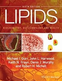
Lipids: Biochemistry, Biotechnology and Health
Sixth Edition
(formerly Lipid Biochemistry: An Introduction, Editions 1–5)
BY
Michael I. Gurr
John L. Harwood
Keith N. Frayn
Denis J. Murphy
Robert H. Michell
© 1971, 1975, 1980 Michael I. Gurr and A.T. James; 1991 Michael I. Gurr and John L. Harwood; 2002 Michael I. Gurr, John L. Harwood and Keith N. Frayn; 2016 Michael I. Gurr, John L. Harwood, Keith N. Frayn, Denis J. Murphy and Robert H. Michell
Registered office
:
John Wiley & Sons, Ltd, The Atrium, Southern Gate, Chichester, West Sussex, PO19 8SQ, UK
Editorial offices:
9600 Garsington Road, Oxford, OX4 2DQ, UKThe Atrium, Southern Gate, Chichester, West Sussex, PO19 8SQ, UK111 River Street, Hoboken, NJ 07030-5774, USA
For details of our global editorial offices, for customer services and for information about how to apply for permission to reuse the copyright material in this book please see our website at www.wiley.com/wiley-blackwell.
The right of the author to be identified as the author of this work has been asserted in accordance with the UK Copyright, Designs and Patents Act 1988.
All rights reserved. No part of this publication may be reproduced, stored in a retrieval system, or transmitted, in any form or by any means, electronic, mechanical, photocopying, recording or otherwise, except as permitted by the UK Copyright, Designs and Patents Act 1988, without the prior permission of the publisher.
Designations used by companies to distinguish their products are often claimed as trademarks. All brand names and product names used in this book are trade names, service marks, trademarks or registered trademarks of their respective owners. The publisher is not associated with any product or vendor mentioned in this book.
Limit of Liability/Disclaimer of Warranty: While the publisher and author(s) have used their best efforts in preparing this book, they make no representations or warranties with respect to the accuracy or completeness of the contents of this book and specifically disclaim any implied warranties of merchantability or fitness for a particular purpose. It is sold on the understanding that the publisher is not engaged in rendering professional services and neither the publisher nor the author shall be liable for damages arising herefrom. If professional advice or other expert assistance is required, the services of a competent professional should be sought.
Library of Congress Cataloging-in-Publication Data
Names: Gurr, M. I. (Michael Ian), author. | Harwood, John L., author. | FraynK. N. (Keith N.), author. | Murphy, Denis J., author | Michell, R. H., author.Lipid biochemistry. Preceded by (work):
Title: Lipids:Biochemistry, Biotechnology and Health / by Michael I. Gurr, JohnL. Harwood, Keith N. Frayn, Denis J. Murphy, and Robert H. Michell.
Description: 6th edition. | Chichester, West Sussex ; Hoboken, NJ : JohnWiley & Sons Inc., 2016. | Preceded by Lipid biochemistry / by Michael I.Gurr, John L. Harwood, and Keith N. Frayn. 5th ed. 2002. | Includesbibliographical references and index.
Identifiers: LCCN 2016000533 (print) | LCCN 2016002203 (ebook) | ISBN9781118501139 (pbk.) | ISBN 9781118501085 (Adobe PDF) | ISBN9781118501108 (ePub)
Subjects: | MESH: Lipids
Classification: LCC QP751 (print) | LCC QP751 (ebook) | NLM QU 85 | DDC572/.57–dc23
LC record available at http://lccn.loc.gov/2016000533
A catalogue record for this book is available from the British Library.
Wiley also publishes its books in a variety of electronic formats. Some content that appears in print may not be available in electronic books.
Cover images: Left panel: An artery partially occluded by an atherosclerotic plaque (Section 10.5.1). The red stain is for macrophages that are present in the plaque and become foam cells. The green stain is for smooth muscle cells in the arterial wall and capping the plaque. Photo courtesy of Thomas S. Davies and Susan Chazi, Cardiff University, UK from work funded by the British Heart Foundation.
Middle panel: An enterocyte from human jejunum displaying multiple lipid droplets a few hours after consuming a fatty meal (Section 7.1.3). The figure also shows mitochondria (dark) and the microvilli (brush border). Electron micrograph courtesy of Dr M Denise Robertson, University of Surrey, UK from work funded by the Biotechnology and Biological Sciences Research Council (BBSRC). Reproduced, with permission from BMJ Publishing Group Ltd, from MD Robertson, M Parkes, BF Warren et al. (2003) Mobilization of enterocyte fat stores by oral glucose in man. Gut 6: 833–8.
Right panel: Distribution of different molecular species of phosphatidylcholine within developing oilseed rape embryos as revealed by MALDI-MS imaging (Section 9.3.1). Red shows high concentrations and green low. Photo courtesy of Helen Woodfield and Drew Sturtevent from work funded by the BBSRC in Prof. Kent Chapman's laboratory at the University of North Texas, USA.
Background: Gettyimages/manuela schewe-behnisch / eyeem
Preface
Our main aims in writing this book have been, as ever, to aid students and other researchers in learning about lipids, to help staff in teaching the subject and to encourage research in this field. Since the publication of the Fifth Edition in 2002, there have been huge advances in our knowledge of the many aspects of lipids, especially in molecular biology. Far more is now known about the genes coding for proteins involved in lipid metabolism and already techniques of biotechnology are making use of this knowledge to produce specialized lipids on an industrial scale. The new knowledge has also had a far-reaching influence on medicine by revealing the role of lipids in disease processes to a much greater extent than hitherto and allowing for advances in diagnosis and disease prevention or treatment. We have endeavoured to reflect as many of these advances as possible in this new edition. Although modern textbooks of general biochemistry or biology now cover lipids to a greater extent than when our first edition was published in 1971, a book devoted entirely to lipids is able to go into far more detail on all these diverse aspects of the subject and to discuss exciting new developments with greater authority. It should be emphasized here that we have referred to a wide range of organisms – including archaea, bacteria, fungi, algae, ‘higher’ plants and many types of animals and not restricted ourselves to mammalian lipids.
Because of this research activity, we have rewritten large parts of the book and have given it a new title that reflects the fact that it is increasingly difficult to identify old boundaries between subjects such as biochemistry, physiology and medicine. This runs in parallel with changes in university structure: away from narrow ‘departments’ of ‘biochemistry’, zoology', ‘botany’ and the like, towards integrated ‘schools’ of biological sciences or similar structures. The increasing diversity of the subject requires greater specialist expertise than is possible with one or two authors. Accordingly, we have brought two new colleagues on board and one of the original authors has been given the role of coordinating editor to assure, as far as possible, consistency of style, so that we could avoid identifying authors with chapters. The authors have consulted widely among colleagues working in lipids and related fields to ensure that each chapter is as authoritative as possible. We are grateful for their help, which is recorded in the acknowledgements section. As a result, advances in such topics as enzymes of lipid metabolism, lipids in cell signalling, lipids in health and disease, molecular genetics and biotechnology have been strengthened.
The need to include new material has had to be balanced against the need to keep the book to a moderate size, with a price within most students' budgets. Some things had to go! As in the Fifth Edition, we decided to restrict some material of historical interest. Nevertheless, we thought that the inclusion of many short references to historical developments should remain, to add interest and to put certain aspects of lipidology in context. We have also removed some of the material that dealt with analytical procedures so that we could focus more on metabolic, physiological, clinical and biotechnological aspects. Chapter 1 now summarizes lipid analytical methods, with ample references to more specialist literature but has a section on lipidomics to highlight modern approaches to lipid profiling in biological fluids and tissues. This introductory chapter also contains a guide to finding your way around the book, which we hope students will find useful. We shall appreciate comments and suggestions so that future editions can be further improved.
MI GurrJL HarwoodKN FraynDJ MurphyRH Michell
Acknowledgements
Over the years, we have received invaluable assistance from many colleagues in the compilation of this book and our thanks have been recorded in the previous five editions. Their contributions are still significant in this new edition and we are also grateful to the following for helping us with new material.
In Chapter 1, Jules Griffin provided valuable assistance with the lipidomics section. The substantial section on fatty acid biosynthesis has been brought up to date with help from Stuart Smith and his colleague Marc Leibundgut, whose huge expertise has been much appreciated. Many other aspects of Chapter 3 have benefited from the help of John Cronan Jr., Michael Schweizer, Marc Leibundgut and Ivo Fuessner. Bill Christie's wide knowledge of lipid chemistry, nomenclature and analysis has been invaluable throughout the book. Deficiencies in our knowledge of fat-soluble vitamins have been rectified by David Bender (Chapter 6); recent advances in comparative aspects of lipid metabolism by Caroline Pond (Chapter 7); lipids in immunity by Parveen Yaqoob and Philip Calder (Chapter 10); lung surfactant by Fred Possmayer (Chapters 4 & 10) and lipoproteins in human metabolism and clinical practice by Fredrik Karpe and Sophie Bridges (Chapters 7 & 10). Gary Brown and Patrick Schrauwen helped with information on inborn errors of lipid metabolism; Jenny Collins with cancer and lipid metabolism; and Sara Suliman with understanding lipodystrophies (Chapter 10).
Our thanks are due to the Wiley-Blackwell team for guiding us through the intricacies of the publication process. Particular mention should be made of Nigel Balmforth, who has been associated with Lipid Biochemistry from its early days with Chapman & Hall, then Blackwell and finally Wiley. Finally, after the enormous amount of work that goes into writing a book of this complexity, the authors conclude that all ‘i's and ‘t's must have been dotted and crossed. It takes an expert, conscientious and helpful copy-editor to put a stop to this complacency and create a much better product. Martin Noble has done just that. Thank you all.
About the Authors
Michael I. Gurr was Visiting Professor in Human Nutrition at Reading and Oxford Brookes Universities, UK.
John L. Harwood is Professor of Biochemistry in the School of Biosciences, Cardiff University, UK.
Keith N. Frayn is Emeritus Professor of Human Metabolism at the University of Oxford, UK.
Denis J. Murphy is Professor of Biotechnology in the School of Applied Sciences, University of South Wales, UK.
Robert H. Michell is Emeritus Professor of Biochemistry in the School of Biosciences, University of Birmingham, UK.
About the Companion Website
www.wiley.com/go/gurr/lipids
The website includes:
Powerpoint slides of all the figures from the book, to download
Pdfs of all tables and boxes from the book, to download
Updates to Further Reading and additional figures to download
Chapter 1Lipids: Definitions, Naming, Methods and a Guide to the Contents of this Book
1.1 Introduction
Lipids occur throughout the living world in microorganisms, fungi, higher plants and animals. They occur in all cell types and contribute to cellular structure, provide energy stores and participate in many biological processes, ranging from transcription of genes to regulation of vital metabolic pathways and physiological responses. In this book, they will be described mainly in terms of their functions, although on occasion it will be convenient, even necessary, to deal with lipid classes based on their chemical structures and properties. In the concluding section of this chapter, we provide a ‘roadmap’ to help students find their way around the book, so as to make best use of it.
1.2 Definitions
Lipids are defined on the basis of their solubility properties, not primarily their chemical structure.
The word ‘lipid’ is used by chemists to denote a chemically heterogeneous group of substances having in common the property of insolubility in water, but solubility in nonaqueous solvents such as chloroform, hydrocarbons or alcohols. The class of natural substances called ‘lipids’ thus contrasts with proteins, carbohydrates and nucleic acids, which are chemically well defined.
The terms ‘fat’ and ‘lipid’ are often used interchangeably. The term fat is more familiar to the layman for substances that are clearly fatty in nature, greasy in texture and immiscible with water. Familiar examples are butter and the fatty parts of meats. Fats are generally solid in texture, as distinct from oils which are liquid at ambient temperatures. Natural fats and oils are composed predominantly of esters of the three-carbon alcohol glycerol with fatty acids, often referred to as ‘acyl lipids’ (or more generally, ‘complex lipids’). These are called triacylglycerols (TAG, see Section 2.2: often called ‘triglycerides’ in older literature) and are chemically quite distinct from the oils used in the petroleum industry, which are generally hydrocarbons. Alternatively, in many glycerol-based lipids, one of the glycerol hydroxyl groups is esterified with phosphorus and other groups (phospholipids, see Sections 2.3.2.1 & 2.3.2.2) or sugars (glycolipids, see Section 2.3.2.3). Yet other lipids are based on sphingosine (an 18-carbon amino-alcohol with an unsaturated carbon chain, or its derivatives) rather than glycerol, many of which also contain sugars (see Section 2.3.3), while others (isoprenoids, steroids and hopanoids, see Section 2.3.4) are based on the five-carbon hydrocarbon isoprene.
Chapter 2 deals mainly with lipid structures, Chapters 3 and 4 with biochemistry and Chapter 5 with lipids in cellular membranes. Aspects of the biology and health implications of these lipids are discussed in parts of Chapters 6–10 and their biotechnology in Chapter 11. The term ‘lipid’ to the chemist thus embraces a huge and chemically diverse range of fatty substances, which are described in this book.
1.3 Structural Chemistry and Nomenclature
1.3.1 Nomenclature, General
Naming systems are complex and have to be learned. The naming of lipids often poses problems. When the subject was in its infancy, research workers gave names to substances that they had newly discovered. Often, these substances would turn out to be impure mixtures but as the chemical structures of individual lipids became established, rather more systematic naming systems came into being and are still evolving. Later, these were further formalized under naming conventions laid down by the International Union of Pure and Applied Chemistry (IUPAC) and the International Union of Biochemistry (IUB). Thus, the term ‘triacylglycerols’ (TAGs – see Index – the main constituents of most fats and oils) is now preferred to ‘triglyceride’ but, as the latter is still frequently used especially by nutritionists and clinicians, you will need to learn both. Likewise, outdated names for phospholipids (major components of many biomembranes), for example ‘lecithin’, for phosphatidylcholine (PtdCho) and ‘cephalin’, for an ill-defined mixture of phosphatidylethanolamine (PtdEtn) and phosphatidylserine (PtdSer) will be mostly avoided in this book, but you should be aware of their existence in older literature. Further reference to lipid naming and structures will be given in appropriate chapters. A routine system for abbreviation of these cumbersome phospholipid names is given below.
1.3.2 Nomenclature, Fatty Acids
The very complex naming of the fatty acids (FAs) is discussed in more detail in Chapter 2, where their structures are described. Giving the full names and numbering of FAs (and complex lipids) at each mention can be extremely cumbersome. Therefore a ‘shorthand’ system has been devised and used extensively in this book and will be described fully in Section 2.1, Box 2.1. This describes the official system for naming and numbering FAs according to the IUPAC/IUB, which we shall use routinely. An old system used Greek letters to identify carbon atoms in relation to the carboxyl carbon as C1. Thus, C2 was the α-carbon, C3 the β-carbon and so on, ending with the ω-carbon as the last in the chain, furthest from the carboxyl carbon. Remnants of this system still survive and will be noted as they arise. Thus, we shall use ‘3-hydroxybutyrate’, not ‘β-hydroxy-butyrate’ etc.
While on the subject of chain length, it is common to classify FAs into groups according to their range of chain lengths. There is no standard definition of these groups but we shall use the following definitions in this book: short-chain fatty acids, 2C–10C; medium-chain, 12C–14C; long-chain, 16C–18C; very long-chain >18C. Alternative definitions may be used by other authors.
1.3.3 Isomerism in Unsaturated Fatty Acids
An important aspect of unsaturated fatty acids (UFA) is the opportunity for isomerism, which may be either positional or geometric. Positional isomers occur when double bonds are located at different positions in the carbon chain. Thus, for example, a 16C monounsaturated (sometimes called monoenoic, see below) fatty acid (MUFA) may have positional isomeric forms with double bonds at C7-8 or C9-10, sometimes written Δ7 or Δ9 (see Box 2.1). (The position of unsaturation is numbered with reference to the first of the pair of carbon atoms between which the double bond occurs, counting from the carboxyl carbon.) Two positional isomers of an 18C diunsaturated acid are illustrated in Fig. 1.1(c,d). Geometric isomerism refers to the possibility that the configuration at the double bond can be cis or trans. (Although the convention Z/E is now preferred by chemists instead of cis/trans, we shall use the more traditional and more common cis/trans nomenclature throughout this book.) In the cis form, the two hydrogen substituents are on the same side of the molecule, while in the trans form they are on opposite sides (Fig. 1.1a,b). Cis and trans will be routinely abbreviated to c,t (see Box 2.1).
Fig. 1.1 Isomerism in fatty acids. (a) cis-double bond; (b) a trans-double bond; (c) c,c-9,12-18:2; (d) c,c-6,9-18:2.
1.3.4 Alternative Names
Students also need to be aware that the term ‘ene’ indicates the presence of a double bond in a FA. Consequently, mono-, di-, tri-, poly- (etc.) unsaturated FAs may also be referred to as mono-, di-, tri- or poly- (etc.) enoic FAs (or sometimes mono-, di-, tri- or poly-enes). Although we have normally used ‘unsaturated’ in this book, we may not have been entirely consistent and ‘-enoic’ may sometimes be encountered! Furthermore it is important to note that some terms are used in the popular literature that might be regarded as too unspecific in the research literature. Thus shorthand terms such as ‘saturates’, ‘monounsaturates’, ‘polyunsaturates’ etc. will be avoided in much of this text but, because some chapters deal with matters of more interest to the general public, such as health (Chapter 10) and food science or biotechnology (Chapter 11), we have introduced them where appropriate, for example when discussing such issues as food labelling.
1.3.5 Stereochemistry
Another important feature of biological molecules is their stereochemistry. In lipids based on glycerol, for example, there is an inherent asymmetry at the central carbon atom of glycerol. Thus, chemical synthesis of phosphoglycerides yields an equal mixture of two stereoisomeric forms, whereas almost all naturally occurring phosphoglycerides have a single stereochemical configuration, much in the same way as most natural amino acids are of the L (or S) series. Students interested in the details of the stereochemistry of glycerol derivatives should consult previous editions of this book (see Gurr et al. (1971, 1975, 1980, 1991, 2002) and other references in Further reading). The IUPAC/IUB convention has now abolished the DL (or even the more recent RS) terminology and has provided rules for the unambiguous numbering of the glycerol carbon atoms. Under this system, the phosphoglyceride, phosphatidylcholine, becomes 1,2-diacyl-sn-glycero-3-phosphorylcholine or, more shortly, 3-sn-phosphatidylcholine (PtdCho; Fig. 1.2). The letters sn denote ‘stereochemical numbering’ and indicate that this system is being used. The stereochemical numbering system is too cumbersome to use routinely in a book of this type and, therefore, we shall normally use the terms ‘phosphatidylcholine’ etc. or their relevant abbreviations, but introduce the more precise name when necessary.
Fig. 1.2 The stereochemical numbering of lipids derived from glycerol.
1.3.6 Abbreviation of Complex Lipid Names and Other Biochemical Terms
Students will appreciate that the official names of complex lipids (and many other biochemicals) are cumbersome and research workers have evolved different systems for abbreviating them. In this latest edition we have incorporated all abbreviations into the index. At the first mention of each term in the text, we shall give the full authorized name followed by the abbreviation in parentheses. This will be repeated at the first mention in each subsequent chapter. Students should be aware that, unlike the IUB/IUPAC naming system, which is now generally accepted and expected to be used, the abbreviation system is still very much a matter of personal choice. Therefore students may expect to find alternative phospholipid abbreviations in some publications, for example PC, PE, PS and PI for phosphatidylcholine, -ethanolamine, -serine and –inositol, instead of the PtdCho, PtdEtn, PtdSer and PtdIns used here. With very few exceptions we have not defined abbreviations for well-known substances in the general biochemical literature, such as ATP, ADP, NAD(H), NADP(H), FMN, FAD etc.
Another field in which nomenclature has grown up haphazardly is that of the enzymes of lipid metabolism. This has now been formalized to some extent under the Enzyme Commission (EC) nomenclature. The system is incomplete and not all lipid enzymes have EC names and numbers. Moreover, the system is very cumbersome for routine use and we have decided not to use it here. You will find a reference to this nomenclature in Further reading should you wish to learn about it.
Since the last edition was published in 2002, there have been huge advances in molecular biology and, in particular, in identifying the genes for an ever-increasing number of proteins. Where appropriate, we have referred to a protein involved in human lipid metabolism, of which the gene has been identified and have placed the gene name in parentheses after it (protein name in Roman, gene name in Italic script).
1.4 Lipidomics
1.4.1 Introduction
Since the last edition of this book in 2002, there have been very considerable advances in analysing and identifying natural lipids. Much modern research in this field is concerned with the profiling of lipid molecular species in cells, tissues and biofluids. This has come to be known as ‘lipidomics’, similar to the terms ‘genomics’ for profiling the gene complement of a cell or ‘proteomics’ for its proteins.
Some older methods of lipid analysis, presented in previous editions, will be described only briefly here and the student is referred to Further reading for books, reviews and original papers for more detail. Before describing the modern approach to lipidomics, we describe briefly the steps needed to prepare lipids for analysis and the various analytical methods, many of which are still widely used.
1.4.2 Extraction of Lipids from Natural Samples
This is normally accomplished by disrupting the tissue sample in the presence of organic solvents. Binary mixtures are frequently used, for example chloroform and methanol. One component should have some water miscibility and hydrogen-bonding ability in order to split lipid-protein complexes in the sample, such as those encountered in membranes (Chapter 5). Precautions are needed to avoid oxidation of, for example, UFAs. Control of temperature is important, as well as steps to inhibit breakdown of lipids by lipases (see Sections 4.2 & 4.6). The extract is finally ‘cleaned up’ by removing water and associated water-soluble substances (see Further reading).
1.4.3 Chromatographic Methods for Separating Lipids
Once a sample has been prepared for analysis, chromatography can be used to separate its many lipid constituents. A chromatograph comprises two immiscible phases: one is kept stationary by being held on a microporous support; the other (moving phase) percolates continuously through the stationary phase. The stationary phase may be located in a long narrow bore column of metal, glass or plastic (column chromatography), coated onto a glass plate or plastic strip (thin layer chromatography, TLC, see Fig. 1.3) or it may simply be a sheet of absorbent paper (paper chromatography).
Fig. 1.3 Separation of lipid classes by thin-layer chromatography (TLC).
The principle of chromatography is that when a lipid sample (often comprising a very large number of molecular species) is applied to a particular location on the stationary phase (the origin) and the moving phase percolates through, the different components of the mixture partition differently between the two phases according to their differing chemical and physical properties. Some will tend to be retained more by the stationary phase, while others tend to move more with the moving phase. Thus, the components will move apart as the moving phase washes through the system (see Christie, 1997; Christie & Han 2010; and Hammond 1993 in Further reading for more details of the theory of chromatography).
Many types of adsorbent solid can be used as the stationary phase (e.g. silica, alumina). The moving phase may be a liquid (liquid chromatography, LC) or a gas (gas chromatography, GC – the original term gas-liquid chromatography, GLC, is now less used). Particularly good separations may now be achieved by GC (see Fig. 1.4) with very long thin columns packed with an inert support for the stationary phase or in which the stationary phase is coated on the wall of the column. This is useful for volatile compounds or those that can be converted into more volatile ones, such as the methyl esters of FAs (see Sections 2.1.8.1 & 11.2.4.2 for further details of the preparation of FA methyl esters). For less volatile complex lipids, LC in thin columns through which the moving phase is passed under pressure can produce superior separations: this is called high performance liquid chromatography (HPLC).
Fig. 1.4 Separation of fatty acid methyl esters by gas chromatography (GC). The figure shows the FA composition of a lipid extract of heart tissue as measured by GC on a capillary column. To the right of the chromatogram is depicted the conversion of a complex lipid into FA methyl esters in preparation for chromatography. The peaks on the chromatogram are labelled with shorthand abbreviations for FAs (see Box 2.1 for details). Detection is by a flame ionization detector. From JL Griffin, H Atherton, J Shockcor & L Atzori (2011) Metabolomics as a tool for cardiac research. Na Rev Cardiol8: 630–43; p. 634, Fig. 3a. Reproduced with permission of Nature Publishing Group.
Once the components have been separated, they can be collected as they emerge from the column for further identification and analysis (see Section 1.4.4). Compounds separated on plates or strips can be eluted from the stationary phase by solvents or analysed in situ by various means. (Further information on methods of detection can be found in Christie & Han (2010) and Kates (2010) in Further reading.)
The power of modern lipidomics has been made possible by the combination of GC or LC with improved methods of mass spectrometry (MS) to provide detailed and sophisticated analyses of complex natural lipid mixtures and this is the subject of the next section.
1.4.4 Modern Lipidomics Employs a Combination of Liquid Chromatography or Gas Chromatography with Mass Spectrometry to Yield Detailed Profiles of Natural Lipids – the ‘Lipidome’
While individual FAs can be readily measured by gas chromatography-mass spectrometry (GC-MS), the commonest method to perform this analysis relies on cleaving FAs from the head groups that they are associated with and converting them into methyl esters by transesterification. This process is used to make the FAs volatile at the temperature used by GC-MS, but during this process information is lost, particularly about which lipid species are enriched in a given FA.
An alternative is to use LC-MS. In this approach, lipid extracts from biofluids and tissues can be analysed directly. The lipids are dissolved in an organic solvent and injected directly onto the HPLC column. Columns can contain a variety of chemicals immobilized to form a surface (stationary phase) that the analytes interact with. For the analysis of lipids, columns containing long chains of alkyl groups are most commonly used, in particular 8C and 18C columns, which have side-chain lengths of 8 and 18 carbons, respectively. The most commonly used HPLC method is referred to as ‘reverse phase’, whereby lipids are initially loaded onto a HPLC column and then the HPLC solvent is varied from something that is predominantly aqueous to a solvent that is predominantly organic, across what is termed a gradient. The solvents are referred to as the mobile phases. During this process, lipids are initially adsorbed on to the stationary phase, until their solubility increases to the point that they begin to dissolve in the mobile phase. In this manner, polar and nonpolar lipids can readily be separated and typically, in a lipid extract, lipid molecular species would elute in the order of nonesterified fatty acids (NEFAs), phospholipids, cholesteryl esters and TAGs. The chromatography serves two important purposes. Firstly, it reduces the complexity of the subsequent mass spectra generated by the mass spectrometer, making metabolite identification more convenient. Secondly, some metabolites can ionize more readily than others and this can produce an effect called ‘ion suppression’ where one metabolite ionizes more easily and reduces the energy available for the ionization of other species. As a result, the mass spectrometer may detect only the metabolite that ionizes readily and miss the other metabolites that do not readily form ions.
LC-MS is most commonly used with ‘electrospray ionization’ where the analytes are introduced to the mass spectrometer in the form of a spray of solvent. They are accelerated over an electric field across the capillary that introduces them into the mass spectrometer and the nebulization of the spray is often assisted by the flow of an inert gas. The inert gas causes the solvent to evaporate (desolvate), producing a fine spray of droplets. As the solvent evaporates, charges build up in the droplets until they explode into smaller droplets, finally producing an ion that is introduced into the mass spectrometer. While this may sound relatively destructive, this form of ionization is relatively ‘soft’, ensuring that the molecule itself or an adduct (a combination of the molecule and another charged species such as H+, Na+, K+ or other ions present in the solvent) is formed. The ions are then detected by the mass spectrometer (Fig. 1.5).
Fig. 1.5 Separation and identification of heart lipidome by liquid chromatography-mass spectrometry (GC-MS). Intact lipids from an extract of heart tissue have been separated, detected and identified by GC-MS. Chromatography separates the intact lipids according to their polarity and high resolution MS identifies individual lipid molecular species. From JL Griffin, H Atherton, J Shockcor & L Atzori (2011) Metabolomics as a tool for cardiac research. Nat Rev Cardiol8: 630–43; p. 634, Fig. 3b. Reproduced with permission of Nature Publishing Group.
While there are numerous designs of mass spectrometer, two common methods are often used in lipidomics. In high resolution MS, the mass accuracy achievable is so great that chemical formulae can be determined with reasonable precision. This is because only carbon-12 has a mass of exactly 12 atomic mass units, while other nuclides all have masses that slightly differ from a whole number. These mass deficits can be used to predict what nuclides are present and estimate a small number of chemical formulas that may be responsible for the ion. The accuracy of modern high resolution mass spectrometers is so high, often less than 3–5 parts per million, that it is possible in lipidomics to determine what species are being detected by their exact mass and references to databases such as LIPID MAPS (http://www.lipidmaps.org/). However, even in cases where only one formula is identified this could still belong to a range of potential lipid species. For example, if we take the PtdCho (36:2 – i.e. total FA chains of 36 carbon atoms with a total of 2 double bonds), this could be due to a PtdCho containing two C18:1 FAs, one C18:0 and one C18:2 or a variety of other isomers. To further define the chemical structure, fragmentation can be performed. In this process the ion is accelerated through a low pressure of inert gas, producing collisions and fragmentation of the parent ion. The daughter fragments can then be used to work out the parent structure, with head groups and FAs commonly being lost in the process (Fig. 1.6).
Fig. 1.6 Fragmentation of two phosphocholines derived from phosphatidylcholines (PtdChos). This figure demonstrates the further characterization of the lipidome by the technique of ‘tandem MS’. One of the main challenges of LC-MS is lipid identification because of the large numbers of isomers present. In this technique, chromatography is dispensed with altogether and the sample is directly infused into a high resolution MS instrument. Figure 1.6 illustrates the identification of two phosphocholine isomers produced by fragmentation of PtdChos that would have been esterified with 18:0/18:2 and 18:1/18:1 respectively. From JL Griffin, H Atherton, J Shockcor & L Atzori (2011) Metabolomics as a tool for cardiac research. Nat Rev Cardiol8: 630–43; p. 636, Fig. 4a. Reproduced with permission of Nature Publishing Group.
In the other form of commonly used lipidomics, a triple quadrupole mass spectrometer is used. In this instrument, the mass spectrometer consists of three electromagnet gates called quadrupoles. The first is used to select for one ion, which is usually the parent ion of the lipid species being detected. The second quadrupole acts as a fragmentation cell where the ion is fragmented. The third quadrupole then selects a particular fragment ion. While many lipid species may have the same parent mass, it is very unlikely that they will fragment in the same manner and thus this method is highly selective. In addition, these instruments can be made to be quantitative and are particularly appropriate for targeted analyses where a limited number of species is to be assayed. Furthermore, in an approach termed ‘shotgun lipidomics’, the assay can be set up to scan for particular lipid species either by virtue of the head group present (e.g. scanning for PtdCho species) or particular FAs (e.g. identifying lipid species that contain a particular FA such as arachidonic acid, all-c5,8,11,14-20:4, n-6).
More detailed accounts of these methods can be found in Further reading.
1.5 A Guide to the Contents of this Book
The purpose of this section is to provide a ‘roadmap’ to enable students to find their way around and make best use of the information provided in this book.
Continuing the scheme adopted in this chapter, each chapter is divided into numbered sections; the first number of the section will indicate the chapter number. There will be extensive cross-referencing between sections within chapters and between chapters. Although there are several ways we could have arranged the succession of chapters, we hope that the one we have chosen will be a logical one.
At the end of each chapter there is a ‘Key points’ section that provides a concise summary of the principal information in the chapter. This is followed by a section on ‘Further reading’, which provides a selection of useful reviews and also some original research publications to give students a flavour of important and exciting recent advances. Although items in Further reading will be referenced throughout each chapter, there are limited references to specific pieces of literature in the main text. The number of references in Further reading could not be unlimited. We have attempted to cite those most useful that were available at the time of writing but additional references and/or diagrams are available on the companion website. Information in the text will be supplemented with figures and tables, and ‘boxes’ will be used to provide more detail on specific topics where inclusion in the text might interfere with the flow.
Chapter 2 introduces the chemical structures of the different types of lipids in three sections. These deal with (1) FAs, (2) lipids mainly involved in energy storage and (3) those predominantly associated with cellular membranes and also involved in physiological processes such as cell signalling. Of course there will be overlap between these functions: it is impossible (and undesirable) fully to compartmentalize lipid forms and functions. These sections discuss how the chemical structures of lipids relate to their physical and physiological properties and point the way to aspects of their metabolism, function and utilization in subsequent chapters. The FA section contains Box 2.1, which provides useful information on the complex topic of FA nomenclature.
Chapter 3 covers the metabolism of the FAs. This starts with their biosynthesis and discusses up-to-date knowledge of biosynthetic pathways, the enzymes involved in their biosynthesis and the genes coding for them. The degradation of FAs by oxidative pathways is then discussed in detail with particular reference to the generation of metabolic energy. A key section in this and other chapters concerns the all-important matter of how these metabolic pathways are controlled and integrated.
In the discussions of the biosynthesis of the polyunsaturated fatty acids (PUFAs) and their subsequent oxidation to form physiologically active products such as the ‘eicosanoids’, reference will be made to later chapters that describe the role of such molecules in cell signalling (Chapter 8) and as mediators in such physiological processes as immunity and the implications for health and disease (Chapter 10). Some PUFAs described in Chapter 3 are essential components of the diet (‘essential fatty acids’, EFAs) and their roles will be discussed further in Chapter 6.
Chapter 4. Just as Chapter 3 discusses the metabolism of the FAs, Chapter 4 deals with the metabolism of the complex lipids. Many of these (TAGs, phosphoglycerides, glycosylglycerides etc.) are FA esters of glycerol but the chapter also covers the sphingolipids (derivatives of the base sphingosine rather than glycerol, many of which incorporate sugars in the molecule) and the isoprenoids (also called terpenoids), in which the sterols, such as cholesterol and the plant steroids, are included.
The formation of TAGs is related to their role in energy storage in adipose tissue. This has ramifications for influence of dietary fats on the fat stores (Chapter 6) and on the relationships between the energy stores and health problems such as obesity, insulin resistance, diabetes, immune function, cancer and cardiovascular diseases, all of which are discussed in more detail in Chapter 10. Numerous seed oils of commercial importance store TAGs as an energy source. This too, has implications for the type and amounts of lipids in the diet (Chapter 6), their implications for health (Chapter 10) and their biotechnological modification to provide useful products (Chapter 11).
An important section in this chapter discusses the many lipases (see Sections 4.2 & 4.6) that degrade lipids. Some are involved in the digestion of dietary lipids (Chapter 7), many others are involved in modifying the FA composition of lipids to suit the needs of particular cell types and cell structures (Chapters 4, 5, 7, 9 & 10), others are utilized in biotechnological processes (Chapter 11) and yet others are involved in the release of components of lipid molecules that are destined to become cell-signalling molecules (Chapters 8 & 10). Failure to degrade certain glycolipids, mainly owing to gene defects, can result in several lethal diseases of the nervous system that are addressed in Chapter 10.
Failure in the regulation of the metabolism of cholesterol in human beings, as a result of gene defects (Chapters 4 & 7) or dietary imbalance (Chapters 7 & 10) has implications for cardiovascular diseases that are also explored in Chapter 10.
Chapter 4 also mentions the biosynthesis of lipids that have specific functions and points the way to more detailed discussion in later chapters – for example the platelet activating factor and the lung surfactant lipid in Chapter 10.
Chapter 5 discusses the various ways in which different lipids can associate with each other and with proteins as a result of their chemical and physical properties. Such lipid assemblies are crucial to the structure and function of cells and cell organelles and in this chapter, we explore what is currently known about how lipids have shaped the evolution of living cells. Light is cast on the way in which, for example, the evolution of the bacteria and the archaea depended on the development of lipids with quite different chemical structures. Of particular importance is the development of different types of membranes whose lipid composition is crucial to their functions. Membranes are important for the topics discussed in each of the chapters of this book because of their role in cell structure, function and integrity, as a location for many metabolic pathways, their involvement in inter- and intracellular signalling processes, in the trafficking of biochemical substances within and between cells and because the development of many disease processes results from defects in the integrity of many membranes. As well as their presence in membranes, lipids accumulate as droplets (LDs) in cells (see Section 5.5) where they act as energy stores or sources of molecules involved in the mediation of metabolic processes. Some lipid assemblies are involved in processes outside cells, for example in the formation of surface layers with barrier properties (see Section 2.2.4) or, as lipoproteins, in the transport of lipids in the bloodstream (see Sections 7.2 & 10.5).
Chapter 6 discusses the types of lipids in food and the diet and their biological roles. (Chapter 7, which follows, explains how these dietary lipids are digested, absorbed and the digestion products transported in the blood to the tissues of the body.) These two chapters are devoted mainly to human diets but there is also discussion of other simple-stomached animals, such as rats, mice and pigs, which are often used as so-called ‘animal models’. This is because, in animal studies, procedures can be more easily controlled and the experimental design can be more rigorous. The disadvantage is that the biochemistry and physiology may sometimes differ between species, leaving open some doubt as to their relevance to Man.
Much of the food we now eat is processed in some way – industrially and domestically. There is some discussion here of how such processes may affect dietary lipids but reference is made to Chapter 11, which provides more detail on food processing and biotechnological developments. Dietary fats provide metabolic energy and although the subject is introduced here, readers will find more detailed information in Chapter 9. Dietary fats also supply many essential nutrients. This chapter picks up on the EFAs – PUFAs that are essential for health but cannot be made in the body – that were first introduced in Chapters 2 and 3. Also essential for good health are the fat-soluble vitamins, which are required in only milligram or microgram quantities as distinct from the gram or almost gram quantities of the EFAs. While knowledge of them developed in the late 19th and early 20th centuries, it is only in the last few decades that the full extent of their physiological roles as, for example, hormones and signalling molecules and regulators of metabolism has been realized. The molecular biology revolution has indicated the key involvement of some of them in the regulation of gene expression. The chapter ends with a thorough discussion of the role of lipids in foetal and postnatal development.
Chapter 7 describes in detail the processes by which lipid components of the diet are digested and the digestion products absorbed from different parts of the intestinal tract. Once within the intestinal absorbing cells (enterocytes) they are ‘remodelled’ and combined with proteins (‘lipoproteins’) for transport around the body in the bloodstream. The proteins not only help to solubilize the lipids but also direct them to sites of further metabolism. The different types of lipoproteins are described and also the elaborate system for the control of their metabolism and their movement to appropriate tissues. Such a complex system is vulnerable to defects either from gene mutations or from ‘dietary overload’ and the reader is pointed to Chapter 10, which describes the involvement of various lipids in health and disease.
Chapter 8 is concerned entirely with molecules that send signals to different cells of the body. The emphasis in this chapter is mainly on two types: the phosphoinositides and the sphingolipids. Before the mid-1960s, lipids were thought of as having three main biological functions: as structural components of membranes, as energy stores and as a barrier against the environment or providers of insulation. Phosphatidylinositol (PtdIns) was already known as a widespread membrane component but everything changed when it was discovered that inositol phospholipids with additional phosphate groups esterified in different positions on the inositol ring could, when cells were stimulated by agonist molecules such as hormones, be catabolized to yield compounds that sent signals across the membrane that then resulted in a variety of metabolic changes. Even a molecule such as diacylglycerol (DAG), it was then discovered, could act as a ‘messenger’. Similar roles were discovered also for a variety of sphingolipids. Several other lipid molecules with signalling functions are described in other chapters, for example: platelet activating factor (PAF, an ‘ether’ phospholipid) in Chapters 4 and 10; endocannabinoids in Chapters 4 and 8.
Chapter 9 is devoted entirely to the role of lipids as energy stores in animals and plants. The first part goes into detail in the animal storage organs – white and brown adipose tissue. The white form is the main storage tissue for TAGs; it is widely dispersed around the body rather than being a discrete organ like the liver or brain. It contains smaller amounts of other lipid molecules and as well as a storage organ it is now known to have endocrine properties, producing hormones. The uptake of TAGs into the fat cells and their mobilization for energy supply is discussed in relation to the biochemistry already described in Chapters 3 and 4.
The cells of the brown form of adipose tissue contain many small LDs (in contrast to white adipose tissue's unilocular droplet) and these are surrounded by mitochondria that accept FAs released from the fat droplets and oxidize them by the process of β-oxidation, which is described in detail in Chapter 3.
Lipid storage by some plants is important for supplying the metabolic energy for seed development and germination. The different storage locations – fruits, seeds and pollen grains – and the types of lipids involved, are described. Plant storage fats are important in diets (Chapter 6) and require industrial processing (Chapter 11). New methods of introducing genes for the biosynthesis of specific lipids that may not be native to a particular plant are now becoming available (Chapter 11).
Chapter 10 addresses the subject of lipids in health and disease. It opens with a discussion of various inborn errors of metabolism, describing the genetic background and the implications for dietary lipids. There are relevant pointers to other chapters in which the biochemical basics are discussed (Chapters 3, 4, 7 & 9). A section on cancer examines the influence of dietary lipids (both in development and treatment), the roles of specific lipids in physiological functions associated with cancer development and the involvement of the immune system. A whole section is devoted to the ways in which lipids may be involved in aspects of immune function, including their modification of gene expression. Once again there is comprehensive referencing to the biochemistry of lipids in Chapters 3, 4, 5, 7 and 9. The conditions of obesity and diabetes (see also Chapter 9) and disorders of lipoprotein metabolism (in their association with cardiovascular diseases, Chapter 7) are similarly related to preceding biochemical background (Chapters 3 & 4).
Chapter 11


